expressions and significance of tumor suppressor gene pten ... · expressions and significance of...
TRANSCRIPT
Expressions and significance of tumor suppressor gene PTEN and p53 inprostate cancer.
Wei Lu*, Jia-Qiang Wang, Yu-Hong Zhang, Yong Wang, Wen-Jing Yin, Jian-Min Guo, Yi Li, Yan-MinWang, Ji-Hu Lian, Ying-Yuan Gao, Xiao-Ran Wang, Bing-Chen Liu
Department of Urology, Jilin Province People’s Hospital, Changchun, P.R. China
Abstract
The study aim was to investigate the impacts of the expressions of tumor suppressor gene phosphataseand tensin homolog deleted on chromosome ten (PTEN) and p53 on the grading and prognosis ofprostate cancer. Immunohistochemistry was used to detect the expressions of tumor suppressor genePTEN and p53 in 80 prostate cancer and adjacent tissue samples, as well as in 40 normal tissue samples,and the relationships among their expressions, prostate cancer grading, and prognosis were thencompared. p53 was significantly up-regulated in prostate cancer tissue (P<0.05), but PTEN wassignificantly down-regulated (P<0.05). The expression levels of PTEN in the cancer tissues with differentdifferentiation degrees, stages, metastasis, and prognosis exhibited significant differences (P<0.05). Theexpression levels of p53 in the cancer tissues with different differentiation degrees and prognosisexhibited significant differences (P<0.05). There were no correlations between the expressions of p53 andPTEN (P>0.05). Apoptosis-related gene p53 and PTEN participate in the grading of prostate cancer, andalso affect patient’s prognosis, but there are no correlations between these two genes.
Keywords: Prostate cancer, p53, (Phosphatase and tensin homolog) PTEN, Immunohistochemistry.Abbreviations List:PC: Prostate Cancer; PTEN: Phosphatase and tensin homologdeleted on chromosome ten; PI3K: Phosphatidylinositol 3-
Kinase; AKT: Protein Kinase B; WHO: World HealthOrganization; PBS: Phosphate Buffered Saline.
Accepted on July 01, 2017
IntroductionProstate Cancer (PC) is one of the cancers with the highestincidence in males, and studies have shown that benignprostatic hyperplasia is an independent risk factor for PC [1].Although the specific pathogenesis of PC has not been exactlyunderstood yet, the over-expressions of proto-oncogenes andthe mutations of tumor suppressor genes play important rolesin PC [2]. PTEN was originally identified as a tumorsuppressor frequently lost from a region of chromosome 10q23in a variety of human tumors including those of the brain,breast and prostate [3]. PTEN gene is a tumor suppressor genewith bi-specific phosphatase activities, although the tumor-suppressor activity of PTEN depends largely on its lipidphosphatase activity, which opposes phosphatidylinositol 3-kinase (PI3K)/protein kinase B (AKT) activation, thereforeinhibiting cell survival, growth, and proliferation, which wasrelated with the intra-nuclear cycle regulation, at the meantime,it has also been reported that PTEN dephosphorylates focaladhesion kinase, leading to reduced cell migration andspreading in fibroblasts, so it has important tumor suppressingfunctions [4-6]. The p53 gene is the most widely studied tumorsuppressor gene. It could be activated by various genotoxic andcellular stress signals, such as DNA damage, hypoxia,
oncogene activation and nutrient deprivation [7]. Wild-typep53 mediates imperative functions such as regulation of thecell cycle and programmed cell death. The deficiency of p53function by mutation or inactivation abrogates normal cellcycle checkpoints and apoptosis, generating a favourablemilieu for genomic instability and carcinogenesis, but Mulleret al. found that the gene has highest mutation rate [8,9]. Therehave been many studies confirmed that PTEN and p53 areabnormally expressed in prostate cancer tissue, but theexpression correlations between these two genes were stillabsent [10,11]. Therefore, discussing the expressions andcorrelations of these two genes in PC will be important toincrease our understanding of PC.
Materials and Methods
Specimen sourceThe specimens were all sampled from the 80 PC patientsadmitted in our hospital from Jan 2012 to Oct 2013, including52 males and 28 females, aging 23-87 y old, with the mean ageas 62.3 ± 14.0 y. This study was conducted in accordance withthe declaration of Helsinki. This study was conducted withapproval from the Ethics Committee of Jilin Province People’s
ISSN 0970-938Xwww.biomedres.info
Biomed Res- India 28 Volume 28 Issue 15
Biomedical Research 2017; 28 (15): 6730-6734
6730

Hospital. Written informed consent was obtained from allparticipants. The differentiation degrees were: 13 highlydifferentiated cases, 45 moderately differentiated cases, and 22poorly differentiated cases; 46 cases occurred lymph nodemetastasis, and 34 cases did not; TNM stages: 5 cases in stageI, 12 cases in stage II, 34 cases in stage III, and 29 cases instage IV. Inclusion criteria: (1) Met the histological diagnosticcriteria of prostate cancer, World Health Organization (WHO);(2) Without genetic family history of PC; (3) Was notperformed chemotherapy before the surgery. The PC tissue andadjacent tissue were collected from these 80 patients, andanother 40 cases with normal prostate tissue were selected asthe normal controls.
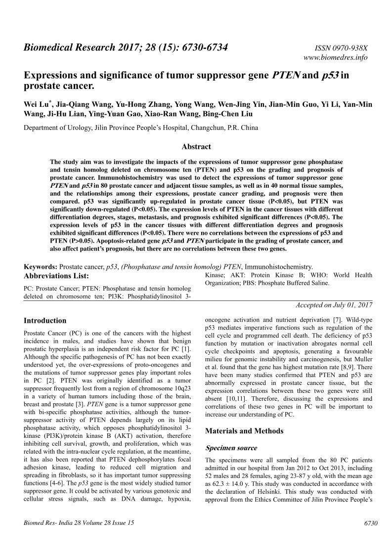
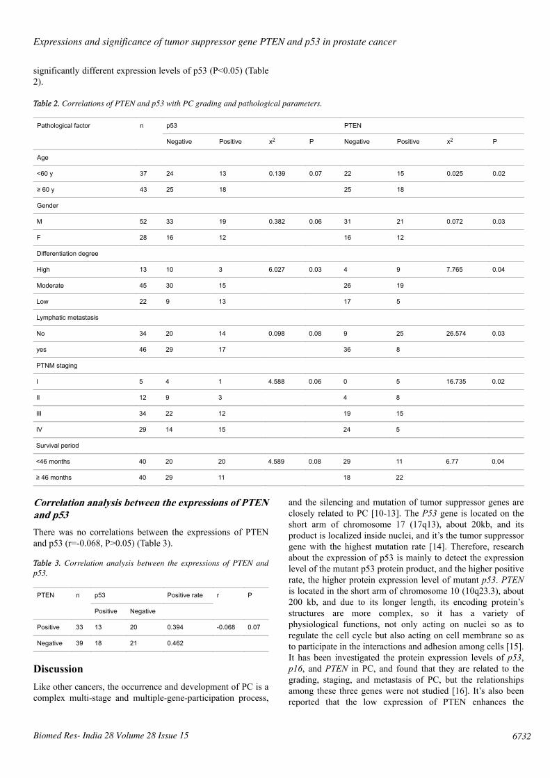
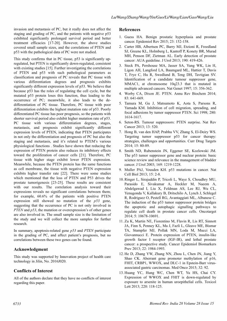
Immunohistochemical stainingThe Super PicTureTM Polymer two-stepimmunohistochemical staining was used. The PTEN and p53antibodies were the mouse anti-human monoclonal antibodiesprovided by Shanghai Sangon Biotech (Shanghai) Co., Ltd.The immunohistochemistry kit was provided by ShanghaiSangon Biotech (Shanghai) Co., Ltd. The control group usedPhosphate‑Buffered Saline (PBS) to replace the primaryantibody, and known positive PC tissue sections were used asthe positive PTEN and p53 controls; the brown particlesappearing in the cytoplasm and nucleus can be seen as thepositive PTEN expression (Figure 1), and those in the nucleican be seen as the positive p53 expression (Figure 2).
Figure 1. Positive immunohistochemical staining of PTEN (x200).
Follow-upThe follow-up used such ways as outpatient visit, telephone,email and mails, and the deadline was Sep 1st, 2015. Thesurvival time, time and location of recurrence and metastasisand death reasons of the patients were recorded. The medianfollow-up time was 46 months (11-50 months), no patientswere lost, and 68 patients are still alive currently.
Figure 2. Positive immunohistochemical staining of p53 (x200).
Statistical analysisSPSS20.0 software package was used for the data analysis andprocessing. The analysis among PTEN, p53, and PC causesused the x2 test, and the correlations between PTEN and p53were analyzed using the Spearman test, with P<0.05considered as statistically significant.
Results
Expressions of PTEN and p53 in PC tissueThere existed significant differences in the expressions ofPTEN and p53 among the above tissue samples (P<0.01),among which p53 was significantly up-regulated in PC tissue,but PTEN was significantly down-regulated (P<0.01) (Table1).
Table 1. Expressions of PTEN and p53 in PC tissue.
Group n p53 PTEN
Positive Negative Positive Negative
PC tissue 80 32 48 33 47
Adjacent tissue 80 15 65 68 12
Normal tissue 40 3 37 40 0
x2 19.566 52.624
P 0.004 0.003
Correlations of PTEN and p53 with PC grading andpathological parametersPC tissue with various differentiation degrees, stages,metastasis, and prognosis exhibited significantly differentexpression levels of PTEN (P<0.05), and PC tissue withvarious differentiation degrees and prognosis exhibited
Lu/Wang/Zhang/Wang/Yin/Guo/Li/Wang/Lian/Gao/Wang/Liu
Biomed Res- India 28 Volume 28 Issue 156731
significantly different expression levels of p53 (P<0.05) (Table2).
Table 2. Correlations of PTEN and p53 with PC grading and pathological parameters.
Pathological factor n p53 PTEN
Negative Positive x2 P Negative Positive x2 P
Age
<60 y 37 24 13 0.139 0.07 22 15 0.025 0.02
≥ 60 y 43 25 18 25 18
Gender
M 52 33 19 0.382 0.06 31 21 0.072 0.03
F 28 16 12 16 12
Differentiation degree
High 13 10 3 6.027 0.03 4 9 7.765 0.04
Moderate 45 30 15 26 19
Low 22 9 13 17 5
Lymphatic metastasis
No 34 20 14 0.098 0.08 9 25 26.574 0.03
yes 46 29 17 36 8
PTNM staging
I 5 4 1 4.588 0.06 0 5 16.735 0.02
II 12 9 3 4 8
III 34 22 12 19 15
IV 29 14 15 24 5
Survival period
<46 months 40 20 20 4.589 0.08 29 11 6.77 0.04
≥ 46 months 40 29 11 18 22
Correlation analysis between the expressions of PTENand p53There was no correlations between the expressions of PTENand p53 (r=-0.068, P>0.05) (Table 3).
Table 3. Correlation analysis between the expressions of PTEN andp53.
PTEN n p53 Positive rate r P
Positive Negative
Positive 33 13 20 0.394 -0.068 0.07
Negative 39 18 21 0.462
DiscussionLike other cancers, the occurrence and development of PC is acomplex multi-stage and multiple-gene-participation process,
and the silencing and mutation of tumor suppressor genes areclosely related to PC [10-13]. The P53 gene is located on theshort arm of chromosome 17 (17q13), about 20kb, and itsproduct is localized inside nuclei, and it’s the tumor suppressorgene with the highest mutation rate [14]. Therefore, researchabout the expression of p53 is mainly to detect the expressionlevel of the mutant p53 protein product, and the higher positiverate, the higher protein expression level of mutant p53. PTENis located in the short arm of chromosome 10 (10q23.3), about200 kb, and due to its longer length, its encoding protein’sstructures are more complex, so it has a variety ofphysiological functions, not only acting on nuclei so as toregulate the cell cycle but also acting on cell membrane so asto participate in the interactions and adhesion among cells [15].It has been investigated the protein expression levels of p53,p16, and PTEN in PC, and found that they are related to thegrading, staging, and metastasis of PC, but the relationshipsamong these three genes were not studied [16]. It’s also beenreported that the low expression of PTEN enhances the
Expressions and significance of tumor suppressor gene PTEN and p53 in prostate cancer
Biomed Res- India 28 Volume 28 Issue 15 6732
invasion and metastasis of PC, but it really does not affect thestaging and grading of PC, and the patients with negative p53exhibited significantly prolonged survival period and bettertreatment efficacies [17-20]. However, the above studiescovered small sample sizes, and the correlations of PTEN andp53 with the pathological data of PC were not studied.
This study confirms that in PC tissue, p53 is significantly up-regulated, but PTEN is significantly down-regulated, consistentwith existing studies [15]. Further investigating the correlationsof PTEN and p53 with such pathological parameters asclassification and prognosis of PC reveals that PC tissue withvarious differentiation degrees and prognosis exhibitssignificantly different expression levels of p53. We believe thatbecause p53 has the roles of regulating the cell cycle, but themutated p53 protein loses this function, thus leading to theoccurrence of PC; meanwhile, it also leads to the de-differentiation of PC tissue. Therefore, PC tissue with poordifferentiation exhibits the highest mutation rate of p53. Poorlydifferentiated PC tissue has poor prognosis, so the patients withshorter survival period also exhibit higher mutation rate of p53.PC tissue with various differentiation degrees, stages,metastasis, and prognosis exhibit significantly differentexpression levels of PTEN, indicating that PTEN participatesin not only the differentiation and prognosis of PC but also thestaging and metastasis, and it’s related to PTEM’s variousphysiological functions . Studies have shown that reducing theexpression of PTEN protein also reduces its inhibitory effectstoward the proliferation of cancer cells [21]. Therefore, PCtissue with higher stage exhibit lower PTEN expression.Meanwhile, because the PTEN protein has the same functionsas cell membrane, the tissue with negative PTEN expressionexhibits higher transfer rate [22]. There were some studieswhich mentioned that the loss of PTEN and P53 drives theprostate tumorigenesis [23-25]. These results are consistentwith our results. The correlation analysis toward theirexpressions reveals no significant correlations between them;for example, 60.6% of the patients with positive PTENexpression still showed no mutation of the p53 gene,suggesting that the occurrence of PC is not only involved inPTEN and p53, the mutation or overexpression’s of other genesare also involved in. The small sample size is the limitation ofthe study and we will collect the more samples for furtherresearch.
In summary, apoptosis-related gene p53 and PTEN participatein the grading of PC, and affect patient’s prognosis, but nocorrelations between these two genes can be found.
AcknowledgmentThis study was supported by Innovation project of health caretechnology in Jilin, No. 2016J020.
Conflicts of InterestAll of the authors declare that they have no conflicts of interestregarding this paper.
References1. Guess HA. Benign prostatic hyperplasia and prostate
cancer. Epidemiol Rev 2015; 23: 152-158.2. Carter HB, Albertsen PC, Barry MJ, Etzioni R, Freedland
SJ, Greene KL, Holmberg L, Kantoff P, Konety BR, MuradMH, Penson DF, Zietman AL. Early detection of prostatecancer: AUA guideline. J Urol 2013; 190: 419-426.
3. Steck PA, Pershouse MA, Jasser SA, Yung WK, Lin H,Ligon AH, Langford LA, Baumgard ML, Hattier T, DavisT, Frye C, Hu R, Swedlund B, Teng DH, Tavtigian SV.Identification of a candidate tumour suppressor gene,MMAC1, at chromosome 10q23.3 that is mutated inmultiple advanced cancers. Nat Genet 1997; 15: 356-362.
4. Worby CA, Dixon JE. PTEN. Annu Rev Biochem 2014;83: 641-669.
5. Tamura M, Gu J, Matsumoto K, Aota S, Parsons R,Yamada KM. Inhibition of cell migration, spreading, andfocal adhesions by tumor suppressor PTEN. Sci 1998; 280:1614-1617.
6. Seton-RS. Tumour suppressors: PTEN surprise. Nat RevCancer 2013; 13: 520.
7. Hong B, van den HAP, Prabhu VV, Zhang S, El-Deiry WS.Targeting tumor suppressor p53 for cancer therapy:strategies, challenges and opportunities. Curr Drug Targets2014; 15: 80-89.
8. Smith ND, Rubenstein JN, Eggener SE, Kozlowski JM.The p53 tumor suppressor gene and nuclear protein: basicscience review and relevance in the management of bladdercancer. J Urol 2003; 169: 1219-1228.
9. Muller PAJ, Vousden KH. p53 mutations in cancer. NatCell Biol 2013; 15: 2-8.
10. Ringer L, Sirajuddin P, Tricoli L, Waye S, Choudhry MU,Parasido E, Sivakumar A, Heckler M, Naeem A,Abdelgawad I, Liu X, Feldman AS, Lee RJ, Wu CL,Yenugonda V, Kallakury B, Dritschilo A, Lynch J, SchlegelR, Rodriguez O, Pestell RG, Avantaggiati ML, Albanese C.The induction of the p53 tumor suppressor protein bridgesthe apoptotic and autophagic signalling pathways toregulate cell death in prostate cancer cells. Oncotarget2014; 5: 10678-10691.
11. Zu K, Martin NE, Fiorentino M, Flavin R, Lis RT, SinnottJA, Finn S, Penney KL, Ma J, Fazli L, Gleave ME, BismarTA, Stampfer MJ, Pollak MN, Loda M, Mucci LA,Giovannucci E. Protein expression of PTEN, insulin-likegrowth factor I receptor (IGF-IR), and lethal prostatecancer: a prospective study. Cancer Epidemiol BiomarkersPrev 2013; 22: 1984-1993.
12. He D, Zhang YW, Zhang NN, Zhou L, Chen JN, Jiang Y,Shao CK. Aberrant gene promoter methylation of p16,FHIT, CRBP1, WWOX, and DLC-1 in Epstein-Barr virus-associated gastric carcinomas. Med Onco 2015; 32: 92.
13. Huang YC, Hung WC, Chen WT, Yu HS, Chai CY.Expression of WWOX and FHIT is down-regulated byexposure to arsenite in human uroepithelial cells. ToxicolLett 2013; 220: 118-125.
Lu/Wang/Zhang/Wang/Yin/Guo/Li/Wang/Lian/Gao/Wang/Liu
Biomed Res- India 28 Volume 28 Issue 156733

14. Svane IM, Pedersen AE, Nikolajsen K, Zocca MB.Alterations in p53-specific T cells and other lymphocytesubsets in breast cancer patients during vaccination withp53-peptide loaded dendritic cells and low-doseinterleukin-2. Vaccine 2008; 26: 4716-4724.
15. Nakahata S, Ichikawa T, Maneesaay P, Saito Y, Nagai K,Tamura T, Manachai N, Yamakawa N, Hamasaki M,Kitabayashi I, Arai Y, Kanai Y, Taki T, Abe T, Kiyonari H,Shimoda K, Ohshima K, Horii A, Shima H, Taniwaki M,Yamaguchi R, Morishita K. Loss of NDRG2 expressionactivates PI3K-AKT signalling via PTEN phosphorylationin ATLL and other cancers. Nat Commun 2014; 5: 3393.
16. Nakamura T, Ide H, Eguchi R, Hayashi K, Takasaki K.Concomitant analysis of p16/INK4, cyclin D1, andretinoblastoma protein expression in esophageal squamouscell carcinoma. Hepatogastroenterol 2003; 50: 1321-1326.
17. Serban AI, Stanca L, Geicu OI, Munteanu MC, CostacheM, Dinischiotu A. Extracellular matrix is modulated inadvanced glycation end products milieu via a RAGEreceptor dependent pathway boosted by transforminggrowth factor-β1 RAGE. J Diabetes 2015; 7: 114-124.
18. Yue MM, Lv K, Meredith SC, Martindale JL, Gorospe M,Schuger L. Novel RNA-binding protein P311 bindseukaryotic translation initiation factor 3 subunit b (eIF3b)to promote translation of transforming growth factor β1-3(TGF-β1-3). J Biol Chem 2014; 289: 33971-33983.
19. Suh SO, Chen Y, Zaman MS, Hirata H, Yamamura S,Shahryari V, Liu J, Tabatabai ZL, Kakar S, Deng G, TanakaY, Dahiya R. MicroRNA-145 is regulated by DNAmethylation and p53 gene mutation in prostate cancer.Carcinogenesis 2011; 32: 772.
20. Nesslinger NJ, Shi XB, de Vere White RW. Androgen-independent growth of LNCaP prostate cancer cells ismediated by gain-of-function mutant p53. Cancer Res2003; 63: 2228-2233.
21. Papa A, Wan L, Bonora M, Salmena L, Song MS, HobbsRM, Lunardi A, Webster K, Ng C, Newton RH, KnoblauchN, Guarnerio J, Ito K, Turka LA, Beck AH, Pinton P,Bronson RT, Wei W, Pandolfi PP. Cancer-associated PTENmutants act in a dominant-negative manner to suppressPTEN protein function. Cell 2014; 157: 595-610.
22. Yasui M, Matsuoka S, Ueda M. PTEN hopping on the cellmembrane is regulated via a positively-charged C2 domain.PLoS Comput Biol 2014; 10: 1003817.
23. Wang L, Xiong H, Wu F, Zhang Y, Wang J, Zhao L, Guo X,Chang LJ, Zhang Y, You MJ, Koochekpour S, Saleem M,Huang H, Lu J, Deng Y. Hexokinase 2-mediated Warburgeffect is required for PTEN and p53-deficiency drivenprostate cancer growth. Cell Rep 2014; 8: 1461.
24. Kim J, Roh M, Doubinskaia I, Algarroba GN, Eltoum IE,Abdulkadir SA. A mouse model of heterogeneous, c-myc-initiated prostate cancer with loss of PTEN and p53.Oncogene 2012; 31: 322-332.
25. Uemura H, Kura Y, Ando N, Fukushima E, Hatanaka Y,Yamamoto Y, Shimizu N, Yoshimura K, Nozawa M,Yoshikawa K, Nishio K, Nishio K, De Velasco MA.Abstract 84: functional evaluation of synchronousinactivation of PTEN and p53 in a murine model ofprostate cancer. Cancer Res 2014; 74: 84-84.
*Correspondence toWei Lu
Department of Urology
Jilin Province People’s Hospital
Changchun
P.R. China
Expressions and significance of tumor suppressor gene PTEN and p53 in prostate cancer
Biomed Res- India 28 Volume 28 Issue 15 6734