extracellular matrix modulation is driven by experience ... · by experience-dependent plasticity...
TRANSCRIPT

Extracellular Matrix Modulation Is Drivenby Experience-Dependent Plasticity During StrokeRecovery
Miriana Jlenia Quattromani1 & Mathilde Pruvost2 & Carla Guerreiro1 &
Fredrik Backlund1& Elisabet Englund3
& Anders Aspberg4 & Tomasz Jaworski5 &
Jakob Hakon1& Karsten Ruscher1 & Leszek Kaczmarek5
& Denis Vivien2,6&
Tadeusz Wieloch1
Received: 17 December 2016 /Accepted: 16 February 2017# The Author(s) 2017. This article is published with open access at Springerlink.com
Abstract Following stroke, complete cellular death in theischemic brain area may ensue, with remaining brain areasundergoing tissue remodelling to various degrees.Experience-dependent brain plasticity exerted through anenriched environment (EE) promotes remodelling after centralnervous system injury, such as stroke. Post-stroke tissue reor-ganization is modulated by growth inhibitory molecules dif-ferentially expressed within the ischemic hemisphere, likechondroitin sulfate proteoglycans found in perineuronal nets(PNNs). PNNs in the neocortex predominantly enwrapparvalbumin-containing GABAergic (PV/GABA) neurons,important in sensori-information processing. Here, we
investigate how extracellular matrix (ECM) proteases andtheir inhibitors may participate in the regulation of PNN in-tegrity during stroke recovery. Rats were subjected tophotothrombotic stroke in the motor cortex, and functionaldeficits were assessed at 7 days of recovery. Sham and strokedrats were housed in either standard or EE conditions for 5 days,and infarct volumes were calculated. PNNswere visualized byimmunohistochemistry and counted in the somatosensory cor-tex of both hemispheres. mRNA expression levels of ECMproteases and protease inhibitors were assessed by RT-qPCRand their activity analyzed by gel zymography. PNNs andprotease activity were also studied in brains from stroke pa-tients where similar results were observed. EE starting 2 daysafter stroke and continuing for 5 days stimulated behavioralrecovery of limb-placement ability without affecting infarctsize. EE promoted a decrease of PNNs around PV/GABAneurons and a concomitant modulation of the proteolytic ac-tivity and mRNA expression of ECM proteases and proteaseinhibitors in the somatosensory cortex. This study providesmolecular targets for novel therapies that could support reha-bilitation of stroke patients.
Keywords Stroke recovery . Experience-dependentplasticity . Extracellular matrix . Perineuronal nets .
Somatosensory cortex . Proteases
Introduction
According to the World Health Organization, approximately15 million people suffer stroke worldwide each year. Of these,five million die and another five million are permanently dis-abled, making stroke a global leading cause of death and long-
Electronic supplementary material The online version of this article(doi:10.1007/s12035-017-0461-2) contains supplementary material,which is available to authorized users.
* Miriana Jlenia [email protected]
1 Laboratory for Experimental Brain Research, Division ofNeurosurgery, Department of Clinical Sciences, Lund University,BMC A13, 22184 Lund, Sweden
2 INSERM UMR-S U919, Serine Proteases and Pathophysiology ofthe Neurovascular Unit, Université Caen Basse Normandie, GIPCyceron, F-14074 Caen, France
3 Division of Oncology and Pathology, Lund University Hospital,22185 Lund, Sweden
4 Rheumatology and Molecular Skeletal Biology, Department ofClinical Sciences, Lund University, BMCC12, 22184 Lund, Sweden
5 Laboratory of Neurobiology, Nencki Institute of ExperimentalBiology, 02-093 Warsaw, Poland
6 Department of Clinical Research, Caen University Hospital, CHUCaen, 14000 Caen, France
DOI 10.1007/s12035-017-0461-2Mol Neurobiol (2018) 55:2196–2213
/Published online: 13 March 2017

term disability [1, 2]. Stroke treatment options are restricted tothe few hours after the accident [3, 4], and beyond that thera-peutic window, treatment is limited to supportive care andrehabilitation. Yet, most stroke patients recover to some ex-tent, demonstrating that remarkable brain plasticity is retainedafter the injury and that the recovery process should emerge asa promising target in stroke research [5].
The current experimental evidence strongly suggests thatmultisensory stimulation by changes in the environment (suchas enriched environment (EE)) as well as task-driven experi-ence confer an additive benefit on behavioral recovery afterbrain insults such as stroke, most likely because of their im-pact on brain plasticity [6, 7]. This promising approach hasbeen recently translated into the clinical practice [8–10].
EE provides laboratory animals with novelty and extraspace, allowing different forms of multisensory stimulationranging from social grouping to enhanced motor activity. EEresults in improved cognitive and sensorimotor functions bothin naive rodents and in animals with brain lesions such asthose occurring after a stroke. In animals which have suffereda stroke, robust behavioral effects are seen during recoveryand are probably related not only to neuronal plasticity inthe peri-infarct cortex but also in remote brain areas [7,11–13].
After stroke, a number of growth inhibitory moleculesare differentially expressed within the peri-infarct region,leading to inhibition of neuronal plasticity [14, 15].Interestingly, in areas more distant from the infarct, thereis a reduction in the number of these inhibitory molecules,particularly in chondroitin sulfate proteoglycans (CSPGs)and their formation into dense lattice-like structures termedperineuronal nets (PNNs) [15–18]. PNNs are considered aspecialized form of extracellular matrix (ECM) with a ma-jor role in establishing and stabilizing connectivity in thebrain by restricting plasticity. They are typically localizedaround inhibitory neurons [19], but they have been alsodescribed around excitatory pyramidal neurons [20–22].CSPGs present in PNNs are lecticans and includeaggrecan, brevican, neurocan, and versican, with aggrecanexclusively present in PNNs and therefore being a specificmarker for PNNs in the adult brain [23]. We have recentlyshown that 9 weeks of EE after stroke induce a furtherreduction in the number of these inhibitory molecules, es-pecially Cat-315+ (aggrecan-containing) PNNs [12],supporting the idea that part of the peri-infarct and remotecortex possesses an environment for post-stroke responsestrategies [24]. Several proteases are involved in the re-modelling of ECM and PNNs, including tissue plasmino-gen activator (tPA) [25], type 4 disintegrin and metallopro-teinase with thrombospondin motifs (ADAMTS-4) [25],and matrix metalloproteinases (MMPs) such as MMP-2and MMP-9 [26–28]. tPA is a serine protease present inthe blood, in the brain and at the interface between the
blood and the brain [29]. In the brain parenchyma, tPA isexpressed and released by endothelial cells, neurons, astro-cytes, microglia, and oligodendrocytes. tPA contributes toECM degradation, influence cell migration, neuronal plas-ticity, death and survival of neurons, endothelial cells, andoligodendrocytes [29]. ADAMTS-4 has been described inthe brain parenchyma, mainly in astrocytes, neurons, mi-croglia, and monocyte/macrophages and is involved inseveral central nervous system (CNS) functions, includingneurorepair, remyelination, angiogenesis, and inflamma-tion [30]. Cleavage by tPA produces the active form ofADAMTS-4, which then degrades inhibitory CSPGs, lead-ing to increased neurite growth and subsequent functionalrecovery [25]. The MMP family of proteases act outsidethe cells and are therefore associated with ECM remodel-ling. They are expressed in neurons, glia, and endothelialcells and are involved in a number of physiological andpathological conditions, including development, tissue re-modelling, inflammation, and metastasis [31]. Current datasuggest that MMPs (such as MMP-2 and MMP-9) mediatebeneficial dendritic plasticity and ECM remodeling at de-layed stages after stroke [28]. Multiple studies showed in-creased expression and activity of MMP-2 and MMP-9after brain injury (such as stroke) during the recoveryphase [28, 31]. In particular, MMP-9 has been implicatedin synaptic plasticity, learning and memory, and tissue re-modelling in response to enhanced neuronal activity [32,33]. Inhibitors of these proteases (such as neuroserpin, tis-sue inhibitor of metalloproteinases TIMP-1 and TIMP-3)are also considered strategic in promoting neuronal remod-elling and neuroplasticity in the CNS [29, 34].
The aim of this study was to further investigate the influ-ence of multisensory brain stimulation provided by EE afterstroke on PNN integrity and on changes in expression/activityof MMP-2 and MMP-9, tPA, ADAMTS-4, and theirinhibitors.
Materials and Methods
Rats
A total of 40 adult male Sprague-Dawley rats (8–11 weeks,Charles River) were used in this study and housed under re-verse light conditions with free access to food and water.Physiological data are given in Supplement Table 1.
Human Postmortem Brain Tissue
A total of ten stroke (n = 5) and non-stroke (n = 5) cases wereused in this study. Age, sex, cause of death, and postmortemdelay are given in Supplement Table 2.
Mol Neurobiol (2018) 55:2196–2213 2197

Experimental Stroke
Animals underwent permanent focal ischemia byphotothrombosis (PT) in the left hemisphere as previouslydescribed [12, 35] or sham operation (same procedure as PT,but without illumination). During surgery, the body tempera-ture of the animals was kept at 37 °C using a self-regulatingheating pad. Briefly, isoflurane-anesthetized rats (2% in O2
under spontaneous ventilation) received intravenous injec-tions of the photosensitive dye Rose Bengal (10 mg/ml,Sigma, USA) in the tail vein. The skin above the skull wasincised, and the brain was illuminated through the exposedskull with cold light (KL 1500 LCD, Schott) for 15 min at astereotactically defined position (0.5 mm laterally and +4/−4 mm anterior/posterior to bregma), producing an approxi-mate irradiation area of 8 × 4 mm2. Incisions were sutured,and animals were allowed to awake from anesthesia while ona heating pad and returned to their home cages.
Behavioral Assessment and Randomization
Paw Placement Test Sensorimotor function and touch sen-sation were assessed by the paw placement test, whichprovides information on the tactile/proprioceptive re-sponse to limb stimulation [12, 36]. Testing at all timeswas performed by the same investigator. In brief, every ratwas placed with all paws on a bench surface and hand-held in a horizontal position. Facial contact stimuli wereavoided by supporting the rat’s chin and holding its head45° upward. The paws to be tested were gently pushedalong the edge in order to loose contact with the benchsurface. The ability of the rat to place the limb back onthe bench surface when the animal was moved along itsedge was evaluated with the following score: (1) promptplacement of the limb on the table, (0.5) incomplete
placing of the limb, (0) no response, with persistent ex-tension of the limb and paw.
One day before PT or sham operations, all animals had ascore = 1 in each of the four paws, indicating that no deficit insensorimotor function was present before the surgeries.
Differential Housing Conditions Two days after surgeries,both PT and sham animals were assigned to either group Ior II (n = 20 each) and randomized into (standard) STD orEE cages [12] where they were housed for 5 days. Ratshoused in STD conditions had one cage mate, and rats inenriched cages had up to five mates. Multilevel EE cageswere equipped with various colored objects such as plas-tic tunnels, removable platforms, grids, pipes, ropes, lad-ders, and chains. The disposition of the objects waschanged twice/week.
The animals were divided as described in Fig. 1. Brainsfrom groups I and II were collected and handled accord-ingly to postmortem analyses. Brain samples from group Iwere used for immunohistochemical analyses (infarctmeasurements and cell counting). Animals from group IIwere used for behavioral assessments on day 7, and aftersacrifice, the brains from this group were used for molec-ular biology experiments (gel-zymography, western blot,RT-qPCR).
Brain Preparation from Rats
Group I Rats were anesthetized with 4% isoflurane andtranscardially perfused with 0.9% NaCl followed by coldphosphate-buffered 4% paraformaldehyde (PFA, Sigma,USA), pH 7.4. Brains were postfixed overnight in PFA andequilibrated in phosphate-buffered 30% sucrose for 48 h at4 °C. Brains were sectioned in the coronal plane on a micro-tome at 30-μm intervals. Sections were collected and stored at
Fig. 1 Experimental design. Rats were pre-tested to assess their full limbplacement ability 1 day before photothrombosis (PT) or sham operations.Functional deficits were assessed 2 days after PTand rats were selectivelysorted into differential housing conditions (standard, STD; enriched, EE).
Differential housing was continued for the five following days. At 7 daysof recovery, brains were perfusion-fixed (group I) or frozen (group II) forfurther analyses
Mol Neurobiol (2018) 55:2196–22132198

−20 °C in an antifreeze solution made in phosphate buffercontaining 30% glycerol and 30% ethylene glycol.
Group II Rats were anesthetized with 4% isoflurane and per-fused with cold heparinized 0.9%NaCl. Brains were extractedand immediately frozen by immersion in isopentane and dryice. Brains were then stored at −80 °C until dissection. On theday of the dissection, frozen brains were moved to a −20 °Cchamber and placed in a rat brain matrix. Two brain slices 2-mm-thick (2.20 to −3.30 mm from bregma) were excisedusing a scalpel blade. The primary somatosensory cortex onboth ipsilateral and contralateral hemispheres was cut out ac-cording to Paxinos and Watson [37]. In the ipsilateral hemi-sphere, the sampling started 1 mm after the end of the lesion.Samples were transferred to tubes and stored at −80 °C untilfurther use.
Brain Preparation from Patients
Following autopsy, cortical fresh tissue from the infarct core,peri-infarct, and remote regions were dissected out by a pa-thologist, immediately frozen and stored at −80 °C. For furtherdissection, samples were temporarily moved to a −20 °Cchamber where smaller specimens were excised and storedat −80 °C until protein extraction.
Immunohistochemistry and Immunofluorescence(IHC/IF)
Rat Brain Tissue Free-floating brain slices from group I wererinsed in phosphate-buffered solution (PBS), quenched in 3%H2O2 and 10% methanol for 15 min (IHC) and blocked inblocking solution (5% normal donkey serum, JacksonImmunoResearch, UK, and 0.25% Triton X-100 in PBS) for1 h at room temperature (RT). Sections were incubated over-night at 4 °C with primary antibodies (monoclonal mouseanti-CSPGs, 1:3000 for IHC and 1:1000 for IF, Cat-315/MAB1581, Millipore, USA; goat anti-parvalbumin (PV),1:2500 for IF, PVG-214, Swant, Switzerland) diluted inblocking solution. Following rinses with 2% normal donkeyserum and 0.25% Triton X-100 in PBS, the sections wereincubated with appropriate secondary antibodies (donkeyanti-mouse for IHC, Vector Laboratories, USA; donkey anti-mouse/goat conjugated with either Cy3 or DyLight 488 fluo-rescent dyes for IF, Jackson ImmunoResearch, UK) at a dilu-tion of 1:400 in blocking solution for 90 min at RT. IHCvisualization was achieved via the Vectastain ABC kit(Vector) using 3,3′-diaminobenzidine-tetrahydrochloride-dihydrate (DabSafe, Saveen Werner, Sweden), 8% NiCl, and3% H2O2. Bright-field pictures were acquired using anOlympus BX60 microscope (Sweden). Fluorescent labellingwas imaged using a confocal laser-scanning microscope(Zeiss LSM 510, Germany).
Human Brain Tissue Five-micron-thick formalin-fixed par-affin-embedded slices were evaluated by a pathologist beforeserial sections were stained. The presence of infarct and infarctage was confirmed by microscopy of the tissue in the imme-diate proximity to the areas sampled and analyzed. Brainslices were deparaffinized, immersed in lectin buffer (LB:0.1 M Tris-buffer pH 7.6, 1.45 M NaCl, 0.01 M MgCl2,0.01 M CaCl2) for 1 h and quenched in 1% H2O2 and 90%methanol for 20 min. Following rinses in LB, antigen retrievalwas performed by microwave boiling in 0.01 M citrate bufferpH 6.0 for 15min and slices were left in the solution until theyreached RT. After rinsing in LB, sections were blocked in 1%bovine serum albumin (BSA, Sigma, USA) for 1 h and incu-bated with biotinylatedWisteria floribunda agglutinin (WFA,1:200, L1516, Sigma, USA) for 40 min at 37 °C in LB. Afterrinsing in LB, ABC and DAB steps followed the same proto-col used for rat sections. Bright-field pictures were acquiredusing an Olympus BX60 microscope (Sweden).
Infarct Volume Measurements
For each animal in group I, ten coronal 30-μm-thick brainsections with a distance of 1.0 mm were immunostained witha monoclonal mouse anti-NeuN antibody (MAB377,Millipore, USA) at a dilution of 1:1500. The sampling rangedfrom 4.20 to −3.80 mm from bregma and covered the entirerostro-caudal extension of the lesion. The non-lesioned area ofthe infarcted hemisphere and the non-lesioned contralateralhemisphere were outlined on each brain section using theImageJ software (National Institute of Health, USA). Infarctvolumes were determined by subtracting the area of the non-lesioned ipsilateral hemisphere from that of the intact contra-lateral hemisphere and calculated by volumetric integrationfor each animal [38].
Cell Counting
Bright Field Microscopy In group I, three coronal sectionsper brain (2.20, 0.48, and −3.30 mm relative to bregma) werestained for Cat-315 as described above. Composite micro-graphs of the whole ipsilateral and contralateral hemisphereswere acquired through a ×4 magnification objective using theCellSens Dimension Software (Olympus BX60, Sweden). Anoptical grid was used to define distances and draw the bound-aries of the primary somatosensory cortex in both hemispheresaccording to the Paxinos atlas [37]. The sizes of the areas ofinterest were 6.55 mm2 at 2.20 mm from bregma, 10.90 mm2
at 0.48 mm from bregma, and 8.34 mm2 at −3.30 mm frombregma.
Confocal Microscopy One coronal section per brain(0.48 mm relative to bregma) was double-stained for Cat-315 and parvalbumin as described above. Three areas of
Mol Neurobiol (2018) 55:2196–2213 2199

interest within the somatosensory cortex (layers II–III) wereacquired using a confocal laser-scanning microscope ×20 ob-jective (Zeiss LSM 510, Germany). For analysis, the ImageJsoftware was used to discriminate between the differentfluorophores. Cat-315+ cells, parvalbumin+ cells, and cellspositive for both antibodies were counted. Data are presentedas an average of the three regions of interest (ROIs).
Protein Extraction and Quantification
For group II and human tissue, regions of interest were disso-ciated in TNT buffer, pH 7.4 (50 mM Trizma base, 150 mMNaCl, 0.5% Triton X-100) and centrifuged at 12,000g for15 min at 4 °C. Supernatants were collected and protein de-termination performed with the Pierce BCA protein Assay Kitaccording to the manufacturer (ThermoFisher Scientific,USA). Twenty-microgram aliquots were prepared and storedat −80 °C until further use.
Gel-Zymography Assays
MMP-2 and MMP-9 Zymography Equal amounts of pro-teins (20 μg) were mixed with 5× sample buffer and separatedat 4 °C under non-reducing conditions on 8% SDS-PAGE gelscopolymerized with FITC-labeled gelatin (FITC 200 μg/ml,Sigma, USA; gelatin 20 mg/ml, Sigma, USA) as in [39]. Gelswere washed in 2.5% Triton X-100 (2 × 30 min, RT) to re-move SDS, and then incubated in activation buffer (50 mMTris-HCl pH 7.5, 10 mM CaCl2, 1 μM ZnCl2, 1% TritonX-100, 0.02% NaN3) for 48 h at 37 °C to allow in-gel MMPrenaturation [40]. Zymograms were digitized with aChemiDoc XRS+ system (Bio-Rad, USA) under UV lightand proteinase activity quantified by densitometry (ImageJ,USA).
tPA Zymography Equal amounts of proteins (20 μg) weremixed with 5× sample buffer and separated at 4 °C under non-reducing conditions on 12% SDS-PAGE gels copolymerizedwith casein (10 mg/ml, Sigma, USA) and plasminogen(2.5 μg/ml, Calbiochem, USA). Gels were washed at 4 °Cwith cold 2.5% Triton X-100 (2 × 30 min) and cold distillatedwater (3 × 10 min), and then incubated in activation buffer(100 mM glycine pH 8.3, 10 mM EDTA) for 5.5 h at 37 °C toallow caseinolysis. Gels were stained with Coomassie BlueR-250 and destained with 40%methanol and 10% acetic acid.Zymograms were digitized with a CanoScan 8800F (Canon,USA) and proteinase activity quantified by densitometry(ImageJ, USA).
Western Blot
β-Dystroglycan (β-DG) Equal amounts of proteins (20 μg)were mixed with 5× sample buffer and separated at RT under
reducing conditions on 10% SDS-PAGE gels. Proteins weretransferred onto polyvinylidene difluoride (PVDF) mem-branes (Millipore, USA) for 1 h at RT and blocked with asolution containing 5% non-fat dry milk for 1 h at RT.Membranes were incubated overnight at 4 °C with primaryantibody monoclonal mouse anti β-DG (1:500, ab49515,Abcam, UK) in 5% BSA. On the next day, membranes wereincubated with a horseradish peroxidase (HRP)-conjugatedanti-mouse IgG (1:10,000, A3862, Sigma, USA) for 1 h atRT. Protein bands were visualized by exposing the mem-branes to a CCD camera (LAS1000, Fujifilm, USA) using achemiluminescent HRP substrate kit (Millipore, USA).Membranes were stripped and re-probed with an anti-β-actinHRP-conjugate (1:75,000, A3854, Sigma, USA). After densi-tometric analysis using the ImageJ software (National Instituteof Health, USA), primary antibody levels were normalized toβ-actin expression.
Aggrecan Protein samples (20 μg) were treated with diges-tion buffer containing 100 mM Tris-HCl pH 8.0, 30 mMNaAc, 5 mM EDTA and protease inhibitors (CompleteEDTA-free, Roche, Germany) with (+) or without (−)chondroitinase ABC (ChABC) (0.5 U/ml, Sigma, USA) at37 °C for 3 h to remove chondroitin sulfate chains from theaggrecan proteoglycan. ChABC treatment induces a gel shiftof the protein targets for the Cat-315 antibody, which we usedto detect aggrecan. The shift diminishes the smear of immu-noreactive aggrecan peptides, leaving a marked protein bandat >250 kDa and weaker bands at lower molecular weights.
Proteins were then denatured and processed as above.Western blots for the Cat-315 antibody (1:5000, MAB1581,Millipore, USA) were ran on 4–15% gradient SDS-PAGEgels.
RNA Extraction and Quantification
In group II, total RNAwas isolated from our regions of inter-ests with TRI reagent (Sigma, USA) according to the manu-facturer. Total RNA was treated with TURBO DNase(Ambion, France) to avoid DNA contamination and wasquantified by spectrophotometry (NanoDrop Technologies,USA). First-strand cDNA synthesis was performed from1 μg of total RNA with the M-MLV Reverse Transcriptase(Invitrogen, France) in a total volume of 20 μL with the fol-lowing cycle conditions: 37 °C (50 min); 70 °C (15 min). RT-qPCR was performed from 1 μL of 1:20 diluted cDNA in15 μL total of a 1× solution of iQ SYBR Green Supermix(Bio-Rad, France) containing 200 nM of each primer. Basedon mRNA coding sequences (www.ensembl.org), rat-specificprimers (see Table 1) were designed by using the Primer3Plussoftware (http://www.bioinformatics.nl/cgi-bin/primer3plus/primer3plus.cgi). Assays for the 40 samples were run intriplicate on the CFX96 real-time system c1000 thermal cycler
Mol Neurobiol (2018) 55:2196–22132200
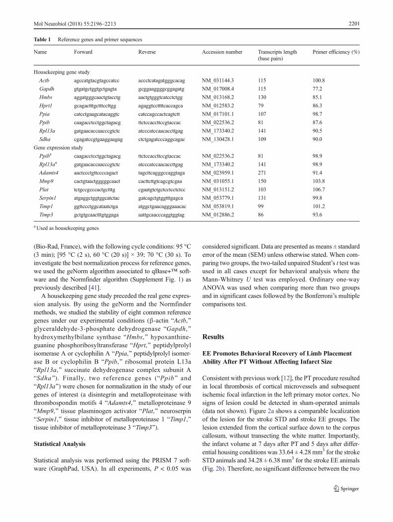
(Bio-Rad, France), with the following cycle conditions: 95 °C(3 min); [95 °C (2 s), 60 °C (20 s)] × 39; 70 °C (30 s). Toinvestigate the best normalization process for reference genes,we used the geNorm algorithm associated to qBase+™ soft-ware and the Normfinder algorithm (Supplement Fig. 1) aspreviously described [41].
A housekeeping gene study preceded the real gene expres-sion analysis. By using the geNorm and the Normfindermethods, we studied the stability of eight common referencegenes under our experimental conditions (β-actin BActb,^glyceraldehyde-3-phosphate dehydrogenase BGapdh,^hydroxymethylbilane synthase BHmbs,^ hypoxanthine-guanine phosphoribosyltransferase BHprt,^ peptidylprolylisomerase A or cyclophilin A BPpia,^ peptidylprolyl isomer-ase B or cyclophilin B BPpib,^ ribosomal protein L13aBRpl13a,^ succinate dehydrogenase complex subunit ABSdha^). Finally, two reference genes (BPpib^ andBRpl13a^) were chosen for normalization in the study of ourgenes of interest (a disintegrin and metalloproteinase withthrombospondin motifs 4 BAdamts4,^ metalloproteinase 9BMmp9,^ tissue plasminogen activator BPlat,^ neuroserpinBSerpin1,^ tissue inhibitor of metalloproteinase 1 BTimp1,^tissue inhibitor of metalloproteinase 3 BTimp3^).
Statistical Analysis
Statistical analysis was performed using the PRISM 7 soft-ware (GraphPad, USA). In all experiments, P < 0.05 was
considered significant. Data are presented as means ± standarderror of the mean (SEM) unless otherwise stated. When com-paring two groups, the two-tailed unpaired Student’s t test wasused in all cases except for behavioral analysis where theMann-Whitney U test was employed. Ordinary one-wayANOVA was used when comparing more than two groupsand in significant cases followed by the Bonferroni’s multiplecomparisons test.
Results
EE Promotes Behavioral Recovery of Limb PlacementAbility After PT Without Affecting Infarct Size
Consistent with previous work [12], the PT procedure resultedin local thrombosis of cortical microvessels and subsequentischemic focal infarction in the left primary motor cortex. Nosigns of lesion could be detected in sham-operated animals(data not shown). Figure 2a shows a comparable localizationof the lesion for the stroke STD and stroke EE groups. Thelesion extended from the cortical surface down to the corpuscallosum, without transecting the white matter. Importantly,the infarct volume at 7 days after PT and 5 days after differ-ential housing conditions was 33.64 ± 4.28 mm3 for the strokeSTD animals and 34.28 ± 6.38 mm3 for the stroke EE animals(Fig. 2b). Therefore, no significant difference between the two
Table 1 Reference genes and primer sequences
Name Forward Reverse Accession number Transcripts length(base pairs)
Primer efficiency (%)
Housekeeping gene study
Actb agccatgtacgtagccatcc accctcatagatgggcacag NM_031144.3 115 100.8
Gapdh gtgatgctggtgctgagta gcggaaggggcggagatg NM_017008.4 115 77.2
Hmbs aggatgggcaactgtacctg aactgtgggtcatcctctgg NM_013168.2 130 85.1
Hprt1 gcagactttgctttccttgg agaggtccttttcaccagca NM_012583.2 79 86.3
Ppia catcctgaagcatacaggtc catccagccactcagtctt NM_017101.1 107 98.7
Ppib caagacctcctggctagacg ttctccaccttccgtaccac NM_022536.2 81 87.6
Rpl13a gatgaacaccaacccgtctc atcccatccaacaccttgag NM_173340.2 141 90.5
Sdha cgagatccgtgaaggaagag ctctgagatcccaggcagac NM_130428.1 109 90.0
Gene expression study
Ppiba caagacctcctggctagacg ttctccaccttccgtaccac NM_022536.2 81 98.9
Rpl13aa gatgaacaccaacccgtctc atcccatccaacaccttgag NM_173340.2 141 98.9
Adamts4 aactccctgttccccagact tagcttcagggccaggtaga NM_023959.1 271 91.4
Mmp9 cactgtaactgggggcaact cacttcttgtcagcgtcgaa NM_031055.1 150 103.8
Plat tctgccgcccactgctttg cgaatgtctgctcctcctctcc NM_013151.2 103 106.7
Serpin1 atgaggctggtggcatctac gatcagctgtggtttgagca NM_053779.1 131 99.8
Timp1 ggttccctggcataatctga atggctgaacagggaaacac NM_053819.1 99 101.2
Timp3 gctgtgcaactttgtggaga aattgcaacccaggtggtag NM_012886.2 86 93.6
a Used as housekeeping genes
Mol Neurobiol (2018) 55:2196–2213 2201

groups was present, consistent with other work in which EEwas initiated at 2 days after PT [12, 13].
On day 2, after stroke and prior to differential housingconditions, all animals subjected to sham operation showedno deficit in the paw placement test (score = 1 for both paws;data not shown). On the contrary, all animals subjected to PTexhibited a clear deficit in both forepaw and hindpaw function(score = 0 in the paws contralateral to the side of the lesion;data not shown). On day 7 of recovery, despite equivalentinfarct volumes, we observed a significant improvement inlimb placement ability (contralateral to the side of the lesion)in the stroke EE group compared with the stroke STD group(Fig. 2c–e). Sham animals did not show any deficit, and theirscores are not included in the graphs. Animals housed for5 days in EE conditions after PT exhibited a significant recov-ery of forepaw function (P < 0.01, Fig. 2c), with 66% of theanimals showing complete recovery (score = 1, four rats out ofsix) and 33% showing improved function (score = 0.5, tworats out of six). Also, 50% of the stroke EE animals exhibit animproved hindpaw sensorimotor function (score = 0.5 in threerats out of six, Fig. 2d). Figure 2e shows the sum of scores forboth paws, with a score = 2 indicating full recovery of thepaws contralateral to the lesion (forepaw = 1 + hindpaw = 1).The entire stroke EE group developed some improved
function 7 days after stroke, with one third of the rats scoring1.5 points, half of them scoring 1 point and one quarter show-ing 0.5 point (P < 0.01). On the contrary, all animals housed inSTD conditions showed complete deficit in both forepaw andhindpaw limb placement ability, described by the lowest scorein the test (score = 0, six rats out of six, Fig. 2c–e).
Taken together, these data show that 5 days of EE condi-tions initiated 2 days after PT induce a robust recovery offorepaw function and improved recovery of hindpawfunction.
PTand EE Diminish the Number of PNNs and Increasethe Level of Cat-315+ Peptides
To determine whether EE modulates the presence of aggrecanin the somatosensory cortex, cell counting was performed tostudy changes in its immunoreactivity at 7 days of recovery(Fig. 3).
Cell counting results are summarized in Fig. 3a (2.20 mmfrom bregma, level 1), Fig. 3b (0.48 mm, level 2), and Fig. 3c(−3.30 mm, level 3). These specific bregma distances alwayspresented an infarct and were selected in order to cover thevast majority of the somatosensory cortex. Results amonggroups were analyzed independently for the ipsilateral and
Fig. 2 Infarct measurements and paw placement test. a Serial coronalslides of a representative brain infarct from the stroke standard (STD) andstroke enriched (EE) groups. b Infarct volume measurements 7 days afterphotothrombosis. Two-tailed unpaired Student’s t test, n = 5 for eachgroup. c–e Limb placement ability of rats subjected to photothromboticstroke (PT, left hemisphere). Scores are shown as individual data pointswith group median and interquartile range. During the pretest (before
surgeries), all rats exhibited a score = 1 in all four paws, indicating nodeficits. At day 2 of recovery, all rats subjected to PT (stroke STD; strokeEE) exhibited a score = 0 on the right forepaw and hindpaws, indicatingfull deficits in the placement of both limbs contralateral to the lesion. cScores for forepaw placement (**P < 0.01), d hindpaw placement, and esum of the scores for both paws (**P < 0.01). Mann-WhitneyU test, n = 6for each group
Mol Neurobiol (2018) 55:2196–22132202

Mol Neurobiol (2018) 55:2196–2213 2203

contralateral hemispheres. In both shams and stroke animals,PNNs were localized mainly between layers II and VI of thecortex and the immunoreactivity revealed a lattice-like struc-ture surrounding the cell body and proximal dendrites. Insham animals, the distribution of PNNs varied substantiallyacross cortical areas where the primary somatosensory cortex,auditory cortex, and retrosplenial cortex resulted richest inPNNs (data not shown). Statistical analysis showed significantdifferences among the four experimental groups in the ipsilat-eral hemisphere of all three levels (P < 0.001, Fig. 3a–c). Ageneral decrease in the number of PNNs was seen after strokeand a further decrease in the number of nets was seen in thestroke EE group. In the contralateral hemisphere, these differ-ences were not present among the groups and the number ofPNNs was not affected by stroke. Importantly, differentialhousing conditions did not alter the number of PNNs in shamanimals.
In the ipsilateral hemisphere of level 1 (Fig. 3a), posthoc comparisons showed significant difference betweenthe sham STD and stroke STD groups (P < 0.001). Cellcounting from level 1 show a reduction of 51 and 72% inthe number of PNNs after stroke or stroke and EE condi-tions, respectively, compared to shams. In level 2 (Fig. 3b),the analysis showed differences similar to level 1, and here,the comparison between the stroke STD and the stroke EEgroups reached statistical significance (sham STD vsstroke STD P < 0.05; stroke STD vs stroke EE P < 0.01).Cell counting from level 2 indicates a reduction in thenumber of PNNs of 28 and 67% in the stroke STD groupand stroke EE group, respectively, compared to shams.Comparable results were seen in level 3 (Fig. 3c) (shamSTD vs stroke STD P < 0.05; stroke STD vs stroke EEP < 0.01). In this level, the reduction seen in the numberof PNNs was 27 and 62% when comparing shams withstroke STD and stroke EE, respectively.
Figure 3d–i′ shows representative micrographs of PNNsin the rat cortex. Figures 3d–e and d′–e′ (in higher magnifi-cation) illustrates the robust presence of nets in sham ani-mals, thick and dispersed between layers II and IV.Figures 3f–g displays the nets of a stroke STD animal. Inthis case, the nets are quite reduced in number in the ipsilat-eral hemisphere compared to shams or the contralateral side.Also, the thickness of the nets seems altered by the strokesince they appear thinner (Fig. 3f′ and g′) compared to theshams animals. PNNs of a stroke EE rat are shown inFig. 3h–i, where they appear barely visible in the ipsilateralhemisphere. Figure 3h′ displays the morphological appear-ance of some very thin and interrupted nets in the ipsilateralhemisphere of a stroke EE rat, which appear almost degrad-ed compared to the contralateral side (Fig. 3i′).
To determine whether the EE-induced reduction of PNNsin the cortex after PT is accompanied by a change in thecortical levels of the aggrecan protein, western blot studieswere performed on cortical homogenates 7 days after PT and5 days after EE. Figure 3j shows representative western blotsof protein samples from the somatosensory cortex of the ipsi-lateral hemisphere. Samples were treated with (+) or without(−) ChABC. The Cat-315 antibody detected a smear of immu-noreactivity and multiple bands between ∼200 and 330 kDaon the western blots, which were quantified. Densitometricanalyses of the sham groups did not show significant differ-ences between the STD and EE groups (Fig. 3k). Likewise,among the stroke groups, protein levels were not altered in theabsence of ChABC. Treatment with ChABC sharpened theprotein bands and removed much of the smear above the mainprotein band at 250 kDa (Fig. 3j). In ChABC-treated samples,the integrated level of Cat-315+ peptides increased by 35%(P < 0.05) in the stroke EE group compared to the strokeSTD group (Fig. 3k).
In summary, these data show that PT induces a reduction inthe number of PNNs in the ipsilateral hemisphere of the so-matosensory cortex visible 7 days after surgery. Interestingly,PT and 5 days of EE conditions further reduce the number ofPNNs, which is visible in two of the three levels analyzed. Theenhanced decrease of PNNs under EE conditions is accompa-nied by an increase in the levels of Cat-315+ aggrecan andwhat appears to be aggrecan fragments.
EE Promotes a Decrease of Cat-315+ PNNsAround PV/GABA Neurons
To reveal the identity of neurons that were enwrapped withPNNs in the somatosensory cortex, we performed a doubleimmunofluorescent staining of PNNswith Cat-315 (aggrecan)and PV (marker for a subset of GABAergic neurons). InFig. 4, we show the results from the counting of nets stainedwith Cat-315, PV, and the nets that express both markers.
�Fig. 3 Bright-field cell counting, microscopy, and western blot ofaggrecan in the rat somatosensory cortex. Cell counting of Cat-315+
PNNs at 7 days of recovery at different distances from bregma (a2.20 mm, b 0.48 mm,c −3.30 mm). Results among groups wereanalyzed independently for the (IPSI) ipsilateral and (CONTRA)contralateral hemispheres. IPSI: a sham STD vs stroke STD(***P < 0.001); b sham STD vs stroke STD (*P < 0.05), stroke STDvs stroke EE (**P < 0.01); c sham STD vs stroke STD (*P < 0.05), strokeSTD vs stroke EE (**P < 0.01). One-way ANOVA and Bonferroni’smultiple comparisons test, n = 5 for each group. d-i' Representativebright-field micrographs of Cat-315+ PNNs immunoreactivity in thesomatosensory cortex. d'-i' Higher magnification. Black arrows arepointing at Cat-315+ PNNs degraded by stroke. j Representative Cat-315 western blots of protein homogenates from the somatosensorycortex of the IPSI hemisphere. Homogenates were digested with (+) orwithout (−) ChABC. k Densitometric analyses of Cat-315 western blotswhere data were normalized toβ-actin expression. Significant differenceswere found in the comparison stroke STD (+) vs stroke EE (+), *P < 0.05.Two-tailed unpaired Student’s t test, n = 3 per group. AU arbitrary units,STD standard, EE enriched
Mol Neurobiol (2018) 55:2196–22132204

Figure 4a displays the results for the ipsilateral and contra-lateral hemispheres. In the ipsilateral hemisphere, the Cat-315+ PNN counting showed statistical significance whencomparing our four experimental groups (P < 0.001) andmainly between stroke STD vs stroke EE (P < 0.001). Thenumber of PNNs was reduced in the stroke EE group; how-ever, shams did not present this reduction, suggesting thatdifferential housing conditions Bper se^ are not responsiblefor the decrement seen in the stroke EE group. Cell countingrevealed a 61% reduction of PNNs in the stroke EE comparedto shams. When analyzing for differences in PV+ cells, we didnot observe any statistical significance. When looking at thenumbers of nets positive for both markers, we found statisticalsignificance (P < 0.01) and especially when comparing strokeSTD vs stroke EE groups (P < 0.01). The stroke EE group had50% fewer PNNs compared to shams and stroke STD groups.In the contralateral hemisphere, no significant difference wasobserved between the experimental groups and relative to thedifferent marker expressions.
A representative double immunohistochemical staining inFig. 4b reveals that most of the PV+ cells (described in green)are surrounded by Cat-315+ PNNs (depicted in red) in a rep-resentative rat from the sham STD group. Figure 4c–c″
displays these details in higher magnification, and thiscoexpression is also seen in the rest of the experimental groups(data not shown).
Overall, these results reveal that the number of PV+ cells inthe somatosensory regions is neither altered by stroke nor byour EE conditions. Therefore, the reduction in the number ofPNNs enwrapping PV+ cells that we observe after stroke andEE conditions cannot be attributed to a relative reduction inthe number of PV+ cells.
MMP-9 and tPA Proteolytic Activities Are Increased AfterPT and EE Conditions
To examine the influence of EE on the proteolytic activity ofECM proteases such as endogenous gelatinases (MMP-2 andMMP-9) and tPA, gel-zymography was performed in samplesfrom the somatosensory cortex of both ipsilateral and contra-lateral hemispheres (Fig. 5).
MMPs gelatinolytic activity produced two bands at∼72 and ∼92 kDa, respectively (Fig. 5a). Since theproforms and active forms of MMP-2 and MMP-9 werenot distinguishable on the zymogram, the two bands werequantified as total MMP-2 and MMP-9 activity,
Fig. 4 Confocal cell-counting andmicroscopy of parvalbumin (PV)+ andPV-aggrecan+ PNNs in the rat somatosensory cortex. a Results from thecounting of nets stained with PV, Cat-315, and the nets that expressedboth markers at 7 days of recovery (0.48 mm from bregma). Means areintended as mean average number of cells for three regions of interest(ROIs). Results among groups were analyzed independently for the Cat-315, PV, and merged stainings and for the ipsilateral hemisphere (IPSI)
and contralateral hemisphere (CONTRA). IPSI: Cat-315 staining, strokeSTD vs stroke EE (***P < 0.001); merged staining, stroke STD vs strokeEE (**P < 0.01). bRepresentative double immunostaining for PV (green)and Cat-315 (red) from the somatosensory cortex of a sham STD rat, c-c''higher magnification. One-way ANOVA and Bonferroni’s multiplecomparisons test, n = 5 for each group. STD standard, EE enriched(color figure online)
Mol Neurobiol (2018) 55:2196–2213 2205

respectively. As shown in Fig. 5b, EE led to an increase inMMP-9 activity in stroke EE animals compared to strokeSTD animals in the ipsilateral hemisphere (P < 0.01, 40%increase).
tPA casein-plasminogen zymography produced one clearband on a dark background at ∼70 kDa whichwe quantified astotal tPA activity (Fig. 5c). As seen in Fig. 5d, tPA proteolyticactivity was increased in the ipsilateral hemisphere of stroke
EE animals, when compared to stroke STD animals (P < 0.05,12% increase).
These results demonstrate that exposure to an EE for 5 daysafter PT elevates the activity of the ECM proteases MMP-9and tPA in the ipsilateral somatosensory cortex but has noeffect on MMP-2 activity.
Since the activity of MMP-9 was increased by EE, we nextstudied the protein levels of β-dystroglycan, a candidate
Fig. 5 MMP-2, MMP-9, and tPA proteolytic activities in the ratsomatosensory cortex. a Representative FITC-gel zymography ofMMP-2 and MMP-9 appear as two clear bands on a dark backgroundat 72 and 92 kDa (MMP-2 and MMP-9, respectively). b Densitometricanalyses of MMP-2 and MMP-9 activities in the ipsilateral (IPSI) andcontralateral (CONTRA) hemispheres. Significant differences werefound in MMP-9 activity in the IPSI hemisphere of stroke injured rats:stroke STD vs stroke EE (MMP-9, **P < 0.01). c Representative casein-plasminogen zymography of tPA appear as one clear band on a darkbackground at 70 kDa. d Densitometric analyses of tPA activity in theIPSI and CONTRA hemispheres. Significant differences were found in
tPA activity in the IPSI hemisphere of stroke injured rats: stroke STD vsstroke EE (tPA, *P < 0.05). e Representative western blot of β-dystroglycan, a substrate of MMP-9, appear as two dark bands on aclear background at 43 and 30 kDa. The 43-kDa band represent thefull-length form of the protein and the 30-kDa band represent aproteolytic fragment. f Densitometric analyses of β-dystroglycan proteinexpression in the ipsilateral (IPSI) and contralateral (CONTRA)hemispheres where data were normalized to β-actin expression. IPSI:stroke STD vs stroke EE (30-kDa form, *P < 0.05). Two-tailedunpaired Student’s t test, n = 4 for sham STD and sham EE, n = 5 forstroke STD and stroke EE. AU arbitrary units, STD standard, EE enriched
Mol Neurobiol (2018) 55:2196–22132206

physiological brain target molecule of MMP-9. Figure 5eshows representative western blots of β-dystroglycan in thesomatosensory cortex of both ipsilateral and contralateralhemispheres. The β-dystroglycan antibody detected twobands at ∼43 and ∼30 kDa, respectively. Statistical analyses(Fig. 5f) showed no differences in the levels of the 43-kDaform between both shams and stroke groups, in the ipsilateraland contralateral hemispheres, respectively. The 30-kDa formof β-dystroglycan was increased in the stroke EE group com-pared to the stroke STD group in the ipsilateral hemisphere(P < 0.05, 40% increase). These data indicate that PT + EEconditions induce an increased cleavage of β-dystroglycan, atarget of MMP-9 activity.
RT-qPCR Shows Modulation of ECM Proteaseand Protease Inhibitor mRNA Expressionsin the Somatosensory Cortex After PT
In order to discern if the increased activity of MMP-9 and tPAthat we see in the stroke EE group was due to changes inexpression of respective ECM proteases, mRNA levels ofMmp9, as well as Adamts4, Tpa, and their inhibitors (respec-tively, Timp1, Timp3, and Neuroserpin) were studied by RT-qPCR (Fig. 6). Data from the four experimental groups wereanalyzed relative to each protease or inhibitor of interest. Inboth hemispheres, mRNA for all our ECM proteases and in-hibitors of interest were constitutively expressed in sham andstroke tissue. Figure 6a shows mRNA levels in the ipsilateralhemisphere, where Timp1 mRNA expression after PT in-creased significantly by 70% (P < 0.05 among our four exper-imental groups; sham STD vs stroke STD, P < 0.05).Figure 6b shows mRNA expression in the contralateral hemi-sphere. Here, Adamts4 mRNA expression was statistically45% higher in the stroke STD group compared to the shamSTD group (P < 0.05 among our four experimental groups;sham STD vs stroke STD, P < 0.05).
In summary, PT induces an increase in the mRNA expres-sion of the ECM protease inhibitor Timp1 in the ipsilateralhemisphere. In the contralateral hemisphere, PT induces anincrease in Adamts4 mRNA expression which is then down-regulated by PT + EE conditions. These data suggest a mod-ulation of the expression levels of ECM proteases and prote-ase inhibitors after PT that could potentially influence theECM proteases activity and the degradation/reduction ofPNNs seen after stroke.
Stroke Affects the Structure of PNNs and the Activityof MMP-2, MMP-9, and tPA in the Human Brain
To translate and assess the relevance of our experimentalstudies to stroke patients (Fig. 7), human cortical braintissue of stroke and non-stroke patients was analyzed forthe presence of WFA, a marker for PNNs in the brain
cortex and analyzed with respect to MMP-2, MMP-9,and tPA activities by gel zymography. Figure 7a shows arepresentative low-magnification micrograph corticalbrain section of a control case. In this picture, someWFA+ PNNs are visible and dispersed in the cortical tis-sue of the patient. At higher magnification (Fig. 7a′),the detailed structure of the PNN appears thick, coveringthe cell-soma and a long dendrite. Figure 7b shows arepresentative peri-infarct cortical brain section of a pa-tient who suffered from an acute stroke (a few days beforedeath). Here, the PNNs appear thin and interrupted, barelyvisible, and a similar structure is visible in Fig. 7cwhich shows a representative peri-infarct cortical brainsection of a patient after a chronic stroke (months before
Fig. 6 mRNA expression of ECM proteases and protease inhibitors inthe rat somatosensory cortex. RT-qPCR of Adamts4, Mmp9, Tpa, andtheir respective inhibitors Timp3, Timp1, Neuroserpin in the ipsilateral(IPSI) and contralateral (CONTRA) hemispheres. Data from the fourexperimental groups were analyzed relative to each protease or inhibitorand normalized to the housekeeping genes Ppib and Rpl13a. a IPSI:Timp1, sham STD vs stroke STD *P < 0.05. b CONTRA: Adamts4,sham STD vs stroke STD *P < 0.05, stroke STD vs stroke EE*P < 0.05. One-way ANOVA and Bonferroni’s multiple comparisonstest, n = 4 for sham STD and sham EE, n = 6 for stroke STD andstroke EE. STD standard, EE enriched
Mol Neurobiol (2018) 55:2196–2213 2207

death). Figure 7d shows the activity of MMP-2 andMMP-9 in a control case and after stroke of the humancortex. In the control case, two bands are visible around100 and 70 kDa, in similar positions to our rat study.After stroke, the activity of the two MMPs appears upreg-ulated in the human cortex. Two more bands are visible inthe gel (at >170 and at 130 kDa) showing the activity of
the proteases in the peri-infarct (PI) and a cortical regionremote from the infarct (R). Quantification of the MMP-2and MMP-9 bands shows increased activity of MMP-9 instroke patients compared to non-stroke (Fig. 7e;P < 0.05 in non-stroke vs PI and non-stroke vs R). tPAactivity also appear increased after stroke, in both PI andR regions of the cortex (Fig. 7f). In this case, two bands
Mol Neurobiol (2018) 55:2196–22132208
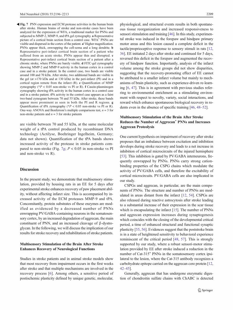
are visible between 70 and 55 kDa, at the same molecularweight of a tPA control produced by recombinant DNAtechnology (Actilyse, Boehringer Ingelheim, Germany;data not shown). Quantification of the tPA bands showsincreased activity of the protease in stroke patients com-pared to non-stroke (Fig. 7g; P < 0.05 in non-stroke vs PIand non-stroke vs R).
Discussion
In the present study, we demonstrate that multisensory stimu-lation, provided by housing rats in an EE for 5 days afterexperimental stroke enhances recovery of paw placement abil-ity, without affecting infarct size. This is accompanied by in-creased activity of the ECM proteases MMP-9 and tPA.Concomitantly, protein substrates of these enzymes are mod-ified as evidenced by a decreased number of PNNsenwrapping PV/GABA-containing neurons in the somatosen-sory cortex, by an increased degradation of aggrecan, the mainconstituent of PNN, and an increased cleavage of β-dystro-glycan. In the following, we will discuss the implication of ourresults for stroke recovery and rehabilitation of stroke patients.
Multisensory Stimulation of the Brain After StrokeEnhances Recovery of Neurological Functions
Studies in stroke patients and in animal stroke models showthat most recovery from impairment occurs in the first weeksafter stroke and that multiple mechanisms are involved in therecovery process [6]. Among others, a sensitive period ofpostischemic plasticity defined by unique genetic, molecular,
physiological, and structural events results in both spontane-ous tissue reorganization and increased responsiveness tosensori stimulation and training [46]. In this study, experimen-tal stroke was induced in the forepaw and hindpaw primarymotor areas and this lesion caused a complete deficit in thetactile/proprioceptive response to sensory stimuli in rats [12,36]. EE initiated 2 days after stroke and continued for 5 days,reversed this deficit in the forepaw and augmented the recov-ery of hindpaw function. Importantly, analysis of the infarctvolume among the stroke groups did not show disparities,suggesting that the recovery-promoting effect of EE cannotbe attributed to a smaller infarct volume but mainly to mech-anisms of brain plasticity, such as experience-driven re-learn-ing [6, 47]. This is in agreement with previous studies refer-ring to environmental enrichment as a stimulating environ-ment with respect to novelty, variety, social interactions, andreward which enhance spontaneous biological recovery in ro-dents even in the absence of specific training [46, 48–52].
Multisensory Stimulation of the Brain After StrokeReduces the Number of Aggrecan+ PNNs and IncreasesAggrecan Proteolysis
One current hypothesis on impairment of recovery after strokeproposes that an imbalance between excitation and inhibitiondevelops during stroke recovery and leads to a net increase ininhibition of cortical microcircuits of the injured hemisphere[53]. This inhibition is gated by PV/GABA interneurons, fre-quently enwrapped by PNNs. PNNs carry strong cation-binding properties of the CSPG chains which modulate theactivity of PV/GABA cells, and therefore the excitability ofcortical microcircuits. PV/GABA cells are also implicated inour study.
CSPGs and aggrecan, in particular, are the main compo-nents of PNNs. The structure and number of PNNs are mod-ulated in areas distant from the infarct [12, 54]. CSPGs arealso released during reactive astrocytosis after stroke leadingto a substantial increase of their expression in the scar tissuewhich is encapsulating the infarct [15]. The number of PNNsand aggrecan expression increases during synaptogenesiswhich coincides with the closing of the developmental criticalperiod, a time of enhanced structural and functional synapticplasticity [55, 56]. Evidences suggest that the poststroke brainis in a state of heightened sensitivity to behavioral experiencereminiscent of the critical period [48, 57]. This is stronglysupported by our study, where a robust sensori-motor stimu-lation provided by EE after stroke induced a reduction in thenumber of Cat-315+ PNNs in the somatosensory cortex ipsi-lateral to the lesion, where the Cat-315 antibody recognizes acarbohydrate epitope carried on the aggrecan core protein [12,42–45].
Generally, aggrecan that has undergone enzymatic diges-tion of chondroitin sulfate chains with ChABC is detected
�Fig. 7 PNN expression and ECM protease activities in the human brainafter stroke. Human brains of stroke and non-stroke cases have beenanalyzed for the expression of WFA, a traditional marker for PNNs andsubjected toMMP-2,MMP-9, and tPA gel zymography. aRepresentativepicture of a cortical brain section from a control case. WFA+ PNNs arevisible and dispersed in the cortex of the patient. a'Higher magnification.PNNs appear thick, enwrapping the cell-soma and a long dendrite. bRepresentative peri-infarct cortical brain section of a patient whosuffered from an acute stroke. PNNs appear thin and disrupted. cRepresentative peri-infarct cortical brain section of a patient after achronic stroke, where PNNs are barely visible. d FITC-gel zymographyshowing MMP-2 and MMP-9 activity in the human cortex in a controlcase and in a stroke patient. In the control case, two bands are visiblearound 100 and 70 kDa. After stroke, two additional bands are visible inthe gel (at >170 kDa and at 130 kDa) in the peri-infarct (PI) and in acortical region remote from the infarct (R). e Quantification of MMPzymography (*P < 0.05 non-stroke vs PI or R). f Casein-plasminogenzymography showing tPA activity in the human cortex in a control caseand in a stroke patient. tPA activity in the control case appears as a fainttwo-band zymogram between 70 and 55 kDa. After stroke, these bandsappear more prominent as seen in both the PI and R regions. gQuantification of tPA zymography (*P < 0.05 non-stroke vs PI or R).One-way ANOVA and Bonferroni’s multiple comparisons test, n = 3 fornon-stroke patients and n = 3 for stroke patients
Mol Neurobiol (2018) 55:2196–2213 2209
with a marked band at >250 kDa, but often multiple weakerbands at lower molecular weights can also be identified onwestern blots [58]. Here, we demonstrate that aggrecan, akey component of PNNs, undergoes increased digestion fol-lowing stroke and EE conditions even in cortical regions re-mote from the lesion. Because aggrecan can be cleaved atmultiple sites, several fragments were identified on the west-ern blots from shams and stroke animals. The increased diges-tion suggests aggrecan proteolysis as an early step in PNNsloss (visible here 7 days after stroke and after 5 days of EE),similarly to what has been described after status epilepticus[45].
Recently, the effects of manipulating CSPGs using ChABChave been investigated after stroke in vivo [59]. It was dem-onstrated that ChABC treatment was able to promote forepawsensorimotor recovery and degrade PNNs which may reacti-vate plasticity. These findings are in line with our results,where environmental stimulation drives functional recoveryand plasticity through the degradation of PNNs, which aremainly enwrapping PV/GABA-containing neurons. PV/GABA cells are key regulators of synchronization of neuronalactivity in cerebral microcircuits. These microcircuits, gatedby PV/GABA interneurons, play an important role in process-ing information for many tasks such as motor control, cogni-tion, and sensory perception [60], of importance in functionalrecovery after stroke [61, 62]. Here, we show that under EEconditions, PV-containing neurons were enwrapped by fewerPNNs. The remaining PNNs had poor structural integrity,suggesting that a digestion process may be responsible forthe loss of PNNs. Finally, we demonstrated the presence ofPNNs in the human brain, which appear similar to the onesseen in our experimental model. In control patients, these netswere thick and enwrapping the entire cell soma and proximaldendrites, as reported earlier [63]. After stroke, the nets ap-peared thin and disrupted, as seen in the rat brains after exper-imental stroke. The degradation of PNNs could change thekinetics and fast-spiking characteristics of the PV/GABA cells[60]. Together, these data strongly suggest that multisensorystimulation provided by EE induces a degradation ofaggrecan-containing PNNs, opening-up a critical period forplasticity after stroke in the adult brain.
Multisensory Stimulation of the Brain After StrokeActivates ECM Proteases
ECM proteases and their inhibitors are involved in PNNmod-ulation after stroke. PNNs can be envisaged as targets forpharmacological interventions as observed in seizure genesis[64]. Different proteases have been proposed to contribute toECM modulation and degradation after brain injury, amongothers tPA, ADAMTS-4, MMP-2, and MMP-9 [25, 28–31].In our study, the activity of tPA andMMP-9 is increased in theipsilateral somatosensory cortex of the stroke EE group
compared to the stroke STD group. These proteases mightthen be involved in the remodelling of ECM structures andchanges in the integrity of PNNs that we observe after strokeand EE conditions in regions remote from the infarct. We alsoshowed an increased activity of tPA and MMPs after stroke inhuman samples, suggesting similar processes in the humanbrain following stroke. tPA expression and activity in thehealthy human cortex have been reported earlier [65] andMMP-2 and MMP-9 activities have been studied in the ische-mic core of stroke patients [66]. Our results in stroke patientshighlight a remarkable presence of tPA, MMP-2, and MMP-9activities in peri-infarct zones and most importantly in regionsremote from the infarct. This suggests a predominant remod-elling of the abovementioned areas after stroke in patientswhich persists for months after the event.
Despite data implicatingMMP-9 in neuronal/synaptic plas-ticity, not many synaptic targets for its enzymatic activity haveyet been identified. However, recent studies have suggestedthat this enzyme may digest the full-lenght β-dystroglycan torelease a 30-kDa product, mainly expressed in the postsynap-tic membrane and in close proximity to MMP-9 [32]. β-Dystroglycan is a cell surface protein protruding from post-synaptic membrane and bound (probably throughout the syn-aptic cleft) to presynaptic neurexins, implicated in neurotrans-mitter release and long-term potentiation (LTP) [67]. In thisstudy, we observed increased levels of the 30-kDa form of β-dystroglycan, which may have been generated by MMP-9,similar to previous reports where enhanced neuronal activitywas challenged [32, 68]. One explanation of this result couldbe that the multisensory stimulation provided by EE induced aMMP-9 activation-dependent cleavage of the full-length formof β-dystroglycan in the ipsilateral hemisphere. A fine struc-tural immuno-colocalization of β-dystroglycan and MMP-9has been previously shown by electron microscopy in the ratbrain [32]. In addition, β-dystroglycan cleavage is abolishedafter a MMP-9 inhibitor treatment [32] and in MMP-2 andMMP-9 knockout mice [69], further suggesting a role ofMMP-9 in β-dystroglycan cleavage.
MMP activity is tightly controlled by endogenous tissueinhibitors of MMPs (TIMPs), a family of proteins comprisedof four members (TIMP-1 to TIMP-4), which inhibit multipleMMPs and display different interactions and substrate speci-ficity with MMP proenzymes [70]. tPA activity in the CNS isregulated by neuroserpin, a member of the serine proteaseinhibitor (serpin) family that is secreted from the growth conesof neurons [71]. It is important to note that protease expressionis not always indicative of net proteolytic activity, since thelatter is an outcome of expression levels, posttranslational ac-tivation, and protease inhibition [34]. Interestingly, mRNAexpression analysis showed an upregulation of Timp1 in theipsilateral hemisphere after stroke. Timp1 has been shown tobe upregulated following focal ischemia, a process that couldbe part of a general neuronal response that mediates tissue
Mol Neurobiol (2018) 55:2196–22132210

reorganization [72]. In our study, the upregulation of Timp1supports this hypothesis and suggests that the observed chang-es in MMP-9 and tPA activities are regulated by the expres-sion of the corresponding inhibitors rather than by translationof ECM protease genes. In the contralateral hemisphere,Adamts4 mRNA expression was upregulated after stroke,but not after stroke and EE conditions, a tendency also seenin Timp3 and Neuroserpin. This suggests a transcallosal influ-ence of the injured hemisphere on the contralateral hemi-sphere protease mRNA expressions, which is prevented bymultisensory stimulation exerted through an EE. Adamts4mRNA expression is upregulated in astrocytes following tran-sient middle cerebral artery occlusion in the rat [73], but this isthe first study reporting their modulation in regions remotefrom the lesion after permanent stroke. Similarly, Timp3 andNeuroserpin are upregulated in penumbral cortical neuronsfollowing stroke in the ipsilateral hemisphere [74, 75].
Conclusions
In conclusion, this study reports that experience-dependentplasticity induced by housing rats in an EE after stroke reducesthe number and structure of aggrecan-containing PNNs sur-rounding PV/GABA neurons in areas remote from the ische-mic lesion. This reduction is accompanied by an upregulationin the activity of ECM proteases such as tPA and MMP-9 anda modulation in the expression of ECM proteases and inhibi-tors such as Adamts4 and Timp1. Similar results were seen inbrain tissue from stroke patients. We conclude that multisen-sory stimulation of the brain after stroke, which provides en-hanced recovery of lost function, involves the modulation ofECM proteases and protease inhibitors. Elucidating the mech-anisms of ECM remodelling after stroke may contribute to thedevelopment of new therapies supporting rehabilitation ofstroke patients.
Acknowledgements The authors wish to acknowledge Carin Sjölund,Kerstin Beirup, Zsuzsanna Szepesi, Sara Martinez de Lizarrondo, BenoitD. Roussel, and Yannick Hommet for excellent technical assistance andadvice on methods. This work was supported by the Swedish ResearchCouncil (TW 2011-2652 and 2014-3802), the Hans-Gabriel and AliceTrolle-Wachtmeister Foundation (TW), the Strategic Research AreaMultiPark (TW), the Pia Ståhl Foundation (TW), the RoyalPhysiographic Society of Lund (TW and MJQ), and the STROKERiksforbundet (MJQ).
Compliance with Ethical Standards Animal experiments were ap-proved by the Malmö-Lund animal review board (ethical permit M25-12) and reported according to the ARRIVE guidelines. Human braintissue was used with the approval of the Lund Ethical Review Boardfor research involving humans (Dnr 2011/80). All studies were performedin a blinded fashion to the investigator who performed the experiments.
Open Access This article is distributed under the terms of the CreativeCommons At t r ibut ion 4 .0 In te rna t ional License (h t tp : / /
creativecommons.org/licenses/by/4.0/), which permits unrestricted use,distribution, and reproduction in any medium, provided you giveappropriate credit to the original author(s) and the source, provide a linkto the Creative Commons license, and indicate if changes were made.
References
1. Mozaffarian D, Benjamin EJ, Go AS et al (2016) Heart disease andstroke statistics—2016 update. Circulation 133:e38–360. doi:10.1161/CIR.0000000000000350
2. World Health Organization (2015) World report on ageing andhealth. doi: 10.1017/CBO9781107415324.004
3. Hacke W, Kaste M, Bluhmki E et al (2008) Thrombolysis withalteplase 3 to 4.5 hours after acute ischemic stroke. N Engl J Med359:1317–1329. doi:10.1056/NEJMoa0804656
4. Kim J-T, FonarowGC, Smith EE et al (2017) Treatment with tissueplasminogen activator in the golden hour and the shape of the 4.5-hour time-benefit curve in the national United States get with theguidelines-stroke population. Circulation 135:128–139. doi:10.1161/CIRCULATIONAHA.116.023336
5. Carmichael ST (2015) The 3 Rs of stroke biology: radial, relayed,and regenerative. Neurotherapeutics 13:1–12. doi:10.1007/s13311-015-0408-0
6. Wieloch T, Nikolich K (2006) Mechanisms of neural plasticityfollowing brain injury. Curr Opin Neurobiol 16:258–264. doi:10.1016/j.conb.2006.05.011
7. Mering S, Jolkkonen J (2015) Proper housing conditions in exper-imental stroke studies-special emphasis on environmental enrich-ment. Front Neurosci 9:1–8. doi:10.3389/fnins.2015.00106
8. Janssen H, Ada L, Karayanidis F et al (2012) Translating the use ofan enriched environment poststroke from bench to bedside: studydesign and protocol used to test the feasibility of environmentalenrichment on stroke patients in rehabilitation. Int J Stroke 7:521–526. doi:10.1111/j.1747-4949.2011.00727.x
9. Janssen H, Ada L, Bernhardt J et al (2014) An enriched environ-ment increases activity in stroke patients undergoing rehabilitationin a mixed rehabilitation unit: a pilot non-randomized controlledtrial. Disabil Rehabil 36:255–262. doi:10.3109/09638288.2013.788218
10. White JH, Bartley E, Janssen H et al (2015) Exploring stroke sur-vivor experience of participation in an enriched environment: aqualitative study. Disabil Rehabil 37:593–600. doi:10.3109/09638288.2014.935876
11. Murphy TH, Corbett D (2009) Plasticity during stroke recovery:from synapse to behaviour. Nat Rev Neurosci 10:861–872. doi:10.1038/nrn2735
12. Madinier A, Quattromani MJ, Sjölund C et al (2014) Enrichedhousing enhances recovery of limb placement ability and reducesaggrecan-containing perineuronal nets in the rat somatosensory cor-tex after experimental stroke. PLoS One 9:e93121. doi:10.1371/journal.pone.0093121
13. Quattromani M, Cordeau P, Ruscher K et al (2014) Enriched hous-ing down-regulates the toll-like receptor 2 response in the mousebrain after experimental stroke. Neurobiol Dis 66:66–73. doi:10.1016/j.nbd.2014.02.010
14. Soleman S, FilippovMA,Dityatev A, Fawcett JW (2013) Targetingthe neural extracellular matrix in neurological disorders.Neuroscience 253:194–213. doi:10.1016/j.neuroscience.2013.08.050
15. Katsman D, Zheng J, Spinelli K, Carmichael ST (2003) Tissuemicroenvironments within functional cortical subdivisions adjacentto focal stroke. J Cereb Blood FlowMetab 997–1009. doi: 10.1097/01.WCB.0000084252.20114.BE
Mol Neurobiol (2018) 55:2196–2213 2211

16. Hobohm C, Günther A, Grosche J et al (2005) Decomposition andlong-lasting downregulation of extracellular matrix in perineuronalnets induced by focal cerebral ischemia in rats. J Neurosci Res 80:539–548. doi:10.1002/jnr.20459
17. Karetko-Sysa M, Skangiel-Kramska J, Nowicka D (2011)Disturbance of perineuronal nets in the perilesional area afterphotothrombosis is not associated with neuronal death. ExpNeurol 231:113–126. doi:10.1016/j.expneurol.2011.05.022
18. Härtig W, Appel S, Suttkus A et al (2016) Abolished perineuronalnets and altered parvalbumin-immunoreactivity in the nucleusreticularis thalami of wildtype and 3xtg mice after experimentalstroke. Neuroscience 337:66–87. doi:10.1016/j.neuroscience.2016.09.004
19. Dityatev A, Schachner M, Sonderegger P (2010) The dual role ofthe extracellular matrix in synaptic plasticity and homeostasis. NatRev Neurosci 11:735–746. doi:10.1038/nrn2898
20. Carstens KE, PhillipsML, Pozzo-Miller L et al (2016) Perineuronalnets suppress plasticity of excitatory synapses on CA2 pyramidalneurons. J Neurosci 36:6312–6320. doi:10.1523/JNEUROSCI.0245-16.2016
21. Hausen D, Brückner G, Drlicek M et al (1996) Pyramidal cellsensheathed by perineuronal nets in human motor and somatosenso-ry cortex. Neuroreport 29:1725–1729
22. Härtig W, Derouiche A, Welt K et al (1999) Cortical neurons im-munoreactive for the potassium channel Kv3.1b subunit are pre-dominantly surrounded by perineuronal nets presumed as a buffer-ing system for cations. Brain Res 842:15–29. doi:10.1016/S0006-8993(99)01784-9
23. Morawski M, Brückner G, Arendt T, Matthews RT (2012)Aggrecan: beyond cartilage and into the brain. Int J Biochem CellBiol 44:690–693. doi:10.1016/j.biocel.2012.01.010
24. Carmichael ST (2006) Cellular and molecular mechanisms of neu-ral repair after stroke: making waves. Ann Neurol 59:735–742. doi:10.1002/ana.20845
25. Lemarchant S, Pruvost M, Hébert M et al (2014) tPA promotesADAMTS-4-induced CSPG degradation, thereby enhancingneuroplasticity following spinal cord injury. Neurobiol Dis 66:28–42. doi:10.1016/j.nbd.2014.02.005
26. Rosell A, Lo EH (2008) Multiphasic roles for matrix metallopro-teinases after stroke. Curr Opin Pharmacol 8:82–89. doi:10.1016/j.coph.2007.12.001
27. Lee S-R, Tsuji K, Lee S-R, Lo EH (2004) Role of matrix metallo-proteinases in delayed neuronal damage after transient global cere-bral ischemia. J Neurosci 24:671–678. doi:10.1523/JNEUROSCI.4243-03.2004
28. Zhao B-Q, Wang S, Kim H-Y et al (2006) Role of matrix metallo-proteinases in delayed cortical responses after stroke. Nat Med 12:441–445. doi:10.1038/nm1387
29. Lemarchant S, Docagne F, Emery E et al (2012) tPA in the injuredcentral nervous system: different scenarios starring the same actor?Neuropharmacology 62:749–756. doi:10.1016/j.neuropharm.2011.10.020
30. Lemarchant S, Pruvost M, Montaner J et al (2013) ADAMTSproteoglycanases in the physiological and pathological central ner-vous system. J Neuroinflammation 10:133. doi:10.1186/1742-2094-10-133
31. Verslegers M, Lemmens K, Van Hove I, Moons L (2013) Matrixmetalloproteinase-2 and -9 as promising benefactors in develop-ment, plasticity and repair of the nervous system. Prog Neurobiol105:60–78. doi:10.1016/j.pneurobio.2013.03.004
32. Michaluk P, Kolodziej L, Mioduszewska B et al (2007) ??-Dystroglycan as a target for MMP-9, in response to enhanced neu-ronal activity. J Biol Chem 282:16036–16041. doi:10.1074/jbc.M700641200
33. Stamenkovic V, Stamenkovic S, Jaworski T, et al (2016) The extra-ce l lu lar matr ix glycoprote in tenasc in-C and matr ix
metalloproteinases modify cerebellar structural plasticity by expo-sure to an enriched environment. Brain Struct Funct 1–23. doi: 10.1007/s00429-016-1224-y
34. Cunningham LA, Wetzel M, Rosenberg GA (2005) Multiple rolesfor MMPs and TIMPs in cerebral ischemia. Glia 50:329–339. doi:10.1002/glia.20169
35. Watson BD, Dietrich WD, Busto R et al (1985) Induction of repro-ducible brain infarction by photochemically initiated thrombosis.Ann Neurol 17:497–504. doi:10.1002/ana.410170513
36. De Ryck M, Van Reempts J, Duytschaever H et al (1992)Neocortical localization of tactile/proprioceptive limb placing reac-tions in the rat. Brain Res 573:44–60. doi:10.1016/0006-8993(92)90112-M
37. Paxinos G, Watson C (1998) The rat brain in stereotaxic coordi-nates, Fourth Edi. Academic Press
38. Swanson RA, Morton MT, Tsao-Wu G et al (1990) Asemiautomated method for measuring brain infarct volume. JCereb Blood FlowMetab 10:290–293. doi:10.1038/jcbfm.1990.47
39. Hattori S, Fujisaki H, Kiriyama T et al (2002) Real-timezymography and reverse zymography: a method for detecting ac-tivities of matrix metalloproteinases and their inhibitors usingFITC-labeled collagen and casein as substrates. Anal Biochem301:27–34. doi:10.1006/abio.2001.5479
40. Szklarczyk A, Lapinska J, Rylski M et al (2002) Matrixmetalloproteinase-9 undergoes expression and activation during den-dritic remodeling in adult hippocampus. J Neurosci 22:920–930
41. Bruckert G, Vivien D, Docagne F, Roussel BD (2015)Normalization of reverse transcription quantitative PCR data dur-ing ageing in distinct cerebral structures. Mol Neurobiol 53:1540–1550. doi:10.1007/s12035-015-9114-5
42. Matthews RT, Kelly GM, Zerillo CA et al (2002) Aggrecanglycoforms contribute to the molecular heterogeneity ofperineuronal nets. J Neurosci 22:7536–7547
43. Lander C, Kind P, Maleski M, Hockfield S (1997) A family ofactivity-dependent neuronal cell-surface chondroitin sulfate proteo-glycans in cat visual cortex. J Neurosci 17:1928–1939
44. Dino MR, Harroch S, Hockfield S, Matthews RT (2006)Monoclonal antibody Cat-315 detects a glycoform of receptor pro-tein tyrosine phosphatase beta/phosphacan early in CNS develop-ment that localizes to extrasynaptic sites prior to synapse formation.Neuroscience 142:1055–1069. doi:10.1016/j.neuroscience.2006.07.054
45. Rankin-Gee EK, McRae PA, Baranov E et al (2015) Perineuronalnet degradation in epilepsy. Epilepsia 56:1124–1133. doi:10.1111/epi.13026
46. Zeiler SR, Krakauer JW (2013) The interaction between trainingand plasticity in the poststroke brain. Curr Opin Neurol 26:609–616. doi:10.1097/WCO.0000000000000025
47. Pekna M, Pekny M, Nilsson M (2012) Modulation of neural plas-ticity as a basis for stroke rehabilitation. Stroke 43:2819–2828. doi:10.1161/STROKEAHA.112.654228
48. Biernaskie J, Chernenko G, Corbett D (2004) Efficacy of rehabilita-tive experience declines with time after focal ischemic brain injury. JNeurosci 24:1245–1254. doi:10.1523/JNEUROSCI.3834-03.2004
49. Ohlsson A-L, Johansson BB (1995) Environment influences func-tional outcome of cerebral infarction in rats. Stroke 26:644–649
50. Nygren J,Wieloch T (2005) Enriched environment enhances recov-ery of motor function after focal ischemia in mice, anddownregulates the transcription factor NGFI-A. J Cereb BloodFlow Metab 25:1625–1633. doi:10.1038/sj.jcbfm.9600157
51. Ruscher K, Shamloo M, Rickhag M et al (2011) The sigma-1 re-ceptor enhances brain plasticity and functional recovery after ex-perimental stroke. Brain 134:732–746. doi:10.1093/brain/awq367
52. Ruscher K, Kuric E, Liu Y et al (2013) Inhibition of CXCL12signaling attenuates the postischemic immune response and
Mol Neurobiol (2018) 55:2196–22132212
improves functional recovery after stroke. J Cereb Blood Flow&. Metab 33:1225–1234. doi:10.1038/jcbfm.2013.71
53. Carmichael ST (2012) Brain excitability in stroke: the yin and yangof stroke progression. Arch Neurol 69:161–167. doi:10.1001/archneurol.2011.1175
54. Carmichael ST, Archibeque I, Luke L et al (2005) Growth-associated gene expression after stroke: evidence for a growth-promoting region in peri-infarct cortex. Exp Neurol 193:291–311.doi:10.1016/j.expneurol.2005.01.004
55. Wang D, Fawcett J (2012) The perineuronal net and the control ofCNS plasticity. Cell Tissue Res 349:147–160. doi:10.1007/s00441-012-1375-y
56. Galtrey CM, Fawcett JW (2007) The role of chondroitin sulfate pro-teoglycans in regeneration and plasticity in the central nervous sys-tem. Brain Res Rev 54:1–18. doi:10.1016/j.brainresrev.2006.09.006
57. Hensch TK (2005) Critical period plasticity in local cortical circuits.Nat Rev Neurosci 6:877–888. doi:10.1038/nrn1787
58. Struglics A, Hansson M (2012) MMP proteolysis of the humanextracellular matrix protein aggrecan is mainly a process of normalturnover. Biochem J 446:213–223. doi:10.1042/BJ20120274
59. Soleman S, Yip PK, Duricki DA, Moon LDF (2012) Delayed treat-ment with chondroitinase ABC promotes sensorimotor recoveryand plasticity after stroke in aged rats. Brain 135:1210–1223. doi:10.1093/brain/aws027
60. Kubota Y (2014) Untangling GABAergic wiring in the corticalmicrocircuit. Curr Opin Neurobiol 26:7–14. doi:10.1016/j.conb.2013.10.003
61. Zeiler SR, Gibson EM, Hoesch RE et al (2013) Medial premotorcortex shows a reduction in inhibitory markers and mediates recov-ery in a mouse model of focal stroke. Stroke 44:483–489. doi:10.1161/STROKEAHA.112.676940
62. Inácio AR, Ruscher K, Wieloch T (2011) Enriched environmentdownregulates macrophage migration inhibitory factor and in-creases parvalbumin in the brain following experimental stroke.Neurobiol Dis 41:270–278. doi:10.1016/j.nbd.2010.09.015
63. Virgintino D, Perissinotto D, Girolamo F et al (2009) Differentialdistribution of aggrecan isoforms in perineuronal nets of the humancerebral cortex. J Cell Mol Med 13:3151–3173. doi:10.1111/j.1582-4934.2009.00694.x
64. Pollock E, Everest M, Brown A, Poulter MO (2014)Metalloproteinase inhibition prevents inhibitory synapse reorgani-zation and seizure genesis. Neurobiol Dis 70:21–31. doi:10.1016/j.nbd.2014.06.003
65. Teesalu T, Kulla A, Simisker A et al (2004) Tissue plasminogenactivator and neuroserpin are widely expressed in the human centralnervous system. Thromb Haemost 92:358–368. doi:10.1160/TH02-12-0310
66. Clark AW, Krekoski CA, Bou SS et al (1997) Increased gelatinaseA (MMP-2) and gelatinase B (MMP-9) activities in human brainafter focal ischemia. Neurosci Lett 238:53–56. doi:10.1016/S0304-3940(97)00859-8
67. Dzwonek J, Rylski M, Kaczmarek L (2004) Matrix metalloprotein-ases and their endogenous inhibitors in neuronal physiology of theadult brain. FEBS Lett 567:129–135. doi:10.1016/j.febslet.2004.03.070
68. Szepesi Z, Bijata M, Ruszczycki B et al (2013) Matrix metallopro-teinases regulate the formation of dendritic spine head protrusionsduring chemically induced long-term potentiation. PLoS One 8:e63314. doi:10.1371/journal.pone.0063314
69. Agrawal S, Anderson P, Durbeej M, et al (2006) Dystroglycan isselectively cleaved at the parenchymal basement membrane at sitesof leukocyte extravasation in experimental autoimmune encephalo-myelitis. 203:1007–1019. doi: 10.1084/jem.20051342
70. Gardner J, GhorpadeA (2003) Tissue inhibitor of metalloproteinase(TIMP)-1: the TIMPed balance of matrix metalloproteinases in thecentral nervous system. J Neurosci Res 74:801–806. doi:10.1002/jnr.10835
71. Miranda E, Lomas DA (2006) Neuroserpin: a serpin to think about.Cell Mol Life Sci 63:709–722. doi:10.1007/s00018-005-5077-4
72. Rivera S, Ogier C, Jourquin J et al (2002) Gelatinase B and TIMP-1are regulated in a cell- and time-dependent manner in associationwith neuronal death and glial reactivity after global forebrain ische-mia. Eur J Neurosci 15:19–32. doi:10.1046/j.0953-816x.2001.01838.x
73. Cross AK, Haddock G, Stock CJ et al (2006) ADAMTS-1 and -4 areup-regulated following transient middle cerebral artery occlusion inthe rat and their expression is modulated by TNF in cultured astro-cytes. Brain Res 1088:19–30. doi:10.1016/j.brainres.2006.02.136
74. Wallace JA, Alexander S, Estrada EY et al (2002) Tissue inhibitorof metalloproteinase-3 is associated with neuronal death in reperfu-sion injury. J Cereb Blood Flow Metab 22:1303–1310. doi:10.1097/00004647-200211000-00003
75. YepesM, SandkvistM,WongMK et al (2000) Neuroserpin reducescerebral infarct volume and protects neurons from ischemia-induced apoptosis. Blood 96:569–576. doi:10.1161/01.str.26.3.361
Mol Neurobiol (2018) 55:2196–2213 2213