extrapolation of the benzene inhalation unit risk estimate ... · a scientifically rigorous method...
TRANSCRIPT
NCEA-W-0517
Extrapolation of the Benzene Inhalation Unit Risk Estimate to the Oral Route
of Exposure
November, 1999
U.S. Environmental Protection Agency Washington, DC

ii
DISCLAIMER
Mention of trade names or commercial products does not constitute endorsement or
recommendation for use.
ABSTRACT
A simple method for extrapolation of benzene-induced cancer risk from the inhalation to
oral route is proposed. The method is based on the relative efficiency of benzene absorption
across routes of exposure, especially pulmonary and gastrointestinal barriers. There exists
substantial literature on pulmonary absorption in humans and a few laboratory animal species.
Data on oral absorption in humans are lacking; hence extrapolation is based on gastrointestinal
absorption studies in several experimental animal species. Currently, available physiologically
based pharmacokinetic (PBPK) animal models are not useful for human risk extrapolation. A
review of the relevant literature suggests absorption efficiencies of 50% and 100% for inhalation
and oral routes of exposure, respectively. Application of these absorption factors to the current
inhalation unit risk range of 2.2 × 10⁻⁶ – 7.8 × 10⁻⁶ per µg/m3 results in a proposed range for the
oral unit risk of 4.4 × 10⁻⁷ to 1.6 × 10⁻⁶/µg/L.
ii

iii
CONTENTS
LIST OF TABLES . . . . . . . . . . . . . . . . . . . . . . . . . . . . . . . . . . . . . . . . . . . . . . . . . . . . . . . . . iv
PREFACE . . . . . . . . . . . . . . . . . . . . . . . . . . . . . . . . . . . . . . . . . . . . . . . . . . . . . . . . . . . . . . . . . v
AUTHORS, CONTRIBUTORS, AND REVIEWERS . . . . . . . . . . . . . . . . . . . . . . . . . . . . . . . vi
1. INTRODUCTION . . . . . . . . . . . . . . . . . . . . . . . . . . . . . . . . . . . . . . . . . . . . . . . . . . . . . . . . 1
2. ORAL ABSORPTION . . . . . . . . . . . . . . . . . . . . . . . . . . . . . . . . . . . . . . . . . . . . . . . . . . . . 2
3. DERMAL ABSORPTION . . . . . . . . . . . . . . . . . . . . . . . . . . . . . . . . . . . . . . . . . . . . . . . . . 4
4. PULMONARY ABSORPTION . . . . . . . . . . . . . . . . . . . . . . . . . . . . . . . . . . . . . . . . . . . . . 5
4.1. RATS AND MICE . . . . . . . . . . . . . . . . . . . . . . . . . . . . . . . . . . . . . . . . . . . . . . . . . . . 5
4.2. HUMANS . . . . . . . . . . . . . . . . . . . . . . . . . . . . . . . . . . . . . . . . . . . . . . . . . . . . . . . . . . 5
4.2.1. Hunter and Colleagues (Hunter, 1966; Hunter, 1968;
Hunter and Blair, 1972) . . . . . . . . . . . . . . . . . . . . . . . . . . . . . . . . . . . . . . . . . 6
4.2.2. Nomiyama and Nomiyama (1974) . . . . . . . . . . . . . . . . . . . . . . . . . . . . . . . . . . 6
4.2.3. Pekari et al. (1992) . . . . . . . . . . . . . . . . . . . . . . . . . . . . . . . . . . . . . . . . . . . . . 7
4.2.4. Sherwood (1988) . . . . . . . . . . . . . . . . . . . . . . . . . . . . . . . . . . . . . . . . . . . . . . . 8
4.2.5. Srbova et al. (1950) . . . . . . . . . . . . . . . . . . . . . . . . . . . . . . . . . . . . . . . . . . . . . 8
4.2.6. Teisinger et al. (1952) (data reported in Fiserova-Bergerova et al., 1974) . . . . 8
4.2.7. Yu and Weisel (1998) . . . . . . . . . . . . . . . . . . . . . . . . . . . . . . . . . . . . . . . . . . . 9
5. DISCUSSION OF ROUTE-TO-ROUTE ABSORPTION . . . . . . . . . . . . . . . . . . . . . . . . . . 9
6. POTENTIAL ISSUES . . . . . . . . . . . . . . . . . . . . . . . . . . . . . . . . . . . . . . . . . . . . . . . . . . . . 10
6.1. DOSE-DEPENDENCY OF ABSORPTION . . . . . . . . . . . . . . . . . . . . . . . . . . . . . . . 10
6.2. TIME-DEPENDENCY OF ABSORPTION . . . . . . . . . . . . . . . . . . . . . . . . . . . . . . . 11
6.3. GENDER DEPENDENCE OF ABSORPTION . . . . . . . . . . . . . . . . . . . . . . . . . . . . 11
6.4. METABOLISM AND TOXICOKINETICS . . . . . . . . . . . . . . . . . . . . . . . . . . . . . . . 12
7. EXTRAPOLATION FROM INHALATION TO ORAL RISK . . . . . . . . . . . . . . . . . . . . . 14
7.1. FURTHER QUESTIONS, AND COMMENTS ON DATABASE ACCURACY . . . 16
REFERENCES . . . . . . . . . . . . . . . . . . . . . . . . . . . . . . . . . . . . . . . . . . . . . . . . . . . . . . . . . . . . 16
iii

iv
LIST OF TABLES
Table 1. Percent of inhaled benzene retained in rats and mice . . . . . . . . . . . . . . . . . . . . . . . . . 6
Table 2. Absorption of inhaled benzene in humans . . . . . . . . . . . . . . . . . . . . . . . . . . . . . . . . . 7
iv

v
PREFACE
This document is a source document for updating the oral cancer unit risk estimate for
benzene in the Integrated Risk Information System (IRIS).
In the development of this document, the scientific literature has been reviewed, key
studies have been evaluated and summarized, and the carcinogenicity and related information are
qualitatively and quantitatively characterized. The relevant scientific literature has been
reviewed through April 1999.
v

vi
AUTHORS, CONTRIBUTORS, AND REVIEWERS
The National Center for Environmental Assessment-Washington Office (NCEA-W),
Office of Research and Development, was responsible for the preparation of this report. An
earlier draft of this report was prepared by the University of California, Berkeley, under EPA
contract no. 8W-3143-NASA.
AUTHORS
David L. Bayliss, NCEA-W
Elinor W. Fanning, University of California, Berkeley
Martyn T. Smith, University of California, Berkeley
Babaseheb Sonawane, NCEA-W
EPA REVIEWERS
Robert Bruce, NCEA-CIN
James Cogliano, NCEA-W
Joyce Donahue, OW
Marina Evans, NHEERL-RTP
Annie Jarabek, NCEA-RTP
Jennifer Jinot, NCEA-W
Leonard Keifer, OPPTS
Robert McGaughy, NCEA-W
Larry Valcovic, NCEA-W
Diana Wong, OW
EXTERNAL REVIEWERS
Patrick W. Beatty, Ph.D. - Chevron Research. & Technology Company, Richmond, CA
David Eastmond, Ph.D. - University of California, Riverside, CA
David Ross, Ph.D. - University of Colorado, Denver, CO
Robert Snyder, Ph.D. - Rutgers University, Rutgers, NJ
vi
1
1. INTRODUCTION
The best available human epidemiological data for evaluation of cancer risk for benzene
derive from studies of occupational inhalation exposure. In order to apply the results of risk
estimates derived from these studies to the estimation of cancer risk arising from oral exposure
to benzene, a rationale for route-to-route extrapolation needs to be established.
A workshop organized by EPA and the ILSI Risk Science Institute concluded that route-
to-route extrapolation for risk assessment is appropriate when similar toxic endpoints are
observed with both routes of exposure and when toxicokinetic data are available (Gerrity and
Henry, 1990). Because of a lack of data on orally exposed humans, it cannot be concluded that
leukemia and related hematopoietic endpoints are associated with the oral route of exposure.
However, in animal models, similar cancers and hematotoxic endpoints occurred in several
studies of both oral and inhalation exposure (ATSDR, 1997). Experimental animal data also
demonstrate that benzene is metabolized to the same products, whether inhaled or ingested.
Therefore, it is reasonable to extrapolate from inhalation to oral cancer risk.
Extrapolation from an inhalation to an oral slope factor in the earlier IRIS entry for
benzene was based on conversion between the standard intake factors for air and water (U.S.
EPA, 1999). The extent of absorption after oral exposure was assumed, by default, to be
equivalent to absorption from inhalation exposure. A data-based extrapolation would improve
upon this default approach.
A scientifically rigorous method for route-to-route extrapolation involves the development
of a pharmacokinetic model to predict the concentration of the ultimate carcinogen in bone
marrow (the target tissue for benzene's carcinogenic effects) under a variety of different human
exposure scenarios. There are currently several inadequacies in the scientific database required
for this approach. No pharmacokinetic models that include metabolism and distribution to the
bone marrow are available that have been adequately validated for humans (Smith and Fanning,
1997). A major difficulty is that the particular chemical species responsible for the induction of
leukemia in benzene-exposed people and animals is not known with certainty; leukemogenesis
may well involve more than one metabolite or combination of metabolites (Smith, 1996).
Most experts agree that benzene metabolites, or by-products of their formation, are
responsible for benzene leukemogenesis. This suggests that extrapolation between routes of
exposure could be based on a dose defined as the total quantity of benzene metabolized in the
body after uptake of equivalent amounts, a somewhat simpler metric than delivered dose of the
unknown ultimate carcinogenic compound(s). However, the kinetics of metabolite formation and
clearance after inhalation, ingestion, or dermal exposure of benzene are not known for humans.
The many uncertainties involved in using animal-based models to predict dosimetry for humans
1

2
may preclude a risk assessment application for PBPK models dependent on animal-derived data.
Using a PBPK model developed primarily with high-dose animal data is not likely to improve the
accuracy of risk estimates based on human data.
Therefore, a simple approach to route-to-route extrapolation is perhaps the most
scientifically defensible approach at this time. This report summarizes published literature
addressing the absorption of benzene after inhalation exposure in humans and laboratory animals,
and after oral exposure to animals. No relevant data were located for absorption of benzene after
ingestion in humans. Using the best estimates of the relative absorption efficiencies across the
pulmonary and gastrointestinal barriers as the basis of route-to-route extrapolation, an oral slope
factor is derived from the inhalation slope factor currently documented in the IRIS database.
2. ORAL ABSORPTION
Benzene is absorbed by rabbits, hamsters, mice, and rats following administration by oral
gavage. In an early study in rabbits, 90% of the radioactivity from a single bolus dose was
recovered in urine and exhaled air (Parke and Williams, 1953). Medinsky et al. (1984)
determined gastrointestinal absorption by comparing the percentage of administered dose excreted
in urine, feces, and exhaled air after gavage to the percentages resulting from intraperitoneal (i.p.)
injection to rats. Furthermore, Sabourin and colleagues administered radiolabeled benzene orally,
by corn oil gavage, and intraperitoneally to rats and mice (Sabourin et al., 1987). The details of
the study are as follows:
Sprague-Dawley and F344/N rats and B6C3F1 mice (3-4 per dose) were dosed by oral
gavage (0.5 through 300 mg/kg body weight) or i.p. injection (0.5 or 150 mg/kg) with [14C]
benzene (approximately 10 uCi per rat and 2 uCi per mouse. The [14C] benzene was administered
in corn oil. Urine and feces were collected at 4, 8, 16, 24, 32, and 48 hr after dosing.
The percentages of the dose excreted by each route were similar following gavage or i.p.
injection. Gastrointestinal absorption of benzene was determined by comparing excretion routes
following oral or i.p. administration at the same dose and was essentially 100% for F344/N rats,
Sprague-Dawley rats, and B6C3F1 mice at 0.5 or 150 mg/kg body weight. Therefore, the dose of
benzene absorbed is equal to the amount administered by gavage. At all dose levels tested, most
of the [14C] benzene and metabolites were excreted in the urine and in the expired air. In both rats
and mice, at doses below 15 mg/kg > 80% of the 14C was excreted in the urine. In F344/N and
Sprague-Dawley rats, the excretion of 14C in the expired air following oral or ip administration
plateaued between 16 and 24 hr. However, in B6C3F1 mice, no further expiration of 14C occurred
beyond 4 hr. Cumulative urinary excretion of 14C plateaued at 16 to 32 hr in all three species.
2
3
Cumulative urinary 14C excretion displayed large animal variability at early time points due to the
voluntary nature of urination.
Urinary 14C can be used as a measure of net [14C] benzene metabolism. Less than 5% of
the 14C in urine from animals treated with 0.5-150 mg/kg [14C] benzene was extractable into
ethylacetate, indicating that >95% of the 14C was due to water-soluble metabolites. For F344/N
rats and B6C3F1 mice, metabolism of [14C] benzene, as indicated by urinary 14C, appeared to be
linearly related to dose up to 15 mg/kg. For both species, nonlinearity of metabolite formation
versus dose became apparent somewhere between 15 and 50 mg/kg and an increasing amount of
the administered [14C] benzene was eliminated in the expired air above 15 mg/kg. At doses up to
50 mg/kg, F344/N rats and B6C3F1 mice excreted almost equal amounts of benzene equivalents
per kg body weight in the urine. In F344/N rats, urinary 14C continued to increase with dose
above 50 mg/kg, albeit not linearly above 15 mg/kg. In B6C3F1 mice, total metabolite formation
(urinary 14C) plateaued above 50 mg/kg at 0.42 mmol benzene equivalents/kg body weight. This
may have been due in part to the faster elimination of [14C] benzene in the expired air by mice
compared with that by rats. In mice, 71% of the initial dose of [14C] benzene was eliminated in
the exhaled air at the 150 mg/kg dose, whereas in rats only 50% was exhaled at this
concentration.
Results of gastrointestinal absorption studies indicated that, for all three species (F344/N
and Sprague-Dawley rats and B6C3F1 mice), virtually 100% of an orally administered dose of
benzene was absorbed. The small amount of 14C found in feces following i.p. injection of [14C]
benzene may be due to biliary excretion.
Sabourin et al., (1987) suggested that absorption of benzene could be determined based
on the method of Medinsky et al. (1984) using the equation:
Percentage absorption = 100 - Fpo + Fip[(Upo + Apo)/(Uip + Aip)].
In this formula, F is the percentage of the initial dose in feces, U is the percentage of the initial
dose in urine, A is the percentage of the initial dose in the expired air, po refers to gavage (per
os)-treated animals, and i.p. refers to intraperitoneally injected animals.
In a recent study, rats, mice, and hamsters were treated by oral gavage (in corn oil) with a
range of benzene doses that overlapped, but extended lower than the dose range used in the
Sabourin study cited above (Mathews et al., 1998). Each dose for rats, mice, and hamsters
contained 12–26, 4–21, and 3–22 uCi radiolabel, respectively. Additionally, all dose formulations
contained an appropriate amount of unlabeled benzene and corn oil in a single dose volume of 5
mL/kg (rats and hamsters) or 25 mL/kg (mice) and these doses were administered by intragastric
gavage. Results of the study demonstrated that benzene was readily absorbed, metabolized, and
3

4
eliminated primarily in urine by each species studied. However, the routes of elimination were
somewhat altered by dose. At the high dose of 100 mg/kg a significant portion of the dose was
eliminated in breath. Elimination of a dose of 100 mg/kg in breath ranged from 22% in mice to
50% in the rats. A complete absorption from the gastrointestinal tract was confirmed in all three
species. Both studies report a greater proportion of metabolites excreted in urine at low doses,
with a shift to greater amounts of unmetabolized benzene excreted in exhaled air at high doses.
This result suggests that saturation of metabolism occurs at doses greater than approximately 100
mg/kg; however, at the oral doses at which humans are likely to be exposed, the animal results
suggest a linear increase in total metabolite production with exposure level.
In humans, oral exposure occurs by ingestion of benzene-contaminated food or water. No
relevant animal studies are available that allow a comparison of absorption between gavage and
drinking water administration. Theoretically, benzene ingested in drinking water could be subject
to volatilization loss from the stomach, which would be suppressed by the oil vehicle used in the
animal gavage experiments. On the other hand, it might be expected that a greater proportion of
large bolus doses would escape absorption, and pass through in the feces, while smaller doses
would be better absorbed. The fact that essentially complete absorption was observed even at
high gavage doses in the Sabourin et al. (1987) and Mathews et al. (1998) studies suggests that,
in the absence of data to the contrary, it is reasonable to assume complete absorption of benzene
ingested by humans.
3. DERMAL ABSORPTION
Several studies conducted both with humans and experimental animals indicate that
benzene is readily absorbed through the skin from both liquid and vapor phases and the
percentage of absorption of the applied doses is usually less than 1% (Franz, 1983, 1984; Maibach
and Anjo, 1981; Susten et al., 1985). In in vivo experiments on four human volunteers, when
0.0026 mg/cm2 of C14-benzene was applied to forearm skin, approximately 0.05% of the applied
dose was absorbed (Franz, 1983, 1984). Absorption was rapid, with more than 80% of the total
excretion of the absorbed dose occurring in the first 8 hours after application. Calculations were
based on urinary excretion data and no correction was made for the amount of benzene that
evaporated from the applied site before absorption occurred. In addition, the percentage of
absorbed dose excreted in urine that was used in the calculation was based only on data from
rhesus monkeys and may not be accurate for humans. Tsuruta (1989) reported that dermal
absorption of benzene increased linearly with dose in hairless mice exposed to benzene vapors and
estimated that skin absorption of benzene by humans would be 3.7% that of inhalation exposure
at the same concentration.
4
5
4. PULMONARY ABSORPTION
Pulmonary absorption of volatile organic compounds is not expected to be complete;
some portion of the inhaled concentration is exhaled from the lung without entering systemic
circulation. Experimental evidence confirms incomplete absorption of benzene in both animals
and humans.
4.1. RATS AND MICE
In the Sabourin study cited above, rats and mice were also exposed to benzene by
inhalation (Sabourin et al., 1987). The results are summarized in Table 1. Mice and rats were
exposed for 6 hours to 13, 29, and 130 ppm benzene by inhalation. Rats were also exposed to
260 and 870 ppm for 6 hours, while mice were exposed to one high dose of 990 ppm for 6 hours.
The total inhaled dose of benzene was computed from the exposure concentration and measured
breathing rate. The amount of benzene retained was then computed as a fraction of this quantity,
based on the amount of benzene remaining in the carcass or excreted in urine and feces. Benzene
taken up but subsequently excreted in exhaled air is not counted in the absorbed fraction. This
definition of absorption is distinct from that used in the subsequent discussion of human data, but
may be examined for rough comparison. Therefore, this absorption information from animal data
may underestimate true exposure in humans. The percent of inhaled benzene retained in rats and
mice will be found in Table 1.
4.2. HUMANS
There is a significant database on benzene in exhaled breath of humans exposed to
benzene in occupational, environmental, or experimental situations. Occupational and
environmental exposure is generally quite variable from individual to individual and over time.
This variability renders estimation of the actual exposure received quite complicated in many
situations. Therefore, we focus here on studies of controlled human exposures to known
concentrations of benzene for known duration.
5

6
-- --
Table 1. Percent of inhaled benzene retained in rats and mice Exposure concentration (ppm) Average percentage retained after 6
hours (n=3) Rats Mice Rats Mice 13 11 33 50 29 29 44 52 130 130 23 38 260 22 870 990 15 9.7
Source: Sabourin et al., 1987.
Chamber studies are often designed to study the excretion of benzene and/or its
metabolites in exhaled air. While useful information concerning half-life of benzene in the body
and elimination kinetics can be obtained from the postexposure period, concurrent measurements
of exposure concentration (Cinh) and benzene in exhaled air (Cexh) are necessary to compute
instantaneous absorption factors. It should be noted that a fraction of the benzene absorbed may
not be metabolized, thus, underestimating absorption. For this report, the percent of benzene
absorbed is defined simply as: 100 * (Cinh - Cexh)/Cinh. In some of the publications we
reviewed, concentration data were reported in different formats, and the numbers were converted
to the units in Table 2 to facilitate comparison across studies. The results are summarized in
Table 2.
4.2.1. Hunter and Colleagues (Hunter, 1966; Hunter, 1968; Hunter and Blair, 1972)
In the first paper of this series, absorption of 47% was reported for one male subject
exposed for 24 minutes to a concentration “a little above the threshold value of 25 ppm” (Hunter,
1966). In the next paper, one male subject exposed for 2 and 4 hours to approximately 30 ppm
absorbed 55%-60% of the inhaled concentration (Hunter, 1968). Hunter and Blair (1972)
exposed 5 male subjects for 2-3 hours to concentrations ranging from about 30 to 100 ppm.
However, inhaled and exhaled air concentrations are not reported for the time during exposure,
except for one subject (Table 2). The time of sampling was not given; neither was it clear
whether the data represent a single sample or an average of multiple samples. For this single
subject, exposed over a period of 5 days to concentrations ranging from 21 to 32 ppm, the
percent absorbed (computed as above) ranged from 53% to 63%. It is not clear whether this is a
different subject from the previous report.
4.2.2. Nomiyama and Nomiyama (1974)
Nomiyama and Nomiyama (1974) determined both “retention” and “uptake” of benzene.
6

7
Table 2. Absorption of inhaled benzene in humans Number of samples per
Percent absorbed, Exposure Exposure Number of exposure Study average (range) concentration duration subjects period
Teisinger et al., 48% n.a. 5 hr 14 n.a. 1952, as cited in Fiserova-Bergerova et al., 1974 Hunter, 1966 47% 25-30 ppm 24 min. 1 n.a. Hunter, 1968 (55%-60%) approx. 30 ppm 2 hr, 4 hr 1
(2 exposures) n.a. Hunter and (53%-63%) 21-32 ppm 3-4 hr 1 (10 exposures) 1 Blair, 1972 Nomiyama and 30% (SD 6.7) 52-62 ppm 4 hr 6 3 Nomiyama, 1974 Pekari et al., 52% (SD 7.3) 1.7 ppm 4 hr 3 6 1992 48% (SD 4.3) 10 ppm 4 hr 3 6 Srbova et al., 50%-62% (one 100 ppm 90 min 1 7 1950 subject) 20%-50% 47-110 ppm 2-3 hr 23 every 15 min
(reported group range after 2 hours)
Yu and Weisel, 64% (range: 48%- 32-69 ppm (in 30 min 3 4 1998 73%) tobacco smoke) 120 min 3 7
n.a. = not available.
Their calculation of retention is equivalent to the definition of absorption used in this report. Six
subjects, three male and three female, were exposed to benzene concentrations ranging from 52
to 62 ppm for 4-hour periods. Exhaled air was sampled every hour. The authors report average
retention to be 30.2%. This figure is somewhat lower than the other studies discussed here.
However, the data indicate that a potential explanation is that absorption was averaged over the 3,
3.5, and 4-hour time points only. The percent absorption was time-dependent in these
experiments: absorption was high early in exposure, and approached a steady state only after 3
hours. According to the data plotted by the authors, the average absorption at the 1-hour time
point was approximately 60% for women and 45% for men. A decrease to approximately 43%
and 35%, respectively, occurred at the 2-hour time point.
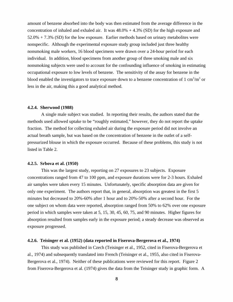
4.2.3. Pekari et al. (1992)
Pekari et al. (1992) developed a reliable and specific method for biologically monitoring
benzene in blood. Subjects were exposed to benzene in air at 10 cm3/m3 and 1.7 cm3/m3. The
7
8
amount of benzene absorbed into the body was then estimated from the average difference in the
concentration of inhaled and exhaled air. It was 48.0% + 4.3% (SD) for the high exposure and
52.0% + 7.3% (SD) for the low exposure. Earlier methods based on urinary metabolites were
nonspecific. Although the experimental exposure study group included just three healthy
nonsmoking male workers, 16 blood specimens were drawn over a 24-hour period for each
individual. In addition, blood specimens from another group of three smoking male and six
nonsmoking subjects were used to account for the confounding influence of smoking in estimating
occupational exposure to low levels of benzene. The sensitivity of the assay for benzene in the
blood enabled the investigators to trace exposure down to a benzene concentration of 1 cm3/m3 or
less in the air, making this a good analytical method.
4.2.4. Sherwood (1988)
A single male subject was studied. In reporting their results, the authors stated that the
methods used allowed uptake to be “roughly estimated,” however, they do not report the uptake
fraction. The method for collecting exhaled air during the exposure period did not involve an
actual breath sample, but was based on the concentration of benzene in the outlet of a self-
pressurized blouse in which the exposure occurred. Because of these problems, this study is not
listed in Table 2.
4.2.5. Srbova et al. (1950)
This was the largest study, reporting on 27 exposures to 23 subjects. Exposure
concentrations ranged from 47 to 100 ppm, and exposure durations were for 2-3 hours. Exhaled
air samples were taken every 15 minutes. Unfortunately, specific absorption data are given for
only one experiment. The authors report that, in general, absorption was greatest in the first 5
minutes but decreased to 20%-60% after 1 hour and to 20%-50% after a second hour. For the
one subject on whom data were reported, absorption ranged from 50% to 62% over one exposure
period in which samples were taken at 5, 15, 30, 45, 60, 75, and 90 minutes. Higher figures for
absorption resulted from samples early in the exposure period; a steady decrease was observed as
exposure progressed.
4.2.6. Teisinger et al. (1952) (data reported in Fiserova-Bergerova et al., 1974)
This study was published in Czech (Teisinger et al., 1952, cited in Fiserova-Bergerova et
al., 1974) and subsequently translated into French (Teisinger et al., 1955, also cited in Fiserova-
Bergerova et al., 1974). Neither of these publications were reviewed for this report. Figure 2
from Fiserova-Bergerova et al. (1974) gives the data from the Teisinger study in graphic form. A
8
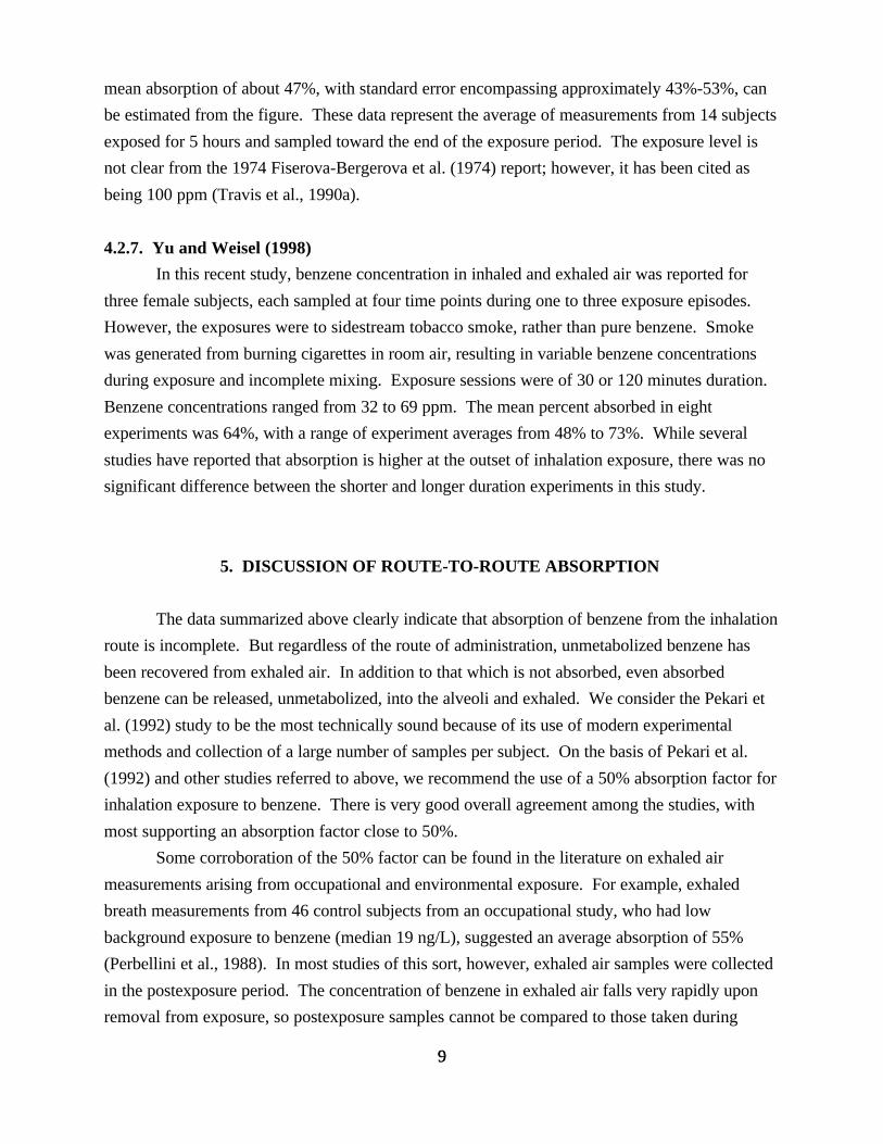
9
mean absorption of about 47%, with standard error encompassing approximately 43%-53%, can
be estimated from the figure. These data represent the average of measurements from 14 subjects
exposed for 5 hours and sampled toward the end of the exposure period. The exposure level is
not clear from the 1974 Fiserova-Bergerova et al. (1974) report; however, it has been cited as
being 100 ppm (Travis et al., 1990a).
4.2.7. Yu and Weisel (1998)
In this recent study, benzene concentration in inhaled and exhaled air was reported for
three female subjects, each sampled at four time points during one to three exposure episodes.
However, the exposures were to sidestream tobacco smoke, rather than pure benzene. Smoke
was generated from burning cigarettes in room air, resulting in variable benzene concentrations
during exposure and incomplete mixing. Exposure sessions were of 30 or 120 minutes duration.
Benzene concentrations ranged from 32 to 69 ppm. The mean percent absorbed in eight
experiments was 64%, with a range of experiment averages from 48% to 73%. While several
studies have reported that absorption is higher at the outset of inhalation exposure, there was no
significant difference between the shorter and longer duration experiments in this study.
5. DISCUSSION OF ROUTE-TO-ROUTE ABSORPTION
The data summarized above clearly indicate that absorption of benzene from the inhalation
route is incomplete. But regardless of the route of administration, unmetabolized benzene has
been recovered from exhaled air. In addition to that which is not absorbed, even absorbed
benzene can be released, unmetabolized, into the alveoli and exhaled. We consider the Pekari et
al. (1992) study to be the most technically sound because of its use of modern experimental
methods and collection of a large number of samples per subject. On the basis of Pekari et al.
(1992) and other studies referred to above, we recommend the use of a 50% absorption factor for
inhalation exposure to benzene. There is very good overall agreement among the studies, with
most supporting an absorption factor close to 50%.
Some corroboration of the 50% factor can be found in the literature on exhaled air
measurements arising from occupational and environmental exposure. For example, exhaled
breath measurements from 46 control subjects from an occupational study, who had low
background exposure to benzene (median 19 ng/L), suggested an average absorption of 55%
(Perbellini et al., 1988). In most studies of this sort, however, exhaled air samples were collected
in the postexposure period. The concentration of benzene in exhaled air falls very rapidly upon
removal from exposure, so postexposure samples cannot be compared to those taken during
9
10
exposure. Wallace et al. (1993) reported an absorption fraction of 70% for benzene, based on
measurements of exhaled air for “several hundred” nonsmokers in the TEAM studies. The
inhaled air concentration used to compute this fraction was the average concentration over the
preceding 12 hours. Using observed measureents of breath and preceding 12-hr air exposures for
several hundred nonsmokers in the TEAM studies, Wallace and his coworkers (1993) calculated a
fraction of the air concentration are about 0.2 to 0.3 for benzene.
A recent PBPK modeling study applied data on benzene in blood and exhaled air supplied
by Pekari and colleagues (1992) to a model describing benzene disposition in the body (Bois et
al., 1996). After fitting model parameters to the data set, the model predicted that 57% of
benzene in inhaled air is metabolized in the body. Since at low exposure levels, a majority of the
absorbed benzene is metabolized rather than excreted unchanged, the 57% figure can be roughly
compared to the 50% absorption factor that Pekari and colleagues (1992) estimated from their
measurements. Until the model is further validated by application to other human data, we
recommend the use of actual measurements.
The general agreement of the animal and human data provides additional support for the
exposure results from Pekari et al. (1992). The two low exposure concentrations in Table 1
overlap with the range of concentrations tested in human studies. At these lower concentrations,
inhalation absorption efficiency is similar in animals and humans.
An estimate of 50% absorption by inhalation is also consistent with other estimates in the
literature. ACGIH (1998) cites the conclusion in Rusch et al. (1977) that approximately 46% of
inhaled benzene is absorbed in humans. Another estimate, based on the studies of Hunter (1966,
1968), Nomiyama and Nomiyama (1974), and Srbova et al. (1950) cited above, was 47% (Owen,
1990). The latter estimate was adopted by MacIntosh and colleagues for use in a recent
population-based exposure model for benzene (MacIntosh et al., 1995). An analysis of short-term
exposure limits for benzene assumed 50% absorption by inhalation (Paxman and Rappaport,
1990). Since dermal absorption was found to be usually less than 1% of applied dose (Franz,
1983, 1984; Maibach and Anjo, 1981; Susten et al., 1985), the effect of dermal absorption upon
the total absorbed from all routes is considered minimal. Thus, there is a general consensus in the
literature that supports replacing the default assumption of equivalent absorption by oral and
inhalation routes by an inhalation absorption estimate of 50%.
6. POTENTIAL ISSUES
6.1. DOSE-DEPENDENCY OF ABSORPTION
Animal studies covered a wide range of inhalation concentrations. A decrease in the
fraction of the exposure concentration absorbed was observed in both mice and rats as inhaled
10

11
concentration increased from 29 to 130 ppm (Table 1), suggesting that metabolic saturation for
animals may begin in this concentration range. In a recent inhalation study in Sprague-Dawley
rats, a shift in clearance from chamber air was seen between concentrations in a much lower
range, raising the possibility that metabolism becomes saturated at concentrations as low as 10
ppm (Yoshida et al., 1998). At steady state, uptake is equal to clearance from the blood.
Saturated metabolism would result in reduced apparent absorption efficiency of benzene due to
limitations of blood benzene concentrations. While air benzene concentrations used in controlled
human exposure studies collectively covered nearly two orders of magnitude, no dose dependency
can be observed when the studies are taken together. There is some indication that the high
exposure levels (up to 110 ppm) used in the Srbova et al. (1950) study may have resulted in lower
absorption (the lower end of the range was 20%); however, the analytical methods in this early
work may not be accurate. It is not clear whether the lack of evidence of saturation in the human
studies is because exposure levels did not reach those used in animal studies or because
substantial interstudy and interindividual variability obscures any possible relationship among
these studies, with their generally very small sample sizes. The results of the TEAM studies may
indicate that higher fractions of inhaled concentrations are absorbed at very low doses.
6.2. TIME-DEPENDENCY OF ABSORPTION
Data from several of the chamber studies indicates that there is a lag time between the
onset of exposure and the time at which steady-state blood concentration is reached. Most
studies averaged the absorption percentages from early and late exposure phases together. In an
excretion study (not considered above, because only postexposure exhaled air was sampled) it
was found that benzene accumulated over a 5-day period in which a subject was exposed each day
(Berlin et al., 1980). This suggests that the chamber studies may not be of sufficient length to
reach a true equilibrium. Pulmonary absorption efficiency in chronically exposed people, or
workers exposed for longer intervals than were subjects of chamber studies, could be lower than
suggested by the relatively short-term exposure studies discussed above. A lower inhalation
absorption efficiency would result in an inversely proportionate higher unit risk estimate.
6.3. GENDER DEPENDENCE OF ABSORPTION
The Nomiyama and Nomiyama (1974) study found that women had higher initial
absorption of benzene, although at equilibrium the percent absorbed was similar to men. The Yu
and Weisel study was performed on female subjects and reported some of the highest estimates of
absorption. Sato et al. (1975) exposed 5 men and 5 women to 25 ppm benzene for 2 hours.
Exhaled air concentrations were measured for the postexposure period only. Clearance of
benzene appeared to be slower in women, a finding the authors attributed to differences in body
11

12
fat. It is possible that the observations of Yu and Weisel (1998), and Nomiyama and Nomiyama
(1974) can be explained by a slower approach to steady-state conditions in women because of
more extensive partitioning into fat. Because of the paucity of detailed data on female subjects,
however, whether there are significant gender differences in absorption kinetics remains unclear.
6.4. METABOLISM AND TOXICOKINETICS
The metabolism of benzene is required for expression of benzene toxicity. Evidence has
recently been reviewed by several investigators (Snyder and Hedli, 1996; Ross, 1996; Rangan and
Snyder, 1997) and has been summarized (U.S. EPA, 1998). Metabolites postulated as responsible
for benzene toxicity include the following: (i) benzene oxide; (ii) open- ringed metabolites such as
trans, trans muconaldehyde; (iii) polyphenolic metabolites such as hydroquinone, catechol, 1,2,4-
trihydroxy-benzene, and their quinone oxidation products; and (iv) combination of metabolites.
The characterization of a single benzene metabolite or a combination of metabolites that
is/are responsible for the pathological effects of benzene has not been well established to date. A
clear concern remains that low-level exposure to benzene may potentially result in acute
myelogenous leukemia (AML) in humans. A better understanding of the role of benzene and its
metabolites and their dosimetry in target organs would be of significant value in characterizing the
exposure and dose-response relationship. In particular, this involves the collection of data on the
concentration of key metabolites in bone marrow, including phenol, hydroquinone,
muconaldehyde, and benzene oxide, and how it may relate to the level of benzene to which an
individual is exposed. There are significant uncertainties if any nonlinearity in the exposure dose-
response relationship and shape of the curve for these metabolites is to be assumed for benzene-
induced AML, particularly in extrapolating from high-level exposures to low-level environmental
exposures of concern.
There have been several attempts to develop PBPK models to refine the understanding of
the interactions of benzene metabolism and toxicity. The first PBPK model for benzene was
developed by Sato (Sato and Nakajima, 1979; Sato, 1988). Subsequently, more PBPK complex
models have been developed, to take into account differences in benzene metabolism between
species and individuals, using both experimental data and simulations, by Medinsky et al. (1989),
Travis et al. (1990a, b), and Bois et al. (1991a).
The Medinsky model (Medinsky et al., 1989) was one of the first PBPK models to be
developed for benzene. It was based on an earlier PBPK model developed by Ramsey and
Anderson (1984). The model describes and predicts the fate of benzene and determines if
differences in the metabolic pathways between rats and mice could explain the differences in
toxicity between these species. Bois et al. (1991b) applied the data of Rickert et al. (1979) to the
Medinsky model and found that the Medinsky model tended to overestimate benzene uptake.
12

13
Since the original model was published, additional compartments have been added to reflect the
advancing understanding of benzene metabolism, and specific biochemical and toxicokinetic
parameters have been refined to reflect age, sex, and species-specific differences (Schlosser et al.,
1993; Seaton et al., 1994; McMahon et al., 1994; Kenyon et al., 1995). Using metabolic
parameters derived from human liver samples in vivo, the steady-state concentration of phenol
predicted by the model varied sixfold (0.38-2.17 nM) and predicted hydroquinone concentrations
varied fivefold (6.66-31.44 nM). The predicted concentrations for mice were higher than the
range for humans, but the rat values were within the predicted concentrations for humans. On
this basis the authors suggested that the rat may be a good model for humans with respect to
tissue dosimetry for these benzene metabolites.
Travis et al. (1990a, b) developed a model to describe the pharmacokinetics of benzene in
rats, mice, and humans. Metabolism of benzene was assumed to follow Michaelis-Menten
kinetics in all species and was assumed to occur primarily in the liver, and to a lesser extent in the
bone marrow. Physiological parameters were derived from the literature, and metabolic
parameter values were determined by fitting model output to available measured data, using visual
inspection as a measure of fit. Model simulations for humans were compared to data from Berlin
et al. (1980), Nomiyama and Nomiyama (1974), Sherwood (1972), Sato et al. (1975), and
Teisinger and Fiserova-Bergerova (1955). The Travis model successfully simulated benzene in
blood and exhaled air from several of these studies. However, the Travis model is limited because
it does not predict the kinetics of benzene metabolites (Bois et al., 1991b). The quantity of phenol
in urine from one older study (Teisinger and Fiserova-Bergerova, 1955) was modeled, but the
quality of the fit is unclear.
The model developed by Bois and Paxman (1992) provided strong evidence that the
exposure rate had a strong influence on the rate of formation of several important metabolites of
benzene. The Bois and Paxman (1992) model was validated against the data of Cassidy and
Houston (1984), Sabourin et al. (1987, 1988, 1989), and Sawahata and Neal (1983). Simulation
results indicated that the model may over- or underestimate the level of urinary metabolites.
More recent efforts on improving the original PBPK model of Bois and Paxman (1992) have
focused on defining the physiological pharmacokinetic parameter distributions needed to develop
models useful in risk assessment (Spear et al., 1991; Spear and Bois, 1994; Watanabe and Bois,
1996; Bois et al., 1996). Bois recently applied his model to the human data collected by Pekari
and colleagues (1992). By using a statistical fitting process that explicitly accounts for
interindividual variability and by using a Bayesian approach, posterior distributions were derived
for model parameters. The median estimate for the fraction of benzene metabolized after
inhalation exposure in this study was 52%, the mean was 57%, and the 95% confidence interval
was 47%–67%. Since the fraction metabolized is expressed as a fraction of the benzene taken
13

14
into the lungs via alveolar air, much of the unmetabolized benzene is simply not absorbed. This
finding is not inconsistent with an absorption efficiency of approximately 50%.
While current PBPK models discussed above may provide insights as to putative toxic
metabolites and potential biochemical mechanisms, they are not sufficiently developed as to
reduce scientific uncertainty (Medinsky et al., 1995, 1996), particularly concerning the prediction
of metabolite formation in humans. One of the most important next steps is to apply the existing
models to data on human metabolism, such as data resulting from biomonitoring of workers for
urinary metabolites.
Thus, we conclude that, while they are important tools for understanding dosimetry in
animals, these models are not sufficiently refined to provide a significant advantage for
extrapolation between oral and inhalation exposure to humans. The key areas for improvement
appear to be the inclusion of the kinetics of the putative toxic metabolites of benzene or their
stable precursors. If benzene metabolites, such as hydroquinone/benzoquinone or
muconaldehyde, or benzene oxide are the toxic species for bone marrow toxicity and AML, then
PBPK models need to include descriptions of their kinetics and validated to predict the risk from
exposure to benzene.
Furthermore, one could potentially examine the relationship between benzene dosimetry
and benzene-induced effects in experimental animals that might be considered precursor events of
human risk for AML. However, there is no confirmed animal model for AML induced by
inhalation or oral exposure to benzene, thus, such modeling efforts are of limited use for human
risk extrapolation. Potential role of- and dosimetry data on trans, trans-muconaldehyde (Smith,
1996) and/or benzene oxide (Lindstrom et al, 1997) in relation to benzene-induced toxicity in
bone marrow remain to be examined.
7. EXTRAPOLATION FROM INHALATION TO ORAL RISK
EPA’s quantitative estimate for the cancer risk associated with inhalation exposure to
benzene was recently updated (U.S. EPA, 1998). The new inhalation unit risk estimate is
reported as a range, from 2.2 × 10⁻⁶ to 7.8 × 10⁻⁶ per µg/m3 (U.S. EPA, 1999). The standard
factors (70 kg person breathing 20 m3 per day and drinking 2 liters of tap water per day) pertain
to the typical adult population as evaluated in the late 1970s when the EPA risk assessment
procedures were established. With the publication of the Exposure Factors Handbook (U.S.
EPA, 1997), information about the population variability of drinking water and air and food
intakes became widely available. It showed that the mean tap-water intake across the U.S.
population is 1.4 liters per day, and that 2 liters per day is the 84th percentile of the intake,
14

15
meaning that 84% of the population drinks 2 liters per day or less (U.S. EPA, 1997). For
inhalation rates, the mean value is 15 cubic meters per day for males and 11 cubic meters per day
for females (U.S. EPA, 1987), but the population distribution was not presented. For body
weights, the mean values are 76 kilograms for males, 65 kilograms for females, and 72 for the
sexes combined. To extrapolate to oral risk, the inhalation unit risk range is first converted to
units of dose (µg/kg/day). Using the standard air intake factor of 20 m3/day, the standard weight
estimate of 70 kg, and the 50% absorption factor for inhalation exposure determined above, the
dose from 1 µg/m3 continuous daily exposure is:
1 µg/m3 * 20 m3/day * 0.5 * 1/70 kg = 0.143 µg/kg/day
The risk estimate range is then divided by this dose, to generate an oral slope factor in units of
inverse dose:
risk/(µg/kg/day) = 2.2 x 10⁻⁶/0.143 µg/kg/day to 7.8 × 10⁻⁶/0.143 µg/kg/day
= 1.54 × 10⁻⁵ to 5.45 × 10⁻⁵ per µg/kg/day
Assuming 100% absorption and a standard intake of 2 L/day, the concentration in drinking water
that would produce a dose of 1 µg/kg/day is:
1 µg/kg/day * 70 kg * (2 L/day)-1 = 35 µg/L
Thus, the oral unit risk, in units of risk/(µg/L) would be:
(1.54 × 10⁻⁵ to 5.45 × 10⁻⁵)/35 µg/L = 4.4 × 10⁻⁷ to 1.6 × 10⁻⁶/µg/L
Note: This estimate is a risk factor for ingested benzene, and is not sufficient to account for total
exposure to drinking water. For development of a drinking water safe concentration, the risk due
to inhalation of volatilized benzene from drinking water and to dermal uptake must be added to
the ingestion risk (Beavers et al., 1996; Lindstrom et al., 1994). Development of a corrected
intake factor to account for total exposure to drinking water is beyond the scope of this report.
In using the standard values for the following risk estimate, it should be recognized that
we are not accounting for the population variability of these factors and that we are over-
estimating the mean intake rates of air and tap water, although 70 kilograms is close to the
population mean body weight. If we had attempted to estimate the population mean risk for tap
water contamination, those values would be 70% of those estimated below, and the inhalation
15
16
risks for males and females would be 75% and 55%, respectively, of those estimated below.
If one assumes a 20% respiratory absorption rate, the lowest value in a group of subjects
(range 20% to 50%), found in one study (Srbova et al., 1950), then the oral unit risk range
becomes 1.10 × 10⁻⁶ to 3.89 × 10⁻⁶. This may represent an upper bound on the risk range.
7.1. FURTHER QUESTIONS, AND COMMENTS ON DATABASE ADEQUACY
A substantial literature provides information on pulmonary absorption in humans. The
animal study selected for this report provides excellent information in two species for both
inhalation and oral absorption. However, data on oral absorption from drinking water exposure
would be a useful addition.
While the human data demonstrate good agreement indicating that approximately one-
half of inhaled benzene is absorbed into the bloodstream at exposure concentrations between 1
and 100 ppm, considerable interindividual variability was observed in all studies that reported on
multiple subjects. Many factors, including activity level, pulmonary health, and metabolic
clearance, are likely to influence the amount of benzene actually taken up in a diverse population
exposed by the inhalation route. To date, characterization of the extent of variability is limited.
The simple absorption ratio approach taken to route-to-route extrapolation here cannot
account for differences in disposition of benzene after it crosses the pulmonary or gastrointestinal
barrier. First-pass metabolism of ingested benzene may have significant effects on the dose of
benzene metabolites that reaches the target bone marrow cells (Sabourin et al., 1989).
Leukemogenic metabolites may be produced more efficiently after ingestion, but on the other
hand, rapid clearance of benzene and metabolites after ingestion may be a mitigating factor. The
data are inadequate to address these questions for humans at this time, but a variety of biomarkers
of benzene exposure can help to address questions of internal dose of benzene metabolites.
Biomarker data, together with further development of PBPK models, using human data to define
parameters wherever possible, may provide improved dose metrics for benzene risk assessment in
the near future.
REFERENCES
ACGIH. (1998) TLVs and BEIs. Threshold limit values for chemical substances and physical agents.
Committee on the TLVs, American Council of Governmental Industrial Hygienists. Cincinnati, OH: ACGIH.
ATSDR (Agency for Toxic Substances and Disease Registry), Public Health Service, U.S. Department of Health
and Human Services. (1997) Toxicological profile for benzene. Available from ATSDR, Atlanta, GA and the
16

17
National Technical Information Service, Springfield, VA; PB-93-182384AS.
Beavers, JD; Himmelstein, JS; Hammond, SK; et al. (1996) Exposure in a household using gasoline-contaminated
water. J Occup Environ Med 38:35-38.
Berlin, M; Gage, JC; Gullberg, B; et al. (1980) Breath concentration as an index of the health risk from benzene.
Scan J Work Environ Health 6:104-111.
Bois, FY; Paxman, DG. (1992) An analysis of exposure rate effects for benzene using a physiologically based
pharmacokinetic model. Regul Toxicol Pharmacol 15:122-136.
Bois, FY; Woodruff, TJ; Spear, RC. (1991a) Comparison of three physiologically based pharmacokinetic models
of benzene disposition. Toxicol Appl Pharmacol 110:79-88.
Bois, FY; Smith, MT; Spear, RC. (1991b) Mechanisms of benzene carcinogenesis: application of a physiological
model of benzene pharmacokinetics and metabolism. Toxicol Lett 56:283-298.
Bois, FB; Jackson, ET; Pekari, K; et al. (1996) Population toxicokinetics of benzene. Environ Health Perspect
104(6):1405-1411.
Cassidy, MK; Houston, JB. (1984) In vivo capacity of hepatic and extra hepatic enzymes to conjugate phenol.
Drug Metab Dispos 12:619-624.
Fiserova-Bergerova, V; Vlach, J; Singhal, K. (1974) Simulation and prediction of uptake, distribution, and
exhalation of organic solvents. Br J Ind Med 31:45-52.
Franz, TJ. (1983) Percutaneous absorption. On the relevance of in vitro data. J Invest Dermatol 64:190-195.
Franz, TJ. (1984) Percutaneous absorption of benzene. In: MacFarland, HN; Holdsworth, CE; MacGregor, JA; et
al., eds. Advances in modern environmental toxicology. Vol VI. Applied Toxicology of Petroleum Hydrocarbons.
Princeton, NJ: Princeton Scientific Publishers, Inc. pp. 61-70.
Gerrity, TR; Henry, CJ, eds. (1990) Principles of route-to-route extrapolation for risk assessment: proceedings of
the workshops on principles of route-to-route extrapolation for risk assessment; 1990; Hilton Head, SC and
Durham, NC. New York: Elsevier.
Hunter, CG. (1966) Aromatic solvents. Ann Occup Hyg 9:191-198.
Hunter, CG. (1968) Solvents with reference to studies on the pharmacodynamics of benzene. Proc R Soc Med
61:913-915.
Hunter, CG; Blair, D. (1972) Benzene: pharmacokinetic studies in man. Ann Occup Hyg 15:193-201.
17

18
Kenyon, EM; Seeley, ME; Janzen, D; et al. (1995) Dose-, route-, and sex-dependent urinary excretion of phenol
metabolites in B6C3F1 mice. J Toxicol Environ Health 44:219-233.
Lindstrom, AB; Highsmith, VR; Buckley, TJ; et al. (1994) Gasoline-contaminated ground water as a source of
residential benzene exposure: a case study. J Expo Anal Environ Epidemiol 4:183-195.
Lindstrom, AB;Yeowell-O’Connell, K; Waidyanatha, S; et al. (1997) Measurement of benzene oxide in the blood
of rats following administration of benzene. Carcinogenesis 18:1637-1641.
MacIntosh, DL; Xue, J; Ozkaynak, H; et al. (1995) A population-based exposure model for benzene. J Expo Anal
Environ Epidemiol 5:375-403.
Maibach, HI; Anjo, DM. (1981) Percutaneous penetration of benzene and benzene contained in solvents used in
the rubber industry. Arch Environ Health 36:256-260.
Mathews, JM; Etheridge, AS; Mathews, HB. (1998) Dose-dependent metabolism of benzene in hamsters, rats and
mice. Toxicol Sci 44:14-21.
McMahon, TF; Medinsky, MA; Birnbaum, LS. (1994) Age-related changes in benzene disposition in male
C57BL/6N mice described by a physiologically based pharmacokinetic model. Toxicol Lett 74:241-253.
Medinsky, MA; Bond, JA; Dutcher, JS; et al. (1984) Disposition of [14C] 2,3-dichloropropene in Fischer 344 rats
after oral or intraperitoneal administration. Toxicol Lett 23:119-125.
Medinsky, MA; Sabourin, PJ; Lucier, G; et al. (1989) A physiological model for simulation of benzene
metabolism by rats and mice. Toxicol Appl Pharmacol 99:193-206.
Medinsky, MA; Kenyon, EM; Schlosser, PM. (1995) Benzene: A case study in parent chemical and metabolite
interactions. Toxicology 105:225-233.
Medinsky, MA; Kenyon, EM; Seaton, MJ; et al. (1996) Mechanistic considerations in benzene physiological
model development. Environ Health Perspect 104(6):1399-1404.
Nomiyama, K; Nomiyama, H. (1974) Respiratory retention, uptake and excretion of organic solvents in man. Int
Arch Arbeitsmed 32:75-83.
Owen, BA. (1990) Literature-derived absorption coefficients for 39 chemicals via oral and inhalation routes of
exposure. Regul Toxicol Pharmacol 11:237-252.
Parke, DV; Williams, RT. (1953) Studies in detoxication. The metabolism of benzene. (a) The formation of
phenylglucuronide and phenylsulphuric acid from [14C] benzene (b) The metabolism of [14C] phenol. Biochem J
55:337-340.
18

19
Paxman D, Rappaport, SM. (1990) Analysis of OSHA's short-term-exposure limit for benzene. Regul Toxicol
Pharmacol 11:275-287.
Pekari, K; Vainiotalo, S; Heikkila, P; et al. (1992) Biological monitoring of occupational exposure to low levels of
benzene. Scand J Work Environ Health 18:317-322.
Perbellini, L; Faccini, GB; Pasini, F; et al. (1988) Environmental and occupational exposure to benzene by analysis
of breath and blood. Br J Ind Med 45:345-352.
Ramsey, JC; Anderson, ME. (1984) A physiologically based description of the inhalation pharmacokinetics of
styrene in rats and humans. Toxicol Appl Pharmacol 73:159-175.
Rangan, U; Snyder, R. (1997) Scientific update on benzene. Ann NY Acad Sci 837:105-113.
Rickert, DE; Baker, TS; Bus, JS; et al. (1979) Benzene disposition in the rat after exposure by inhalation.
Toxicol Appl Pharmacol 49:417-423.
Ross, D. (1996) Metabolic basis of benzene toxicity. Eur J Haematol 57:111-118.
Rusch, GM; Leong, BK; Laskin, S. (1977) Benzene metabolism. J Toxicol Environ Health 2:23-36.
Sabourin, PJ; Chen, BT; Lucier, G; et al. (1987) Effect of dose on the absorption and excretion of [14C]benzene
administered orally or by inhalation in rats and mice. Toxicol Appl Pharmacol 87:325-336.
Sabourin, PJ; Bechtold, WE; Birnbaum, LS; et al. (1988) Differences in the metabolism and disposition of
inhaled [3H]benzene by F344/N rats and B6C3F1 mice. Toxicol Appl Pharmacol 94:128-140.
Sabourin, PJ; Bechtold, WE; Griffith, WC; et al. (1989) Effect of exposure rate, and route of administration on
metabolism of benzene by F344 rats and B6C3F1 mice. Toxicol Appl Pharmacol 99:421-444.
Sato, A. ( 1988) Toxicokinetics of benzene, toluene and xylenes. In: Fishbein, L; O'Neill, IK, eds. Environmental
carcinogens: methods of analysis and exposure measurement. Vol. 10, Benzenes and alkylated benzenes. Lyon,
France:
IARC Scientific Publ. No. 85, pp. 47-64.
Sato, A; Nakajima, T. (1979) Dose-dependent metabolic interaction between benzene and toluene in vivo and in
vitro. Toxicol Appl Pharmacol 48:249-256.
Sato, A; Nakajima, T; Fujiwara, Y; et al. (1975) Kinetic studies on sex difference in susceptibility to chronic
benzene intoxication - with special reference to body fat content. Br J Ind Med 32:321-328.
19

20
Sawahata, T; Neal, RA. (1983) Biotransformation of phenol to hydroquinone and catechol by rat liver
microsomes. Mol Pharmacol 23:453-460.
Schlosser, PM; Bond, JA; Medinsky, MA. (1993) Benzene and phenol metabolism by mouse and rat liver
microsomes. Carcinogenesis 14:2477-2486.
Seaton, MJ; Schlosser, PM; Bond, JA; et al. (1994) Benzene metabolism by human liver microsomes in relation
to cytochrome P450 2E1 activity. Carcinogenesis 15:1799-1806.
Sherwood, RJ. (1972) Benzene: the interpretation of monitoring results. Ann Occup Hyg 15:409-421.
Sherwood, RJ. (1988) Pharmacokinetics of benzene in a human after exposure at about the permissible limit. Ann
NY Acad Sci 534:635-647.
Smith, MT. (1996) The mechanism of benzene-induced leukemia: a hypothesis and speculations on the causes of
leukemia. Environ Health Perspect 104(6):1219-25.
Smith, MT; Fanning, EW. (1997) Report on the workshop entitled “Modeling chemically induced leukemia-
implications for benzene risk assessment.” Leuk Res 21:361-374.
Snyder, R; Hedli, CC. (1996) An overview of benzene metabolism. Environ Health Perspect 104(6):1165-1171.
Spear, RC; Bois, FY. (1994) Parameter variability and the interpretation of physiologically based
pharmacokinetic modelling results. Environ Health Perspect 102:61-66.
Spear, RC; Bois, FY; Woodruff, T; et al. (1991) Modeling benzene pharmacokinetics across three sets of animal
data: parametric sensitivity and risk implications. Risk Anal 11:641-654.
Srbova, J; Teisinger, J; Skramovsky, S. (1950) Absorption and elimination of inhaled benzene in man. Arch Ind
Hyg Occup Med 2:1-8.
Susten, AS; Dames, BL; Burg, JR; Niemeier, RW. (1985) Percutaneous penetration of benzene in hairless mice:
an estimate of dermal absorption during tire-building operations. Am J Ind Med 7:323-335.
Teisinger, J; Fiserova-Bergerova, V. (1955) Valeur comparée de la determination des sulfates et du phenol
contenus dans l&urine pour l&évaluation de la concentration du benzène dans l&air. Arch des Maladies
Professionelles 16:221-232.
Teisinger, J; Fiserova-Bergerova, V; Kudrna, J. (1952) The metabolism of benzene in man. Pracovni Lekarstvi
4:175-188.
Travis, CC; Quillen, JL; Arms, AD. (1990a) Pharmacokinetics of benzene. Toxicol Appl Pharmacol 102:400-420.
20

21
Travis, CC; White, RK; Wards, RC. (1990b) Interspecies extrapolation of pharmacokinetics. J Theor Biol
142:285-304.
Tsuruta, H. (1989) Skin absorption of organic solvent vapors in nude mice in vivo. Ind Health 27:37-47.
U.S. EPA (Environmental Protection Agency). (1997) Exposure factors handbook. 3 vols. National Center for
Environmental Assessment, Washington, DC. EPA/600/P-95/002Fa, Fb, Fc. Available from:
http://www.epa.gov/ncea and the National Technical Information Service, Springfield, VA, PB-98-124217.
U.S. EPA. (1998) Carcinogenic effects of benzene: an update. Prepared by the National Center for Environmental
Assessment, Washington, DC. EPA/600/P-97/001F. Available from: http://www.epa.gov/ncea and the National
Technical Information Service, Springfield, VA; PB
U.S. EPA. (1999) Integrated Risk Information System (IRIS). National Center for Environmental Assessment,
Washington, DC. http://www.epa.gov/iris.
Wallace, L; Pellizzari, E; Gordon, S. (1993) A linear model relating breath concentrations to environmental
exposures: application to a chamber study of four volunteers exposed to volatile organic chemicals. J Expo Anal
Environ Epidemiol 3(1):75-103.
Watanabe, KH; Bois, FY. (1996) Interspecies extrapolation of physiological pharmacokinetic parameter
distributions. Risk Anal 16:741-754.
Yoshida, T; Andoh, K; Fukuhara, M. (1998) Estimation of absorption of environmental contaminants in low-level
exposure by pharmacokinetic analysis. J Toxicol Environ Health 54:145-158.
Yu, R; Weisel, CP. (1998) Measurement of benzene in human breath associated with an environmental exposure. J
Expo Anal Environ Epidemiol 6(3):261-277.
21