eye basic anatomy - ort.org.ilbrd4.ort.org.il/~ksamuel/improc.31651/lectures.for students/12... ·...
TRANSCRIPT

Dr. Samuel Kosolapov. Image Processing. Lectures : 2 - Eye Basic Anatomy
Eye Basic Anatomy
Eye: Can be “extracted” (still operates)
Pair of Eyes: Redundancy, reliability, 3D (Stereo)
Six Muscles attached to the sclera {eye cup, בן העין}
– control the movements of the eye
(point to the object of interest)

Dr. Samuel Kosolapov. Image Processing. Lectures : 2 - Eye Basic Anatomy
Eye Basic Anatomy: Eye can be “extracted” and “disassembled” (for study)

Dr. Samuel Kosolapov. Image Processing. Lectures : 2 - Eye Basic Anatomy
Simplified Midline View of the Eye (To explain how eye focuses)
Width ~ 25 mm
Depth ~ 25 mm
Height ~ 23 mm
Focal Length ~ 25 mm
(attention: n~1.4)

Dr. Samuel Kosolapov. Image Processing. Lectures : 2 - Eye Basic Anatomy
How eye focuses

Dr. Samuel Kosolapov. Image Processing. Lectures : 2 - Eye Basic Anatomy
How eye focuses

Dr. Samuel Kosolapov. Image Processing. Lectures : 2 - Eye Basic Anatomy
How eye focuses: Normal Vision-Focusing on the Retina

Dr. Samuel Kosolapov. Image Processing. Lectures : 2 - Eye Basic Anatomy
From Eye to the Brain
How does our brain receive this information?
Once the image is clearly focused on the sensitive part of the retina,
energy in the light that makes up that image creates an electrical signal.
Nerve impulses can then carry information about that image
to the brain through the optic nerve.
Dr. Samuel Kosolapov. Image Processing. Lectures : 2 - Eye Basic Anatomy
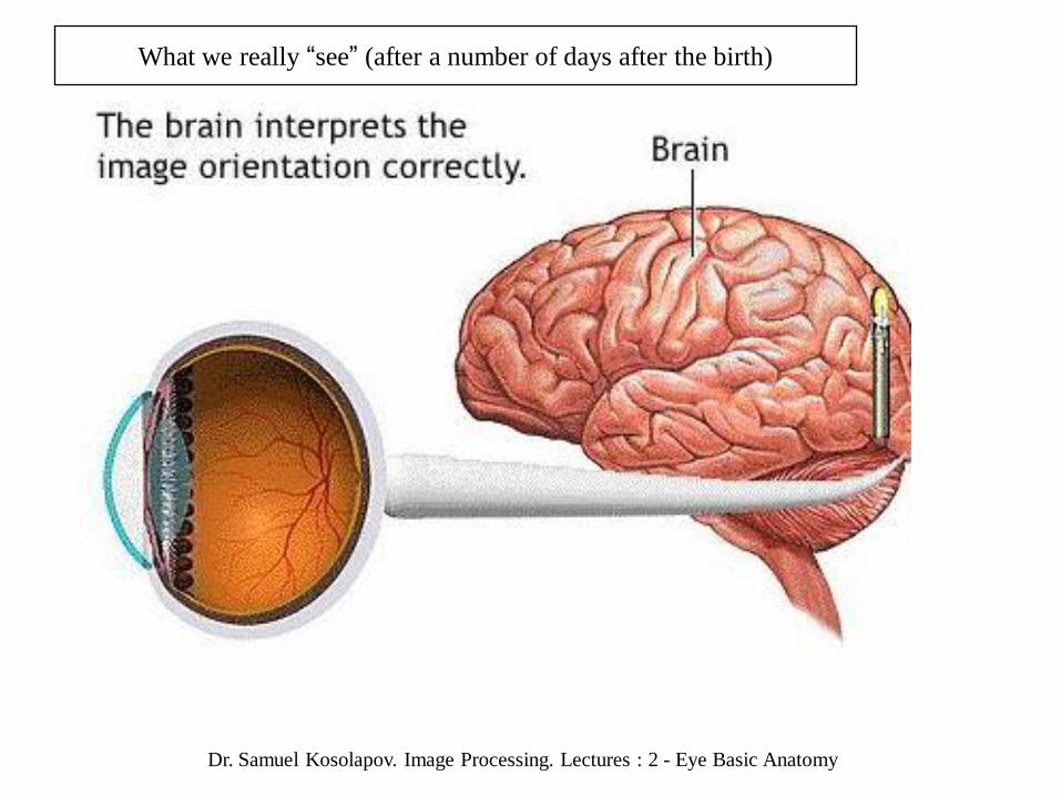
What we really “see” (after a number of days after the birth)

Dr. Samuel Kosolapov. Image Processing. Lectures : 2 - Eye Basic Anatomy
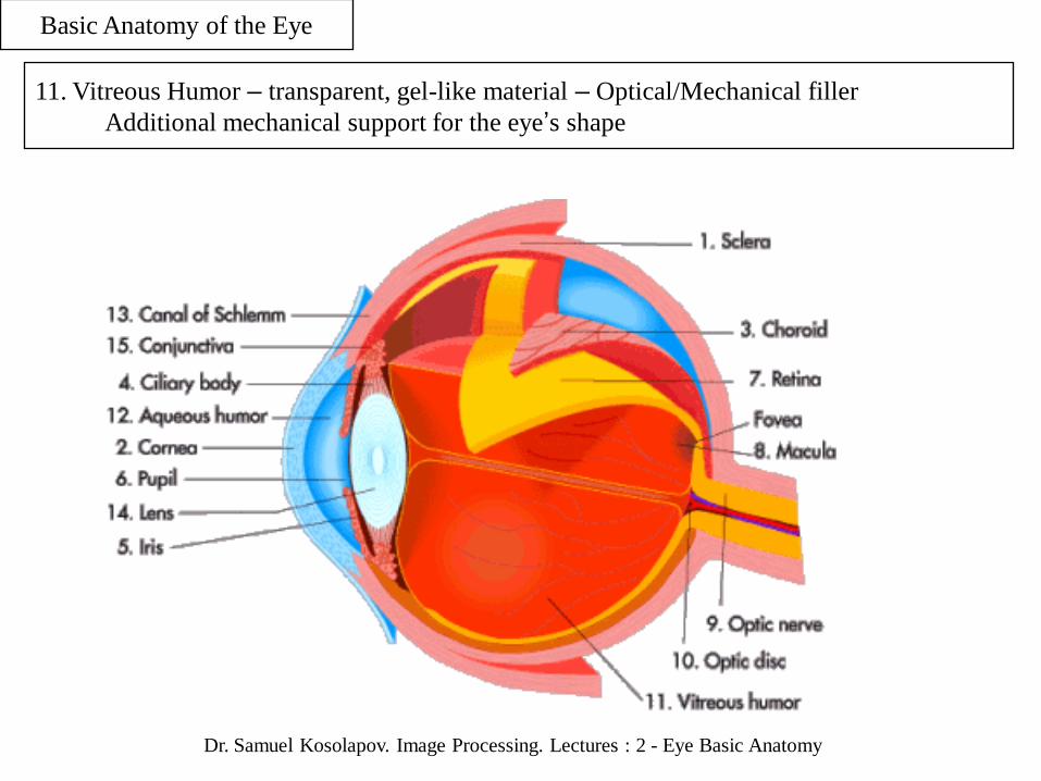
Basic Anatomy of the Eye
1. Sclera {בן העין} – Outermost layer of the eye.
Maintains the shape of the eye. Non-transparent to light.

Dr. Samuel Kosolapov. Image Processing. Lectures : 2 - Eye Basic Anatomy
15. Conjunctiva – { לחמית( אנטומיה )} (Wrong pointer in the basic image)The conjunctiva is merely a thin, transparent membrane covering the cornea,
and yet its function is vital – it protects the eye from airborne debris.
This is actually only one of the protective features of the human eye.
Others include the orbit (or eye socket), the eyelashes {ריס}
and, quite surprisingly, the eyebrows {גבה}
– their function being to stop sweat {“water”, זעה} from running into the eye.

Dr. Samuel Kosolapov. Image Processing. Lectures : 2 - Eye Basic Anatomy
Basic Anatomy of the Eye
2. Cornea { קרנית( אנטומיה } – Front part of the Sclera(1) which IS transparent to light
Light entering the eye pass Conjunctiva and Cornea first.

Dr. Samuel Kosolapov. Image Processing. Lectures : 2 - Eye Basic Anatomy
Basic Anatomy of the Eye
12. Aqueous Humor – transparent watery material - Optical Filler
SHAPE of the cornea and n: defines optical properties of the eye (~70% of the focusing):
Effective Lens: nair – naqueous humor 12, Rcornea
Idea: Change Rcornea by (say) laser cerathomy

Dr. Samuel Kosolapov. Image Processing. Lectures : 2 - Eye Basic Anatomy
Basic Anatomy of the Eye
5. Iris {צמצם ,diapfragm} and 6. Pupil {אישון}
The iris is the colored part of the eye.
The color of the iris is determined by the color of the connective tissue and pigment cells.
Less pigment makes the eyes blue; more pigment makes the eyes brown.
The iris is an adjustable diaphragm around an opening called the pupil.
Iris Pupil

Dr. Samuel Kosolapov. Image Processing. Lectures : 2 - Eye Basic Anatomy
Iris Pupil
Basic Anatomy of the Eye
The iris has two muscles:
The dilator muscle makes the iris smaller and therefore the pupil larger,
allowing more light into the eye;
the sphincter muscle makes the iris larger and the pupil smaller,
allowing less light into the eye.
Pupil size can change from ~ 2 millimeters to ~8 millimeters.
This means that by changing the size of the pupil,
the eye can change the amount of light that enters it by 30 times.
Goals Equivalent of the Goals of Diaphragm/Iris in the Camera:
Change amount of light entering the eye
Larger focal depth in the sunny day

Dr. Samuel Kosolapov. Image Processing. Lectures : 2 - Eye Basic Anatomy
The pupil is basically a circular hole in the middle of the iris,
which regulates the amount of light that passes through into the retina.
In the dark, as the amount of natural light diminishes, the pupil will expand
- allowing as much of the little light that there is to pass through.
In the day, when everything is brighter,
the pupil will constrict to limit the amount that passes through.
Surplus: Better Focal Depth (why am I without glasses now ?)
Bright Light Dim Light

Dr. Samuel Kosolapov. Image Processing. Lectures : 2 - Eye Basic Anatomy
Other things can also cause the pupil to dilate.
Most common among these are: rapidly changing from distant to 'near' focus; drugs;
and when we something that arouses or interests us
(an effect replicated by women in medieval times by putting belladonna in their eyes,
but please don't try this at home!
Atropine : for eye tests (to see periphery of the retina better)
More about Iris/Pupil operation:
Closed Loop System;
Indirect Indicator of the Brain operation
Provide Flash (Step input) – See the Output (Transfer Function)
{ Rise Time, Pulsations }

Dr. Samuel Kosolapov. Image Processing. Lectures : 2 - Eye Basic Anatomy
Basic Anatomy of the Eye
14. Lens {עדשה}
The lens is a clear, bi-convex structure about 10 mm (0.4 inches) in diameter.
Accommodation : The lens CAN changes shape (by using muscles of the the ciliary body 4).
The lens is used to fine-tune vision. (~30% of the “focusing”)
Dr. Samuel Kosolapov. Image Processing. Lectures : 2 - Eye Basic Anatomy
Basic Anatomy of the Eye
11. Vitreous Humor – transparent, gel-like material – Optical/Mechanical filler
Additional mechanical support for the eye’s shape

Dr. Samuel Kosolapov. Image Processing. Lectures : 2 - Eye Basic Anatomy
Basic Anatomy of the Eye
7. Retina – light sensitive element: Converts light/image into (set of) electrical signals
Can be DETACHED (still work ~ 24 hours)
Dr. Samuel Kosolapov. Image Processing. Lectures : 2 - Eye Basic Anatomy
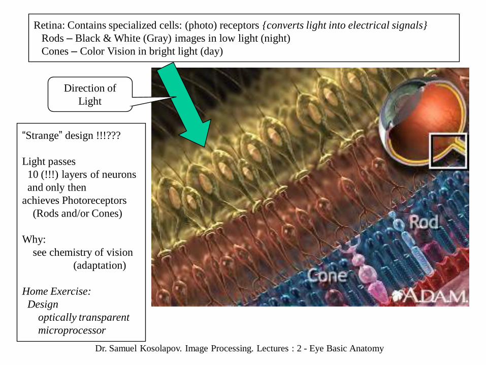
Retina: Contains specialized cells: (photo) receptors {converts light into electrical signals}
Rods – Black & White (Gray) images in low light (night)
Cones – Color Vision in bright light (day)
Direction of
Light
“Strange” design !!!???
Light passes
10 (!!!) layers of neurons
and only then
achieves Photoreceptors
(Rods and/or Cones)
Why:
see chemistry of vision
(adaptation)
Home Exercise:
Design
optically transparent
microprocessor

Dr. Samuel Kosolapov. Image Processing. Lectures : 2 - Eye Basic Anatomy
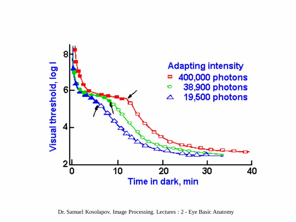
Resolution

Dr. Samuel Kosolapov. Image Processing. Lectures : 2 - Eye Basic Anatomy
Resolution

Dr. Samuel Kosolapov. Image Processing. Lectures : 2 - Eye Basic Anatomy

Dr. Samuel Kosolapov. Image Processing. Lectures : 2 - Eye Basic Anatomy

Dr. Samuel Kosolapov. Image Processing. Lectures : 2 - Eye Basic Anatomy

Dr. Samuel Kosolapov. Image Processing. Lectures : 2 - Eye Basic Anatomy

Dr. Samuel Kosolapov. Image Processing. Lectures : 2 - Eye Basic Anatomy

Dr. Samuel Kosolapov. Image Processing. Lectures : 2 - Eye Basic Anatomy

Dr. Samuel Kosolapov. Image Processing. Lectures : 2 - Eye Basic Anatomy

Dr. Samuel Kosolapov. Image Processing. Lectures : 2 - Eye Basic Anatomy
Carrot b-Carotene
Cut here Vitamin A

Dr. Samuel Kosolapov. Image Processing. Lectures : 2 - Eye Basic Anatomy

Dr. Samuel Kosolapov. Image Processing. Lectures : 2 - Eye Basic Anatomy

Dr. Samuel Kosolapov. Image Processing. Lectures : 2 - Eye Basic Anatomy

Dr. Samuel Kosolapov. Image Processing. Lectures : 2 - Eye Basic Anatomy
Wiring

Dr. Samuel Kosolapov. Image Processing. Lectures : 2 - Eye Basic Anatomy
Wiring

Dr. Samuel Kosolapov. Image Processing. Lectures : 2 - Eye Basic Anatomy

Dr. Samuel Kosolapov. Image Processing. Lectures : 2 - Eye Basic Anatomy

Dr. Samuel Kosolapov. Image Processing. Lectures : 2 - Eye Basic Anatomy
Dr. Samuel Kosolapov. Image Processing. Lectures : 2 - Eye Basic Anatomy

Dr. Samuel Kosolapov. Image Processing. Lectures : 2 - Eye Basic Anatomy
It has been commonly claimed that the vertebrate eye is functionally suboptimal,
because photoreceptors in the retina are oriented away from incoming light.
"In fact it is stupidly designed,"
writes the influential neo-Darwinian theorist George Williams
Cephalopod (squid and octopus) retina: "wired correctly,“
with its photoreceptors facing towards the light, and with its nerves
neatly tucked away behind the photoreceptor layer"
However, there are excellent functional reasons
for vertebrate photoreceptors to be oriented as they are.
Photoreceptor structure and function is maintained by a critical tissue,
the retinal pigment epithelium (RPE),
which recycles photopigments,
removes spent outer segments of the photoreceptors,
provides an opaque layer to absorb excess light,
and performs additional functions.
These aspects of the structure and function of the vertebrate eye
have been ignored in evolutionary arguments about suboptimality,
yet they are essential for understanding how the eye works.

Dr. Samuel Kosolapov. Image Processing. Lectures : 2 - Eye Basic Anatomy
What happens when light reaches the retina?
The retina is a complex part of the eye, and yet only the very back of it is sensitive to light.
Amazingly, this part of the retina has only the area of roughly the size of a ten pence coin,
and is packed with photosensitive cells called rods and cones.
Cones are the cells responsible for daylight vision,
of which there are three different kinds – each responding to a different wavelength of light.
One responds to red light, one to green light and one to blue light.
It is these cones which allow us to see in colour and detail.
Rods, on the other hand, are responsible for night vision.
They are sensitive to light but not to wavelength information (colour).
In darkness, the cones do not function at all – so we need rods in order to see things,
even if it is only in shades of grey.

Dr. Samuel Kosolapov. Image Processing. Lectures : 2 - Eye Basic Anatomy
Basic Anatomy of the Eye
8. Macula / Fovea (optical center of the eye)
Macula – Center of the retina. Fovea (centralis) – Center of the macula (contains cones only)

Dr. Samuel Kosolapov. Image Processing. Lectures : 2 - Eye Basic Anatomy
10. Optic Disk; 11. Optic Nerve; Blind Spot:
The blind spot is the point in the eyeball at the head of the optic nerve.
This is where nerve fibres and blood vessels leave the eye,
and as a result there are no photosensitive cells.
Any images, therefore, that are projected onto the blind spot
are simply not registered (“seen”) by the brain
Hard decision during design:
Decision #1. Last layer of the “pre-processor” positioned BEFORE the photoreceptors layer
Decision #2. Brain (main processor) positioned AFTER photoreceptor layer
Question: How to connect “pre-processor’ and “main processor” with a cable ???
“Solution” – Drill a hole and pass the cable through the hole !!!

Dr. Samuel Kosolapov. Image Processing. Lectures : 2 - Eye Basic Anatomy

Dr. Samuel Kosolapov. Image Processing. Lectures : 2 - Eye Basic Anatomy

Dr. Samuel Kosolapov. Image Processing. Lectures : 2 - Eye Basic Anatomy

Dr. Samuel Kosolapov. Image Processing. Lectures : 2 - Eye Basic Anatomy
Normal Vision: Focusing on the Retina (Image is Upside-Down Image)
3D 2D
Focusing in the Eye:
The eye is often compared with a camera.
In order for us to see anything,
an image must be sent to an area at the back of the eye called the retina.
Of course, just like a camera, the image must first be focused.
The images we see are no more than light reflected from the object we are looking at.
Dr. Samuel Kosolapov. Image Processing. Lectures : 2 - Eye Basic Anatomy
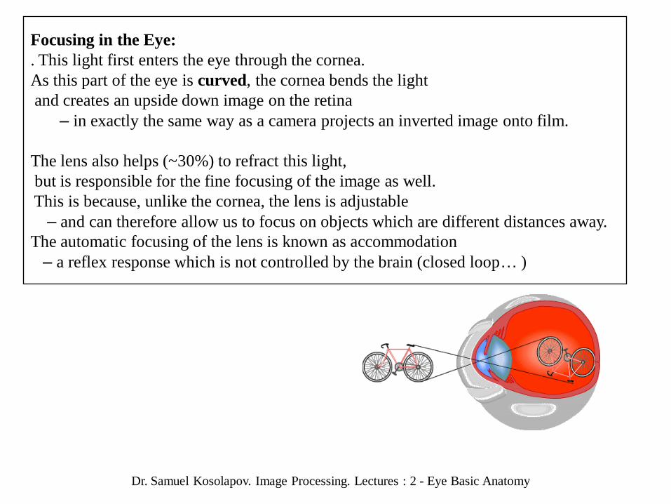
Focusing in the Eye:
. This light first enters the eye through the cornea.
As this part of the eye is curved, the cornea bends the light
and creates an upside down image on the retina
– in exactly the same way as a camera projects an inverted image onto film.
The lens also helps (~30%) to refract this light,
but is responsible for the fine focusing of the image as well.
This is because, unlike the cornea, the lens is adjustable
– and can therefore allow us to focus on objects which are different distances away.
The automatic focusing of the lens is known as accommodation
– a reflex response which is not controlled by the brain (closed loop… )

Dr. Samuel Kosolapov. Image Processing. Lectures : 2 - Eye Basic Anatomy
ACCOMODATION: Lens (by operating proper muscles – 4 chilary body)
changes its shape (and thus focal length) {try co create something like this}
The ciliary body comprises two parts
– the ciliary process
and the ciliary muscle. which causes the lens to change shape.
If the eye is focusing on a distant object the muscles relax,
causing the ligaments to tighten and the lens to lengthen. Rlens is larger less “focusing”
When we focus on an object nearby the muscles tighten,
the ligaments slacken, and the lens shortens Rlens is smaller more “focusing”
.
Distant Object Nearby Object

Dr. Samuel Kosolapov. Image Processing. Lectures : 2 - Eye Basic Anatomy
But Retina is not flat (3D sphere) !!!! What about focusing and Field of view ???
Good Engineering: Excellent Field Of View of the Human/Vertebrate Eye
(compare with camera)
Field of View ~ 180 degrees !!!

Dr. Samuel Kosolapov. Image Processing. Lectures : 2 - Eye Basic Anatomy
Field of View (Angle of view)
Different animals have different fields of view, depending on the placement of the eyes.
Humans have a 180-degree forward-facing field of view,
Some birds have a complete 360-degree field of view.
In addition the vertical range of the field of view may vary.
The range of visual abilities is not uniform across a field of view,
and varies from animal to animal.
For example, binocular vision, which is important for depth perception,
only covers 140 degrees of the field of vision in humans;
the remaining peripheral 40 degrees have no binocular vision
(because of the lack of overlap in the images from either eye for those parts of the field of view).
The afore-mentioned birds would have a scant 10 or 20 degrees of binocular vision.
Home exercise:
Design CCD (or CMOS) positioned on the internal surface of the sphere
Hint: last models of photo cameras have some curvature in the horizontal direction

Dr. Samuel Kosolapov. Image Processing. Lectures : 2 - Eye Basic Anatomy
Short sight (Myopia) means
that the image is focused
in front of the retina
Reasons: either the eyeball is elongated
or the lens too thick
OR: Muscles are too weak
for PROPER accommodation
Long sight means
that the image is focused
behind the retina
Focusing problems:

Dr. Samuel Kosolapov. Image Processing. Lectures : 2 - Eye Basic Anatomy
nair = 1.000
nwater = 1.33
ncornea = 1.376
(Why can’t we see well
in the water ?)
naqueous_humor = 1.336
Pupil Diameter : 2-8 mm
~ 0.1 sec
Crystalline Lens:
Diameter: 9 mm
Thickness: 4 mm
~ 22000 fine layers (like onion)
nlens varies from 1.386-1.406
(depending on layer/position,
compare with gradient index (GRIN) lens !!!)
nvitreous_humor = 1.337
Summary: Optical Structure & Properties of the Eye

Dr. Samuel Kosolapov. Image Processing. Lectures : 2 - Eye Basic Anatomy
Gullstrand's Eye Model (1862-1930)

Dr. Samuel Kosolapov. Image Processing. Lectures : 2 - Eye Basic Anatomy

Dr. Samuel Kosolapov. Image Processing. Lectures : 2 - Eye Basic Anatomy
Practical Optical Model of the Eye (for exercises)
Thin lens with F=17.1 mm (front focal length: 24mm/1.4)

Dr. Samuel Kosolapov. Image Processing. Lectures : 2 - Eye Basic Anatomy
The physiology of vision determines how we perceive images,
regardless of whether they are analog or digital.
Similar to most digital image sensors,
the human eye processes images by dividing light into its three primary colors
, red, green and blue.
The human eye is limited to perceiving approx. 40 shades of grey, 200 colors,
and a resolution of 10-14 line pairs per mm (at a viewing distance of 25 cm or 10 in.).
Digital images are primarily characterized by resolution and color depth.
Resolution means clarity and sharpness of an image, commonly expressed in dots per inch.
Color (or bit) depth (typically expressed in bits)
indicates how many colors an image can contain (e.g. 8 bits = 256 colors).
Human eye resolution is 1 arcmin.
At closest focus (10 cm), that's 30 microns or 1 billion pixels per square meter.
According to tests, human eye resolution is about 0.3 arc minute
(of course it's depends on many conditions),
that's about 0.087 mm (about 300 dpi) at one meter distance,
and about 100 dpi at 3 meters distance.
That means for a 42" 16:9 screen, at 3 meters distance it should have about 3600x2000.

Dr. Samuel Kosolapov. Image Processing. Lectures : 2 - Eye Basic Anatomy
Why ultraviolet vision?
Many insect-pollinated flowers appear to the honeybee quite different
from the way they appear to us.
The sharp contrasts between flowers that appear similar to us partly
explains the efficiency with which honeybees secure nectar
from only one species of flower
at a time even when other species are also in bloom.
The photos (courtesy of Dr. Eisner) show
a blackeyed susan photographed
in visible light (left) and under ultraviolet light (right).