eye flourecein angiography
TRANSCRIPT


Mohamed Abdel-Aziz (MSc,ICO,Egyption Board)
Mataryia Teaching Hospital
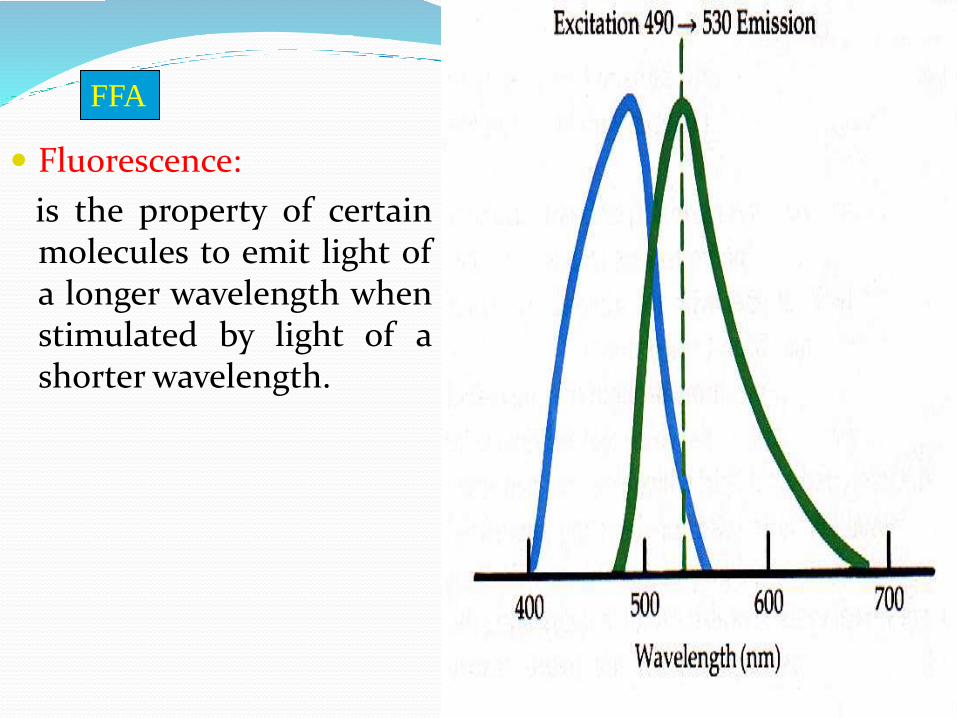
Fluorescence:
is the property of certainmolecules to emit light ofa longer wavelength whenstimulated by light of ashorter wavelength.
FFA

Characteristics of Fluorescein Alkaline solution
Highly fluorescent
Absorbs blue light (490 nm)
Emits yellow-green (530nm)
Removal from blood by kidneys and liver within24-48 hrs.
injection of 5 cc of 10% Na Fl, or 3 cc of 25%solution
FFA

Hazards Minimal relatively safe drug
Temporary tan skin color
Fl. Urine discoloration
Transient nausea and vomiting
Laryngeal edema
Rarely – Syncope, resp. or Cardiac arrest
Rx – I.V. Cortisone
A physician in the 1st few minutes
FFA

Anatomic Considerations

FFA
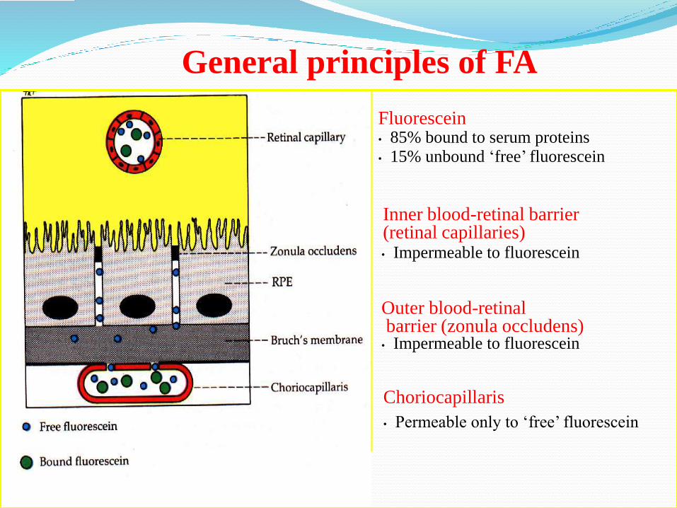
General principles of FA
Fluorescein• 85% bound to serum proteins• 15% unbound ‘free’ fluorescein
• Impermeable to fluorescein
Outer blood-retinal barrier (zonula occludens)
• Impermeable to fluorescein
Choriocapillaris
• Permeable only to ‘free’ fluorescein
Inner blood-retinal barrier(retinal capillaries)

Filters
1.Blue excitation filter
2.Yellow-green filter
FFA

Angiographic phases
Angiographic phases:
Five angiographic phases:
• Pre arterial (choroidal 9-15 seconds)
• Arterial
• Arteriovenous(capillary)
• Venous
• Recirculation
FFA

1-Choroidal flush 2-Arterial phase

3-Arteriovenous phase

4-Venous phase
Mid Phase Late Phase

Interpretation of FA
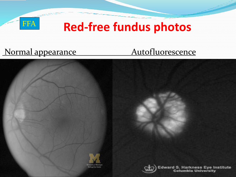
Red-free fundus photos
Normal appearance Autofluorescence
FFA

Abnormal angiographic findings
Hypofluorescence:
Filling defect
Blocking defect
Hyperfluorescence :
window defect
Leakage
Pooling
Staining

Hypofluorescence
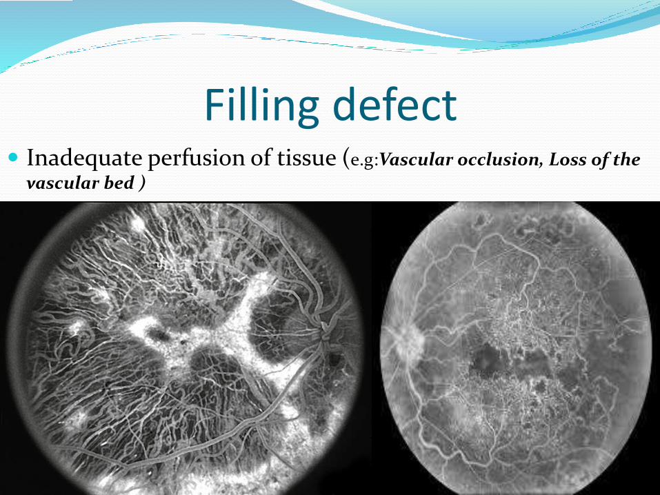
Filling defect Inadequate perfusion of tissue (e.g:Vascular occlusion, Loss of the
vascular bed )
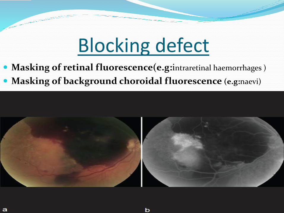
Blocking defect Masking of retinal fluorescence(e.g:intraretinal haemorrhages )
Masking of background choroidal fluorescence (e.g:naevi)
Hyperfluorescence
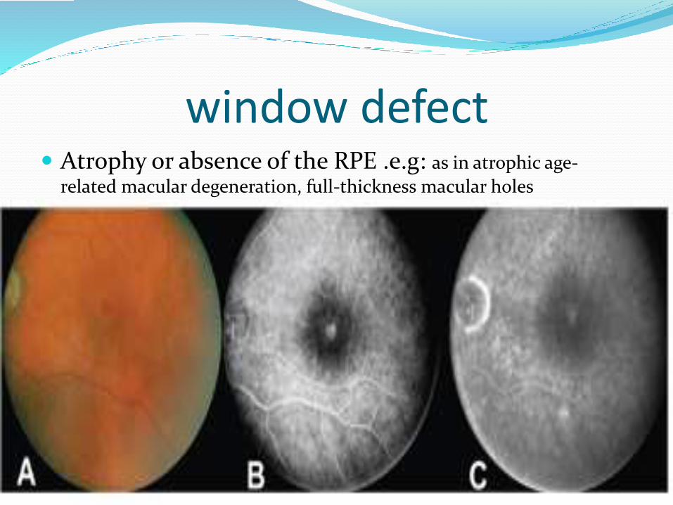
window defect Atrophy or absence of the RPE .e.g: as in atrophic age-
related macular degeneration, full-thickness macular holes

Pooling breakdown of the outer blood–retinal barrier (RPE tight
junctions)
A) In the sub-retinal space: increases in intensity and area, the maximum extent remaining relatively well-defined.(e.g:CSR)

Pooling B ) In the sub-RPE space: early hyper-fluorescence which
increases in intensity but not in size .(e.g:PED)
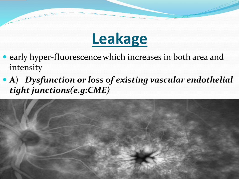
Leakage early hyper-fluorescence which increases in both area and
intensity
A) Dysfunction or loss of existing vascular endothelial tight junctions(e.g:CME)

Leakage B) Primary absence of vascular endothelial tight
junctions (e.g:CNV,PDR)

Staining prolonged retention of dye in tissue(e.g:drusen,
exposed sclera )
Systematic approach to reporting
angiograms
a- Patient's age and gender.
b-Right or left eyes.
c-Comment on any color and red free images.
d -Timing of filling, especially arm-to-
eye transit time.
e-Briefly scan through the sequence of images