eye health no · 2013-06-08 · community eye health vol 14 no. 40 2001 vitamin a deficiency,...
TRANSCRIPT

CommunityEEyyee HHeeaalltthh
A N I N T E R N A T I O N A L J O U R N A L T O P R O M O T E E Y E H E A L T H W O R L D W I D E
S U P P O R T I N G V I S I O N 2 0 2 0 : T H E R I G H T T O S I G H T
Community
53
Clare Gilbert
MD MSc FRCOphthSenior Lecturer
International Centre for Eye Health
Institute of Ophthalmology
11–43 Bath Street
London, EC1V 9EL, UK
The main issues in relation to blindness
in children relate to a better understand-
ing of the epidemiology, which has led to
improved priority setting. In this article the
most recent epidemiological data will be
presented, the consequences for the Vision
2020 programme will be discussed, and
research priorities considered.
Definitions
A blind child is an individual aged less than
16 years, who has a visual acuity in the bet-
ter eye of <3/60. However, many studies do
not use this definition, which makes it diffi-
cult to compare the findings of different
studies.
Prevalence and Incidence
The prevalence of blindness in children
(i.e., the proportion of the child population
who are blind), varies from approximately
0.3/1,000 children in wealthy regions of
the world, to 1.2/1,000 in the poorer
countries / regions.1 Blindness in
children is more common in poor
regions for two main reasons: firstly,
there are diseases and risk factors
which can lead to blindness from
causes that do not now occur in
industrialised countries (e.g., measles,
vitamin A deficiency, ophthalmia
neonatorum, malaria), and, secondly,
there are fewer well equipped eye
departments with ophthalmologists,
nurses and ophthalmic paramedics
trained in managing treatable causes
of blindness (e.g., cataract and glau-
coma). The incidence is therefore
higher, and fewer blind children have
their sight restored.
Incidence data are very difficult to
obtain, but it has been estimated that there
are 8 new blind children for every 100,000
children each year in industrialised coun-
tries. The figures are likely to be higher in
developing countries.
Magnitude of Blindness
Globally there are estimated to be 1.4 mil-
lion children who are blind, and around
three quarters live in developing countries.
Although the actual number of children
who are blind is much smaller than the
number of adults blind, e.g., from cataract,
the number of years lived with blindness by
blind children is almost the same as the total
number of ‘blind years’ due to age-related
cataract. The high number of blind years
resulting from blindness during childhood
is one of the reasons why the control of
childhood blindness is a priority of the
WHO/IAPB Vision 2020: The Right to
Sight programme.2
Causes of Blindness in Children
The available data suggest that there is wide
regional variation in the major causes of
blindness in children. Tables 1 and 2 show
the causes of blindness obtained from
examining over 10,000 blind children, with
the causes classified using the World Health
Organization's classification system.3 These
data do not take account of children who are
‘blind’ from refractive errors.
In wealthy parts of the world lesions of
the central nervous system predominate,
while in poorer countries corneal scarring
as a result of acquired diseases are the most
important causes. Table 3 shows estimates
of the number of blind children by anatomi-
cal site, and by underlying cause.
Editorial: New Issues in Childhood Blindness Clare Gilbert 53
IOLs in Children David Yorston 57
The Increasing Problem of ROP Andrea Zin 58
Guidelines for Setting up a Low Vision Hasan Minto
Programme for Children & Haroon Awan 60
Training Ophthalmologists for
Child Eye Care Centres Anil K Mandal 62
Evaluation of Training Detlef Prozesky 65
Vernal Keratoconjunctivitis in Uganda Medi Kawuma 66
Blindness in Kenya Jerry Harrell, Dean Larson,
Esther Menza, Ammon Mboti 68
Children’s eye care in TanzaniaPhoto: Sue Stevens

Commun it yEEyyee HHeeaalltthh
Volume 14 Issue No. 40 2001
International Centre for Eye HealthInstitute of OphthalmologyUniversity College London11– 43 Bath StreetLondon EClV 9EL
Tel: 00 44 (0)207 608 6909/6910/6923Fax: 00 44 (0)207 250 3207E-mail: See box on this pageWeb-site: www.jceh.co.uk
World Health Organization Collaborating Centre for Prevention of Blindness
EditorDr Murray McGavin
Nurse ConsultantMs Susan Stevens
Administrative DirectorMs Ann Naughton
Editorial SecretaryMrs Anita Shah
Editorial Review CommitteeDr Allen FosterDr Clare GilbertProfessor Gordon JohnsonDr Darwin MinassianDr Ian MurdochDr Daksha PatelDr Richard WormaldDr Ellen SchwartzDr David Yorston
Language and CommunicationConsultant Professor Detlef Prozesky
Consulting EditorsDr Harjinder Chana (Mozambique)Dr Parul Desai (UK)Dr Virgilio Galvis (Colombia)Professor M Daud Khan (Pakistan)Professor Volker Klauss (Germany)Dr Susan Lewallen (Canada) Dr Donald McLaren (UK)Dr Angela Reidy (UK)Professor I S Roy (India)Professor Hugh Taylor (Australia)Dr Randolph Whitfield, Jr (Kenya)
Typeset byRegent Typesetting, London
Printed byThe Heyford Press Ltd.
On-line edition by OASIS/Xalt
ISSN 0953-6833
E d i t o r i a l
Community Eye Health Vol 14 No. 40 2001
© Journal ofCommunity Eye Health
InternationalCentre for Eye Health, London
Articles may be photocopied, reproduced or translated provided these are not used
for commercial or personal profit. Acknowledgements should be made to the
author(s) and to the Journal of Community Eye Health.
Readersareaskedtousethefollowing specifiedE-mailaddressesonly whencontacting
the Journal or other Departments at ICEH:
Journal mailing list requests – [email protected]
Journal editorial – [email protected]
Resource Centre (information service) – [email protected]
Research enquiries – [email protected]
Courses – [email protected]
Regional Variation in the Magnitude
and Major Causes of Blindness in
Children
It is possible to combine what we know
about the prevalence of blindness in
children with data on causes, and apply this
to a total population of one million people
(Table 4). This information is perhaps more
useful for planning. Figure 1 shows these
data.
Avoidable Causes
In all regions of the world there are causes
which are amenable to primary, secondary
and tertiary prevention, but the proportions
vary from region to region (Table 5).
Vision 2020 Priorities
Given these findings, the following condi-
tions are priorities for control:4
• Corneal scarring, due to measles,
vitamin A deficiency,harmful traditional
eye medicines, and ophthalmia
neonatorum: priorities in poor and very
poor regions
• Cataract and glaucoma: important
treatable causes in all regions
• Retinopathy of prematurity, a condition
which is preventable and treatable;
important in middle income countries,
and in urban centres in developing
countries
• Refractive errors: treatable cause in all
regions
• Lowvision:servicesneedtobeexpanded
or developed in all regions.
Targets for disease control
The following targets have been agreed for
disease control:
1. Reduce the global prevalence of
childhood blindness from 0.75/1,000
children to 0.4/1,000 children.
2. Elimination of corneal scarring caused
by vitamin A deficiency, measles, or
ophthalmia neonatorum.
3. Elimination of new cases of congenital
rubella syndrome.
4. All children with congenital cataract to
receive appropriate surgery, with imme-
diate and effective optical correction, in
suitably equipped specialist centres.
54
The Journal of Community Eye Health is pu blished four times a year.FREE TO DEVELOPING COUNTRY APPLICANTS
2002/2003 Subscription Rates for Applicants Elsewhere
1 Year: UK£28 / US$45 2 Years: U K £ 5 0/U S $ 8 0
(4 Issues) (8 Issues)
To place your subscription, please send an international cheque/banker’s order made
payable to UNIVERSITY COLLEGE LONDON or credit card details with a note
of your name, full address and occupation (in block capitals please) to:
Journal of Community Eye Health, International Centre for Eye Health,
Institute of Ophthalmology, 11–43 Bath Street, LONDON, EC1V 9EL, UK
Tel: 00 44 (0)20 7608 6910 Fax: 00 44 (0)20 7250 3207
E-mail: [email protected] Website: www.jceh.co.uk
EEyyee HHeeaalltthh

5. All babies at risk of retinopathy of pre-
maturity to have fundus examination,
by a trained observer, 6-7 weeks after
birth. Cryotherapy or laser treatment to
be provided for all those with treatable
disease.
6. All school children to receive a simple
vision screening examination, with
glasses provided for all those with sig-
nificant refractive error. This should be
integrated into the school health pro-
gramme.
Human resource development
The implications and recommendations
for human resources development are as
follows:
1. Ensure that prevention of childhood
blindness is an explicit aim of all
primary health care programmes.
2. Ensure that all secondary level eye
clinics have facilities to provide approp-
riate spectacles for children with refrac-
tive errors.
3. Train one refractionist per 100,000
population by 2010.
4. Train at least one low vision worker for
every 20 million children, by 2010, and
for every 5 million by 2020.
5. Train one paediatric-orientated ophthal-
mologist for every 50 million popula-
tion by 2010, and one per 10 million
population by 2020.
Appropriate technology & infrastructure
There is the following need for appropriate
technologyandinfrastructuredevelopment:
1. Development of low cost, high quality
low vision devices, which should be
widely available, even in low income
countries.
2. Establish a network of specialist ‘child-
hood blindness’ tertiary centres.
In this editionofthe Journal of Community
Eye Health there are articles which address
some of the priority causes of blindness in
children. The article on cataract discusses
the relative merits of intraocular lens
implantation in children, as a means of cor-
recting their aphakia. The article on retino-
pathy of prematurity from Brazil highlights
how screening programmes need to be
expanded in Latin America if blindness
from ROP is to be brought under control.
Research Issues
Corneal scarring. The control of diseases
that cause corneal scarring lies in primary
healthcare,publichealth interventions,and
childsurvivalprogrammes. However,there
is still a need to develop cost effective,
sustainable interventions at the community
and household level for the control of
E d i t o r i a l
55
Wealthiest Region Poorest RegionRegion EME FSE LAC MEC China India OAI SSANumber examined: * 504 1,007 866 1,131 2,283 850 1,702
Globe 10 12 12 14 26 25 21 9Cornea 1 2 8 8 4 27 21 36Lens 8 11 7 20 19 11 19 9Uvea 2 5 2 4 1 5 3 5Retina 25 44 47 38 25 22 21 20Optic nerve 25 15 12 8 14 6 7 10Glaucoma 1 3 8 5 9 3 6 6Other (incl. CNS) 28 8 4 3 2 1 2 5
Total: 100 100 100 100 100 100 100 100
Table 1: Regional Variation in the Causes of Blindness in Children:
Descriptive Classification by World Bank Region (%)
* data from published studies
EME = Established Market Economies; FSE = Former Socialist Economies;
LAC = Latin America and Caribbean; MEC = Middle East Crescent;
OAI = Other Asia and Islands; SSA = Sub-Saharan Africa.
Wealthiest Region Poorest RegionRegion EME FSE LAC MEC China India OAI SSANumber examined: * 504 1,007 866 1,131 2,211# 850 1,702
Hereditary 45 18 22 53 31 26 27 20Intrauterine 7 6 8 2 0 2 3 3Perinatal 24 28 28 1 2 2 9 6Childhood 10 5 10 7 14 28 14 34Unknown 14 43 32 37 53 42 47 37
Total: 100 100 100 100 100 100 100 100
Table 2: Regional Variation in the Causes of Blindness in Children:
Aetiological Categories by World Bank Region (%)
* data from published studies # aetiological data not available for 72 children
Community Eye Health Vol 14 No. 40 2001
VAD = vitamin A deficiency ON = ophthalmia neonatorum
HTEM = harmful traditional eye medicines
Fig. 1: Pattern of Blindness in Children by Level of
Socio-Economic Development
ROP
Preventable corneal scarring from VAD, measles, ON, HTEM
Treatable cataract, glaucoma
Unavoidable causes: anomalies, genetic disease, CNS lesions

Community Eye Health Vol 14 No. 40 2001
vitamin A deficiency, interventions that do
not depend on vitamin A supplementation.
Cataract. Cataract surgery is much more
difficult in children, and very few clinical
trials have been undertaken to explore the
optimum management. Further research is
also needed into the aetiology of cataract in
different parts of the world, as well as qual-
itative research to investigate barriers to
the uptake of cataract surgery.
Retinopathy of prematurity. The pattern of
disease in middle income countries seems
to be different from that currently seen in
industrialised countries. There is a need for
research into risk factors in different set-
tings, and the validity of different methods
of screening for threshold disease needs to
be investigated.
Low vision. There are very few studies of
low vision in children. It is not really
known how common it is, and what the
major causes are at the population level.
There are virtually no studies which have
addressed the issue of best low vision
devices for children.
Diseases of unknown cause. There are
many blinding eye diseases where the
underlying cause is not known, e.g., con-
genital anomalies of the eye. Research is
needed to try and clarify the relative contri-
bution of genetic and environmental risk
factors.
References
1 Gilbert C E, Foster A. Childhood blindness inthe context of VISION 2020 – The Right to
Sight. Bull WHO 2001; 79: 227–232.
2 World Health Organization, Geneva. GlobalInitiative for the Elimination of Avoidable
Blindness. Geneva WHO/PBL/97.61.3 Gilbert C, Foster A, Negrel D, Thylefors B.
Childhood blindness: a new form for recording
causes of visual loss in children. Bull WHO
1993; 71: 485-489.
4 World Health Organization, Geneva. Preventingblindness in children: Report of WHO/IAPB sci-
entific meeting (Unpublished document
WHO/PBL/00.77).
✩ ✩ ✩
56
Editorial
High income Middle income Low incomeBlind = 90,000 Blind = 290,000 Blind = 1,020,000
ROP 9,000 (10%) Cataract 45,000 (15%) Corneal scar 200,000 (20%)
Teratogens 5,400 (6%) ROP 29,000 (10%) Cataract 133,000 (13%)
Cataract 5,400 (6%) Glaucoma 17,000 (6%) Glaucoma 60,000 (6%) Glaucoma 2,000 (2%) Teratogens 12,000 (4%) Optic atrophy* 60,000 (6%)
Total Total Totalavoidable: 21,800 (24%) avoidable: 103,000 (35%) avoidable: 453,000 (45%)
Table 5: Regional Variation in Avoidable Causes of Blindness
* due to infections, trauma and tumours
High income Middle income Low income Very low income
% population 20% 30% 40% 50% childrenNumber of 200,000 300,000 400,000 500,000 childrenBlindness 0.3/1000 0.6/1,000 0.9/1,000 1.2/1,000 prevalenceNo. of blind 60 180 360 600 children
Cause % Cause % Cause % Cause %CNS/Other 80 CNS/Other 55 CNS/Other 60 CNS/Other 35ROP 10 ROP 25 ROP 0 ROP 0Cat/glaucoma 10 Cat/glaucoma 20 Cat/glaucoma 20 Cat/glaucoma 15Scarring 0 Scarring 0 Scarring 20 Scarring 50
Table 4: Estimates of the Magnitude and Major Causes of Blindness in
Children per Million Total Population
CNS = central nervous system ROP = retinopathy of prematurity Cat = cataract
Short Courses
Table 3: Estimates of Number of Blind Children
by Anatomical Site, and Underlying Aetiology
Anatomical Number Examples Aetiological Number Examplessite affected category affected
Retina 381,000 Dystrophies Hereditary 423,000 CataractCornea 231,000 Scarring Childhood 260,000 MeaslesWhole eye 230,000 Microphthalmos Perinatal 151,000 Ophthalmia
neonatorumLens 170,000 Cataract Intrauterine 50,000 RubellaOptic nerve 167,000 Optic atrophy Unknown 516,000 PhthisisGlaucoma 68,000 GlaucomaUvea 50,000 AniridiaOther 103,000 Cortical blindness
Total: 1,400,000 1,400,000
Management Training for Eye Care Managers (2 weeks, March 2002)
Contact: Keerti Bhyusan Pradhan, LAICO-Aravind Eye Care System
1-Anna Nagar,Madurai-625020, Tamilnadu, India
Phone: 0091 (0)452-537580; Fax: 0091 (0)452-530984; Email: [email protected]
Course for Cataract Surgical Rate Workers and Their Trainers (1 week, April 2002)
Contact: International Training and Consultancy Program
PO Box 23.310, Dar es Salaam, Tanzania
Tel. 00 255 (0)22 2601 544 or 00 255 (0)22 2601 543. E-mail: [email protected]

David Yorston
FRCS FRCOphthSpecialist Registrar
Moorfields Eye Hospital
London, UK
Medical Advisor
Christian Blind Mission International
IOLs in Children
The insertion of an intraocular lens (IOL)
is a routine part of cataract surgery in
adults, even in many developing countries.
It is now over fifty years since the first IOL
was implanted. However, until recently,
IOLs were not widely used in children.
As evidence for the long-term safety of
IOLs in adults accumulates, there is a
growing willingness to use IOLs in
children. Provided the zonule is stable and
the eye is not inflamed, IOL implantation
is already routine in children over five
years old, and increasingly in children
between two and five years. The use of
IOLs in children under two years remains
very controversial. One reason for this is
that the eye changes very rapidly in young
children. In a three month old baby, an
IOL power of 28-30D may be required for
emmetropia. However, this will lead to
significant myopia in later life. Unlike a
spectacle lens or contact lens, it is not
simple to change the power of an IOL.
Secondly, the diameter of the lens in an
infant is 2mm less than an adult lens. This
makes it difficult to implant a standard
adult IOL into the capsular bag. The maxi -
mumdiameteroftheIOLshouldnotexceed
12 mm. Smaller IOLs designed for use in
children can be obtained. The lens should
be placed in the capsular bag to reduce
intraocular inflammation and the risk of
complications such as aphakic glaucoma.
Possible Complications of IOLs in
Children
Fibrinous uveitis. A major problem with
implantation of IOLs in children is the
increased inflammatory response, particu-
larly in heavily pigmented eyes. A fibri-
nous uveitis is often seen about 3-7 days
after surgery. This may be controlled by
intensive topical steroids (hourly Gutt.
prednisolone 1%), or by orbital floor,
steroid depot injections. In a developing
country, it is probably sensible to keep the
child in hospital for a week after surgery to
ensure early detection and treatment of
uveitis. The risks of steroids should be
considered. Despite steroid treatment,
some children will develop a
dense fibrous membrane, which
may requiresurgical removal.
Astigmatism and residual refrac-
tive error. While an IOL corrects
the aphakia, it does not correct
astigmatism, and there will almost
certainly be some residual spheri-
cal error. This means that specta-
cles will still be necessary to
achieve the best possible vision.
These spectacles will be lighter,
and easier to wear than the thick
lenses required for spectacle cor-
rection of aphakia. As they are rel-
atively low powered lenses, there are fewer
optical aberrations. It is important that the
parents – and the medical and nursing staff
– realise that insertion of an IOL does not
mean that glasses will not be required.
We recommend that children should
be refracted within a month of IOL implan-
tation. Thereafter, children should be
refracted every four months until they are
two years old, and then every six months.
If there is any amblyopia, it may be wise to
refract even more often. Refraction must
be carried out by someone who is skilled at
accurate retinoscopy.
In young children, the glasses should
leave the child slightly myopic (approx.
–1.0D) as this will give them a good depth
of field, and most things and people they
want to look at will be within one or two
metres. As the child gets older, and starts to
read small print, an additional reading cor-
rection will be required.
Unsuitable eyes. Some eyes are unsuitable
for IOL insertion. For example, microph-
thalmic eyes may be too small to receive an
IOL. It is inadvisable to insert an IOL if the
corneal diameter is less than 9mm. Eyes
with chronic uveitis – associated with juve-
nile rheumatoid arthritis and rubella syn-
drome, for example – should never have an
IOL, as the presence of an IOL may wors-
en the intraocular inflammation.
If the surgery is difficult, and it appears
that it may be impossible to implant an IOL
withoutdamagingthecorneal endothelium,
itisbetter toleavethechild aphakicwithan
intact cornea rather than risk later corneal
decompensation.
Advantages of IOLs in Children
The major advantage of an IOL is that it
provides permanent continuous correction
of the aphakia. This may be important in
preventing amblyopia, and encouraging
normal visual development. Although
glasses are necessary to obtain the best
vision, uncorrected pseudophakic vision is
probably better than uncorrected aphakic
vision.
Which Lens Should Be Used?
Many different materials and designs of
intraocular lens (IOL) are available.
Although anterior chamber lenses have
been shown to be safe and effective in
adults, there is no evidence confirming
their safety in children. It is recommended
that anterior chamber lenses should not be
used in children at this time.
Polymethyl methacrylate (PMMA) has
been the material of choice for all IOLs
until recently. Coating the PMMA lens
with heparin greatly reduces intraocular
inflammationandfibrinformation.Ifcoated
lenses are not available, intra-operative
addition of heparin to the infusion fluid
may be beneficial. There are now silicon
IOLs, hydrophilicacrylic IOLs,and hydro-
gel IOLs. Some of these new materials
may have specific advantages in children,
particularly increased bio-compatibility,
and a reduced risk of uveitis. However, this
has not yet been proven. Before inserting
an IOL made of a newer material, consider
that it may need to last for 60 years, and we
have barely ten years experience of most of
the newer IOL materials compared to 50
years experience of PMMA.
Which Power of IOL?
The most difficult question is what power
of IOL to use. In children over five years,
where biometry is available, the IOL that
will come closest to emmetropia should be
used. If biometry is not available, and there
is no information on the previous refractive
state of the eye, then the standard power
adult IOL (usually 21–22 dioptres) should
beused.
In children between two and five, it is
Review Article
Community Eye Health Vol 14 No. 40 2001 57
I n t r a o c u l a r L e n s (I O L) I m p l a n t s i n C h i l d r e n
Healthyeyes(left).Bilateralcongenitalcataract(right)Photos: Murray McGavin & Allen Foster

Community Eye Health Vol 14 No. 40 2001
IOLs
usual to leave them with 1–2 D (dioptres)
of hypermetropia, as this should come
close to emmetropia later in life. If no bio-
metry is available, a 23–24 D IOL is used.
In children under two there is no clear con-
sensus regarding appropriate IOL power.
Correction of aphakia in an infant will
probably require a 28-30 D IOL. This is
likely to lead to myopia in later life. It is
currently recommended that children under
two should have an IOL with a 20% under
correction. This means that if biometry
shows that the child needs a 30 D IOL, a 24
D IOL is inserted. The disadvantage of this
is that it leaves the child with significant
hypermetropia, which may lead to a
blurred retinal image and abnormal visual
development.
Even with an IOL, children will have no
accommodation, and it is important to pro-
vide reading correction, or bifocals, for
older children. Although multi-focal IOLs
are available, there are few reports of their
effectiveness in children.
Conclusion
IOLs are increasingly regarded as the best
treatment for aphakia in all age groups.
However, insertion of an IOL into a child
can be a difficult procedure, and, if there
are serious complications, the vision may
be permanently lost. It is possible that good
pseudophakia may be better than good
aphakia. However, it is absolutely certain
that bad pseudophakia is much worse than
good aphakia. ❏
58
Andrea Zin MD MScPaediatric Ophthalmologist
Department of Neonatology
Fernandes Figueira Institute
Oswaldo Cruz Foundation
Avenue Rui Barbosa 716
Famengo CEP 22250–020
Rio de Janeiro, Brazil
Background
Retinopathy of prematurity (ROP) is an
important cause of avoidable childhood
blindness in industrialised countries. It is
also emerging as a problem in econom-
icallydevelopingpartsoftheworldbecause
of the ever increasing survival of low, and
very low birth weight infants, especially in
urban settings.
Many studies have investigated risk fac-
tors for ROP, and the major parameters are
low birth weight and pre-term birth. In
addition, the infant retina has been shown
to be very susceptible to fluctuating oxy-
gen levels. The development of ROP
seems to be determined by the immaturity
of the infantile retina and how early the
damage to the tissues starts. Other factors
related are hypoxia, septicaemia, acidosis,
vitamin E deficiency and intraventricular
haemorrhage.1
Retinopathy of prematurity has been
reported to be responsible for 4.1–38%
of severe visual impairment/blindness
(SVI/BL) in Latin America.2 Countries
with intermediate infant mortality rates
(10–60 per 1000 live births) seem to have
the highest proportion of childhood blind-
ness due to ROP. These are middle-income
countriesthat are introducing or expanding
intensive neonatal care services in private
andgovernmentsectors.Survivingneonates
are generally not screened or treated for
ROP, thus increasing the prevalence of
blindness and severe visual
impairment. Industrialised
countries have infant mort-
ality rates of less than 10 per
1000 and good neonatal
intensive outcomes. In these
settings ROP accounts for
6–18% of childhood blind-
ness.2
There is little data on the
proportion of premature, low
birth weight babies who have the different
stages of ROP, as well as little data on the
proportion of childhood blindness due to
ROP in Brazil. As ROP seems to be associ-
ated with infant survival, these rates may
reflect overall mortality rates for each
region (Table1). There is neither a national
programme for ROP screening nor avail-
able official childhood blindness registra-
tion data. As an isolated initiative, some
public and private institutions perform
screening for ROP using different guide-
lines.
The data presented in this paper come
from examining babies in one neonatal unit
in Rio de Janeiro, Brazil over a 3-year
period (1998–2000). The data have been
extrapolated to estimate the number of
babies at risk of ROP in Rio, and in Brazil
as a whole. The implications for screening
programmes in Brazil are discussed.
Recent Data from One Neonatal Unitin Rio de Janeiro, Brazil
The Fernandes Figueira Institute /Oswaldo
Cruz Foundation (IFF/FIOCRUZ), Rio de
Janeiro, is one of the national reference
centres for maternal and infant care.
Screening for ROP has been undertaken
since 1998. Criteria for screening are a
birth weight of less than or equal to 1500 g
and gestational age less than 33 weeks. The
first exam was performed 4 - 6 weeks after
birth, and follow-up depended on the
retinal findings at the time of each exami-
nation. Treatment by cryotherapy to the
avascular peripheral retina was indi-
cated when threshold disease was diag-
nosed.
Data collected from all surviving prema-
ture infants born between January 1998
and December 2000 are shown in Table 2.
Any stage of ROP was diagnosed in 62.4%
The Increasing Problemof Retinopathy ofP r e m a t u r i t y
Review Article
Retinopathy of prematurityPhotos: Clare Gilbert & Iris Medical Instruments
Region 0–6 days 7-27 days > 28 days Total
North 49.64 12.37 37.99 100 Northeast 39.71 10.87 49.42 100Southeast 52.23 14.04 33.73 100South 46.88 12.58 40.54 100 Centre-west 51.06 14.09 34.85 100Total 47.11 12.66 40.23 100
Table 1: Mortality Rate/Age < 1 Year/ Region (Brazil)

Community Eye Health Vol 14 No. 40 2001
Retinopathy of Prematurity
59
and threshold disease in 7.3% of the 157
examined babies. The vast majority of
babies needing treatment weighed <1,000g
at birth, but 4 babies with birth weights of
1000–1210g developed Stage III or Stage
III plus disease.
Extrapolation of Findings to Rio de
Janeiro and Brazil
In order to determine the present situation
in Rio de Janeiro County, data on all live
births were collected for the year 2000 with
the cooperation of the Municipal Secretary
of Health. Rio de Janeiro has a population
of approximately 5.6 million people, with
103,000 live births/year. It is estimated that
931 neonates with a birth weight less than
1500g survived to 6 weeks (survival rate
of 62.7%) in the public hospitals, and
required screening for ROP. Only 5 % of
these 931 neonates are known to have been
screened and treated (Table 3). There are
no data for the private units, but it is esti-
mated that around 202 neonates under
1500g survived in 2000. In the whole
county in the year 2000, therefore, there
were approximately 1133 babies at risk of
ROP; approximately 60 – 80 babies would
have required treatment, and up to 40 may
have become needlessly blind.
Brazil is a large country with huge
regional variation in levels of socio-
economic development, and a population
of approximately 160 million people. It is
estimated that of the 3 million live births in
2000, 1% have a birth weight of less than
1500g. Based on an overall mortality rate
of 40%, there are estimated to be 18,000
survivors at risk of ROP each year, all of
whom require screening for ROP. There is
some variation in the proportion of babies
with threshold disease, but it is likely that
5–7% of those at risk will benefit from
treatment, i.e., 900 –1260 babies each year.
If all these neonates were not screened or
treated,450 – 630wouldbecomeneedlessly
blind. Considered in terms of blind-years
(i.e., number of individuals blind x esti-
mated life expectancy), this translates to
approximately 22,500 – 31,500 blind years
annually. Blind babies have a lifetime of
blindness ahead, with all the social, econo-
mic and emotional costs entailed.
Implications for Screening
An increased incidence of blindness from
ROP can be expected in Brazil, as services
for neonates are being introduced. To
prevent this, there is a need to promote
some specific disease control measures,
development ofhumanresources aswell as
appropriate technology and infrastructure
according to the Vision 2020 programme.3
There is an evident need to start a screening
programme. To assure better communica-
tion among the involved group as well as
awareness of the importance of screening,
workshopsaboutretinopathyofprematurity
for all health care professionals, including
neonatologists, nurses and social workers
should be organised. Provision of adequate
equipment is recommended as none of the
units have an indirect ophthalmoscope or
28 D lens for diagnostic purposes, nor do
they have cryo or laser devices for treat-
ment. In addition, well trained ophthal-
mologists are required to perform indirect
ophthalmoscopy in pre-term babies as
well as treat threshold disease. However,
hindrances in implementing a programme
in an integrated fashion, with adequate
planning, are predictable in such a large
countryasBrazil.AccordingtoZiakasetal,
a co-ordinated regional strategy can imp-
rove the implementation of national guide-
lines for screening ROP, which in the UK
resulted in a higher uptake for babies most
at risk.4
Screeningguidelinespertainingtoindus-
trialised countries may prove inappropriate
in middle-income economies, which may
have lower standards of neonatal care and
poorer neonatal outcomes. In these situa-
tions larger, less pre-term babies who
might be at risk of ROP would not be
included in a screening programme. Since
2001 a new protocol has been adopted,
which was developed by the NO-ROP
GROUP. The establishment of new pro-
grammes throughout Latin America pro-
vides an excellent opportunity to collect
data from all the countries involved using
standard screening criteria, examination
methods and definitions of disease. The
new screening criteria are birth weight less
than 1750g and all who have had 30 or
more days of oxygen, regardless of birth
weight or gestational age. The first exami-
nation should be at 6 – 7 weeks after birth.
Blindness and visual impairment have
important socio-economic implications.
Blindness occurring in infancy is a long-
lasting burden, both in terms of social
dependence and lost productivity. There-
fore, a public health intervention that saves
the sight of even a relatively small number
of infants and children provides significant
savings, while ensuring a better quality of
life for those affected.
Ackowledgments
The author would like to express her grati-
tude to Dr Clare Gilbert and Dr Allen
Foster for their advice.
References
1 Gilbert C. Retinopathy of Prematurity: Epidemi-
ology. J Comm Eye Health 1997; 10: 22–24.
2 Gilbert C, Rahi J, Eckstein M, O’Sullivan
J, Foster A. Retinopathy of prematurity in
middle-income countries. The Lancet 1997; 350:
12–14.
3 Gilbert C, Foster A. Bull WHO 2001; 79(3):227–232.
4 Ziakas NG, Cotrell DG, Milligan DWA,
Pennefather PM, Bamashmus MA, Clarke MP.
Regionalisation of retinopathy of prematurity
(ROP) screening improves compliance with the
guidelines: an audit of ROP screening in the
Northern Region of England. Br J Ophthalmol
2001; 85: 807–810.
✩ ✩ ✩
< 1000 g 1000 – 1500 g Total No. Screened
Number % Number % Number %No ROP 2 4 57 54 59 37.6ROP I 7 14 22 21 29 18.5ROP II 27 52 22 21 49 31.2ROP III 5 10 3* 3 8 5.1ROP III plus 8 15 1* 1 9 5.7ROP IV 1 2 0 0 1 0.6 ROP V 2 4 0 0 2 1.3Total 52 100 105 100 157 100
Table 2: ROP Stages/Birthweight 1998-2000 (IFF)
*birthweight 1010g to 1210g
Live Survivors %births Survival
9 County 925 604 65.3Hospitals 3 State 185 101 54.6Hospitals3 Federal 301 177 58.8Hospitals2 University 74 49 66.2HospitalsTotal 1485 931 62.7
Table 3: Number of Live Births in Rio
de Janeiro City Weighing < 1,500g
from Jan-Dec 2000 and Survival
Rates in the Different Public Neonatal
Intensive Care Units
Community Eye Health Vol 14 No. 40 200160
Review Article
Hasan Minto
DipOpt FAAOTechnical Consultant on Low Vision
Sight Savers International
Haroon Awan
MBChB MMedOphthCountry Representative Pakistan
Sight Savers International
House No 2, Street No. 10
F-7/3, Islamabad
Pakistan
Introduction
Blindness and low vision are major causes
of morbidity and have profound effects on
the quality of life for many people. They
inhibit mobility and economic well-being
of the individuals affected as well as their
families. Childhood blindness (CBL) is
one of the challenges faced by the world
generally and developing countries in par-
ticular. In industrialised countries, certain
mechanisms for normal schooling and
socio-economic rehabilitation of visually
impaired children exist. However, in
developing countries due to scarce resour-
ces and traditional taboos, these children
are rarely able to attend the normal educa-
tional institutions. Vision 2020: The Right
to Sight, has recognised CBL and low
vision and refraction as important strategic
themes for control of avoidable blind-
ness.
This paper attempts to provide some
guidelines on how a low vision programme
for children could be set up in a developing
country.
Clinical Definition
A person with low vision is one who has
impairment of visual function even after
treatment and/or standard refractive cor-
rection and has an acuity of less than 6/18
to light perception or a visual field less
than ten degrees from the point of fixation,
but who uses or is potentially able to use
vision for the planning and execution of a
task.1
Functional Definition
Anne Corn, in 1989, defined Low Vision
as ‘a level of vision that with standard cor -
rection hinders an individual in the plan-
ning and/or execution of a task, but which
permits enhancement of the functional
vision through the use of optical or non-
optical devices, environmental
modifications and/or techni-
ques’. Natalie Barraga, in 1983,
designated children with low
vision as those who have limi-
tations in distance, but are able
to see objects and materials
within a few inches or at a
maximum of a few feet away.
There is now an increasing
acceptance of a behavioural
function rather than a medical
basis for low vision. However,
children in developing coun-
tries are rarely encouraged to
develop the use of residual
vision and its existence is often
ignored by medical and education staff.
The challenge for us is to recognise ways
which allow partially sighted children to
benefit from their residual vision through
the provision of appropriate services,
materialsand devices.
Ten Steps in Developing a Low Vision
Programme for Children
Step 1: Establish a Need
The need can be established using direct or
anecdotal evidence. This may be in the
form of census surveys that give the pro-
portion of children under 16 years, national
prevalence of blindness surveys, surveys of
schools for the blind, blind registry’s, and
regional estimates of prevalence of child-
hood blindness. The mean global preva-
lence of childhood blindness/severe visual
impairment (BI/SVI) is 0.75/1000 and the
prevalence of low vision is about twice that
number.2 While establishing a need at
national level, it is also helpful to deter-
mine the magnitude at the provincial and
district levels where appropriate.
Step 2: Situation Analysis of Available
Infrastructure, Human Resources and
Technology
The next step in the sequence of planning
is to conduct a situation analysis of the
available infrastructure (eye care services,
education institutions, social welfare ser-
vices, organisations of and for the blind).
Human resources available for service
delivery to the visually impaired at tertiary
level (ophthalmologists, optometrists, spe-
cial education/resource/itinerant teachers
and orthoptists), at secondary level (oph-
thalmic medical assistants, nurses, refrac-
tionists, teachers, orientation and mobility
instructors) and at primary level (commu-
nity based rehabilitation workers, commu-
nity health workers and social workers) are
assessed. Appropriate technology opportu-
nities available, i.e., current level of optical
services and its capacity to produce assort-
ed low vision devices, are noted. The situa-
tion analysis should also identify what cur-
rent legislation/laws ensure the rights of
disabled persons and how they can be
utilised effectively.
The situation analysis will identify the
most suitable cadres at the tertiary, sec-
ondary and primary levels on whom the
service can be based. The review of the
infrastructure will determine where the
services will be based so as to ensure
maximum utilisation. An analysis of the
technology available will help in determin-
ing what can be produced/procured locally
and what will be needed from external
sources.
Step 3: Gap Analysis of Available
Resources
As a precursor to a low vision programme,
a concept will have to be developed that
determines what is available and what
needs to be achieved over a certain period
of time, and the difference between these
would be the gap analysis. This could be in
the form of training that needs to be
imparted to existing cadres to be able to
perform a low vision assessment,3 pre-
scribe low vision devices (LVDs) or manu-
facture low cost LVDs.4,5 It may also
involve determining means and ways to
make the best use of existing infrastruc-
ture, e.g., space in an eye department for a
low vision clinic.
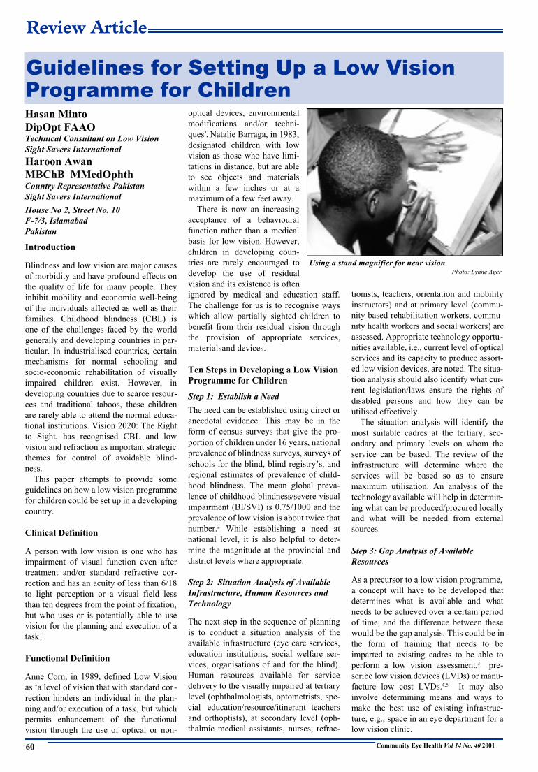
Guidelines for Setting Up a Low VisionProgramme for Children
Using a stand magnifier for near visionPhoto: Lynne Ager

Low Vision in Children
Community Eye Health Vol 14 No. 40 2001 61
Step 4: Develop a Plan for Low Vision with
Short, Medium and Long Term Objectives
The situation analysis and the gap analysis
together will form the basis for develop-
ment of a plan for low vision services for
children. Usually, this forms part of a more
comprehensive low vision programme. It is
useful to define short, medium and long
term objectives. Examples of short term
objectives could be training of core cadres
in low vision, awareness workshops for
eye care professionals, and including a low
vision component in existing training pro-
grammes, e.g., paramedics and teachers,
and standardising curricula to incorporate
low vision. Medium term objectives may
include establishment of low vision clinics,
networking of service providers, and
development/enhancement of the local
capacity to produce LVDs. In the long
term, the low vision concept and compo-
nent should be fully integrated into a
national comprehensive eye care pro-
gramme with a measured increase in the
quality and coverage of service.
Step 5: Identify and Mobilise Resources
Even though the main emphasis on devel-
oping a low vision programme remains the
best and effective utilisation of existing
resources, nevertheless, some external sup-
port will still be required in the form of
training of national focal/resource persons
in low vision and setting up of low vision
clinics (supply of equipment). The differ-
ent components of the plan (short, medium
and long term) should be costed and fund-
ing sought from the government, non-gov-
ernmental organisations, community based
and service organisations and commercial
enterprises willing to support programmes
for disabled persons.
Step 6: Pilot the Programme in a
Defined Setting or Area
As in most new programmes, it is better to
pilot the plan first in a defined setting or
area to test the concepts proposed and iden-
tify deficiencies. The piloting phase could
conceivably be done by identifying a dis-
trict that has a secondary level eye unit,
availability of optical services and an edu-
cational institution willing to participate in
this programme. Access to a tertiary eye
department and existence of an on-going
community based rehabilitation program-
me in the area are definite added advan-
tages.
Step 7: Develop Local Expertise for
Production of Assessment Materials and
LVDs
Simple optical and non-optical low vision
devices and assessment materials can be
produced in most countries where basic
optical services exist. The assessment
materials can be developed using a desk
top computer with laser printer. A semi-
skilled technician with basic optical
knowledge can be trained in a short time to
produce low vision devices. Most of the
materials involved in the production of low
vision devices are usually available locally
and may include PVC pipes and optical
lenses. The issue of non-availability of
optical lenses in higher power and aberra-
tions associated with these lenses can be
overcome by combining 2 or 3 low pow-
ered lenses to produce a higher power
system.
Step 8: Network with Other Service
Providers of the Visually Impaired
The role of low vision service as a bridge
between medical, educational and rehabili-
tative services has been recognised. The
low vision centre in the district can act as a
referral point for the child to access other
services that may be required, e.g., orienta-
tion and mobility training, early interven-
tion, and peer support groups. One way in
which this could be brought about is to
hold networking meetings between the dif-
ferent service providers and develop a con-
sensus on the methods for detection of the
visually impaired child in the community,
referral to a low vision clinic for assess-
mentandprescriptionofLVDs,appropriate
placement in school, access to statutory
benefits and elimination of barriers to care.
Step 9: Replicate the Pilot Model by
Integration into the National Vision 2020
Programme
The lessons learnt from the piloting phase
can then be employed to develop a larger
programme within the framework of a
national Vision 2020 programme. This will
ensure its lateral integration with other eye
care related activities, removing the need
to set up a vertical programme, and so
promote its long term sustainability.
Step 10: Maintain the Dynamic
Character of the Programme and
Increase Coverage
The low vision programme thus developed
as part of a national comprehensive eye
care plan should be dynamic in character
with an ability to absorb changes in tech-
nology, move towards sustainability and
have within it a mechanism for reporting,
monitoringandevaluation.Thegoalshould
be to increase the coverage of the service
and continually improve its quality.
Conclusion
Most specialties in ophthalmology are
costly to develop and require specially
trained people and sophisticated equip-
ment. Low vision as a specialty is one area
that can easily be initiated in any ophthal-
mic, educational or optometric set-up with
a minimum of investment and training.
Most of the devices used for assessment
can be produced locally using indigenously
available materials and appropriate tech-
nology. The use of simple magnifiers can
help children pursue education in normal
stream schools and improve their quality.
Each country can identify its own rele-
vant existing human resources and train
them in a short period of time to provide
low vision care in a school, hospital or
clinic setting. Standard manuals on produc-
tion of inexpensive low vision devices can
provide instruction in making these
devices. As experience is gained, and with
some input from external sources, a cost
effective and sustainable low vision ser-
vice can be developed. It would be prefer-
able to plan the development of any such
service so that it is capable of fitting in the
ongoing national health, educational and
social welfare programmes. This will not
only ensure its sustainability and cost con-
tainment but also its early acceptability and
implementation.
References
1 The Management of Low Vision in Child-
hood. Proceedings of WHO/PBL Consultation,
Bangkok, July 1992. WHO, Geneva, 1993.
2 Prevention of blindness in children. Report of a
WHO/IAPB scientific meeting. Hyderabad,
April 1999. WHO/PBL/00.77.
3 World Health Organization. Assessment of low
vision in developing countries. WHO/PBL/
95.48.
4 World Health Organization. How to make spec-
tacles at low cost. Spoerer P. WHO/PBL/ 95. 50.
5 Management of Low Vision for Developing
Countries. Minto H, Awan H R. Pangraphics,
Islamabad, 1995.
✩ ✩ ✩
Further Reading
An article which complements this
report was written by Lynne Ager in a
previous issue of the Journal.
Ager L. Optical Services for Visually
Impaired Children. J CommEyeHealth
1998; 11: 38–40
Editor
Review Article
Community Eye Health Vol 14 No. 40 200162
Anil K Mandal MDDirector
Jasti V Ramanamma
Children’s Eye Care Centre
L V Prasad Eye Institute
L V Prasad Marg, Banjara Hills
Hyderabad 500 034
India
Childhood Blindness and Vision 2020
Since children constitute only 3% of the
world’s blind population, childhood blind-
ness has not been given its due importance
during allocation of health resources.
However, the control of blindness in
children has been included as a priority
within the World Health Organization’s
(WHO) Vision 2020: The Right To Sight
programme.1 Vision 2020 will be imple-
mented through the following activities:2
1. Specific disease control measures.
2. Human resource development.
3. Development of appropriate technology
and infrastructure.
The main priorities for action are:
• Elimination of vitamin A deficiency
• Treatment of congenital cataract,
glaucoma, retinopathy of prematurity
• Serious refractive errors.
This will be achieved through:
• Promotion of primary health care (PHC)
• Developing specialist children’s eye
services, including surgery and low
vision clinics
• School screening.
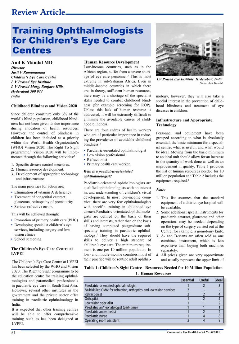
The Children’s Eye Care Centre at
LVPEI
The Children’s Eye Care Centre at LVPEI
has been selected by the WHO and Vision
2020: The Right to Sight programme to be
the education centre for training ophthal-
mologists and paramedical professionals
in paediatric eye care in South-East Asia.
However, several other institutes in the
government and the private sector offer
training in paediatric ophthalmology in
India.
It is expected that other training centres
will be able to offer comprehensive
training such as has been deisigned at
LVPEI.
Human Resource Development
Low-income countries, such as in the
African region, suffer from a severe short-
age of eye care personnel.3 This is most
extreme in sub-Saharan Africa. Even in
middle-income countries in which there
are, in theory, sufficient human resources,
there may be a shortage of the specialist
skills needed to combat childhood blind-
ness (for example screening for ROP).
Unless this lack of human resource is
addressed, it will be extremely difficult to
eliminate the avoidable causes of child-
hood blindness.
There are four cadres of health workers
who are of particular importance in reduc-
ing the prevalence of avoidable childhood
blindness:2
• Paediatric-orientated ophthalmologist
• Low vision professional
• Refractionist
• Primary health care worker.
Who is a paediatric-orientated
ophthalmologist?
Paediatric-orientated ophthalmologists are
qualified ophthalmologists with an interest
in, and understanding of, children’s visual
development. In most low-income coun-
tries, there are very few ophthalmologists
with specific training in childhood eye
disease.Paediatric-orientatedophthalmolo-
gists are defined on the basis of their
skills and interests, rather than on the basis
of having completed postgraduate sub-
specialty training in paediatric ophthal-
mology.2 They should have the required
skills to deliver a high standard of
children’s eye care. The minimum require-
ment is one per 10 million population. In
low- and middle-income countries, most of
their practice will be routine adult ophthal-
mology, however, they will also take a
special interest in the prevention of child-
hood blindness and treatment of eye
diseases in children.
Infrastructure and Appropriate
Technology
Personnel and equipment have been
grouped according to what is absolutely
essential, the basic minimum for a special-
ist centre; what is useful, and what would
be ideal. Moving from the basic minimum
to an ideal unit should allow for an increase
in the quantity of work done as well as an
improvement in quality. Table 1 provides
the list of human resources needed for 10
million population and Table 2 includes the
equipment required.2
Note:
1. This list assumes that the standard
equipment of a district eye hospital will
be available.
2. Some additional special instruments for
paediatric cataract, glaucoma and other
operations may be needed, depending
on the type of surgery carried out at the
Centre, for example, a goniotomy knife.
3. A- and B-scans are often sold as one
combined instrument, which is less
expensive than buying both machines
separately.
4. All prices given are very approximate
and usually represent the upper limit of
Training Ophthalmologistsfor Children’s Eye CareC e n t r e s
Essential Useful IdealPaediatric-orientated ophthalmologist 1 2 3Multiskilled OMA: for refraction, orthoptics and low vision services 2 - -Refractionist 2 2 4Orthoptist 1 2 4Low vision specialist 1 1 2Paediatrician/neonatologist (part-time) 1 1 1Paediatric anaesthetist 1 2 2Paediatric nurse 1 4 8Operating room assistant 2 4 8
Table 1: Children’s Sight Centre - Resources Needed for 10 Million Population
1. Human Resources
LV Prasad Eye Institute, Hyderabad, IndiaPhoto: Anil Mandal

Community Eye Health Vol 14 No. 40 2001
Training for Children’s Eye Care Centres
63
a range of likely prices. In most cases, it
will be possible to obtain equivalent
equipment at a lower cost.
Training Fellowship Programme for
Paediatric Ophthalmology
L V Prasad Eye Institute (LVPEI) is a
premier eye care facility in South Asia, and
is a non-profitable charitable institution
which started in 1987. It has state-of-the-
art facilities for providing high quality eye
care to the community.
The Jasti V Ramanamma Children's Eye
Care Centre, was established in 1997, and
is the first of its kind in India. The Centre
has the infrastructure and facilities for
integrated management of all eye diseases
that afflict children. The Centre offers
excellent diagnostic facilities, including
examination of children under anaesthesia,
and evaluation/management of children
with visual impairment or blindness. It has
active collaboration with other institu-
tions in India and abroad for research and
education. The Centre has established an
interdisciplinary approach for the compre-
hensive management of paediatric eye dis-
eases, and is engaged in training ophthal-
mologists.
1. Need for the course
It is estimated that there are 1.4 million
blind children in the world and half a mil-
lion children become blind every year.
Childhood blindness accounts for 75 mil-
lion blind years. Thus, childhood blindness
is the second largest cause of blind-person
years. Childhood blindness has also been
estimated to cause one-third of the 75 mil-
lion dollars total economic loss from blind-
ness. Recent population-based studies in
the State of Andhra Pradesh, India
(APEDS) indicate that one out of every
1,000 children is blind, and at least half of
this blindness is readily avoidable.
Throughout the State it is estimated that
10,000 children are ‘blind’ due to uncor-
rected refractive errors which can be easily
corrected by a pair of spectacles. Another
4,000 children are blind due to corneal
scarring (vitamin A deficiency) and 2,700
are blind due to cataract. If these figures
are extrapolated to the entire country, then
there would be about 260,000 (0.26 mil-
lion) children who are blind in India.
Although there are no accurate data on the
causes of childhood blindness in India,
there could be about 28,600 (11%) children
blind due to cataract/aphakia and another
7,800 (3%) children could be blind due to
glaucoma.2
The Global Initiative for Elimination of
Avoidable Blindness (Vision 2020: The
Right to Sight), by definition seeks to elim-
inate avoidable blindness. It is estimated
that 40% of childhood blindness is
avoidable. It is against this background that
L V PrasadEyeInstitutehasstartedtraining
fellowships in paediatric ophthalmology.
2. Objectives
The objective of the training programme is
to train paediatric ophthalmic teams from
large eye care centres in the Asian region
and beyond in the management of congeni-
tal cataract and other paediatric eye
problems, and to provide equipment and
supplies to selected institutions.
3. Training format
Training is being conducted at two levels.
Shorttermtrainingfor 3 monthsisprovided
for experienced ophthalmologists so they
can acquire special skills in paediatric
ophthalmology. In order to develop a team
approach, anaesthetists and nurses from the
same institutions will also be trained for
the same 3 month period. The second level
of trainingisfor12months,andisintended
for younger ophthalmologists as a fellow-
ship in paediatric ophthalmology with a
view to develop paediatric ophthalmology
units in selected countries.
The short term training programme will
have 10 candidates who are senior ophthal-
mologists from WHO/IAPB member coun-
tries – two each from Bangladesh, India
and Indonesia and one each from
Myanmar, Nepal, Sri Lanka and Thailand.
For the long term programme, 10 young
ophthalmologists from the same member
countries will be identified and selected by
the Regional Chairman, IAPB, South-East
Asia and the Director of Education, L V
Prasad Eye Institute.
(a) Three Month Training Programmes
Short term trainees will primarily focus on
acquiring skills in the management of pae-
diatric cataract, but they will also gain
experience in corneal and glaucoma surgi-
cal procedures, by observation. Trainees
will have their surgical skills assessed
before they are allowed to perform inde-
pendent surgery on paediatric eyes. It will
generally not be possible to train candi-
dates in pars plana lensectomy since that
would require proper vitreo-retinal surgical
experience.
During the 3 months, trainees will spend
6 weeks with two consultants who spe-
cialise in paediatric cataract surgery.
During the remaining 6 weeks trainees will
spend the first 3 days of each week in the
paediatric ophthalmology clinic observing
the strabismology work, and the remaining
3 days of the week observing the manage-
ment of glaucoma, screening for ROP, and
paediatric ophthalmic plastic surgery.
Approx. Unit Essential Useful IdealCost (US $)
For use in operating theatre:Operating microscope (co-axial) 10,000 1 2 4Cryotherapy machine with paediatric probes 10,000 1 1 1Vitrectomy machine 20,000 1 2 2Paediatric anaesthesia equipment 10,000 1 1 1CT/MRI (access to) - 1 1
For use in outpatients and/or theatre*:Laser (diode-indirect) 30,000 1 2 2Tonometer- Perkins or Tonopen 3,000 1 2 2Indirect ophthalmoscope 1500 3 4 6Slit-lamp portable 2000 1 1 2A-scan ultrasound 5,000 1 1 2B-scan ultrasound 10,000 1 1 2Keratometer-hand held 3,000 1 2 2
For use in outpatients:Tests of visual acuity for pre-verbal children 500 4 6 8Colour vision tests 200 1 1 1Automated visual fields 5,000 - 1 1YAG laser 15,000 - 1 1Refractometer 3,000 - - 1Low vision equipment 5,000 1 1 1Electrodiagnostic equipment 25,000 - 1 1
Table 2: Children’s Sight Centre – Resources Needed for 10 Million Population
2. Equipment
* children often need to be examined under anaesthesia

Community Eye Health Vol 14 No. 40 2001
Training for Children’s Eye Care Centres
64
(b) One Year Fellowship Programme
Training content
The areas where the trainees would be
given exposure include:
• Adult and paediatric cataract surgery
• Paediatric glaucoma procedures
• Strabismus surgery
• Exposure to treating ROP
• Exposure to ophthalmic plastic surgery
and tumours
• Management of low vision and visual
rehabilitation in children
• Genetic counselling.
Training schedule
The training will start on 1st January and
1st July each year. The first candidates
started their fellowship on 1st July 2001.
The admission year and month and number
of candidates is shown in Table 3 below:
Training rotation
During the first month trainees will spend
time learning basic adult cataract surgery,
and will also gain experience in cataract-
related investigative procedures such as
biometry. Also the trainees are expected to
work in the wet lab during this period.
The following is the schedule of the
trainees during their one-year fellowship
programme:
• Basic cataract surgical skills - 1 month
• Anterior segment including cornea and
cataract surgery - 3 months
• Strabismusandpaediatricophthalmology
- 3 months
• Paediatric glaucoma - 2 months
• Ophthalmoplasty and tumours - 1 month
• Retina - 1 month
• Repeat posting in cornea and anterior
segment – 1 month
Evaluation
Each candidate will be asked to keep a
record of all the cases which they per-
formed on their own, or at which they
assisted. At the end of each posting the fel-
low’s performance will be evaluated by the
faculty at L V Prasad.
Post-Training Support
A maximum of US$ 20,000 has been
allocated for the equipment at each centre
from where the long term fellows will be
trained. These long term trainees will be
encouraged to visit LVPEI in future as
part of our observer fellowship programme
to keep their knowledge up-dated.
References
1 Rao G N. Ophthalmology in India. Arch Oph-
thalmol 2000; 118: 1431–1432.
2 World Health Organization. Preventing blind-
ness in children. Report of a WHO/IAPB scien-
tific meeting. WHO/PBL/00.77. Geneva: WHO,
2000.
3 Global Initiative for the Elimination of
Avoidable Blindness. Geneva, World Health
Organization, 1998 (unpublished document
WHO/PBL/97.61).
✩ ✩ ✩
Table 3: Admission Year and Month / Number of Candidates
Year/MonthLong Term (1 Year) July 2001 Jan 2002 July 2002 Jan 2003 July 2003No. of candidates 2 2 2 2 2
Short Term (3 months) July 2001 Oct 2001 Jan 2002 April 2002 July 2002No. of candidates 2 2 2 2 2
ROYAL COLLEGE OF OPHTHALMOLOGISTS17 Cornwall Terrace, Regent’s Park, London NW1 4QW, UK
Diploma Examination in Ophthalmology
DRCOphth
ANNOUNCING A CHANGE
TO THE STRUCTURE
From November 2001, there has been no Practical
Refraction section in the Diploma Examination
The New Diploma Examination (DRCOphth) is a test
of ophthalmic knowledge including relevant basic
sciences and clinical skills for candidates who have
worked in ophthalmology for one year (full-time or
equivalent). This work experience need not have been
gained in the UK.
Information, Exams syllabi, Applications from:
The Head of the Examinations Department at
the above address
Or Tel: 00 44 (0) 20 7935 0702
Or Fax: 00 44 (0) 20 7487 4674
Or E-mail: [email protected]
Or visit the College website www.rcophth.ac.uk
UK and Overseas Examination Calender 2002
Exam Dates of Examination Location Closing Date
Part 1 8–9 April India 22 February
MRCOphth 22–23 April UK 11 March
7–8 October UK, India 26 August
Part 2 9–11 April India 22 February
MRCOphth 17–21 June UK 6 May
9–10 October India 26 August
4–8 November UK 23 September
Part 3 4–8 March UK 21 January
MRCOphth 11–12 April India 22 February
9–13 September UK 29 July
10–11 October India 26 August
DRCOphth 27–28 June UK 16 May
18–19 November UK 7 October
Overseas Location:
• Aravind Eye Hospital, Madurai, Tamil Nadu, India

Detlef Prozesky
MBChB MCommH PhDProfessor
Community Based Education
Faculty of Medicine
University of Pretoria
PO Box 667, Pretoria 0001
South Africa
'Quality assurance' and 'appraisal' are
words we often see nowadays. Training
institutions in many countries are trying to
improve the quality of the training they
provide. This is not new - good teachers
have always looked critically at the way
they teach, in order to do it better.
What is 'evaluation'? Broadly, it means
looking carefully at something that we are
worried about, and then making a judge-
ment about it. We usually do this because
we want to improve the thing we are look-
ing at. The following diagram shows this
process more clearly:
We call this the 'evaluation cycle'. Once
you have seen if your plan of improvement
is working you identify a new problem in
your teaching, and evaluate that again.
This is clearly a kind of research.
Perhaps you feel: 'I'm not an evaluator! I
can't do this kind of research!' Another way
of looking at evaluation is that you use
your common sense to judge what you (and
other teachers) do in your work. If you
think back you will see that you have often
done it – but this article should help you
to do it more systematically. Also, you
don't have to evaluate everything at once.
You can choose a small part of your
work which seems to be giving particular
trouble, and start by looking at that. What
might this be?
Things Teachers Evaluate
The curriculum: what we teach
Curricula get out of date easily, and people
just go on year after year teaching the same
material.
Here are some areas you might want to
look at:
• The overall curriculum: is it complete?
Does it contain all the knowledge and
skills that the students are going to need
to perform their job?
• The overall curriculum: is it overloaded?
Are you teaching a lot of 'nice to know'
and 'nice to do' material, instead of con-
centrating on the 'must know' and 'must
be able to do’ material?
• The content of individual lessons: do
they contain what the curriculum
planners intended them to? Do they
emphasise priorities, and leave out the
rest? Does the teacher present the
material in a sequence which helps
students to understand it more easily?
The lesson process: how we teach
• The teaching methods: are they
appropriate for the domain of the
material you are teaching? Do students
learn skills by seeing a demonstration,
and then practising the skill personally?
• How well do teachers use these
methods? Are lectures well prepared and
skilfully delivered, and do they interest
and involve the students? Are practicals
well organised, with checklists, and do
all students get feedback about their
performance?
• About the teaching aids that are used in
class: is their quality good? Are they
well used - do they help the learning
process?
• About the handouts and written
documents that are used: do they focus
on priorities? Are they clearly written,
using simple language? Are they
suitably illustrated?
The assessment: how we test our
students
• Is the assessment valid? Is it suitable for
the 'domain' of the subject matter (for
example, do we assess skills by
observing students perform them)? Does
it mostly contain the 'must know' and
'must be able to do' material? Does it
cover most of the important topics?
• Is the assessment reliable? Are there
good marking schedules and checklists,
to guide the examiners so that they give
fair marks?
• What is the 'assessment curriculum'? - in
other words, does the assessment make
students learn those things which we
consider to be the priorities?
These are just some of the possibilities.
Of course, you will decide from your situa-
tion what you should be looking at.
Instruments to Collect Information
for Evaluation
Once we have identified a problem we
need to collect more information about it.
How do we do this? There are a number of
'instruments' that we commonly use, to col-
lect data for evaluation:
Document study
Here we examine written curricula, time-
tables, lesson plans, visual aids, handouts,
exam papers and so on. We compare them
to a standard that we have set beforehand.
This can be done in an unstructured way
(by reading them and gaining an overall
impression), or more structured (by mak-
ing a checklist beforehand, of things we are
looking for in the document). One special
kind of document study is the 'readability
test', where we check how easily students
are able to read and understand the hand-
outs and textbooks we give them.
Observing practice
Here we sit in during classroom and practi-
cal teaching, and observe what is going on.
Once again we can do this is an unstruc-
tured way (by writing down what happens,
and analysing it afterwards) or a structured
way (by having a checklist of things we
would like to see, and checking if these
happen). We can also ask colleagues or
even students to observe us, as we teach.
Questionnaires
We use these when we want to know peo-
ple's opinion about an aspect of a training
course - practical arrangements, the rele-
vance of the material that is taught, what
happens in class and so on. Again, ques-
tionnaires can be unstructured (asking the
respondents to write general comments on
how they feel about the topic) or structured
(giving questions with pre-prepared
answers, from which they have to choose
the one they prefer). There are some spe-
cial kinds of questionnaires we use:
• The 'student happiness questionnaire'
(see box on next page)
• Diaries: we ask teachers or students to
keep diaries of their experiences on the
course.
Interviews (with individuals) and
discussions (with groups)
These are useful when we want informa-
tion from people about aspects of our
courses and teaching, but in more depth
and detail. We carefully prepare some
questions, and put them to the persons
Community Eye Health Vol 14 No. 40 2001
Teaching and Learning
65
Evaluation of Training
We check if ourimprovementsare working
We identify a problem
We collectinformationabout theproblem
We make ajudgementabout theproblem
We takeaction to
improve thesituation

Community Eye Health Vol 14 No. 40 2001
Evaluation of Training
66
concerned. Then we record exactly what
they say (by hand or with a tape recorder)
and analyse the information afterwards.
What were the main points that the respon-
dents raised?
Who Should Evaluate?
Who is best placed to evaluate teaching
practice? Do you do it yourself (an 'insid-
er'), or do you get someone else to do it for
you (an 'outsider')? Do you evaluate your
own practice, or that of your colleagues?
The advantages of doing it yourself, about
your own work, is that you understand it
thoroughly – the background, the players,
the details. The disadvantage is that you
are used to looking at your work in a cer-
tain way, and it is difficult to see it objec-
tively - so an outsider coming with a fresh
viewmaybemoreuseful.Outsidersusually
want to be paid though!
Three Points
When someone asks you to do an evalua-
tion you must be opportunistic. Of course
you are going to collect specific data with
instruments you have prepared. However,
you should use every opportunity to get
additional information. Talk to everyone
you meet (and write down what they say);
look at notice boards and classroom walls
(making notes of relevant information), go
into the course filing cabinet and read rele-
vant documents. In this way you gain a
deeper understanding, which helps you to
make the right judgements.
One of the aims of evaluation is to find
and clarify problems. The problem is that
many people find it difficult to accept that
they have been making mistakes. You,
therefore, have to present your judgements
- your feedback - in a sensitive way. Start
by listing all the good things that you found
(and you will find them). Then, once you
have affirmed the persons you are evaluat-
ing, you can mention the deficiencies in a
polite and non-judgmental way.
Here are two books on evaluation which I
have found very helpful:
• Harris D and Bell C. (1986).
Evaluating and Assessing for Learning.
London: Kogan Page.
• Hopkins D. (1989). Evaluation for
School Development. Milton Keynes:
Open University Press.
And Finally ..
Here is a thought. The great philosopher
Socrates said that the best teachers are like
stinging flies. They make their students
uncomfortable, by asking the really impor-
tant, really difficult questions – then make
the students find the answers to those
themselves. What do you think?
✩ ✩ ✩
Medi Kawuma MDAssociate Professor
Department of Ophthalmology
Makerere University
PO Box 7072, Kampala
Uganda
Introduction
Vernal keratoconjunctivitis (VKC) is a
recurrent, bilateral, interstitial inflamma-
tion of the conjunctiva, resolving sponta-
neously after a course of several years and
characterised by giant papillae (with a cob-
blestone appearance) on the tarsal conjunc-
tiva, a discrete or confluent gelatinous
hypertrophy of the limbal conjunctiva, and
a distinct type of keratitis. It is associated
with intense itching, redness or
brownness, lacrimation, photo-
phobia and a mucinous, ropy dis-
charge containing eosonophils.
VKC is a type 1 hypersensi-
tivity reaction but with addition-
al immune mechanisms involved
in its pathogenesis.
VKC has a global distribution
with a widely varying incidence.
It is less common in northern Europe and
North America, and more common in the
Africancontinent,the Mediterraneancoun-
tries, in Central and South America, and
the Indian subcontinent. 1,2
All patients in this study were indige-
nous Africans, and all had been exposed
to similar environmental conditions.
The high prevalence of VKC in Uganda,
and the lack of data on the pattern and typi-
cal clinical picture of this medical condi-
tion provided the impetus for this study.
Subjects and Methods
This study was carried out in two places, at
the eye clinic, Department of Ophthal-
The Clinical Picture of Vernal Kerato-Conjunctivitis in Uganda
Vernal keratoconjunctivitis affecting the limbus (left),the conjunctiva and cornea (right). The conjunctivahas pronounced papillae (‘cobblestones’) and fluorescein dye shows a corneal ulcer
Photos: John Sandford-Smith
Clinical Report
The 'StudentHappinessQuestionnaire'
It is common practice to present students
with a questionnaire at the end of a part
of a curriculum, or at the end of a term.
The students are asked what they liked or
disliked about the content; the teaching;
the assessment; the practical arrange-
ments. This can provide useful informa-
tion about problems. However, it must be
used with caution:
• Students may be wrong – for example,
since they lack a wider understanding
they may believe (wrongly) that some
of the course content is unimportant
• Teachers may believe that this is all the
evaluation you need to do. If it is
routinely done, year after year, teachers
tend to ignore the findings.
Ready-Made Data Collection
Instruments
Other teachers who have gone before us
can help us with evaluation. It is often
possible to find ready-made data collec-
tion instruments, which deal with a vari-
ety of common problems in teaching. A
good source of these is the booklet, 53
Ways to Evaluate Your Teaching by
Gibbs, Gibbs and Habeshaw (published
by Educational Technologies, Bristol,
UK). Such instruments have been tested
and refined, and should provide us with
useful information. Of course we don't use
them uncritically - they usually need to be
adapted a little to fit our own situation.

Community Eye Health Vol 14 No. 40 2001
Vernal Keratoconjunctivitis
67
mology, Mulago National Hospital, and at
the Medicare Clinic, a private eye consult-
ing clinic in the capital city, Kampala, over
a period of seven years, 1989 to 1996. A
total of 420 patients with VKC were diag-
nosed on the grounds of their history, the
presence of characteristic symptoms and
on the basis of their clinical features.
Clinical signs varied from mild to severe.
The history of each patient was taken
including a record of age, sex, place of res-
idence, change of place of residence, age at
onset of the disease, seasonal variations,
associated allergic or ‘atopic’ illnesses,
family history of VKC or associated aller-
gic or atopic conditions.
Each patient was thoroughly examined
with a slit-lamp or a binocular 3x loupe.
Refraction with determination of visual
acuity, retinoscopy, and assessment of the
cornea with the placido disk to determine
the presence of keratoconus were carried
out. Patients who had any other keratocon-
junctival disease were excluded from the
study. All male patients underwent a full
systematic examination.
Patients were reviewed once a month,
and the period of follow-up ranged from a
minimum of three months to a maximum
of six years.
Each patient received treatment to
relieve symptoms, hydrocortisone eye
drops or prednisone eye drops, whichever
was available at the time. If both were
available, one or the other was given,
picked at random by the clinic dispensing
nurse.
Results
Of the 420 patients, 231 (55%) were males
and 189 (54%) were female.
Table 1 displays the age and sex distrib-
ution at the onset of VKC; the highest
incidence of VKC occurred in the age
group 5–9 years, and the lowest 20 years
and above.
Corneal complications occurred in 231
(55%) patients; 196 had minor complica-
tions and 35 major complications or associ-
ations. The minor complications usually
consisted of superficial punctate kerato-
pathy,orotherepithelialdisturbance.Major
complications or associations consisted of
superior pannus and 15 with huge plaque
formation which had already compromised
patients’ visual acuity.
Patients with VKC often give a history
of allergy or of atopic diseases such as
allergic rhinitis, asthma, or hay fever 3 but
in my study, co-existing allergic conditions
could be detected in only 47 (11%)
patients. Of the 47 patients, 37 gave a
history of asthma, rhinitis or both. The
remaining 10 had atopic dermati-
tis.
All the three sub-types of
VKC, namely limbal, mixed, and
palpebral were seen. The most
dominant sub-type is limbal,
which constituted 75% of all
patients. This was followed by
the mixed type which was 18% of
the total. Palpebral sub-type VKC
was diagnosed in only 7% of the
patients.
No patient presented with
either hypogonadism or hypoad-
renalism (see below).
Discussion
I found no disproportionate high preva-
lence of the disease in male patients. Some
previous workers have found a high preva-
lence of the disease in male patients.1,2,4
Some experimental work has shown that
VKC is associated with hypogonadism and
hypoadrenalism in males. Clinical evi-
dence for this is based on findings in a
Japanese population.6 However, my own
findings in this study do not support the
stated association.
Since 80 of my patients were under 15
years of age, this disease in Uganda is one
of young persons, resolution mostly occur-
ring at puberty.
Over 70% of all patients noted signifi-
cant seasonal variations of their symptoms;
more than 85% reported accentuation of
symptoms during the hotter, drier months.
Seasonal variability was marked in all the
three sub-types, but was most marked in
the limbal sub-type.
Limbal or mixed VKC had proportion-
ately more corneal complications than the
palpebral type. This finding agrees with
those of Easty and others working in
Europe and the Middle East. 7,8 The high
incidence of corneal involvement in limbal
VKC is most likely due to poor limbal
nutrition, affecting the nutrition of the
avascular cornea. Some of my patients had
keratoconus; other studies have reported
even higher incidences.9
The often described typical appearance
of changes on the tarsal conjunctiva in
tarsal VKC, that of cobblestone formation,
was hardly seen. Only one patient out of
the 29 with palpebral VKC presented with
a mild form of cobblestone formation.
Instead, there was a red to pink uniform
colour, completely concealing all tarsal
conjunctival vessels.
There is a well established association of
atopic disease with VKC. Several authors
who have investigated the exact nature of
this relationship have come to varying con-
clusions.3,10 A low incidence of atopic dis-
ease in VKC in Uganda has been recorded.
Relatively low incidences of atopic dis-
eases in VKC have been recorded in other
parts of the world. 5,8 There are no previous
studies in either Uganda or other East
African countries to make comparisons.
References
1 Khan M D,KundiN,SaedN,GulabA,Nazeer AF. A study of 530 cases of vernal conjunctivitisfrom the North West Frontier Province of
Pakistan. Pakistan J Ophthalmol 1986; 2:111–114.
2 Dahan E, Appel R. Vernal keratoconjunctivitisin the black child, and its response to therapy. Br
J Ophthalmol 1983; 67: 688-692.
3 Tuft S J, Kemeny S M, Dart J K G, Buckley R J. Clinical features of atopic keratoconjunc-tivitis. Ophthalmology 1991; 98: 150–158.
4 Cameron J A, Al-Rajhi A A, Badr I A. Cornealectasia in vernal keratoconjunctivitis. Ophthal-
mology 1989; 96: 1615-1623.5 Aoki H. Relations of eye disease with thymus
and endocrine glands especially with regard toexperimental allergic conjunctivitis. Acta Soc
Ophthalmol Jap 1968; 72: 1949-1965.
6 Easty D L, Birkinshaw M, Merrett T. Immuno-logical investigations in vernal eye disease.Trans Ophthalmol Soc UK 1980; 100: 76-82.
7 Emarah M H. Vernal keratoconjunctivitis, diag-nosis and management. Bull Ophthalmol Soc
Egypt 1985; 78: 5-10.8 Khan M D, Kundi N, Saeed N, Gulab A, Nazaar
AF. Incidence of keratoconus in spring catarrh.Br J Ophthalmol 1988; 72: 41–43.
9 Tuft S J, Dart J K G, Kemeny M. Limbal vernal
keratoconjunctivitis: clinical characteristics andimmunoglobin E expression compared withpalpebral vernal conjunctivitis. Eye 1989; 3:420–427.
✩ ✩ ✩
Table 1: Age and Sex Distribution
of Patients
Age Group Sexin years Male (%) Female (%) Total (%) 0–4 38 (9) 29 (7) 67 (16)5-9 88 (21) 64 (15) 152 (36)10–14 59 (14) 59 (14) 118 (28)15-19 15 (3) 32 (8) 47 (11)20 & above 9 (2) 28 (7) 37 (9)
Papillae of vernal conjunctivitis in an 18 year oldAfghan male. The patient also had trachoma
Photo: John D C Anderson

68 Community Eye Health Vol 14 No. 40 2001
Clinical Report
Jerry Harrell MD DTM&HOphthalmologist
N Dean Larson MDOphthalmologist
Esther MenzaClinical Officer
Ammon MbotiClinical Assistant
Lighthouse for Christ Eye Centre
PO Box 81465, Mombasa
Kenya
Patients and Methods
During the course of a seven-month period
in 1998 and 1999, 617 new patients with
unilateral or bilateral blindness were evalu-
ated as to the underlying cause. This was
done at the Lighthouse for Christ Eye
Centre in Mombasa, Kenya. These patients
came not only from the Mombasa area, but
from the entire coastal region – from the
frontier with Somalia in the north to the
border with Tanzania in the south. Others
were from towns in the interior along the
highway to Nairobi. Many of these had
been made aware of the medical and surgi-
cal care available at the Lighthouse
through our mobile clinics.
Upon arrival at the Lighthouse each
patient was registered and then a history of
vision problems was obtained. Visual
acuity was measured using the Snellen E
chart at six metres. Patients were then
examined with a slit-lamp, an applanation
tonometer and either a direct or indirect
ophthalmoscope. Where question existed
regarding the degree of vision loss, the
patient was referred to the clinic refraction-
ist. With the exception of twelve patients
seen by the clinical officer, all others were
evaluated by one of the ophthalmologists
at the Lighthouse. Patients with visual acu-
ity of less than 3/60 in one eye, but 3/60 or
better in the other eye were considered uni-
laterally blind. Those with less than 3/60 in
both eyes were considered bilaterally
blind. When compound causes for blind-
ness were present in one eye, the most pre-
ventable or avoidable one was selected as
the main cause of the blindness. This same
principle was applied when different caus-
es led to blindness in both eyes.
Results
During the study, 617 patients were found
to be blind in one or both eyes. Of these,
374 (60.6%) were unilaterally blind and
243 (39.4%) were bilaterally blind.
There were 365 male patients (59.2%)
and 252 females (40.8%).
The age distribution of the patients is
shown in Figure 1. The increase in the
number of blind patients after the age of 50
is due to the greater prevalence of cataract
and glaucoma in that segment of the popu-
lation. Of the 153 bilaterally blind patients
due to cataracts, 137 (89.5%) were age 51
or above. Likewise, of the 29 bilaterally
blind patients due to glaucoma, 22 (75.9%)
were 51 or older.
Thecausesofblindnessinbothunilateral
and bilateral cases are noted in Figures 2
and 3. Cataract (33.4%), trauma (25.1%),
retinal disease (9.1%) and leucoma (7.8%)
were the most frequent causes of unilateral
blindness (Table 1). Cataract (63.0%),
glaucoma (12.0%), retinal disease (7.0%)
and optic atrophy (3.7%) were the most
common causes of bilateral blindness
(Table 2).
Discussion
The 3:2 ratio of male to female patients
(365:252) is similar to that found in other
studies. This probably does not represent
actual prevalence rates, but is more likely
due to a greater tendency on the part of
men to seek evaluation and surgical treat-
ment.
Age-related cataract was the greatest
single cause of treatable blindness among
patients at the Lighthouse. Unfortunately,
treatment is sought late. One indicator of
this is the fact that one out of every five
bilaterally blind cataract patients over the
age of 50 had only light perception in each
eye.
In our experience most glaucoma
patients are first seen when they seek help
because of vision loss, often very profound
loss. Among the 29 bilaterally blind glau-
coma patients, 13 had lost all perception of
light in one or both eyes. Such findings
indicate that there is a considerable delay
in making the diagnosis of this disease.
Injuries were a major cause of unilateral
blindness, especially among younger
patients. The most frequent causes of trau-
matic blindness were injuries from sticks
(41%), fists (8%), stones (7%), thorns (5%)
and knives or pangas (4%). Other blinding
A Clinic-based Survey of Blindness in Kenya
Dr Roberta Walter-Fincke, outpatientclinic, Lighthouse for Christ Eye Centre
Dr Larson and Dr Downing in theoutpatient clinic
Photos: Jerry Harrell
Male Female Total %Cataract 78 47 125 33.4Trauma 55 39 94 25.1Retinal disease 20 14 34 9.1Leucoma 17 12 29 7.8Glaucoma 15 11 26 7.0Corneal ulcer 15 6 21 5.6Optic atrophy 12 5 17 4.5Uveitis 6 0 6 1.6Capsule opacity 1 3 4 1.1Amblyopia 2 2 4 1.1Enucleation 1 1 2 0.5Phthisis 2 0 2 0.5Ocular tumour 0 2 2 0.5Other CNS 1 0 1 0.3Post-operative 1 0 1 0.3endophthalmitisUncertain 1 5 6 1.6
Total 227 147 374 100
Table 1: Causes of Unilateral Blindness
Male Female Total %Cataract 83 70 153 63.0Glaucoma 16 13 29 12.0Retinal disease 10 7 17 7.0Optic atrophy 3 6 9 3.7Leucoma 4 3 7 2.9Trauma 6 0 6 2.5Aphakia 3 2 5 2.1Cortical Blindness 4 1 5 2.1Uveitis 2 0 2 0.8Microphthalmia/ 1 1 2 0.8AnophthalmiaPhthisis 1 0 1 0.4Ocular tumour 1 0 1 0.4Pituitary tumour 0 1 1 0.4Other CNS 3 1 4 1.6Uncertain 1 0 1 0.4
Total 138 105 243 100
Table 2: Causes of Bilateral Blindness

Blindness in Kenya
Community Eye Health Vol 14 No. 40 2001 69
injuries were due to the following: an
insect sting, a goat's horn, a kick from a
cow, a fall into a fire, a bullet, a crocheting
needle, football contusions and a beating
with a rifle butt by bandits in Somalia.
Although not a direct injury to the eyes,
a 28 year old Somali male related his bilat-
eral blindness to the bite of a viper (the
‘aweso’). Following the bite on his left
hand he developed haematuria and noted
decreasing vision. Ten months later he had
become totally blind. Visual acuity testing
at our clinic showed no light perception
(right eye) and light perception without
projection (left eye). Slit-lamp examination
revealed an opaque, vascularized mem-
brane immediately posterior to the lens in
both eyes. Presumably, membrane forma-
tion occurred secondary to vitreous haem-
orrhage. While we generally see one or
two spitting cobra injuries each year, this
was the first case of blindness related to
envenomation by a viper.
Leucomata accounted for 7.8% of uni-
lateral blindness and active corneal ulcers
were responsible for another 5.6%. If con-
sidered together, corneal disease was the
third most common cause of unilateral
blindness. Among bilateral cases, corneal
leucomata constituted 2.9% of the total
number of the blind. Severe xerophthalmia
was evident in two small children with
corneal perforations and, based on histo-
ries, was most likely the cause of other
leucomata.
Retinal pathology accounted for 9.1% of
the cases of unilateral blindness and 7% of
those with bilateral blindness. Among the
varied entities were retinal detachment (9),
diabetic retinopathy (6), retinitis pigmen-
tosa (2) and retinopathy of prematurity (1).
There were five cases of cortical blind-
ness in children. In each there had been a
preceding febrile illness which was stated
to have been malaria. Cortical blindness
does occur in about 4% of the cases of
cerebral malaria. Professor Kevin Marsh of
the Malaria Research Institute at Kilifi
stated in February, 1999 that there is quite
oftensomerecoveryofvision after a period
of several months.
Treatment for malaria was a likely cause
of optic atrophy and blind-
ness in at least four adults.
In only one case was the
identity of the medication
known, although the exact
quantity was not recalled.
T h i sp a t i e n tw a s a 3 5y e a ro l d
Swahili female who lived in
‘Old Town’, Mombasa. She
described an attack of
malaria, accompanied by
delirium, which took place a
little over two weeks prior
to her clinic visit. Three
days after the onset she
received an injection of
chloroquine and then took
quinine tablets. A few days
later she noted a marked
loss of vision over a period
of several hours. When seen
at the Lighthouse she had no
perception of light (right
eye) and light perception
without projection (left eye).
There was an afferent pupil-
lary defect of the right eye
and pallor of both optic
nerves (right>left).
Conclusion
There needs to be a greater
awareness on the part of the
public concerning:
1. The benefits of cataract
surgery. Hopefully, this will lead to the
restoration of good vision prior to inca-
pacity through blindness.
2. The need for early detection of
glaucoma. By the time vision loss has
occurred it is often too late for any
effective treatment.
3. The need for the addition of vitamin A
containing fruits and vegetables to the
daily food intake. The staple cornmeal
‘ugali’ is all too often supplemented by
inadequate quantities of such foods.
4. The potential dangers of antimalarials.
These are easily obtained and most
often taken without the guidance of a
physician.
The main patient waiting room at the
central clinic and the patient assembly
areas on mobile clinics should be utilized
to a much greater degree for educational
purposes.
Acknowledgements
The authors would like to thank Dr Sharon
Bolin for preparation of the figures and
Mrs Betty Harrell for the typing of the
manuscript.
References
1 Cofie G, Tenkorang J, Thomson I. Blindness in
Ghana- a hospital-based survey. J Comm Eye
Health 1991; 7: 9-10.
2 Landry P. Blindness in the Western Province of
Cameroon. J Comm Eye Health 1993; 12:
30–31.
3 Loewenthal R, Pe’er J. A prevalence survey of
ophthalmic diseases among the Turkana tribe in
northwest Kenya. Br J Ophthalmol 1990; 74:
84–88.
4 Manjunatha HR. Blindness at the FISH Clinic,
Kingston, Jamaica, West Indies. J Comm Eye
Health 1995; 16: 30–31.
5 Thomson I. A clinic based survey of blindness
and eye disease in Cambodia. Br J Ophthalmol
1997; 81: 578-580.
Dr Ingrid Gralle in the operating roomPhoto: Jerry Harrell
Mobile clinic examination, KenangoPhoto: Jerry Harrell
Fig. 1: Age Distribution for Blindness
Fig. 2: Major Causes of
Unilateral Blindness
Fig. 3: Major Causes of
Bilateral Blindness

Li Wern Voon
Jovina See
Tien Yin Wong
Purpose – To describe the epidemiology of
ocular trauma from the perspective of the
emergency service of a large tertiary
hospital in Singapore.
Methods – A prospective survey was con-
ducted over a 3 month period (August to
October 1997) on all patients seen at the
ophthalmic unit at the Singapore General
Hospital’s emergency service. Data on
clinical presentation, type and cause of
injury and use of eye protective devices
(EPD) were collected via a standardised
interview and examination.
Results – A total of 870 persons presented
with a diagnosis of ocular trauma, out of
the 1631 patients seen during the study
Abstracts
Community Eye Health Vol 14 No. 40 200170
Paul J Foster
Gordon J Johnson
Aims – To derive preliminary estimates for
the number of adults in China suffering
from glaucoma, and project the burden of
visual morbidity attributable to primary
and secondary glaucoma.
Methods – Age and sex specific data from
two population surveys were applied to US
Census Bureau population estimates for
urban and rural China. It was assumed that
data from Singapore were representative of
urban China, and those from Mongolia
were representative of rural China.
Results – It was estimated that 9.4 million
people aged 40 years and older in China
have glaucomatous optic neuropathy. Of
this number, 5.2 million (55%) are blind in
at least one eye and 1.7 million (18.1%) are
blind in both eyes. Primary angle closure
glaucoma (PACG) is responsible for the
vast majority (91%) of bilateral glaucoma
blindness in China. The number of people
with the anatomical trait predisposing to
PACG (an ‘occludable’ drainage angle) is
in the region of 28.2 million, and of these
9.1 million have significant angle closure,
indicated by peripheral anterior synechiae
or raised intraocular pressure.
Conclusions – This extrapolation of data
from two east Asian countries gives an
approximate number of people in China
suffering from Glaucoma. It is unlikely
that this crude statistical model is entirely
accurate. However, the authors believe the
visual morbidity from glaucoma in China
is considerable. PACG is probably the
leading cause of glaucoma blindness in
both eyes, and warrants detailed investiga-
tion of strategies for prevention.
Published courtesy of :
Br J Ophthalmol 2001; 85: 1277–1282
Glaucoma in China: How Big is the Problem?
Gamal Ezz Al Arab
Nagah Tawfik
Reda El Gendy
Wagida Anwar
Paul Courtright
Background – Evidence of widespread
distribution of trachoma in Egypt had not
been clarified as previous surveys were
limited to individual communities which
may not have been representative of the
general population. The Nile Delta of
Egypt presents a unique environment for
trachoma to persist. Economic improve-
ments in the past decade have affected
even the poorest rural environments; avail-
ability of electricity is now found in many
rural communities. Availability of water in
the Nile Delta has always been good but
poor hygiene conditions have been the
primary factor in trachoma transmission. A
survey of trachoma was undertaken in
MenofiyagovernoratetodetermineifEgypt
should be identified as trachoma endemic
and targeted for trachoma control efforts.
Methods – A multistage random cluster
study design was used with the target pop-
ulation defined as adults aged 50 and over
and children aged 2–6 years from through-
out the governorate. Among pre-school
children only trachoma was graded while
among adults presenting visual acuity and
cause of vision loss or blindness were also
recorded. Adults were interviewed regard-
ing past trichiasis surgery; those currently
with trichiasis or history of trichiasis
surgery were also interviewed regarding
outcome of surgery.
Results – A total of 3272 children aged 2–6
and 3322 adults age 50+ were enumerated.
Among the children 81.3% were examined
and among the adults 73.0% were exam-
ined. Active trachoma follicles (TF) and/or
intense inflammation (TI) was found
among 36.5% (95% confidence interval
(CI) 34.7-38.3%) of the children. TI was
1.89 (95% CI 1.22–2.94) times more com-
mon in rural children compared to urban
children. The prevalence of trichiasis (TT)
in adults was 6.5%; women had an age
adjusted odds of trichiasis of 1.68 (95% CI
1.18-2.39) compared to men. Trichiasis
was 2.11 times (95% CI 1.33-3.37) more
common in rural Menofiya compared to
urban Menofiya. TT accounts for blindness
(presenting vision <3/60) in 8% of patients
and accounts for 13.2% of visual impair-
ment. Overall, trichiasis surgical coverage
was 34.4%, slightly higher among men
than women. The outcome of trichiasis
surgery was poor in 44.4% of cases.
Conclusion – Trachoma is a serious public
health problem in Menofiya governorate
and a significant contributor to vision loss.
These findings would suggest that contin-
ued poor hygienic conditions in rural
Egypt have limited the reduction of active
trachoma even in the face of significant
improvements in socio-economic status.
Furthermore, the high proportion of trichi-
asis surgery cases with a poor outcome
would indicate a need to reassess current
surgical practices in Egypt and improve
training and monitoring.
Published courtesy of :
Br J Ophthalmol 2001; 85: 1406-1410
The Burden of Trachoma in the Rural Nile Delta ofEgypt: a Survey of Menofiya Governorate
The Epidemiology of Ocular Trauma in Singapore:Perspective from the Emergency Service of a LargeTertiary Hospital

Abstracts
Community Eye Health Vol 14 No. 40 2001 71
period. Compared with non-trauma cases,
trauma cases were more likely to be male
(odds ratio (OR): 4.2, 95% confidence
interval (95% CI): 3.2, 5.4), non-residents
(OR: 6.2, 95% CI: 3.7, 10.5), younger than
40 years of age (OR: 3.2, 95% CI: 2.7, 4.1)
and less likely to require follow-up or hos-
pital admission (OR: 0.2, 95% CI: 0.2,
0.3). The three most common types of
injuries were superficial foreign body
(58.2%), corneal abrasion (24.9%) and
blunt trauma (12.6%), while open globe
injury occurred in only 17 cases (2%).
Comparison with a 10% random sample of
all cases seen in the previous 9 months (n =
284) revealed no significant time variation
in the types of injuries (p = 0.63). Work-
related injuries accounted for 590 (71.4%)
cases, where grinding, cutting metal and
drilling were the specific activities in more
than 90% of the cases. In appropriate set-
tings, only 21.7% of patients with work-
related injuries used EPD; 43.7% were pro-
vided with EPD, but did not use them at the
time of injury; and the remaining 34.6%
reported that EPD were not provided.
Conclusions – Ocular trauma at the emer-
gency service level in Singapore involved
mainly young non-resident men, were
work-related and associated with well-
defined activities, and were generally
minor. The low prevalence of EPD use
reinforces the need for a review of the
design and implementation of occupational
eye safety programmes, especially among
non-resident workers.
Published courtesy of :
Eye 2001; 15: 75-81
Dear Editor
Dr Prozesky has expertly described why
and how learning should be assessed
(J Comm Eye Health 2001; 14: 27-28). It
should be emphasised that assessing com-
petence in a workplace situation follows
the same designs although this assessment
is often summative and based on the
principles of evidence. In assessing compe-
tence, one is concerned with whether the
evidence collected (through observation,
MCQ, checklists, or oral examination) is
current, authentic and sufficient to declare
a candidate competent in performing a
specific task. That is when the use of
OSPE/OSCE is very helpful for the pur-
pose of assessment because it is possible to
assess in a given scenario the knowledge,
the skills and the attitude of candidates.
Historical evidence (reports, testimonials,
work history) is also considered in the
assessment of competence but its value is
limited by its authenticity which can be
questionable. As teachers move from
didactic to problem-based learning
methodology, the assessment of compe-
tence becomes a critical issue. Assessment
skills will then become not only necessary
but also a specialised area with qualified
assessors, moderators and verifiers work-
ing alongside teachers or trainers as
partners. This is the system that is already
implemented to some extent in countries
including UK, Australia, Singapore and the
United States, to name but a few.
Mr Ntambwe Malangu
Lecturer
Medunsa School of Pharmacy
South Africa
Dear Editor
I did not know that there was room for
arguments on whether or not to do ICCE. I
thought that all efforts are towards IOL
after lens extraction, preferably by the
ECCE method. Sadly, in those regions
where no surgeon exists to do ECCE with
IOL or he/she exists but there is no relevant
equipment, ICCE may be performed.
I will restrict myself to the Africa that I
know and have worked in – East, West and
Central. In all these regions, I have found
that there exists a backlog of unoperated
cataracts (according to surveys and epi-
demiological projections)) but there are no
(or insignificant) surgery waiting lists in
eye departments. The pressure is not on
surgery time but community awareness
and mobilisation campaigns to increase
cataract surgery uptake. Backlog or no
backlog, Africa or Asia, I would rather take
15 minutes on ECCE with PC IOL than 3
minutes on ICCE with no implant (and no
sutures).
With proper distribution of existing
resources within countries in our region -
human resources and equipment - every
patient needing and willing to have
cataract surgery should have lens extrac-
tion with IOL inserted.
To increase cataract surgery uptake we
need to demonstrate improved quality of
service. In my catchment area we used to
get resistance to surgery because nearly
every elder could name somebody who
was blinded by cataract surgery - over a
decade ago! Today, nearly every other
patient who comes for cataract surgery is
on recommendation of our former, satis-
fied cataract patient. There was a change
from ICCE to ECCE with PC IOL.
Cataract surgical uptake is increasing by
about 20% every year.
I agree with my old friend and ICEH
classmate, Dr Mmbaga, that some vast
regions still do not have microscopes or
surgeons trained in ECCE with IOL and
that ICCE may therefore be a good solu-
tion. But we must sing the song how inap-
propriate the technique is and how the rele-
vant local NGOs and Ministries of Health
must make acquisition of the very afford-
able equipment a top priority.
Internally, within the countries, we
should retrain ICCE surgeons in ECCE
with PC IOL technique.
Dr Kenneth Kagame
PO Box 14, Ruharo Eye Centre
Mbarara, South Western Uganda
Letters to the Editor
Assessment ofLearning VersusCompetence
Cataract Surgery
CCOOMMMMUUNNII TTYY EEYYEE HHEEAALLTTHH CCOOUURRSSEESSProfessorGordonJohnson. InMarch,2002 Professor Johnson will retire after 15years service at the International Centre for Eye Health,London. We shall allgreatly miss his good and wise leadership.
Community Eye Health Courses. There are no plans to continue Courses in Community Eye Health (CEH) at the Institute ofOphthalmology beyond August 2002. There are, however, plans for a proposed, new Community Eye Health Course in London, beginninginSeptember 2002.
Dr Allen Foster. Dr Foster, who is based at the London School of Hygiene and Tropical Medicine, is currently conducting short, oneweek Courses in Community Eye Health for the Vision 2020 programme. These short Courses are now taking place in 12 Centresaround the world.
Dr Foster should be contacted for information relating to both the new, planned CEH Course in London and the Courses in Centres abroad.
Dr Allen Foster, CRU/ITD, LSHTM, Keppel St., London WC1E7HT, UK. E mail: [email protected]

Commun it yEEyyee HHeeaalltthh
supported by
Christian Blind Mission International
Sight Savers International
Conrad N. Hilton Foundation
International Glaucoma Association
Tijssen Foundation
Dark and Light Blind Care
The Netherlands
Dutch Society for the
Prevention of Blindness
The West Foundation
Prevention of Childhood Blindness
Community Eye Health Vol 14 No. 40 200172
Ophthalmia neonatorumPhotos: Murray McGavin & Allen Foster
EEyyee HHeeaalltthhNow available on-line
w w w.j c e h . c o . u k
This slides/handbook teaching set
provides a practical tool for teaching
health workers how to recognise, treat
and/or refer children with eye diseases. It
also sets out the measures necessary for
prevention of these diseases, such as
immunisation, health education, improved
nutrition and santitation.
TeachingSlides/Text Set
Vitamin A deficiencyPhotos: Simon Franken, Allen Foster,
Donald McLaren & Gordon Johnson
Congenital glaucomaPhoto: Murray McGavin
Ordering Information:
Developing Country Rate:
UK£15+ £3 (surface)
or £5 (airmail) postage.
Elsewhere: UK£20 + £3 (surface)
or £5 (airmail) postage.
Payment by credit card,
international banker's order or cheques
drawn on UK bank accounts.
Please make cheques payable to
UNIVERSITY COLLEGE LONDON
Monitoring and Evaluation
of Intervention Programmes
for Cataract and for Refractive
Errors in India
by Hans Limburg
The first pre-requisite for improving the
results of surgery is to monitor surgical
outcomes. This book summarises the work
of one of the most expert researchers in
this field. It is a valuable resource for
anyone involved in cataract surgery
delivery. A separate chapter deals with
school screening for refractive errors.
A limited number of copies of this 230-
page book, which was produced as a doc-
toral thesis, are available from the Inter-
national Resource Centre, ICEH, London.
Ordering Information:
Cost: £5.00 + £3.00 (surface)
or £5 (airmail) post & packing.
Payment by credit card,
international banker's order or cheques
drawn on UK bank accounts.
Please make cheques payable to
UNIVERSITY COLLEGE LONDON
Book Review
We express our appreciation to
Dr Terry and Mrs Sara Curran
for their generous gift to
the Journal in 2001
Ordering Address:
International Resource Centre/ICEH,
11–43 Bath Street, London EC1V 9EL
Fax. 00 44 (0) 20 7250 3207;
E-mail: [email protected]
Ordering Address: as given above