eye
TRANSCRIPT


Accessory Accessory structuresstructures
EyebrowsEyebrows - facial expressions, prevent - facial expressions, prevent
glare and perspiration in eyesglare and perspiration in eyes EyelidsEyelids (palpebrae) (palpebrae)
- protect eye- protect eye - allow sleep- allow sleep - separated by palpebral - separated by palpebral
fissurefissure - medial & lateral commissures - medial & lateral commissures
(canthi)(canthi) - contain tarsal plates & tarsal - contain tarsal plates & tarsal
glandsglands ConjunctivaConjunctiva
- transparent mucous - transparent mucous membrane that secretes film membrane that secretes film to prevent eye from dryingto prevent eye from drying

Tears!

The Six Extrinsic Muscles of the Eye
Oblique: Superior and InferiorRectus: Lateral and Medial, Superior and Inferior

Anatomy of the EyeAnatomy of the Eye Three layers (Tunics)Three layers (Tunics)
– Outer fibrous layer (Tunica Fibrosa)Outer fibrous layer (Tunica Fibrosa) ScleraSclera: dense CT: dense CT CorneaCornea: transparent, admits light: transparent, admits light
– Middle vascular layer (Tunica Vasculosa)Middle vascular layer (Tunica Vasculosa) ChoroidChoroid: vascular: vascular Ciliary bodyCiliary body: muscular ring around lens: muscular ring around lens
– Secretes aqueous humorSecretes aqueous humor– Supports iris & lensSupports iris & lens
– Inner layer (Tunica Interna)Inner layer (Tunica Interna) RetinaRetina: contains fovea centralis (detailed images): contains fovea centralis (detailed images)
– Rods (black & white, night vision)Rods (black & white, night vision)– Cones (color, day vision)Cones (color, day vision)– Optic nerve (not receptors, blind spot)Optic nerve (not receptors, blind spot)

Tunica Fibrosa
Tunica Vasculosa
Tunica Interna
Anatomy of the Eye

Optical ComponentsOptical Components Transparent, admit light, refract rays focus images on retina.Transparent, admit light, refract rays focus images on retina.
– CorneaCornea Modified extension of choroid layer
– PupilPupil Round opening in center of iris that allows light in Iris controls diameter, para & symp NS
– thin diaphragm composed mostly of connective tissue and smooth musclethin diaphragm composed mostly of connective tissue and smooth muscle
– Aqueous humorAqueous humor Secreted by ciliary body Flows from posterior chamber to anterior chamber &reabsorbed through canal of
Schlemm (glaucoma if blocked)
– LensLens Suspended by suspensory ligament which attaches to ciliary body
– Vitreous humorVitreous humor Transparent jelly behind lens

Production and Reabsorption of Aqueous Humor
(Canal of Schlemm)

Neuronal layerof Retina

Ophthalmic Exam

TheTheVisual Visual
PathwayPathway
The optic nerves lead to the optic chiasm The optic nerves lead to the optic chiasm where some of the fibers in the nerve where some of the fibers in the nerve trunks cross and some do not.trunks cross and some do not.
The optic tracts then lead to the The optic tracts then lead to the lateral lateral geniculate nuclei of the thalamusgeniculate nuclei of the thalamus. . Each lateral geniculate nucleus actually Each lateral geniculate nucleus actually receives information from both eyes. The receives information from both eyes. The lateral geniculate nuclei, bring together lateral geniculate nuclei, bring together information from both eyes. information from both eyes.
Information is then passed on to the visual Information is then passed on to the visual cortex in the occipital region of the cortex.cortex in the occipital region of the cortex.
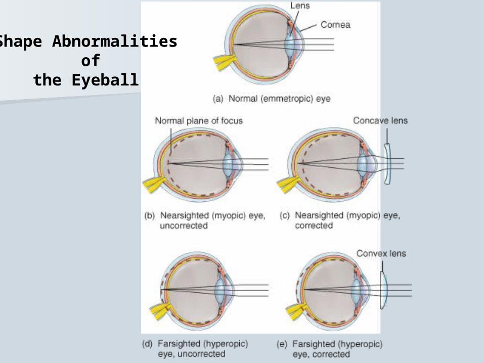
Shape Abnormalities of
the Eyeball


VITREOUSBODY
Vitreous Humor
ANTERIOR CHAMBER (Aqueous Humor)
CORNEA
PUPIL
CILIARY BODY
LENS
OPTIC NERVE
1. Fibrous TunicSCLERA
2. Vascular TunicCHOROID
3. RETINA
THREE LAYERS
IRIS

LACRIMAL GLAND
RIGHT EYE
SUPERIOR RECTUS MUSCLE
INFERIOR RECTUS MUSCLE
LATERAL RECTUS MUSCLE
MEDIAL RECTUS MUSCLE
PUPIL
IRIS behind transparent CORNEA
SCLERA
* SUPERIOR & INFERIOR OBLIQUE MUSCLES are not shownNOSE