eyes, function management drugs for each disorder
TRANSCRIPT
8/3/2019 Eyes, Function Management Drugs for Each Disorder
http://slidepdf.com/reader/full/eyes-function-management-drugs-for-each-disorder 1/19
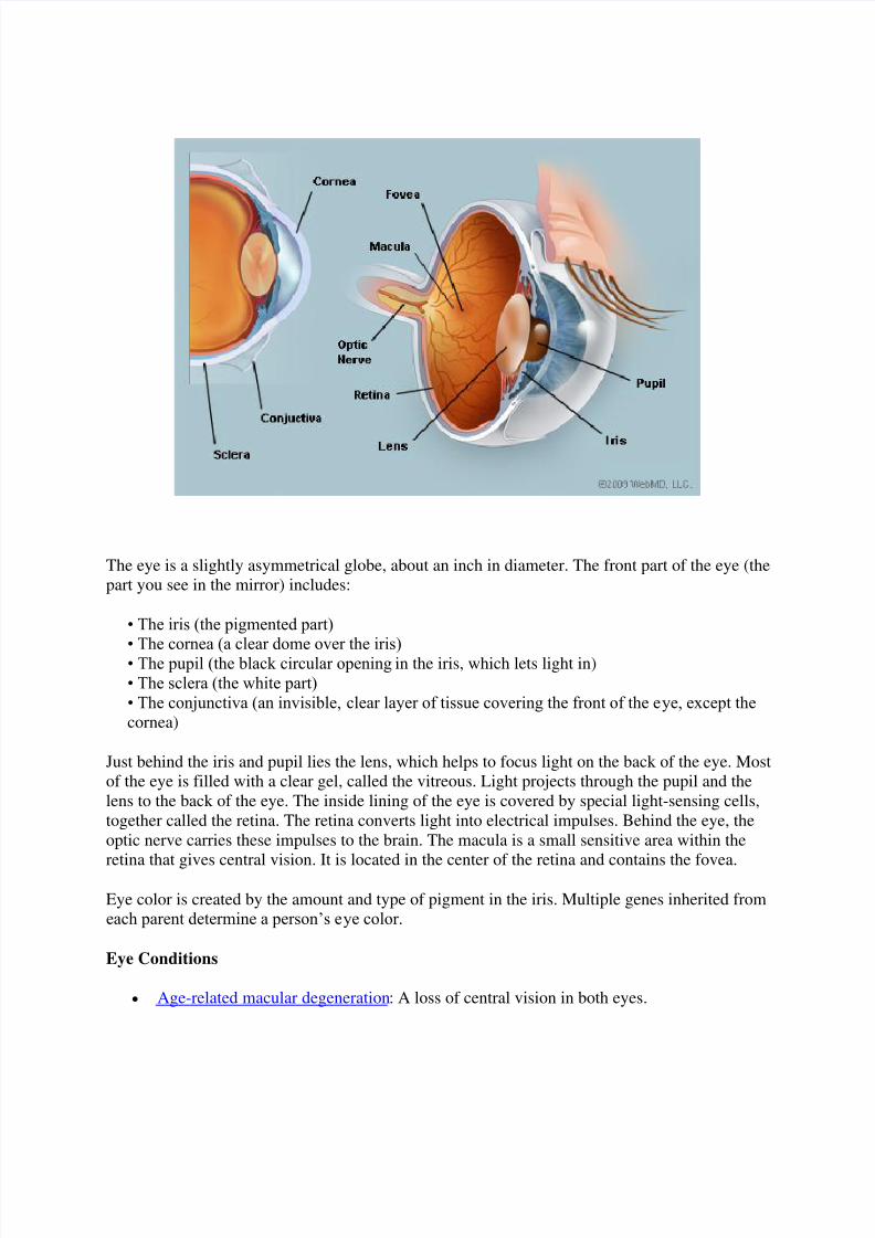
The eye is a slightly asymmetrical globe, about an inch in diameter. The front part of the eye (thepart you see in the mirror) includes:
• The iris (the pigmented part)
• The cornea (a clear dome over the iris) • The pupil (the black circular opening in the iris, which lets light in)• The sclera (the white part) • The conjunctiva (an invisible, clear layer of tissue covering the front of the eye, except thecornea)
Just behind the iris and pupil lies the lens, which helps to focus light on the back of the eye. Mostof the eye is filled with a clear gel, called the vitreous. Light projects through the pupil and thelens to the back of the eye. The inside lining of the eye is covered by special light-sensing cells,together called the retina. The retina converts light into electrical impulses. Behind the eye, theoptic nerve carries these impulses to the brain. The macula is a small sensitive area within theretina that gives central vision. It is located in the center of the retina and contains the fovea.
Eye color is created by the amount and type of pigment in the iris. Multiple genes inherited fromeach parent determine a person’s eye color.
Eye Conditions
Age-related macular degeneration : A loss of central vision in both eyes.

8/3/2019 Eyes, Function Management Drugs for Each Disorder
http://slidepdf.com/reader/full/eyes-function-management-drugs-for-each-disorder 2/19
Myopia (nearsightedness): Inability to see clearly at a distance. The eye is “too long” for the lens, so light isn’t focused properly on th e retina.
Hyperopia (farsightedness): Inability to see near objects clearly. The eye is “too short”for the lens, or certain eye muscles have weakened with age.
Strabismus : The eyes do not point in the same direction. The brain may then favor one
eye, causing decreased vision (amblyopia) in the other eye. Pterygium : A thickened conjunctival mass usually on the inner part of the eyeball. It maycover a part of the cornea, causing vision problems.
Scotoma: A blind or dark spot in the visual field. Amblyopia (lazy eye): One eye sees better than the other, a problem of childhood
development. The weaker eye may or may not “wander.” The weaker eye is called the"lazy eye."
Astigmatism : A defect that causes an inability to properly focus light onto the retina.Astigmatism causes blurry vision that can be corrected with glasses or contact lenses.
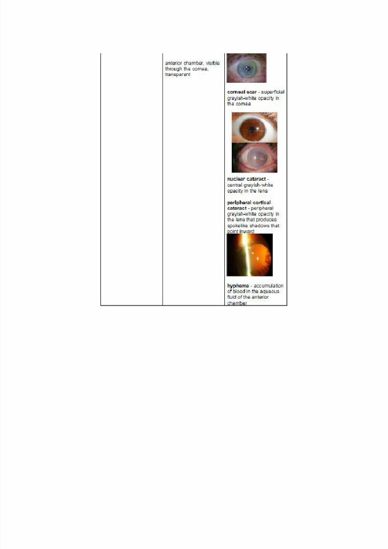
Cataract : A clouding of the lens, which hinders the passage of light through the lens. Conjunctivitis : Also known as "pinkeye,” conjunctivitis is an infection or inflammation
of the conjunctiva. It is usually caused by allergies, a virus, or a bacterial infection. Glaucoma : Increased pressure inside the eye slowly reduces vision. Peripheral vision islost first, often going undetected for years.
Diplopia (double vision): Seeing double can be caused by many serious conditions.Diplopia requires immediate medical attention.
Retinal detachment : The retina comes loose from the back of the eye. Trauma anddiabetes are common causes of this medical emergency.
Diabetic retinopathy : High blood sugar damages blood vessels in the eye. Eventually,weakened blood vessels may overgrow the retina or bleed, threatening vision.
Stye : Bacteria infect the skin on the edge of the eyelid, creating a tender red bump. Chalazion : An oil-making gland gets blocked and swells into a bump. Often confused
with styes, chalazions are not caused by infections. Hyphema : Bleeding into the front of the eye, behind the cornea. Hyphema is usually
caused by trauma. Blepharitis : Inflammation of the eyelids near the eyelashes. Blepharitis is a common
cause of itching or a feeling of grit in the eyes. Corneal abrasion : A scratch on the clear part of the front of the eye. Pain, light
sensitivity, or a feeling of grit in the eye are the usual symptoms. Keratitis : Inflammation or infection of the cornea. Keratitis typically occurs after germs
enter a corneal abrasion. Retinitis : Inflammation or infection of the retina. Retinitis may be a long-term genetic
condition or result from a viral infection. Uveitis (iritis): The colored part of the eye becomes inflamed or infected. An overactive
immune system, bacteria, or viruses can be responsible. Dry eye : Either the eyes don’t produce enough tears, or the tears are of poor quality. Dry
eye can be caused by medical problems such as lupus, scleroderma, and Sjogren'ssyndrome.
Optic neuritis : The optic nerve becomes inflamed, usually from an overactive immunesystem. Painful vision loss in one eye typically results.
Black eye : Swelling and discoloration around the eye as a result of injury to the face.

8/3/2019 Eyes, Function Management Drugs for Each Disorder
http://slidepdf.com/reader/full/eyes-function-management-drugs-for-each-disorder 3/19
http://www.webmd.com/eye-health/picture-of-the-eyes
http://www.nlm.nih.gov/medlineplus/eyesandvision.html
Adult Eye Exams
It's important for adults to have eye exams on a regular basis to check for problems. Regular eyeexams are critical for detecting:
Glaucoma Age-related macular degeneration (AMD) Cataracts Diabetic retinopathy
But everyone needs regular eye exams. This is particularly important if you have risk factors or afamily history of eye problems. Children need their vision checked at 6 months, 3 years, andbefore first grade. Adults should see an eye doctor at least every two years and annually after age60.
Recommended Related to Eye Health
Albinism,Ocular
Important It is possible that the main title of the report Albinism, Ocular is not the name youexpected. Please check the synonyms listing to find the alternate name(s) and disordersubdivision(s) covered by this report.
Read the Albinism,Ocular article > >
Your doctor may recommend more frequent exams if you have a health condition such asdiabetes or high blood pressure, work in a visually demanding job, or take medications that canaffect eyesight.
Preparing for Your Eye Exam
When you call to make an appointment for an eye exam, briefly and clearly describe any vision problem you're having.
Before you go, list questions for the eye doctor. Be prepared to discuss any drugs you're takingand your (and your family's) eye health history.
When you go, take your glasses and/or contact lenses, if you use them, and sunglasses for the triphome with your pupils dilated.

8/3/2019 Eyes, Function Management Drugs for Each Disorder
http://slidepdf.com/reader/full/eyes-function-management-drugs-for-each-disorder 4/19
During Your Eye Exam
Before your eye exam, the eye doctor or an office staff member will take your medical andvision history.
Your eye exam may take from half an hour to an hour. It will evaluate both your vision and thehealth of your eyes.
You'll likely have all or most of the following eye tests (you may also have more specialized eyetests):
Eye muscle movement test : To test muscle strength and control, the doctor will ask you tovisually track a target in different directions and observe your eye movements.
Cover test : This is a check for how well your eyes work together. As you stare at a small targetsome distance away, the doctor will cover and uncover each eye to observe how much your eyes
move, watching for an eye that turns away from the target (strabismus). The test may be repeatedwith a target close to you.
External exam and pupillary reactions : The doctor will watch the reactions of your pupils tolight and objects at close distance. At the same time, the doctor will check the exterior eye,looking at things such as the condition of the white of the eyes and the position of your eyelids.
Visual acuity test : You'll sit in front of an eye chart, with letters that get smaller as you readdown each line. You cover each eye in turn and, using the other eye, read aloud, going down thechart, until you can't read the letters anymore.
Retinoscopy : The eye doctor may shine a light in your eyes and flip lenses in a machine(phoropter) that you look through while staring at a large target, such as a big "E," or the doctormay use an automated machine (refractor) for the same purpose. By checking the way lightreflects from your eyes, the doctor gets an approximate idea of the lens prescription you neednow.

8/3/2019 Eyes, Function Management Drugs for Each Disorder
http://slidepdf.com/reader/full/eyes-function-management-drugs-for-each-disorder 5/19

8/3/2019 Eyes, Function Management Drugs for Each Disorder
http://slidepdf.com/reader/full/eyes-function-management-drugs-for-each-disorder 6/19

8/3/2019 Eyes, Function Management Drugs for Each Disorder
http://slidepdf.com/reader/full/eyes-function-management-drugs-for-each-disorder 7/19

8/3/2019 Eyes, Function Management Drugs for Each Disorder
http://slidepdf.com/reader/full/eyes-function-management-drugs-for-each-disorder 8/19

8/3/2019 Eyes, Function Management Drugs for Each Disorder
http://slidepdf.com/reader/full/eyes-function-management-drugs-for-each-disorder 9/19

8/3/2019 Eyes, Function Management Drugs for Each Disorder
http://slidepdf.com/reader/full/eyes-function-management-drugs-for-each-disorder 10/19

8/3/2019 Eyes, Function Management Drugs for Each Disorder
http://slidepdf.com/reader/full/eyes-function-management-drugs-for-each-disorder 11/19
8/3/2019 Eyes, Function Management Drugs for Each Disorder
http://slidepdf.com/reader/full/eyes-function-management-drugs-for-each-disorder 12/19

8/3/2019 Eyes, Function Management Drugs for Each Disorder
http://slidepdf.com/reader/full/eyes-function-management-drugs-for-each-disorder 13/19

8/3/2019 Eyes, Function Management Drugs for Each Disorder
http://slidepdf.com/reader/full/eyes-function-management-drugs-for-each-disorder 14/19

8/3/2019 Eyes, Function Management Drugs for Each Disorder
http://slidepdf.com/reader/full/eyes-function-management-drugs-for-each-disorder 15/19

8/3/2019 Eyes, Function Management Drugs for Each Disorder
http://slidepdf.com/reader/full/eyes-function-management-drugs-for-each-disorder 16/19
Eye Conditions And Disorders
If you have any of these eye conditions you may require emergency medical attention: sudden
loss of vision in one eye; sudden hazy or blurred vision; flashes of light or black spots in yourvision; Halos or rainbows around light; curtain-like blotting out of vision; and Loss of peripheral(side) vision.
Would you give up smoking if your cigarette pack had this warning? Studies have shown that people whocurrently smoke are two to five times more likely to develop AMD than non-smokers or past smokers. AMD -Age related Macular Degeneration is the leading cause of blindness in people 65 and over.
Four of the most common eye conditions.Myopia - (nearsightedness)
People with Myopic vision usually have eyeballs that are too large for their lens and cornea tofocus light properly on their retina. Eyeglasses and contact lenses can usually correct thisproblem.
Hyperopia - (farsightedness)
Hyperopia vision is caused by the eyeball being too small for the lens and cornea to focus lightproperly on the retina. Eyeglasses and contact lenses can usually correct this problem.
Presbyopia - (aging eyes)
As people age, they often begin to have difficulty focusing their eyes for reading or close work. Itis usually corrected with reading glasses. Some people may need bifocal of trifocal lenses.
Astigmatism - (distorted vision)
People with Astigmatism have irregularly shaped corneas. It is usually corrected with eyeglassesor contact lenses.
Most Common Causes of Vision LossCataracts
There are over 1 million Cataract operations performed annually in the USA. Cataract surgery isan outpatient procedure with a very high success rate. Due to the lack of modern medicaltechnology in the developing world, it is also the world's leading cause of blindness. Over 16million people are blind from cataracts.
Age-Related Macular Degeneration - (AMD)

8/3/2019 Eyes, Function Management Drugs for Each Disorder
http://slidepdf.com/reader/full/eyes-function-management-drugs-for-each-disorder 17/19
This is a degenerative disease of the macula; the macula is the part of the retina responsible forcentral vision. There is no way yet of repairing the vision that has been lost, but if detected earlylaser surgery can help slow the progression of the disease. (AMD) is the leading cause of visionloss in people over age 65. Eight million people are legally blind from macular degenerationworldwide and as the population ages this number is expected to grow.
Glaucoma
This disease increases the fluid pressure inside the eye, leading to loss of side vision andeventually total blindness. The increased pressure destroys the optic nerve. With earlydetection, it can be kept under control with pressure reducing eye drops and surgery. Chancesof developing it increase with age. There are over five million people blind from glaucomaworldwide.
Diabetic Retinopathy
This complication of diabetes is a leading cause of blindness among middle-aged Americans. Thelonger a person has had diabetes the more apt they are to develop diabetic retinopathy. Lasersurgery can slow the progression of this disease along with management of blood glucose levels.There are 2.4 million people blind from retinopathy worldwide.
Retinitis Pigmentosa - (RP)
This rare inherited degenerative disease slowly destroys the retina. Signs of (RP) first show up inearly childhood. The side vision is lost first. The Disease progresses over many years leaving theperson with only a small portion of their central vision. There is on cure for (RP) yet. There are1.6 million people blind from (RP) worldwide.
Eye Injuries
Over 1 million people are blind worldwide from eye injuries. 90% of injuries can be avoided byusing proper eye protection. If you are reading this page now because you or a loved one has aneye injury, turn your computer off and get medical attention. Any eye injury is a medicalemergency.
Optic Nerve Hypoplasia
With this birth defect the optic nerve that connects the eye to the brain has not developed
properly, it is underdeveloped. This happens before birth leaving the child with mild to severvision loss. In most cases, there is no known cause.
Retinopathy of Prematurity - (ROP)
This is an eye disease of premature babies. Soon after birth, abnormal blood vessels start togrow on the retina eventually destroying it. No one is quite sure what causes this disease, butpremature birth and low birth weight are thought to be the main causes. With modern medical

8/3/2019 Eyes, Function Management Drugs for Each Disorder
http://slidepdf.com/reader/full/eyes-function-management-drugs-for-each-disorder 18/19
procedures, only 400 babies a year go blind from this condition. In the 1950's the number of babies blind from (ROP) reached epidemic proportions.
Neurological Visual Impairment (NVI)
Children with (NVI) have normal eyes, but a part of the brain responsible for seeing is damaged.It can be caused from a lack of oxygen before, during, and after birth. Diseases like meningitis ortraumatic brain injury can also cause it. Many of the children with (NVI) are multihandicapped.Vision loss can be temporary or last a lifetime.
Ocular Albinism
With this inherited birth defect, people have no pigment or reduced pigment in their eyes, skinand hair. This pigment called melanin is needed for the full development of the retina. Childrenwith this condition have mild to sever vision loss and are very light sensitive.
Coloboma
Coloboma is a Greek word, which means mutilation. People with Coloboma are missing aportion of the structure of the eye. The human eye develops between the fourth and fifteenthweek of pregnancy. As the eye starts to develop, a gap opens on the underside of each eye bud.This gap provides a way for the developing eye to be nourished. This gap, called the opticfissure, has to close before the eye is fully developed. Coloboma is the incomplete closer of theoptic fissure. Gaps can occur in the eyelid, iris, ciliary body, lens, retina, macula, choroid andoptic disc. It can cause from mild to sever vision loss. Coloboma occurs in about 1 in 10,000births.
Preventable Vision Loss
Over 90% of the world's blind people live in developing countries. Many diseases can beprevented with education in basic hygiene, proper nutrition and medical care. Conditionsunheard of in the modern world like: Trachoma, Onchocerciasis (river blindness) and nutritionalblindness (vitamin A deficiency), are the leading causes of vision loss in these countries.
http://www.99main.com/~charlief/vi/disorders.html

8/3/2019 Eyes, Function Management Drugs for Each Disorder
http://slidepdf.com/reader/full/eyes-function-management-drugs-for-each-disorder 19/19