fabrication of magneto-controlled moveable architecture to develop reusable electrochemical...
DESCRIPTION
Fabrication of magneto-controlledmoveable architecture to developreusable electrochemical biosensorsTRANSCRIPT
Synthesis of Au/Graphene OxideComposites for Selective and SensitiveElectrochemical Detection of AscorbicAcidJian Song, Lin Xu, Ruiqing Xing, Qingling Li, Chunyang Zhou, Dali Liu & Hongwei Song
State Key Laboratory on Integrated Optoelectronics, College of Electronic Science and Engineering, Jilin University, 2699 QianjinStreet, Changchun, 130012, People’s Republic of China.
In this work, we present a novel ascorbic acid (AA) sensor applied to the detection of AA in human sera andpharmaceuticals. A series of Au nanoparticles (NPs) and graphene oxide sheets (Au NP/GO) compositeswere successfully synthesized by reduction of gold (III) using sodium citrate. Then the Au NP/GOcomposites were used to construct nonenzymatic electrodes in practical AA measurement. The electrodethat has the best performance presents attractive analytical features, such as a low working potential of10.15 V, a high sensitivity of 101.86 mA mM21 cm22 to AA, a low detection limit of 100 nM, goodreproducibility and excellent selectivity. And more,it was also employed to accurately and practically detectAA in human serum and clinical vitamin C tablet with the existence of some food additive. The enhancedAA electrochemical properties of the Au NP/GO modified electrode in our work can be attributed to theimprovement of electroactive surface area of Au NPs and the synergistic effect from the combination of AuNPs and GO sheets. This work shows that the Au NP/GO/GCEs hold the prospect for sensitive and selectivedetermination of AA in practical clinical application.
Ascorbic acid (AA), commonly called vitamin C, is one of the most important nutrients and effectiveantioxidant in protecting human from oxidative stress1–3. Today, AA is widely used in biomedicalchemistry, diagnostics and the identification of food ingredients. The concentration of AA is usually
kept in a low level, so accurate detection of AA content is of great importance to guarantee health conditions andfood security4,5. However, some other accompanied electroactive compounds of similar electrochemical prop-erties with AA, such as dopamine (DA) and uric acid (UA) glucose, and other oxidizable species, complicate theirelectrochemical identification, especially in biological environment6,7. Hence, the clear separation of the electro-chemical signals of AA from these compounds is still a great challenge.
In the past few decades, electrochemical techniques have been used to detect small biomolecules owing to theirhigh sensitivity, rapid response, flexibility, and low expense8,9. In an electrochemical system, the AA voltammetricpeak can be separated from that of interfering species using functional material modified electrodes10,11.Compounding two different reasonable selected materials is an effective way to further improve the electrochem-ical properties12–14. Specifically, GO sheets have been considered as a suitable substrate for assembling nanoma-terials because of their large surface area, excellent thermal and mechanical properties and chemical modificationcapability. In addition, they exhibit specific properties such as hydrophilicity, multiple oxygen moieties, andcontrollable electronic properties15–18. Meanwhile, in recent years, Au NPs have been widely investigated andapplied to molecular catalysis, biosensors, and mutifunctional reagents due to their quantum size effect19–21.Therefore, there is a potential development of a non-enzymatic electrochemical biosensor for AA involving thecomplementary use of GO sheets and Au NPs. For example, Tian et al. used a Au NP-b-cyclodextrin-graphenemodified electrode to detect AA, DA and UA22. The detection limit for AA (based on S/N 5 3) is 10 mM and thelinear response range is 30–2000 mM. Zhang et al. used a facile one-step method to fabricate nanoflower-likedendritic Au NPs and polyaniline composite nanosheets and applied them on glass carbon electrode as an AAsensor14. A linear relation between the current response and the AA concentration was obtained between 10 mMand 12 mM, with a detection limit of 8.3 mM (S/N 5 3) and the sensitivity of 25.69 mA mM21 cm22. However,
OPEN
SUBJECT AREAS:ANALYTICAL
BIOCHEMISTRY
CHEMICAL MODIFICATION
Received21 August 2014
Accepted25 November 2014
Published17 December 2014
Correspondence andrequests for materials
should be addressed toL.X. ([email protected])or D.L. ([email protected])
SCIENTIFIC REPORTS | 4 : 7515 | DOI: 10.1038/srep07515 1
there have been very few reports on the detection of AA in real-lifehuman sera or pharmaceuticals to demonstrate the practical applica-tions of AA biosensors.
In this work, we have chemically reduced Au(III) using sodiumcitrate to form Au NPs of approximately 13–16 nm average diameteron GO sheets23. These Au NP/GO composite nanomaterials werethen immobilized on the glass carbon electrodes (GCE) for AAdetection. These modified electrodes were used to monitor AA con-centration in real-life human sera and pharmaceuticals.
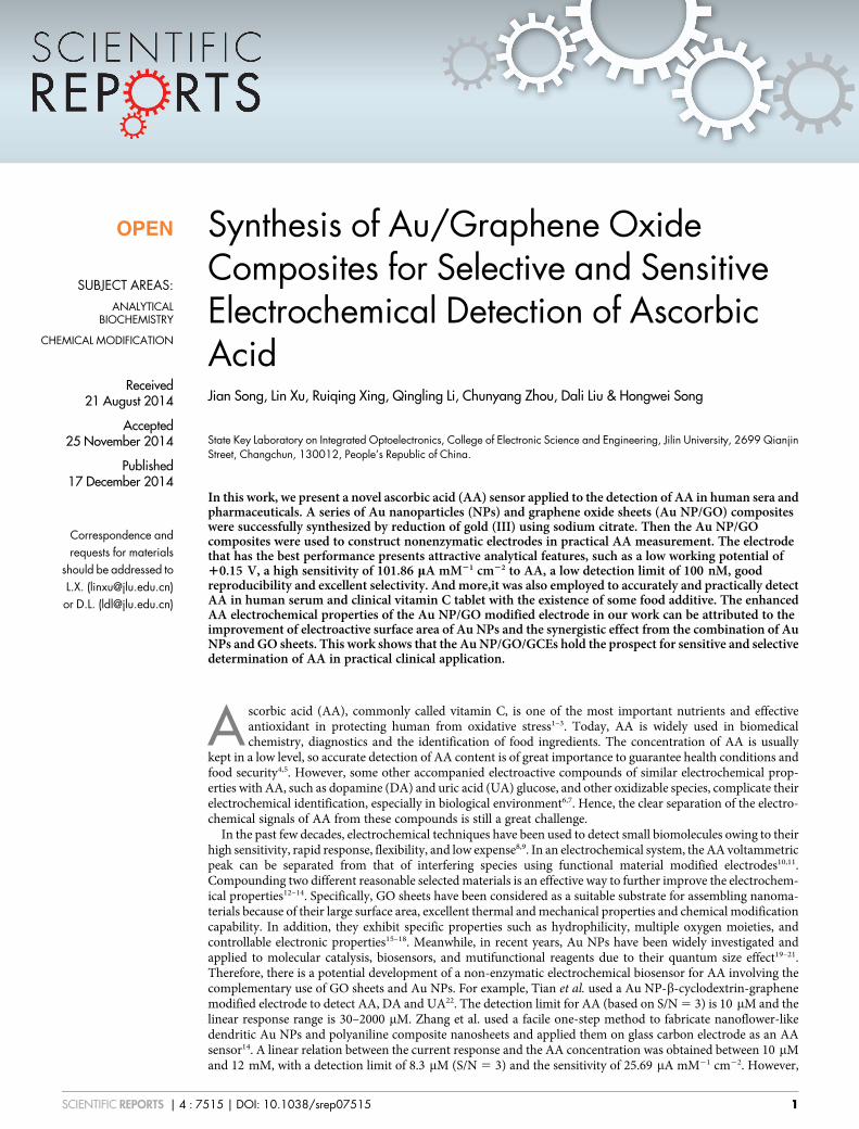
Results and DiscussionCharacterization of the Au NP/GO composites. The morphologyof the GO sheets used in this work was first examined bytransmission electron microscope (TEM) technique, and theresults are shown in Fig. 1a. The GO sheets exhibit flake-like shapeand is few-layer flexible wrinkled. The smooth and planar surfaceindicates that GO sheets can provide a high surface to volume ratioand a 2D structure, which is necessary for loading Au NPs. Then theGO sheets were surface loaded with different amount of Au NPs(Fig. 1b–1e). As can be seen, the loaded Au demonstrates regularsmall NP morphology and can grow on the GO sheet independently.The size histograms of Au NPs in each sample were carefullymeasured and shown in Fig. S2. The size distribution of Au NPsshows a slightly growing trend from S1 to S4 and the average sizeof S1, S2, S3 and S4 are determined to be 13.3, 14.2, 15.3 and 16.0 nm,respectively. Besides, as the amount of initial HAuCl4 graduallyincreased, the amount of Au NPs loaded on the GO sheets alsoincreased. Note that when the amount of initial HAuCl4 reached
20 mg, the Au NPs loaded on the GO sheet tended to aggregatetogether (Fig. 1e). Fig. 1f shows the high resolution TEM(HRTEM) image, in which the interplanar distances can be clearlyseen. The as marked interplanar distances are 0.235 nm and0.202 nm, corresponding to the (111) and (200) face of face-centered cubic Au, respectively. The inset of Fig. 1f is the selectedarea electron diffraction (SAED) pattern of Au NP/GO composites(S3). The diffraction patterns irregularly distribute and thediffraction rings can be observed, which indicates this sampleyields a polycrystalline structure24. The conditions of the othersamples are the same.
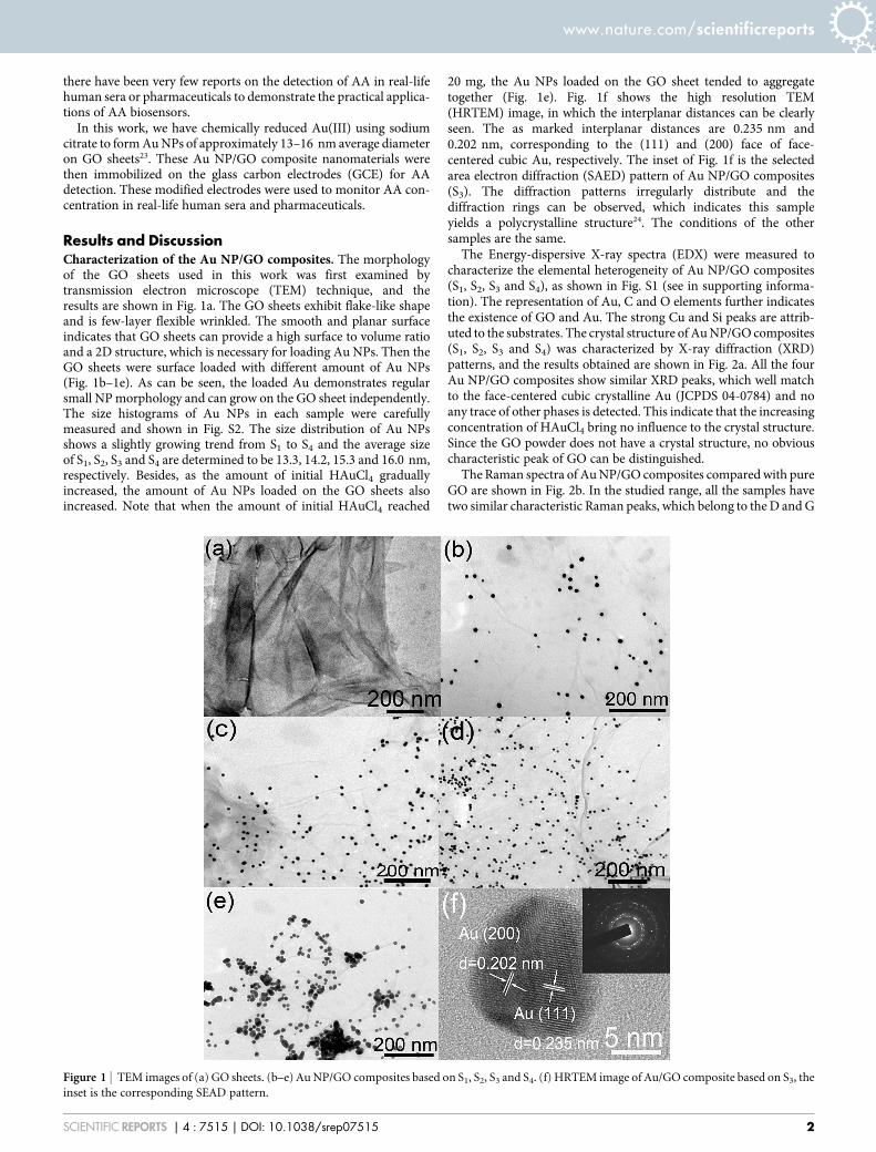
The Energy-dispersive X-ray spectra (EDX) were measured tocharacterize the elemental heterogeneity of Au NP/GO composites(S1, S2, S3 and S4), as shown in Fig. S1 (see in supporting informa-tion). The representation of Au, C and O elements further indicatesthe existence of GO and Au. The strong Cu and Si peaks are attrib-uted to the substrates. The crystal structure of Au NP/GO composites(S1, S2, S3 and S4) was characterized by X-ray diffraction (XRD)patterns, and the results obtained are shown in Fig. 2a. All the fourAu NP/GO composites show similar XRD peaks, which well matchto the face-centered cubic crystalline Au (JCPDS 04-0784) and noany trace of other phases is detected. This indicate that the increasingconcentration of HAuCl4 bring no influence to the crystal structure.Since the GO powder does not have a crystal structure, no obviouscharacteristic peak of GO can be distinguished.
The Raman spectra of Au NP/GO composites compared with pureGO are shown in Fig. 2b. In the studied range, all the samples havetwo similar characteristic Raman peaks, which belong to the D and G
Figure 1 | TEM images of (a) GO sheets. (b–e) Au NP/GO composites based on S1, S2, S3 and S4. (f) HRTEM image of Au/GO composite based on S3, the
inset is the corresponding SEAD pattern.
www.nature.com/scientificreports
SCIENTIFIC REPORTS | 4 : 7515 | DOI: 10.1038/srep07515 2
bands of GO sheets. The disorder-induced D bands at 1351 cm21
arising from sp3-hybridized carbon were observed25. The tangentialstretch G band represents the E2g zone center mode of the crystallinegraphite. Generally, the relative intensity ratio of the D and G bands(ID/IG) depends strongly on the degree of disorder in the graphiticmaterial26. Generally, the ID/IG ratio increases when more defects arebrought into GO. According to Fig. 2b, the ID/IG ratios of the four AuNP/GO composites are calculated to be 0.80, 0.82, 0.81 and 0.82,respectively, which are a little higher than that of pure GO sheet(0.77). This shows that the loading amount of Au NPs has only alittle influence on the ID/IG ratio, implying that the nucleation of AuNPs at GO surfaces introduces very few defects into the GOstructure.
To further confirm the concentration of Au and GO in each com-posite sample, thermogravimetric analysis (TGA) curves were inves-tigated. As shown in Fig. 2c, the mass loss below 200uC (25.7%,19.8%, 16.2% and 8.2% for S1, S2, S3, and S4, respectively) can beattributed to the evaporation of adsorbed water. The slight mass lossbetween 200uC and 400uC (12.4%, 10.3%, 6.0% and 4.4% for S1, S2,S3, and S4, respectively) arose from the decomposition of some resid-ual oxygen-containing groups27. Then the finial significant mass lossoccurred when the Au NP/GO composites were heated above 400uC
(38.6%, 30.8%, 20.5 and 12.9% for S1, S2, S3, and S4, respectively).This was most likely caused by the decomposition of carbon skeletonfrom GO27. Accordingly, the mass percentage of Au in S1, S2, S3 andS4 are calculated to be 24.0%, 39.0%, 57.4% and 75.5%, respectively,which also implies the successful loading of Au NPs on GO sheets.
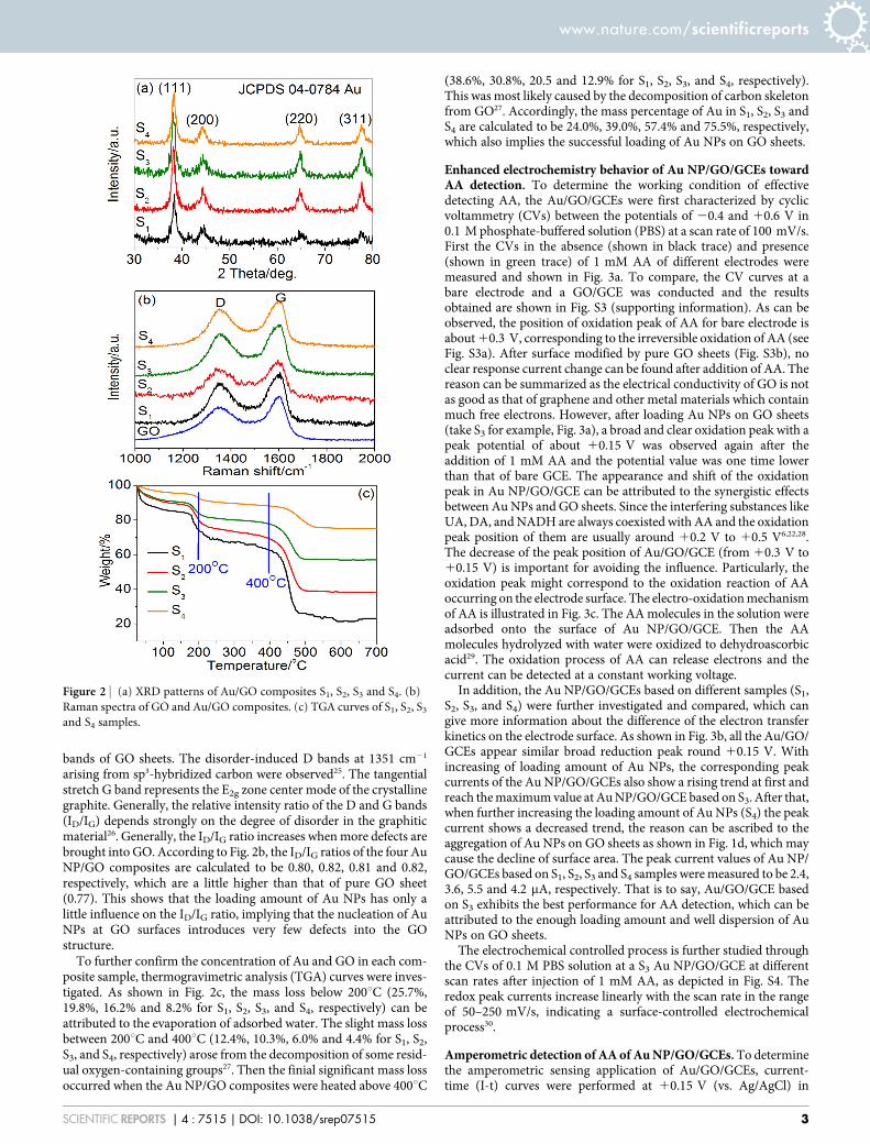
Enhanced electrochemistry behavior of Au NP/GO/GCEs towardAA detection. To determine the working condition of effectivedetecting AA, the Au/GO/GCEs were first characterized by cyclicvoltammetry (CVs) between the potentials of 20.4 and 10.6 V in0.1 M phosphate-buffered solution (PBS) at a scan rate of 100 mV/s.First the CVs in the absence (shown in black trace) and presence(shown in green trace) of 1 mM AA of different electrodes weremeasured and shown in Fig. 3a. To compare, the CV curves at abare electrode and a GO/GCE was conducted and the resultsobtained are shown in Fig. S3 (supporting information). As can beobserved, the position of oxidation peak of AA for bare electrode isabout 10.3 V, corresponding to the irreversible oxidation of AA (seeFig. S3a). After surface modified by pure GO sheets (Fig. S3b), noclear response current change can be found after addition of AA. Thereason can be summarized as the electrical conductivity of GO is notas good as that of graphene and other metal materials which containmuch free electrons. However, after loading Au NPs on GO sheets(take S3 for example, Fig. 3a), a broad and clear oxidation peak with apeak potential of about 10.15 V was observed again after theaddition of 1 mM AA and the potential value was one time lowerthan that of bare GCE. The appearance and shift of the oxidationpeak in Au NP/GO/GCE can be attributed to the synergistic effectsbetween Au NPs and GO sheets. Since the interfering substances likeUA, DA, and NADH are always coexisted with AA and the oxidationpeak position of them are usually around 10.2 V to 10.5 V6,22,28.The decrease of the peak position of Au/GO/GCE (from 10.3 V to10.15 V) is important for avoiding the influence. Particularly, theoxidation peak might correspond to the oxidation reaction of AAoccurring on the electrode surface. The electro-oxidation mechanismof AA is illustrated in Fig. 3c. The AA molecules in the solution wereadsorbed onto the surface of Au NP/GO/GCE. Then the AAmolecules hydrolyzed with water were oxidized to dehydroascorbicacid29. The oxidation process of AA can release electrons and thecurrent can be detected at a constant working voltage.
In addition, the Au NP/GO/GCEs based on different samples (S1,S2, S3, and S4) were further investigated and compared, which cangive more information about the difference of the electron transferkinetics on the electrode surface. As shown in Fig. 3b, all the Au/GO/GCEs appear similar broad reduction peak round 10.15 V. Withincreasing of loading amount of Au NPs, the corresponding peakcurrents of the Au NP/GO/GCEs also show a rising trend at first andreach the maximum value at Au NP/GO/GCE based on S3. After that,when further increasing the loading amount of Au NPs (S4) the peakcurrent shows a decreased trend, the reason can be ascribed to theaggregation of Au NPs on GO sheets as shown in Fig. 1d, which maycause the decline of surface area. The peak current values of Au NP/GO/GCEs based on S1, S2, S3 and S4 samples were measured to be 2.4,3.6, 5.5 and 4.2 mA, respectively. That is to say, Au/GO/GCE basedon S3 exhibits the best performance for AA detection, which can beattributed to the enough loading amount and well dispersion of AuNPs on GO sheets.
The electrochemical controlled process is further studied throughthe CVs of 0.1 M PBS solution at a S3 Au NP/GO/GCE at differentscan rates after injection of 1 mM AA, as depicted in Fig. S4. Theredox peak currents increase linearly with the scan rate in the rangeof 50–250 mV/s, indicating a surface-controlled electrochemicalprocess30.
Amperometric detection of AA of Au NP/GO/GCEs. To determinethe amperometric sensing application of Au/GO/GCEs, current-time (I-t) curves were performed at 10.15 V (vs. Ag/AgCl) in
Figure 2 | (a) XRD patterns of Au/GO composites S1, S2, S3 and S4. (b)
Raman spectra of GO and Au/GO composites. (c) TGA curves of S1, S2, S3
and S4 samples.
www.nature.com/scientificreports
SCIENTIFIC REPORTS | 4 : 7515 | DOI: 10.1038/srep07515 3
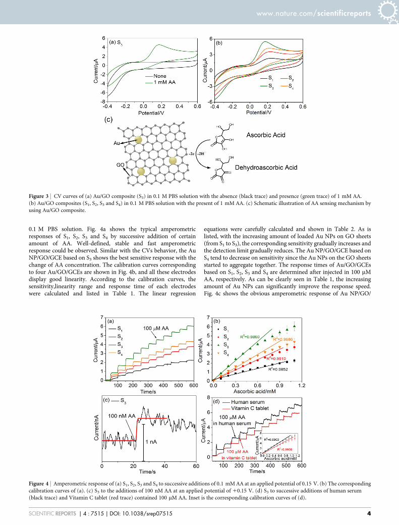
0.1 M PBS solution. Fig. 4a shows the typical amperometricresponses of S1, S2, S3 and S4 by successive addition of certainamount of AA. Well-defined, stable and fast amperometricresponse could be observed. Similar with the CVs behavior, the AuNP/GO/GCE based on S3 shows the best sensitive response with thechange of AA concentration. The calibration curves correspondingto four Au/GO/GCEs are shown in Fig. 4b, and all these electrodesdisplay good linearity. According to the calibration curves, thesensitivity,linearity range and response time of each electrodeswere calculated and listed in Table 1. The linear regression
equations were carefully calculated and shown in Table 2. As islisted, with the increasing amount of loaded Au NPs on GO sheets(from S1 to S3), the corresponding sensitivity gradually increases andthe detection limit gradually reduces. The Au NP/GO/GCE based onS4 tend to decrease on sensitivity since the Au NPs on the GO sheetsstarted to aggregate together. The response times of Au/GO/GCEsbased on S1, S2, S3 and S4 are determined after injected in 100 mMAA, respectively. As can be clearly seen in Table 1, the increasingamount of Au NPs can significantly improve the response speed.Fig. 4c shows the obvious amperometric response of Au NP/GO/
Figure 3 | CV curves of (a) Au/GO composite (S3) in 0.1 M PBS solution with the absence (black trace) and presence (green trace) of 1 mM AA.
(b) Au/GO composites (S1, S2, S3 and S4) in 0.1 M PBS solution with the present of 1 mM AA. (c) Schematic illustration of AA sensing mechanism by
using Au/GO composite.
Figure 4 | Amperometric response of (a) S1, S2, S3 and S4 to successive additions of 0.1 mM AA at an applied potential of 0.15 V. (b) The corresponding
calibration curves of (a). (c) S3 to the additions of 100 nM AA at an applied potential of 10.15 V. (d) S3 to successive additions of human serum
(black trace) and Vitamin C tablet (red trace) contained 100 mM AA. Inset is the corresponding calibration curves of (d).
www.nature.com/scientificreports
SCIENTIFIC REPORTS | 4 : 7515 | DOI: 10.1038/srep07515 4
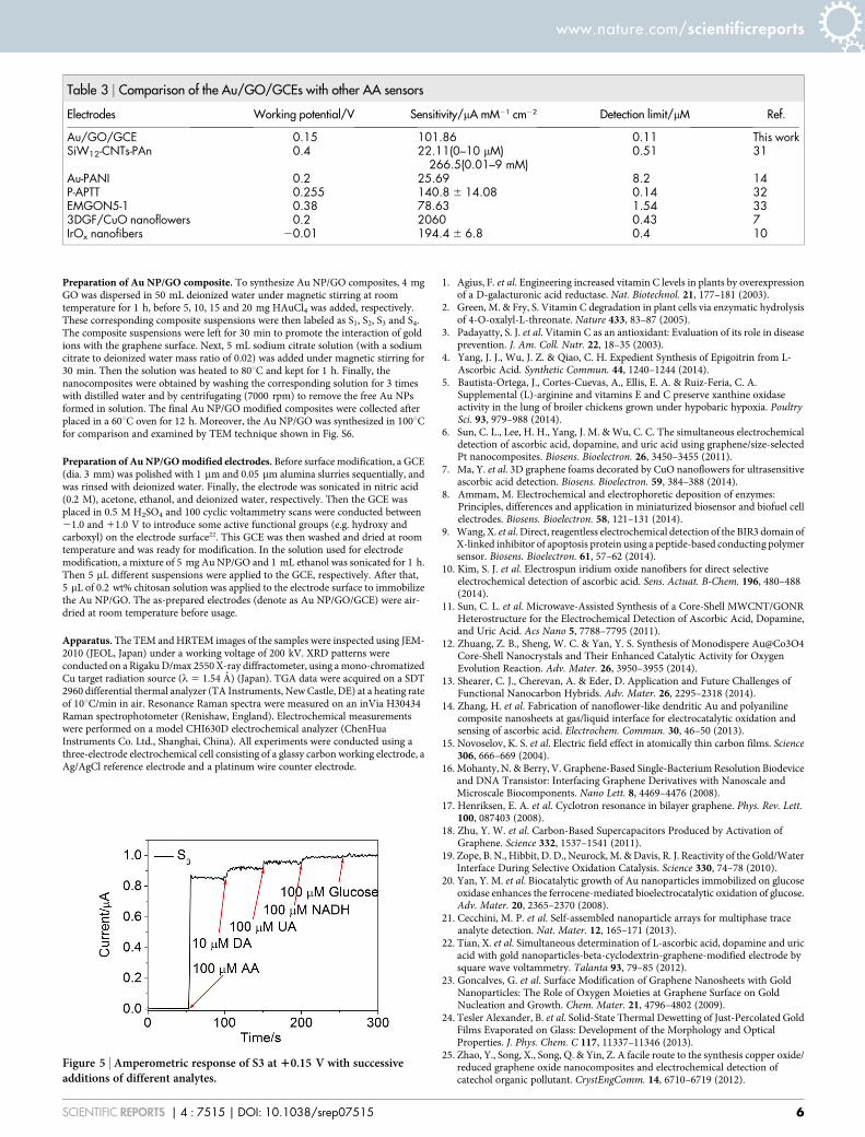
GCE based on S3 after the addition of 100 nM AA with a signal-to-noise ratio (S/N) of ,3 in order to obtain the accurate detection limit.This result is much lower than the previously reported graphene-based electrochemical sensors and electrical sensors assisted byenzymes as listed in Table 3. Besides, as compared, the Au NP/GO/GCE based on S3 in this work also shows high sensitivity, lowdetection limit and large liner range. According to the sensitivityimprovement, two factors need to be considered: On the one hand,the amount of Au NPs influences dominantly on the performancewhen they are gradually increased. The reason can be attributed tothe good electrical conductivity and large surface area introduced byAu NPs. The Au NPs on S4 tend to aggregate together so the surfacearea decreases and the sensitivity is negatively influenced. On theother hand, the GO sheets can provide a large area for the Au NPsloading, so they can grow independently without aggregating in acertain amount range. So the GO sheets also make a greatcontribution to the improvement of the sensitivity.
Moreover, the practical AA sensing ability of Au NP/GO/GCEbased on S3 was further investigated in real human serum with AuNP/GO/GCE based on S3 and in clinical vitamin C tablet. As shownin Fig. 4d, the I–t curve corresponds to human serum was obtainedby adding human serum contained 100 mM AA, and the I–t curvecorresponds to vitamin C tablet was gotten by addition of aqueoussolution of vitamin C tablet which contained 100 mM AA. As can beseen, the amperometric response of the vitamin C tablet is slightlylower than that in human serum. It may caused by the food additivesin vitamin C tablet which may attach on and then fouling the elec-trode surface. The inset of Fig. 4d shows the calibration curves andboth of them display good linearity in the range of 0.1–0.6 mM. Thesensitivities of the Au NP/GO/GCE based on S3 are calculated to be108.82 mAmM21cm22 and 89.76 mAmM21cm22 corresponding tothe human serum and vitamin C tablet environments. The errorranges are 106.87% and 88.12% for the human serum and vitaminC tablet environments after calculated.
Reproducibility, stability, anti-interference property of the AuNP/GO/GCEs. The reproducibility, stability and anti-interferenceproperty of Au NP/GO/GCE based on S3 were further evaluated.For reproducibility, five same Au NP/GO/GCEs based on S3 wereinvestigated at 10.15 V to compare their amperometric currentresponses. The relative standard deviation (R.S.D.) was 8.4%,confirming that the preparation method was highly reproducible.The stability of Au NP/GO/GCE based on S3 was tested once perweek in 0.1 M PBS solution at a scan rate of 100 mV/s and lasted fora month (data not shown). The sensitivity decrease of the electrodewas no more than 10%, indicating a good stability of Au NP/GO/GCE. Since there are many interfering substances coexist with AA inthe reported normal physiological concentration, the selectivity will
be a big restrictive factor for the measurement34–37. Here, anti-interference property were carried out by successive injection of100 mM AA, UA, NADH, glucose and 10 mM DA depending onthe normal physiological concentration, which are the mostcommon coexistence material of AA. The normal physiologicalconcentration for DA is about 100 nM34, which is 100 times lowerthan the concentration we injected. The CV curves obtained bydetecting UA, DA and NADH independently or simultaneouslywere first shown in Fig. S5 in supporting information. As seen inFig. S5a, the oxidation peak positions of UA, DA, and NADH are10.3 V, 10.4 V, and 10.55 V, respectively, which are much higherthan that of AA (10.15 V). When the 1 mM UA, 0.1 mM DA, and1 mM NADH coexist with 1 mM AA, the CV curve shows anobvious oxidation peak at 10.15 V, corresponding to oxide peakof AA. This indicates that the other interfering substances have alittle influence on that of AA. In order to further prove the fact,amperometric response of S3 modified electrode at 10.15 V withsuccessive additions of different analytes are shown in Fig. 5, andthe current responses of interfering species are also calculatedaccurately. Compared to AA, the current responses of interferingspecies are determined to be 4.7% (100 mM UA), 3.5% (100 mMNADH) and 8.2% (10 mM DA) at 10.15 V. It can be concludedthat similar amount of these interfering substances can beneglected. The excellent sensitivity indicates the potential practicalapplication of the as-prepared AA sensor.
ConclusionsIn conclusion, we have successfully synthesized a series of Au NP/GOcomposites. The loading amount of Au NPs on GO sheets control ofAu NP/GO composite has been investigated and the as–preparedsamples are used to detect AA. The optimal sample of these AuNP/GO composites sheets is S3 which presents high sensitivity andlow detection limit compared to the other samples. The sensor alsoshows good reproducibility, excellent selectivity and accurate mea-surement in real serum sample and vitamin C tablet. The reason canbe attributed to the improvement of electroactive surface area of AuNPs on GO sheets and the synergistic effect from the combination ofAu NPs and GO. It is anticipated that the Au NP/GO compositematerial holds great potential for developing novel AA sensors.
MethodsMaterials. All chemicals used were analytical grade and without further purification.GO sheets were purchased from Nanjing XFNANO Materials Tech Co., Ltd. KH2PO4
(99.5%), KCl (99%), Na2HPO4 (99%), NaCl (99.5%) and sodium citrate(C6H5Na3O7?2H2O, 99%) were purchased from Beijing Chemical Works.HAuCl4?3H2O (99.9%), Chitosan (93%), AA (99%), DA (99%), UA (99%) andNADH (97%) were supplied by Sigma-Aldrich. PBS (0.1 M, pH 7.4) was prepared bydispersing 0.2 g KCl, 8 g NaCl, 0.2 g KH2PO4 and 1.54 g Na2HPO4 in 1 L deionizedwater.
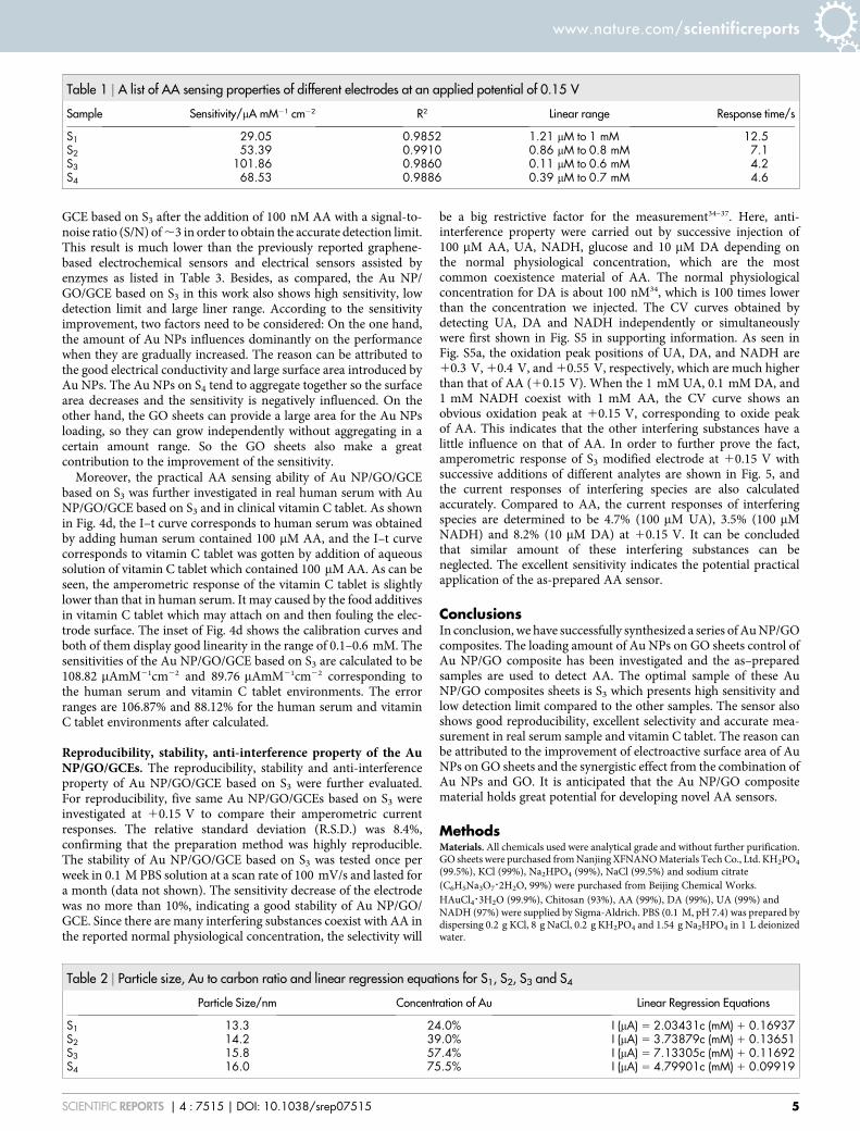
Table 1 | A list of AA sensing properties of different electrodes at an applied potential of 0.15 V
Sample Sensitivity/mA mM21 cm22 R2 Linear range Response time/s
S1 29.05 0.9852 1.21 mM to 1 mM 12.5S2 53.39 0.9910 0.86 mM to 0.8 mM 7.1S3 101.86 0.9860 0.11 mM to 0.6 mM 4.2S4 68.53 0.9886 0.39 mM to 0.7 mM 4.6
Table 2 | Particle size, Au to carbon ratio and linear regression equations for S1, S2, S3 and S4
Particle Size/nm Concentration of Au Linear Regression Equations
S1 13.3 24.0% I (mA) 5 2.03431c (mM) 1 0.16937S2 14.2 39.0% I (mA) 5 3.73879c (mM) 1 0.13651S3 15.8 57.4% I (mA) 5 7.13305c (mM) 1 0.11692S4 16.0 75.5% I (mA) 5 4.79901c (mM) 1 0.09919
www.nature.com/scientificreports
SCIENTIFIC REPORTS | 4 : 7515 | DOI: 10.1038/srep07515 5
Preparation of Au NP/GO composite. To synthesize Au NP/GO composites, 4 mgGO was dispersed in 50 mL deionized water under magnetic stirring at roomtemperature for 1 h, before 5, 10, 15 and 20 mg HAuCl4 was added, respectively.These corresponding composite suspensions were then labeled as S1, S2, S3 and S4.The composite suspensions were left for 30 min to promote the interaction of goldions with the graphene surface. Next, 5 mL sodium citrate solution (with a sodiumcitrate to deionized water mass ratio of 0.02) was added under magnetic stirring for30 min. Then the solution was heated to 80uC and kept for 1 h. Finally, thenanocomposites were obtained by washing the corresponding solution for 3 timeswith distilled water and by centrifugating (7000 rpm) to remove the free Au NPsformed in solution. The final Au NP/GO modified composites were collected afterplaced in a 60uC oven for 12 h. Moreover, the Au NP/GO was synthesized in 100uCfor comparison and examined by TEM technique shown in Fig. S6.
Preparation of Au NP/GO modified electrodes. Before surface modification, a GCE(dia. 3 mm) was polished with 1 mm and 0.05 mm alumina slurries sequentially, andwas rinsed with deionized water. Finally, the electrode was sonicated in nitric acid(0.2 M), acetone, ethanol, and deionized water, respectively. Then the GCE wasplaced in 0.5 M H2SO4 and 100 cyclic voltammetry scans were conducted between21.0 and 11.0 V to introduce some active functional groups (e.g. hydroxy andcarboxyl) on the electrode surface22. This GCE was then washed and dried at roomtemperature and was ready for modification. In the solution used for electrodemodification, a mixture of 5 mg Au NP/GO and 1 mL ethanol was sonicated for 1 h.Then 5 mL different suspensions were applied to the GCE, respectively. After that,5 mL of 0.2 wt% chitosan solution was applied to the electrode surface to immobilizethe Au NP/GO. The as-prepared electrodes (denote as Au NP/GO/GCE) were air-dried at room temperature before usage.
Apparatus. The TEM and HRTEM images of the samples were inspected using JEM-2010 (JEOL, Japan) under a working voltage of 200 kV. XRD patterns wereconducted on a Rigaku D/max 2550 X-ray diffractometer, using a mono-chromatizedCu target radiation source (l 5 1.54 A) (Japan). TGA data were acquired on a SDT2960 differential thermal analyzer (TA Instruments, New Castle, DE) at a heating rateof 10uC/min in air. Resonance Raman spectra were measured on an inVia H30434Raman spectrophotometer (Renishaw, England). Electrochemical measurementswere performed on a model CHI630D electrochemical analyzer (ChenHuaInstruments Co. Ltd., Shanghai, China). All experiments were conducted using athree-electrode electrochemical cell consisting of a glassy carbon working electrode, aAg/AgCl reference electrode and a platinum wire counter electrode.
1. Agius, F. et al. Engineering increased vitamin C levels in plants by overexpressionof a D-galacturonic acid reductase. Nat. Biotechnol. 21, 177–181 (2003).
2. Green, M. & Fry, S. Vitamin C degradation in plant cells via enzymatic hydrolysisof 4-O-oxalyl-L-threonate. Nature 433, 83–87 (2005).
3. Padayatty, S. J. et al. Vitamin C as an antioxidant: Evaluation of its role in diseaseprevention. J. Am. Coll. Nutr. 22, 18–35 (2003).
4. Yang, J. J., Wu, J. Z. & Qiao, C. H. Expedient Synthesis of Epigoitrin from L-Ascorbic Acid. Synthetic Commun. 44, 1240–1244 (2014).
5. Bautista-Ortega, J., Cortes-Cuevas, A., Ellis, E. A. & Ruiz-Feria, C. A.Supplemental (L)-arginine and vitamins E and C preserve xanthine oxidaseactivity in the lung of broiler chickens grown under hypobaric hypoxia. PoultrySci. 93, 979–988 (2014).
6. Sun, C. L., Lee, H. H., Yang, J. M. & Wu, C. C. The simultaneous electrochemicaldetection of ascorbic acid, dopamine, and uric acid using graphene/size-selectedPt nanocomposites. Biosens. Bioelectron. 26, 3450–3455 (2011).
7. Ma, Y. et al. 3D graphene foams decorated by CuO nanoflowers for ultrasensitiveascorbic acid detection. Biosens. Bioelectron. 59, 384–388 (2014).
8. Ammam, M. Electrochemical and electrophoretic deposition of enzymes:Principles, differences and application in miniaturized biosensor and biofuel cellelectrodes. Biosens. Bioelectron. 58, 121–131 (2014).
9. Wang, X. et al. Direct, reagentless electrochemical detection of the BIR3 domain ofX-linked inhibitor of apoptosis protein using a peptide-based conducting polymersensor. Biosens. Bioelectron. 61, 57–62 (2014).
10. Kim, S. J. et al. Electrospun iridium oxide nanofibers for direct selectiveelectrochemical detection of ascorbic acid. Sens. Actuat. B-Chem. 196, 480–488(2014).
11. Sun, C. L. et al. Microwave-Assisted Synthesis of a Core-Shell MWCNT/GONRHeterostructure for the Electrochemical Detection of Ascorbic Acid, Dopamine,and Uric Acid. Acs Nano 5, 7788–7795 (2011).
12. Zhuang, Z. B., Sheng, W. C. & Yan, Y. S. Synthesis of Monodispere Au@Co3O4Core-Shell Nanocrystals and Their Enhanced Catalytic Activity for OxygenEvolution Reaction. Adv. Mater. 26, 3950–3955 (2014).
13. Shearer, C. J., Cherevan, A. & Eder, D. Application and Future Challenges ofFunctional Nanocarbon Hybrids. Adv. Mater. 26, 2295–2318 (2014).
14. Zhang, H. et al. Fabrication of nanoflower-like dendritic Au and polyanilinecomposite nanosheets at gas/liquid interface for electrocatalytic oxidation andsensing of ascorbic acid. Electrochem. Commun. 30, 46–50 (2013).
15. Novoselov, K. S. et al. Electric field effect in atomically thin carbon films. Science306, 666–669 (2004).
16. Mohanty, N. & Berry, V. Graphene-Based Single-Bacterium Resolution Biodeviceand DNA Transistor: Interfacing Graphene Derivatives with Nanoscale andMicroscale Biocomponents. Nano Lett. 8, 4469–4476 (2008).
17. Henriksen, E. A. et al. Cyclotron resonance in bilayer graphene. Phys. Rev. Lett.100, 087403 (2008).
18. Zhu, Y. W. et al. Carbon-Based Supercapacitors Produced by Activation ofGraphene. Science 332, 1537–1541 (2011).
19. Zope, B. N., Hibbit, D. D., Neurock, M. & Davis, R. J. Reactivity of the Gold/WaterInterface During Selective Oxidation Catalysis. Science 330, 74–78 (2010).
20. Yan, Y. M. et al. Biocatalytic growth of Au nanoparticles immobilized on glucoseoxidase enhances the ferrocene-mediated bioelectrocatalytic oxidation of glucose.Adv. Mater. 20, 2365–2370 (2008).
21. Cecchini, M. P. et al. Self-assembled nanoparticle arrays for multiphase traceanalyte detection. Nat. Mater. 12, 165–171 (2013).
22. Tian, X. et al. Simultaneous determination of L-ascorbic acid, dopamine and uricacid with gold nanoparticles-beta-cyclodextrin-graphene-modified electrode bysquare wave voltammetry. Talanta 93, 79–85 (2012).
23. Goncalves, G. et al. Surface Modification of Graphene Nanosheets with GoldNanoparticles: The Role of Oxygen Moieties at Graphene Surface on GoldNucleation and Growth. Chem. Mater. 21, 4796–4802 (2009).
24. Tesler Alexander, B. et al. Solid-State Thermal Dewetting of Just-Percolated GoldFilms Evaporated on Glass: Development of the Morphology and OpticalProperties. J. Phys. Chem. C 117, 11337–11346 (2013).
25. Zhao, Y., Song, X., Song, Q. & Yin, Z. A facile route to the synthesis copper oxide/reduced graphene oxide nanocomposites and electrochemical detection ofcatechol organic pollutant. CrystEngComm. 14, 6710–6719 (2012).
Table 3 | Comparison of the Au/GO/GCEs with other AA sensors
Electrodes Working potential/V Sensitivity/mA mM21 cm22 Detection limit/mM Ref.
Au/GO/GCE 0.15 101.86 0.11 This workSiW12-CNTs-PAn 0.4 22.11(0–10 mM)
266.5(0.01–9 mM)0.51 31
Au-PANI 0.2 25.69 8.2 14P-APTT 0.255 140.8 6 14.08 0.14 32EMGON5-1 0.38 78.63 1.54 333DGF/CuO nanoflowers 0.2 2060 0.43 7IrOx nanofibers 20.01 194.4 6 6.8 0.4 10
Figure 5 | Amperometric response of S3 at 10.15 V with successiveadditions of different analytes.
www.nature.com/scientificreports
SCIENTIFIC REPORTS | 4 : 7515 | DOI: 10.1038/srep07515 6
26. Zhou, M., Zhai, Y. & Dong, S. Electrochemical Sensing and Biosensing PlatformBased on Chemically Reduced Graphene Oxide. Anal. Chem. 81, 5603–5613(2009).
27. Chen, H. et al. Mechanically strong, electrically conductive, and biocompatiblegraphene paper. Adv. Mater. 20, 3557–3561 (2008).
28. Shang, N. G. et al. Catalyst-Free Efficient Growth, Orientation and BiosensingProperties of Multilayer Graphene Nanoflake Films with Sharp Edge Planes. Adv.Funct. Mater. 18, 3506–3514 (2008).
29. Hu, G., Ma, Y., Guo, Y. & Shao, S. Electrocatalytic oxidation and simultaneousdetermination of uric acid and ascorbic acid on the gold nanoparticles-modifiedglassy carbon electrode. Electrochim. Acta 53, 6610–6615 (2008).
30. Dong, X. C. et al. 3D Graphene-Cobalt Oxide Electrode for High-PerformanceSupercapacitor and Enzymeless Glucose Detection. ACS Nano 6, 3206–3213(2012).
31. Zhang, X., Lai, G., Yu, A. & Zhang, H. A glassy carbon electrode modified with apolyaniline doped with silicotungstic acid and carbon nanotubes for the sensitiveamperometric determination of ascorbic acid. Microchim. Acta 180, 437–443(2013).
32. Abdelwahab, A. A., Kim, D., Halappa, N. & Shim, Y. A Selective CatalyticOxidation of Ascorbic Acid at the Aminopyrimidyl Functionalized-ConductivePolymer Electrode. Electroanal. 25, 1178–1184 (2013).
33. Weng, C. J. et al. Synthesis of electroactive mesoporous gold-organosilicananocomposite materials via a sol-gel process with non-surfactant templates andthe electroanalysis of ascorbic acid. J. Mater. Chem. B 1, 4983–4991 (2013).
34. Mo, J. W. & Ogorevc, B. Simultaneous measurement at dopamine and ascorbate attheir physiological levels using voltammetric microprobe based on overoxidizedpoly(1,2-phenylenediamine)-coated carbon fiber. Anal. Chem. 73, 1196–1202(2001).
35. Park, S., Chun, T. & Kim, H. Nonenzymatic glucose detection using mesoporousplatinum. Anal. Chem. 75, 3046–3049 (2003).
36. Yamada, K. et al. The simultaneous measurement of nicotinamide adeninedinucleotide and related compounds by liquid chromatography/electrosprayionization tandem mass spectrometry. Anal. Biochem. 352, 282–285 (2006).
37. Cirillo, P. et al. Uric acid, the metabolic syndrome, and renal disease. J. Am. Soc.Nephrol. 17, S165–8 (2006).
AcknowledgmentsThis work was supported by NSFC (Grant no. 61204015, 81301289, 61177042), Programfor Chang Jiang Scholars and Innovative Research Team in University (No. IRT13018). TheChina Postdoctoral Science Foundation Funded Project (2012M511337 and 2013T60327),the State Key Laboratory of Bioelectronics of Southeast University, the Jilin ProvinceNatural Science Foundation of China (No. 20140101171JC), and Graduate InnovationFund of Jilin University (No. 2014060).
Author contributionsJ.S. conducted the most of investigation for the samples and wrote the main paper. L.X.supervised the project, had given valuable advices on the proceeding of this work, andrevised the manuscript. D.L.L. and H.W.S. had provided precious suggestions on theselection of test. R.Q.X., Q.L.L. and C.Y.Z. supported the characterization of the samples. Allauthors discussed the results and commented on the manuscript at all stages.
Additional informationSupplementary information accompanies this paper at http://www.nature.com/scientificreports
Competing financial interests: The authors declare no competing financial interests.
How to cite this article: Song, J. et al. Synthesis of Au/Graphene Oxide Composites forSelective and Sensitive Electrochemical Detection of Ascorbic Acid. Sci. Rep. 4, 7515;DOI:10.1038/srep07515 (2014).
This work is licensed under a Creative Commons Attribution-NonCommercial-NoDerivs 4.0 International License. The images or other third party material inthis article are included in the article’s Creative Commons license, unless indicatedotherwise in the credit line; if the material is not included under the CreativeCommons license, users will need to obtain permission from the license holderin order to reproduce the material. To view a copy of this license, visit http://creativecommons.org/licenses/by-nc-nd/4.0/
www.nature.com/scientificreports
SCIENTIFIC REPORTS | 4 : 7515 | DOI: 10.1038/srep07515 7