facile solution-based synthesis and optical properties of co3o4 nanoparticles at low-temperature
TRANSCRIPT

Chem. Res. Chin. Univ. 2013, 29(6), 1040—1044 doi: 10.1007/s40242-013-3137-0
——————————— *Corresponding author. E-mail: [email protected] Received March 25, 2013; accepted June 3, 2013. Supported by the National Natural Science Foundation of China(Nos.J1103312, J1210040), the Science and Technology
Project of Changsha City, China(No.k0905033-11) and the Scientific Research Foundation of Henan Institute of Science and Technology, China(No.210010610003).
© Jilin University, The Editorial Department of Chemical Research in Chinese Universities and Springer-Verlag GmbH
Facile Solution-based Synthesis and Optical Properties of Co3O4 Nanoparticles at Low-temperature
LI Yun-ling1, ZHAO Jing-zhe2*, ZHAO Yan2, HAO Xin-li2 and HOU Zhen-yu1 1. College of Chemistry and Chemical Engineering, Henan Institute of Science and Technology,
Xinxiang 453003, P. R. China; 2. College of Chemistry and Chemical Engineering, Hunan University, Changsha 410082, P. R. China
Abstract Cobalt oxide(Co3O4) with different morphologies was achieved by a simple solution-based method. Various parallel experiments show that several experimental parameters, such as the concentrations of NaOH and ethylene glycol(EG), play important roles in the morphological controlling of Co3O4 nanoparticles. A lower concen-tration of NaOH favors quasi-spherical product with a uniform size of about 15 nm, whereas a higher concentration of NaOH generally leads to the formation of nanoplates with wide size distribution. In addition, Co3O4 nanorods were also obtained partially by introducing a certain amount of EG. A possible mechanism was proposed for the selective formation of Co3O4 with various morphologies. X-Ray diffraction(XRD), infrared(IR) spectrometry, scanning elec-tron microscopy(SEM), transmission electron microscopy(TEM) and UV-Vis spectrometry were used to characterize the samples. Keywords Cobalt oxide; Solution-based synthesis; Nanostructure; Optical property
1 Introduction
Over the past decades, nanostructured metal oxide semi-conductors have attracted considerable attention due to their technological application and intriguing properties[1―4]. Among these materials, unitary spinel cobalt oxide(Co3O4) has been extensively investigated[5,6]. As an important magnetic p-type semiconductor with an indirect band gap of 1.5 eV, Co3O4 has been widely used in lithium-ion batteries[7] and electrochemical devices[8] as well as gas sensing[9], catalysis[10] and other appli-cation[11―14].
Presently, the controlled preparation of cobalt oxide nanoparticles has attracted much attention because the size and morphology of the particles have an important influence on their properties and functionality[15]. Moreover, a great number of research projects during the past years have been focused on the control of the morphology and size of the cobalt oxide nanoparticles[16,17]. As is well known, a series of general meth-ods, such as thermal decomposition method[18], micro-wave-assisted reflux method[19], spray pyrolysis[20], sol-gel method[21] and solvothermal synthesis[22] have been developed to prepare the cobalt oxide materials, and a variety of novel shapes of Co3O4 particles, such as hollow microspheres[23], nanoboxes[24], nanobelts[25], nanocubes[26], nanocages[27], nano-rods[28] and nanotues[29] have been reported. He et al.[30,31] syn-thesized mono-dispersed Co3O4 nanoparticles via the thermal decomposition of intermediate product Co(NO3)2⋅7C6H13OH in
long-carbon-chain alcohols. Zeng’s group[32] synthesized free-standing Co3O4 nanocubes with a uniform size of ca. 47 nm in aqueous solution at 95 °C by a nitrate-salt-mediated for-mation route. Kumada et al.[16] synthesized Co3O4 nanorings via a simple hydrothermal method using acetic acid, and as-prepared hexagonal β-Co(OH)2 nanosheets as the precursors. Cheng and co-workers[33] employed solution route to prepare Co(OH)2, which was subsequently heated at 200 °C to obtain porous Co3O4 hierarchical microspheres. Although great pro-gress has been made in this field, most of preparations require surfactants, organic solvents or special instruments. From an environmental and practical viewpoint, it is significant to de-velop a simple, environment-benign and large-scale route for the synthesis of Co3O4 nanoparticles. In this work, we used a facile method to synthesize Co3O4 nanoparticles with different morphologies in aqueous solution at a low temperature. In our previous work, NH3·H2O was used as precipitator for the syn-thesis of high-dispersed Co3O4 nanocubes[34], and here, NaOH was employed as precipitator to obtain Co3O4 nanoparticles by varying NaOH concentration in the presence of ethylene gly-col(EG).
2 Experimental All the chemical reagents used were of analytical grade
purity that were used as staring materials without further puri-fication. In a typical synthesis, first, 40 mL of 0.005 mol/L

No.6 LI Yun-ling et al. 1041
sodium oleate(SOA) and 40 mL of 0.25 mol/L cobalt chloride (CoCl2·6H2O) were mixed together and stirred in a water bath of 90 °C. Then, 2 mol/L NaOH aqueous solution was used to adjust pH value of the mixture to 8―9. After that, 7.5 mL of 1.35%(mass fraction) hydrogen peroxide(H2O2) was dropped into the reaction system. The homogeneous solution was gra- dually turned into black suspension under dynamoelectronical-ly stirred along with the addition of H2O2, indicating the oxida-tion of Co(II) to Co(III) in solution. Co3O4 precipitate was ob-tained after reaction for 5 h, and afterwards the reaction vessel was removed from the water bath and the precipitate was al-lowed to be filtrated. The Co3O4 nanoparticles were obtained by washing the as-prepared precipitate several times with dis-tilled water and then dried in an oven for hours at 60 °C.
The shape and size of the Co3O4 nanoparticles were cha-racterized with JEOL-1230 transmission electron microscopy (TEM) and JEOL JSM-6700F field-emission scamning electron microscopy(SEM). The crystallographic structure and part information on the chemical composition of the Co3O4 nano-particles were identified by powder X-ray diffraction(XRD) on a Shimadzu model XRD-6000 with Cu Kα radiation. The infrared(IR) spectra were recorded on a Nicolet 5PC FTIR spectrometer with KBr pellets. The optical property of the Co3O4 nanoparticles was detected on a SHIMADZU UV-1800 UV-Vis spectrophotometer.
3 Results and Discussion
3.1 Structural Characteristics and Morphology
The phase composition and structure of as-obtained sam-ples were examined by XRD. As shown in Fig.1(A), all the diffraction peaks can be indexed to cubic phase Co3O4(Fd3m, JCPDS: 42-1467). No peaks of any other phases were detected, which indicates the high purity of the product. Average particle size of Co3O4 nanocrystals was calculated via Scherrer formula, according to (311) diffraction peak, to be 13.4 nm in estimation for the sample. IR spectrum of the as-prepared Co3O4 sample is
also given in Fig.1(B) to further confirm the sample structure. The IR absorption peaks at ca. 667 and 584 cm–1 confirm the formation of spinel Co3O4. The broad band centered at 3397 cm–1 and the peak at 1594 cm–1 are assigned to O―H stret-ching and bending modes of water. The absorption peak cen-tered at 2934 cm–1 is assigned to C―H stretching vibrations from SOA.
Fig.1 XRD(A) and IR patterns(B) of the as-prepared samples in the reaction systems with 2 mol/L NaOH aqueous solution
The morphology of the sample was characterized by SEM, TEM and high-resolution TEM(HRTEM). Fig.2(A) gives the typical SEM image of the sample in large scale, indicating relatively good dispersion and uniform size. Fig.2(B) shows a typical TEM image of the Co3O4 sample, from which it can be seen that the as-prepared sample was quasi-spherical nanopar- ticles with uniformed size. The size distribution of these qua-si-spherical nanoparticles is very narrow. And the size of each particle is about 15 nm from inset of Fig.2(B), which is close to the value(13.4 nm) obtained from the XRD analysis. This result is in good agreement with SEM observation. The crystal struc-ture was further characterized with HRTEM[Fig.2(C)]. The HRTEM image shows that the Co3O4 quasi-spherical nanopar-ticles have high crystallinity with a lattice spacing value of 0.24 nm, which agrees with the (311) planes of Co3O4.
Fig.2 SEM(A), TEM(B) and HRTEM(C) images of Co3O4 sample prepared in the reaction systems with 2 mol/L NaOH Inset of (B) corresponds to a single particle.
3.2 Reaction Parameter Effect
According to experimental results, additive agents have significant effects on the morphologies of product. For the typical sample shown in Fig.2, the additive agent is SOA(3%, mass fraction). Fig.3 gives the morphologies of the samples in
the presence of other additive agents instead of SOA. When cetyltrimethylammonium bromide(CTAB, 3%) was used, the obtained sample was nanoparticles with the uniform size(about 20 nm) as that of Co3O4 nanoparticles obtained with SOA as the additive agent[Fig.3(A)]. While EG(3%) was used, the size distribution of the sample was broad from 10 nm to 50 nm

1042 Chem. Res. Chin. Univ. Vol.29
[Fig.3(B)]. However, when diethylamine(DEA) was added into the reaction system, hexagonal flakes with holes in the middle were obtained[signed as dashed line circles in Fig.3(C)]. The size of hexagonal flakes was about 200 nm and the size of
holes was about 30―40 nm. Based on the above results, we can see the morphologies of samples changed a lot with the introduction of different additive agents into the reaction sys-tem.
Fig.3 TEM images of Co3O4 nanoparticles prepared from the reactants with 3% of different additive agents (A) CTAB; (B) EG; (C) DEA. Inset of (C) corresponds to a single particle.
In order to examine the effects of alkali concentration of the primary solution, the concentration of precipitator NaOH was changed from typical 2 mol/L to 5 mol/L. Fig.4(A)―(C) give the SEM images of the samples obtained at different con-centrations of NaOH. When the concentration of NaOH was 3 mol/L, nanoparticles and nanoplates were produced, where we can see a transformation from nanoparticles to nanoplates [Fig.4(A)]. With increasing the concentration of NaOH to 4 mol/L, the nanoplates increased while nanoparticles re-duced[Fig.4(B)]. When the concentration of NaOH reached to 5 mol/L, irregular Co3O4 nanoplates with wide size distribu-
tion(60―200 nm) were prepared[Fig.4(C)]. In order to further confirm the impacts of concentration of hydroxyl or sodium ions, we added sodium chloride(NaCl) with different concen-trations in the reaction system instead of NaOH. Fig.4(D)―(F) show the TEM images at different concentrations of NaCl. As shown in the images, we can see the particle size was reduced to about 5 nm from primary 15 nm with the increase of NaCl concentration, which accords with the literature[32]. This con-trast experiments reveal that the initial alkali concentration of the reaction system plays an important role in the project reac-tion for nanoplates formation.
Fig.4 SEM(A―C) and TEM(D―F) images of Co3O4 obtained in different solutions (A) 3 mol/L NaOH; (B) 4 mol/L NaOH; (C) 5 mol/L NaOH; (D) 3 mol/L NaCl; (E) 4 mol/L NaCl; (F) 5 mol/L NaCl.
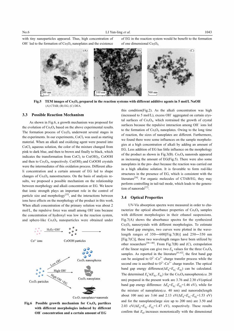
From the above results we can see that alkali concentra-tion of the primary solution determines the reaction time and the morphology of final product in this case. If different addi-tive agents(such as CTAB, EG and DEA) were introduced in the reaction systems, we can obtain products with alterable morphology. Fig.5 shows TEM images of the Co3O4 samples prepared from the reactants with different additive agents in a NaOH aqueous solution of 5 mol/L. As we can see, irregular nanoplates of about 200 nm were obtained with CTAB as addi-
tive agent[Fig.5(A)]. When a certain amount of EG was intro-duced in the solution, the morphology and the size of the Co3O4 obviously changed compared to those of the sample without EG[Fig.4(C)]. We can see nanorods and nanoplates are in the sample[Fig.5(B)]. The nanorods are ca. 100 nm in length and 10 nm in diameter, while the size of nanoplates is about 40 nm. These changes would originate from the substitution of EG for H2O and the changes of the concentration of OH–. However, when DEA was added into the system, relatively regular plates
No.6 LI Yun-ling et al. 1043
with tiny nanoparticles appeared. Thus, high concentration of OH– led to the formation of Co3O4 nanoplates and the existence
of EG in the reaction system would be benefit to the formation of one dimensional Co3O4.
Fig.5 TEM images of Co3O4 prepared in the reaction systems with different additive agents in 5 mol/L NaOH (A) CTAB; (B) EG; (C) DEA.
3.3 Possible Reaction Mechanism
As shown in Fig.6, a growth mechanism was proposed for the evolution of Co3O4 based on the above experimental results. The formation process of Co3O4 underwent several stages in the experiments. In our experiments, CoCl2 was used as starting material. When an alkali and oxidizing agent were poured into CoCl2 aqueous solution, the color of the mixture changed from pink to dark blue, and then to brown and finally to black, which indicates the transformation from CoCl2 to Co(OH)2, CoOOH and then to Co3O4, respectively. Co(OH)2 and CoOOH crystals were the intermediates of this oxidation process. Different alka-li concentration and a certain amount of EG led to shape changes of Co3O4 nanostructures. On the basis of analysis re-sults, we proposed a possible mechanism on the relationship between morphology and alkali concentration or EG. We know that ionic strength plays an important role in the control of particle size and morphology[35], and the interactions between ions have effects on the morphology of the product in this work. When alkali concentration of the primary solution was about 2 mol/L, the repulsive force was small among OH– ions because the concentration of hydroxyl was low in the reaction system, and sphere-like Co3O4 nanoparticles were obtained under
Fig.6 Possible growth mechanism for Co3O4 partilces with different morphologies induced by different OH– concentration and a certain amount of EG
this condition(Fig.2). As the alkali concentration was high (increased to 5 mol/L), excess OH– aggregated on certain crys-tal surfaces of Co3O4, which restrained the growth of crystal surfaces because the repulsive interaction among OH– ions led to the formation of Co3O4 nanoplates. Owing to the long time of reaction, the sizes of nanoplates are different. Furthermore, we found there were some influences on the sample morpholo-gies at a high concentration of alkali by adding an amount of EG. Less addition of EG has little influence on the morphology of the product as shown in Fig.3(B). Co3O4 nanorods appeared as increasing the amount of EG(Fig.5). There were also some nanoplates in the pro- duct because the reaction was carried out in a high alkaline solution. It is favorable to form rod-like structures in the presence of EG, which is consistent with the literature[36]. For organic molecules of CTAB/EG, they may perform controlling in tail-tail mode, which leads to the genera-tion of nanorods[37].
3.4 Optical Properties
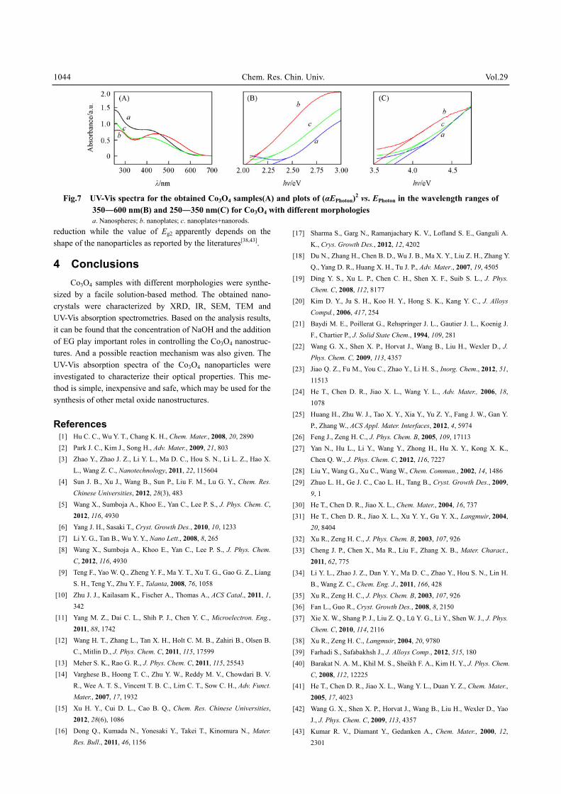
UV-Vis absorption spectra were measured in order to cha-racterize the optical absorbance properties of Co3O4 samples with different morphologies in their ethanol suspensions. Fig.7(A) shows the absorbance spectra for the synthesized Co3O4 nanocrystals with different morphologies. To estimate the band gap energies, two curves were plotted in the wave-length ranges of 350―600[Fig.7(B)] and 250―350 nm [Fig.7(C)], these two wavelength ranges have been utilized by other researchers[38―40]. From Fig.7(B) and (C), extrapolation of the linear region can give two Eg values for the three Co3O4 samples. As reported in the literature[41,42], the first band gap can be assigned to O2–-Co2+ charge transfer process while the second one is ascribed to O2–-Co3+ charge transfer. The optical band gap energy difference(ΔEg=Eg1–Eg2) can be calculated. The determined Eg’s(Eg1, Eg2) for the Co3O4 nanospheres(ca. 20 nm) prepared in the present work are 3.76 and 2.30 eV(optical band gap energy difference: ΔEg=Eg1–Eg2=1.46 eV), while for the mixture of nanoplates(ca. 40 nm) and nanorods(length about 100 nm) are 3.66 and 2.13 eV(ΔEg=Eg1–Eg2=1.53 eV) and for the nanoplates(large size up to 200 nm) are 3.50 and 2.03 eV(ΔEg=Eg1–Eg2=1.47 eV), respectively. These results confirm that Eg1 increases monotonically with the dimensional
1044 Chem. Res. Chin. Univ. Vol.29
Fig.7 UV-Vis spectra for the obtained Co3O4 samples(A) and plots of (αEPhoton)2 vs. EPhoton in the wavelength ranges of 350―600 nm(B) and 250―350 nm(C) for Co3O4 with different morphologies
a. Nanospheres; b. nanoplates; c. nanoplates+nanorods. reduction while the value of Eg2 apparently depends on the shape of the nanoparticles as reported by the literatures[38,43].
4 Conclusions Co3O4 samples with different morphologies were synthe-
sized by a facile solution-based method. The obtained nano-crystals were characterized by XRD, IR, SEM, TEM and UV-Vis absorption spectrometries. Based on the analysis results, it can be found that the concentration of NaOH and the addition of EG play important roles in controlling the Co3O4 nanostruc-tures. And a possible reaction mechanism was also given. The UV-Vis absorption spectra of the Co3O4 nanoparticles were investigated to characterize their optical properties. This me-thod is simple, inexpensive and safe, which may be used for the synthesis of other metal oxide nanostructures.
References
[1] Hu C. C., Wu Y. T., Chang K. H., Chem. Mater., 2008, 20, 2890 [2] Park J. C., Kim J., Song H., Adv. Mater., 2009, 21, 803 [3] Zhao Y., Zhao J. Z., Li Y. L., Ma D. C., Hou S. N., Li L. Z., Hao X.
L., Wang Z. C., Nanotechnology, 2011, 22, 115604 [4] Sun J. B., Xu J., Wang B., Sun P., Liu F. M., Lu G. Y., Chem. Res.
Chinese Universities, 2012, 28(3), 483 [5] Wang X., Sumboja A., Khoo E., Yan C., Lee P. S., J. Phys. Chem. C,
2012, 116, 4930 [6] Yang J. H., Sasaki T., Cryst. Growth Des., 2010, 10, 1233 [7] Li Y. G., Tan B., Wu Y. Y., Nano Lett., 2008, 8, 265 [8] Wang X., Sumboja A., Khoo E., Yan C., Lee P. S., J. Phys. Chem.
C, 2012, 116, 4930 [9] Teng F., Yao W. Q., Zheng Y. F., Ma Y. T., Xu T. G., Gao G. Z., Liang
S. H., Teng Y., Zhu Y. F., Talanta, 2008, 76, 1058 [10] Zhu J. J., Kailasam K., Fischer A., Thomas A., ACS Catal., 2011, 1,
342 [11] Yang M. Z., Dai C. L., Shih P. J., Chen Y. C., Microelectron. Eng.,
2011, 88, 1742 [12] Wang H. T., Zhang L., Tan X. H., Holt C. M. B., Zahiri B., Olsen B.
C., Mitlin D., J. Phys. Chem. C, 2011, 115, 17599 [13] Meher S. K., Rao G. R., J. Phys. Chem. C, 2011, 115, 25543 [14] Varghese B., Hoong T. C., Zhu Y. W., Reddy M. V., Chowdari B. V.
R., Wee A. T. S., Vincent T. B. C., Lim C. T., Sow C. H., Adv. Funct. Mater., 2007, 17, 1932
[15] Xu H. Y., Cui D. L., Cao B. Q., Chem. Res. Chinese Universities, 2012, 28(6), 1086
[16] Dong Q., Kumada N., Yonesaki Y., Takei T., Kinomura N., Mater. Res. Bull., 2011, 46, 1156
[17] Sharma S., Garg N., Ramanjachary K. V., Lofland S. E., Ganguli A. K., Crys. Growth Des., 2012, 12, 4202
[18] Du N., Zhang H., Chen B. D., Wu J. B., Ma X. Y., Liu Z. H., Zhang Y. Q., Yang D. R., Huang X. H., Tu J. P., Adv. Mater., 2007, 19, 4505
[19] Ding Y. S., Xu L. P., Chen C. H., Shen X. F., Suib S. L., J. Phys. Chem. C, 2008, 112, 8177
[20] Kim D. Y., Ju S. H., Koo H. Y., Hong S. K., Kang Y. C., J. Alloys Compd., 2006, 417, 254
[21] Baydi M. E., Poillerat G., Rehspringer J. L., Gautier J. L., Koenig J. F., Chartier P., J. Solid State Chem., 1994, 109, 281
[22] Wang G. X., Shen X. P., Horvat J., Wang B., Liu H., Wexler D., J. Phys. Chem. C, 2009, 113, 4357
[23] Jiao Q. Z., Fu M., You C., Zhao Y., Li H. S., Inorg. Chem., 2012, 51, 11513
[24] He T., Chen D. R., Jiao X. L., Wang Y. L., Adv. Mater., 2006, 18, 1078
[25] Huang H., Zhu W. J., Tao X. Y., Xia Y., Yu Z. Y., Fang J. W., Gan Y. P., Zhang W., ACS Appl. Mater. Interfaces, 2012, 4, 5974
[26] Feng J., Zeng H. C., J. Phys. Chem. B, 2005, 109, 17113 [27] Yan N., Hu L., Li Y., Wang Y., Zhong H., Hu X. Y., Kong X. K.,
Chen Q. W., J. Phys. Chem. C, 2012, 116, 7227 [28] Liu Y., Wang G., Xu C., Wang W., Chem. Commun., 2002, 14, 1486 [29] Zhuo L. H., Ge J. C., Cao L. H., Tang B., Cryst. Growth Des., 2009,
9, 1 [30] He T., Chen D. R., Jiao X. L., Chem. Mater., 2004, 16, 737 [31] He T., Chen D. R., Jiao X. L., Xu Y. Y., Gu Y. X., Langmuir, 2004,
20, 8404 [32] Xu R., Zeng H. C., J. Phys. Chem. B, 2003, 107, 926 [33] Cheng J. P., Chen X., Ma R., Liu F., Zhang X. B., Mater. Charact.,
2011, 62, 775 [34] Li Y. L., Zhao J. Z., Dan Y. Y., Ma D. C., Zhao Y., Hou S. N., Lin H.
B., Wang Z. C., Chem. Eng. J., 2011, 166, 428 [35] Xu R., Zeng H. C., J. Phys. Chem. B, 2003, 107, 926 [36] Fan L., Guo R., Cryst. Growth Des., 2008, 8, 2150 [37] Xie X. W., Shang P. J., Liu Z. Q., Lü Y. G., Li Y., Shen W. J., J. Phys.
Chem. C, 2010, 114, 2116 [38] Xu R., Zeng H. C., Langmuir, 2004, 20, 9780 [39] Farhadi S., Safabakhsh J., J. Alloys Comp., 2012, 515, 180 [40] Barakat N. A. M., Khil M. S., Sheikh F. A., Kim H. Y., J. Phys. Chem.
C, 2008, 112, 12225 [41] He T., Chen D. R., Jiao X. L., Wang Y. L., Duan Y. Z., Chem. Mater.,
2005, 17, 4023 [42] Wang G. X., Shen X. P., Horvat J., Wang B., Liu H., Wexler D., Yao
J., J. Phys. Chem. C, 2009, 113, 4357 [43] Kumar R. V., Diamant Y., Gedanken A., Chem. Mater., 2000, 12,
2301