faculdade de ciências e tecnologias da universidade de coimbra
TRANSCRIPT


Faculdade de Ciências e Tecnologias da Universidade de Coimbra
Isolation, cultivation and antioxidant
capacity of Laetiporus sulphureus
Bruno Miguel Rodrigues Simões
Dissertação no âmbito do Mestrado em Biodiversidade e Biotecnologia Vegetal orientada pela
Professora Doutora Lília Maria Antunes dos Santos, coorientada pelo Professor Doutor António
Manuel Santos Carriço Portugal e apresentada ao Departamento de Ciências da Vida.
Agosto de 2019

3
Agradecimentos
Chegado o fim desta importante etapa não posso deixar de agradecer a todos aqueles que, das mais
diversas formas, contribuíram para a concretização deste trabalho.
À Professora Doutora Lília Maria Antunes dos Santos e ao Professor Doutor António Manuel Santos
Carriço Portugal, orientadora e coorientador deste trabalho, respetivamente, por terem possibilitado a
sua realização, orientação científica, amabilidade e por todos os ensinamentos prestados.
À minha família da Algoteca, Clara, Joana, Maria João, Raquel, Zé e em especial à Mariana pelo apoio,
ensinamentos, amizade e animação ao longo destes dois anos.
Aos colegas do Laboratório de Biologia Molecular, Hugo e Pedro pelos ensinamentos ao longo deste
trabalho.
À Dona Isabel de cima e à Dona Isabel de baixo pelo auxílio prestado, convívio e constante animação
no laboratório.
Aos meus amigos, em especial à Brasileira, aos Patezinhos e às Sexy Ladies pelos bons momentos que
não vou esquecer e que fizeram tudo valer a pena.
Ao meu enfermeiro pela companhia, amizade e amor nestes últimos meses que foram tanto a reta final
como o ponto de partida para ambos.
À minha família, em especial aos meus avós, pais, irmã, madrinha e Kiko, que são tudo para mim, por
estarem sempre presentes, pelo carinho, confiança e por todo o apoio que sempre me proporcionaram
em todos os aspetos da minha vida.
A Coimbra, que me viu crescer!

4
Resumo
Atualmente dependemos dos fungos num sentido mais amplo, devido ao seu papel como
decompositores, mas também em assuntos mais específicos, especialmente relacionados com a
Agricultura e Medicina. Assim, este trabalho focou-se em abordagens biotecnológicas relativas ao
isolamento, cultivo e potencial antioxidante de fungos, com especial ênfase em Laetiporus sulphureus,
um cogumelo comestível com propriedades organoléticas interessantes.
Primeiramente, foram isolados vários fungos com valor comestível/medicinal da região (e algumas
estirpes comerciais), com ênfase em Laetiporus sulphureus. Isto resultou na constituição de uma
biblioteca micológica apelidada de Coleção Micológica de Coimbra (MICOI), atualmente composta por
29 isolados de fungos, 8 de Laetiporus sulphureus. Os resultados obtidos suportam o método de
conservação usado (submersão em água destilada à temperatura ambiente), que se provou eficaz na
manutenção de espécies saprófitas até 5 anos, mas é um método incerto, especialmente para espécies
micorrízicas.
De seguida, o crescimento miceliar de Laetiporus foi avaliado em meio PDA, de modo a selecionar uma
estirpe para cultivo com uma taxa de crescimento alta. Foi escolhida a estirpe MICOI_23, mas o cultivo
em diferentes substratos não levou à ocorrência de frutificação. No entanto, no tratamento de indução
com choque frio, foram observadas formações miceliares distintas. O cultivo de Laetiporus parece ser
bastante seletivo no que toca a estirpes e tendo em conta que apenas existe um caso de cultivo com
sucesso, muito está ainda por melhorar.
Por fim, a atividade antioxidante de estirpes de Laetiporus foi avaliada, recorrendo aos testes ABTS,
FRAP e Folin-C. Antes dos mesmos, o crescimento miceliar em PDB foi testado de modo a compreender
a cinética e a delinear as fases de latência, crescimento exponencial e estacionárias. Os resultados do
teste ABTS, revelaram que todas as estirpes possuem elevadas percentagens de inibição do radical livre
(algumas até 90% de inibição), ou seja, elevada capacidade antioxidante. Com os testes FRAP e Folin-
C, apenas a estirpe MICOI_18 revelou ser relevante, pois foram obtidos valores máximos de 6 mmol
Trolox/kg peso seco e 1 mg GAE/g peso seco, respetivamente. Contudo, os valores obtidos são inferiores
quando comparados com os de outras espécies.
Como resultados principais, foram obtidos dados de crescimento em meio solidificado (taxas de
crescimento) e líquido (curvas de crescimento) das estirpes regionais de Laetiporus. Uma estirpe de
Laetiporus (MICOI_23) revelou-se como potencial candidata a cultivo e os valores obtidos para os testes
antioxidantes confirmaram o potencial antioxidante médio da espécie. Para além disso, o facto de
estarem conservadas (juntamente com outras espécies de cogumelos com interesse biotecnológico)
permite que estas possam ser usadas para investigação futura.
Palavras-chave: Laetiporus sulphureus; coleção micológica; crescimento fúngico; cultivo de
cogumelos; atividade antioxidante; cultura líquida.
5
Abstract
Presently, we depend on fungi on a broader sense, because of their role as decomposers, but also in
specific matters specially relating to agriculture and medicine. Thus, this work was focused on
biotechnological approaches regarding isolation, cultivation and antioxidant potential of fungi, with
special emphasis on Laetiporus sulphureus, an edible mushroom with interesting organoleptic
properties.
Firstly, several regional edible/medicinal fungi (and of some commercial strains), with special emphasis
on Laetiporus sulphureus were isolated. This resulted in the composition of a mycological library named
Mycological Collection of Coimbra (MICOI), that for now holds 29 fungal isolates, 8 of them of
Laetiporus sulphureus. The results supported the used conservation method (submersion in distilled
water at room temperature), which proved effective for the maintenance of saprophytic species for at
least 5 years, but it is not a fool proof method, especially for mycorrhizal species.
Next, strains mycelium growth on PDA medium was assessed, in order to select a strain for cultivation
with a high growing rate. Laetiporus sulphureus (MICOI_23) was chosen, but cultivation on different
substrates did not allow for fruiting. Although, in the cold shock induction treatment, distinct mycelial
formations appeared. Laetiporus cultivation seems to be very strain selective, once there was only one
successful case of cultivation and, as such a lot need to be improved.
Lastly, the antioxidant activity of Laetiporus strains was evaluated through the ABTS, FRAP and Folin-
C assays. Beforehand, mycelium growth on PDB had to be tested, regarding growth kinetics and in order
to delineate the lag, log and stationary phases of its growth. Our results showed that for the ABTS assay,
all strains hold a significant high percentage of inhibition of the ABTS radical (some up to 90%
inhibition), meaning that there is high antioxidant activity. For the FRAP and Folin-C assays, only
MICOI_18 revealed to be a potential interesting strain regarding antioxidant activity, which presented
values of 6 mmol Trolox/kg DW and 1 mg GAE/g DW, respectively. However, the obtained values
were lower when compared with other mushroom species.
As main results, data relating the growth of regional Laetiporus strains on solidified (growth rates) and
liquid (growth curves) medium was obtained. A strain of Laetiporus (MICOI_23) revealed to be a
potential candidate for cultivation and the obtained values for the antioxidant assays confirmed the
species average antioxidant activity. Besides, the fact that the isolates were conserved (along with other
biotechnological interesting mushroom species) allows them to be used in future research.
Keywords: Laetiporus sulphureus; mycological collection; fungal growth; mushroom cultivation;
antioxidant activity; liquid cultures.

6
Table of Contents
Agradecimentos ....................................................................................................................................... 3
Resumo .................................................................................................................................................... 4
Abstract ................................................................................................................................................... 5
Table of Contents .................................................................................................................................... 6
Introduction ............................................................................................................................................. 8
Fungal biology ..................................................................................................................................... 8
Fungal ecology .................................................................................................................................... 8
Fungal diversity & classification ......................................................................................................... 9
Identification of fungi ........................................................................................................................ 11
Edible and medicinal fungi ................................................................................................................ 12
Mushroom cultivation ....................................................................................................................... 14
Antioxidants ...................................................................................................................................... 18
Laetiporus sulphureus ....................................................................................................................... 18
Objectives and thesis layout .............................................................................................................. 20
Materials and Methods .......................................................................................................................... 21
Establishment of mycelium cultures ................................................................................................. 21
Molecular identification .................................................................................................................... 21
Conservation of mycelium cultures ................................................................................................... 21
Mycelium growth on PDA ................................................................................................................ 22
Cultivation trials ................................................................................................................................ 22
Grain spawn ................................................................................................................................... 22
Substrates ...................................................................................................................................... 22
Induction ........................................................................................................................................ 23
Mycelium growth on PDB ................................................................................................................ 23
Antioxidant activity assays ................................................................................................................ 23
ABTS ............................................................................................................................................. 24
FRAP ............................................................................................................................................. 24
Folin-C .......................................................................................................................................... 24
Results ................................................................................................................................................... 26
Mycological Collection of Coimbra (MICOI) .................................................................................. 26
Mycelium growth on PDA ................................................................................................................ 27
Cultivation trials ................................................................................................................................ 29
Mycelium growth on PDB ................................................................................................................ 33
Antioxidant activity assays ................................................................................................................ 35
ABTS ............................................................................................................................................. 35
7
FRAP ............................................................................................................................................. 36
Folin-C .......................................................................................................................................... 37
Discussion ............................................................................................................................................. 38
Mycological Collection of Coimbra (MICOI) .................................................................................. 38
Mycelium growth on PDA ................................................................................................................ 38
Cultivation trials ................................................................................................................................ 38
Mycelium growth on PDB ................................................................................................................ 39
Antioxidant activity assays ................................................................................................................ 40
Final remarks ..................................................................................................................................... 41
Bibliography .......................................................................................................................................... 42

8
Introduction
Fungal biology
Fungi are a unique group of organisms with their distinct biology, from cellular organization to roles in
the ecosystem. They are therefore worthy of a kingdom, which is placed on evolutionary trees alongside
the Plant and even more so to the Animal kingdom, meaning fungi make part of one of the major
evolutionary branches of multicellular organisms (Moore et al. 2011).
Kingdom Fungi is undoubtedly one of the most significant yet forgotten group of organisms. Besides
their major role as recyclers of organic matter (a.k.a. decomposers), fungi impact our lives daily, mostly
but not always, in a compassionate manner and both directly and indirectly. For example, some fungi
act as crop diseases and others form associations with crop species resulting in higher yields. Metabolites
such as mycotoxins, antibiotics, steroids, ciclosporins (which act as immunosuppressants in transplant
surgery) and enzymes (useful for food and drink processing) which we currently depend on, are
produced by fungi. Edible fungi (commonly known as mushrooms, fruitbodies, carpophores,
sporocarps, basidiomata/ascomata, etc.) are themselves direct food sources, but bread, cheese and
fermented goods also have fungi into play and depend on them. Moreover, several fungi can be model
organisms in the study of biological processes and others are significant animal and human pathogens
(Moore et al. 2011, Deacon 2013, Watkinson et al. 2015).
Inherent to all true fungi, there is an assortment of characteristics that differentiates them from all others.
Organisms belonging to Kingdom Fungi are eukaryotic; typically grow as filaments (a.k.a. hyphae) that
elongate at the tip (apical growth), which ultimately by branching give rise to a network of hyphae called
mycelium, but can also grow as single-celled yeasts; fungi are heterotrophs (chemo-
organoheterotrophs), their cell wall prevents phagocytosis but allows the absorption of simple, soluble
nutrients that result from the degradation of complex polymers by secreted enzymes; they present a
distinct cell wall mainly composed of chitin and glucans; the most common form of nuclear status is
haploid with multiple nuclei within the same hyphal compartment; reproduction can be both sexual and
asexual and typically there is the production of spores (Moore et al. 2011, Deacon 2013, Watkinson et
al. 2015).
Fungal ecology
As for their place in the ecosystem, fungi can be divided up into three categories: mutualists, parasites
and saprophytes. Both mutualism and parasitism are forms of symbiotic associations between a fungus
and another organism, being it a plant, algae, animal or another fungus. In mutualistic associations both
parts benefit from the association and the most notorious cases of mutualism in this Kingdom are lichens
(associations between a fungus and usually a green alga and/or cyanobacteria) and mycorrhizae
(associations between fungi and most plant roots, in which fungi help the plant with nutrient and water
uptake by the extension of the surface area of the roots and in return receiving photosynthetic sugars,
that is carbon sources). In parasitic associations, the fungus benefits and the other organism is harmed.
If it is too aggressive then the parasite is termed a pathogen and let it be noted that fungal parasites
account for more than 70% of all crop diseases but on the other hand, they can be helpful in biological
control of pests when the fungus is extremely host specific (Moore et al. 2011)
Saprophytes are responsible for the major part of nutrient recycling since these organisms feed on dead
organic matter, which they degrade with the help of various enzymes for a wide range of polymers (e.g.
9
starch, cellulose, proteins, chitin, keratin and specially wood) and different fungi are adapted to different
types of polymers so in nature saprophytes are found as complex, mixed communities. They can be
divided into primary, secondary and tertiary decomposers, depending on the complexity of the substrate
they are found on (Grimm & Wösten 2018). Wood (a.k.a. plant secondary cell wall) is the most readily
available substrate on the planet, yet both its physical and chemical properties make it an extremely
difficult substrate to degrade. This is due to the presence of lignin (a complex, variable, non-
hydrolyzable and water insoluble aromatic polymer) which prevents the access of enzymes to the rest
of the constituents (hemicelluloses and cellulose), because of its low content in nitrogen or existence of
fungitoxic compounds. Nevertheless, besides these obstacles, some fungi (mainly Basidiomycota) can
degrade it (Moore et al. 2011).
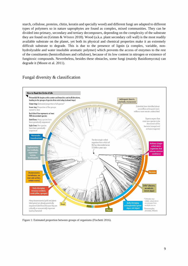
Fungal diversity & classification
Figure 1: Estimated proportion between groups of organisms (Fischetti 2016).

10
The current estimate of existing fungal species stands between 2,2 and 3,8 million and only 120,000 of
these are described (Hawksworth & Luecking 2017). After Arthropods and Bacteria, Fungi make up the
third largest group of living organisms and a lot is yet to be found (Figure 1) (Fischetti 2016).
The division of this monophyletic clade now sets at eight phyla: Cryptomycota, Microsporidia,
Blastocladiomycota, Chytridiomycota, Zoopagomycota, Mucoromycota, Ascomycota and
Basidiomycota (Willis 2018).
The latter two are combined in the subkingdom Dikarya, literally meaning that each hyphal compartment
holds two nuclei. Fruitbody forming fungi belong to this subkingdom and account for 96,000 of the total
described species. The phylum Ascomycota, the largest group of fungi has at least 64 000 species, their
spores (ascospores) are formed within an ascus, usually produced in a complex ascomata (Moore et al.
2011).
Basidiomycota, the phylum on which this thesis is mainly focused, comprises rusts and smuts, jelly,
club, coral and shelf fungi, the common mushrooms, puffballs and stinkhorns. It has 32 000 known
species and is mainly composed by saprophytes and parasites of plants and insects; septate hyphae;
primary mycelium (homokaryotic) followed by secondary mycelium (heterokaryotic) often with clamp
connections over the septa; asexual reproduction by fragmentation and sexual reproduction by
compatible hyphal fusion which ultimately gives rise to basidiomata with basidia, which in turn produce
basidiospores (Moore et al. 2011) (Figure 2).
Figure 2: Sexual life cycle of a Basidiomycete (J.C.P. Hancock).
11
Identification of fungi
Until now, phenotypical characters such as sporocarp, spore and spore-producing structure morphology
were the way to go, identification-wise. This was a major obstacle for progress since only some fungi
produce observable structures and those still spend much of their life cycle as mycelia, making their
identification near impossible (Watkinson et al. 2015). Moreover, this identification may not always do
well for species classification since there is a wide variety of growth patterns and morphological
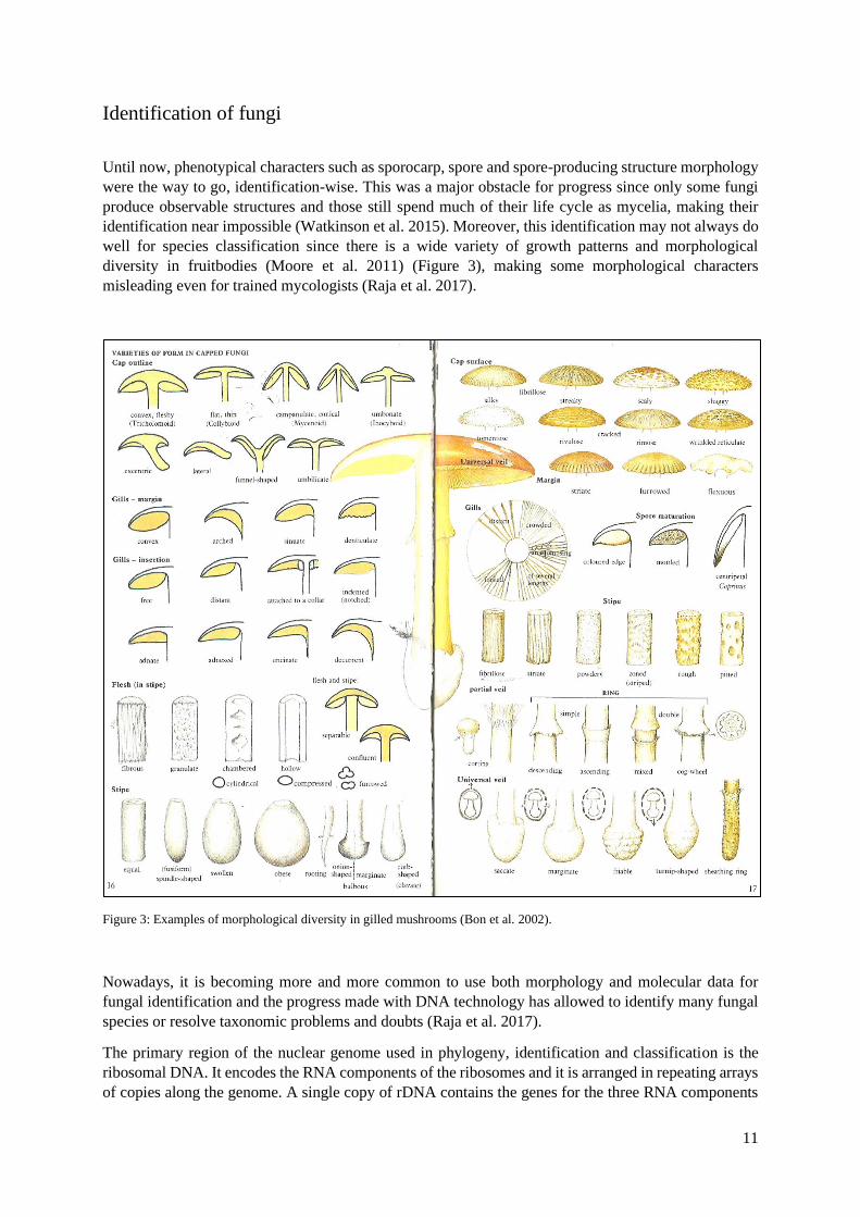
diversity in fruitbodies (Moore et al. 2011) (Figure 3), making some morphological characters
misleading even for trained mycologists (Raja et al. 2017).
Figure 3: Examples of morphological diversity in gilled mushrooms (Bon et al. 2002).
Nowadays, it is becoming more and more common to use both morphology and molecular data for
fungal identification and the progress made with DNA technology has allowed to identify many fungal
species or resolve taxonomic problems and doubts (Raja et al. 2017).
The primary region of the nuclear genome used in phylogeny, identification and classification is the
ribosomal DNA. It encodes the RNA components of the ribosomes and it is arranged in repeating arrays
of copies along the genome. A single copy of rDNA contains the genes for the three RNA components

12
of the ribosome, 18s, 5.8s and 28s RNA (Figure 4). The 28s and 18s can be used in identifications
relating to phylum and domain since they have not varied much through the course of evolution. The
5.8s located between these genes, along with the internal transcribed spacers 1 and 2, called the
internally transcribed spacer (ITS) region, which is removed during the processing of the mRNA, is less
conserved than the coding regions and varies enough between species allowing their identification
(Watkinson et al. 2015, Raja et al. 2017).
Figure 4: Primers for amplification of ITS region (Raja et al. 2017).
Edible and medicinal fungi
Mushrooms have been collected from the wild and used as an integral part of the human diet for
centuries, mainly in the Far East (Sánchez 2010, Rathore et al. 2017). Considered as delicacies and/or
condiments because of their organoleptic properties, namely those concerning their specific flavours,
aromas, textures and aesthetics (Moore et al. 2011, Kalač 2013, Valverde et al. 2015). Their chemical
composition is attractive from the nutritional point of view and can be compared to that of eggs and milk
(Sánchez 2010), making mushrooms a sound food for everyone, especially vegetarians (Moore et al.
2011). It should be noted that chemical composition can be influenced by substrate, environmental
factors, development stage, storage conditions, processing and cooking practices but it can also vary
among and within species (Valverde et al. 2015, Kalač 2013).
Generally, or when unknown, it is assumed that water accounts for 90% of a mushroom’s fresh weight
(FW) meaning the rest 10% are dry matter which is made up of carbohydrates, proteins, lipids, vitamins
and minerals (Sánchez 2010, Moore et al. 2011, Kalač 2013).
Carbohydrates constitute the biggest proportion of the dry matter with about 5-6.5% of a mushroom’s
total dry weight (DW) (Rathore et al. 2017). They include digestible sugars (e.g. mannitol – alcoholic
sugar and glycogen – energy store just as in animals) and non-digestible carbohydrates (e.g. trehalose -
alcoholic sugar and non-starch polysaccharides: 𝛽- glucans, hemicelluloses, pectins and chitin - cell wall
structuring element) which works as fibre (Moore et al. 2011, Kalač 2013, Valverde et al. 2015).

13
Proteins are the main interesting nutrient in mushrooms, with 2-3% of crude protein of total dry matter.
There is, however, extreme variation among species (e.g. 0.8% in Auricullaria auricullaria and 5% in
Fistulina hepatica) (Cheung 2010, Kalač 2013). All 9 essential amino acids to human nutrition are
usually present (amino acid content is a reliable measure of nutritive value) (Moore et al. 2011). Protein
content is above that of vegetables like onions (1.4%), below in meats like chicken (18-20%) and in line
with animal products like milk (2.9-3.3%) (Cheung 2010), but the lack of data on its bioavailability
doesn’t give much support to the protein oriented marketing used to promote mushrooms (Kalač 2013).
Mineral/Ash content is the steadiest value (Kalač 2013), reaching 0.8-1.2% of total dry matter (mainly
potassium, phosphorus, magnesium, calcium, iron, zinc and copper) (Valverde et al. 2015). Mushrooms
are also known to accumulate heavy metals, however, detailed evaluation on the toxicological risk of
such substances is limited (Cheung 2010, Kalač 2013).
Lipid content is low, approximately, 0.2-0.3% of total dry matter. This fraction contains all classes of
lipid compounds from free fatty acids (e.g. linoleic acid - only essential fatty acid to humans and
precursor to the principal aromatic compound of dried mushrooms) (Cheung 2010; Kalač 2013) to
glycerides, sterols, and phospholipids. Mushrooms are cholesterol-free but since most mushroom
cooking practices require the use of butter or oil, these items are ultimately added to their fatty value
(Moore et al. 2011).
The remaining percentage refers to vitamins. Mushrooms are an excellent source of B-complex
vitamins, including vitamin B1 (thiamine), B2 (riboflavin), B3 (niacin), B5 (pantothenic acid), B9
(folates), B12 (cobalamin – classic deficiency in vegetarians), vitamin C (ascorbic acid), pro-vitamin D
(only non-animal source, converted to vitamin D by sunlight) and vitamin E (Cheung 2010, Moore et
al. 2011, Valverde et al. 2015, Rathore et al. 2017).
The total energy is low and is calculated using the equation: kcal = 4 × (g protein + g carbohydrates) +
9 × (g lipids). It usually stands around 350–400 kcal/kg of fresh fruitbodies (Kalač 2013). Elemental
composition of a typical filamentous fungus in relation to macro-elements is, by decreasing order:
carbon, oxygen, nitrogen, hydrogen, phosphorus and sulphur and it can be very helpful when culturing
fungi (Moore et al. 2011).
Table 1: Proximate composition of some common edible mushroom species (Cheung 2010, Kalač 2013). *All data presented
as percentage of dry weight.
Species
Common name
Protein
Fat
Carbohydrate
Fibre
Ash
Agaricus bisporus Button mushroom 2.3-3.4 0.1-0.8 5.1-6.2 0.8-1 0.7-1.2
Auricularia
auricula-judae Wood ear 0.8 0.1 8.1 0.6 0.9
Boletus edulis Cep 2.9 0.3 5.1 0.8 0.5
Cantharellus
cibarius Chanterelle 2.1 0.5 6.4 1.1 0.8
Cordyceps sinensis Caterpillar fungus 2.1 0.8 2.4 - 0.2
Fistulina hepatica Beefsteak Fungus 5 0.1 3.1 - 1.6
Grifola frondosa Maitake 2.1 0.3 5.8 1 0.7
Hericium erinaceus Lion’s mane 2.2 0.3 5.7 0.7 0.9
Lentinula edodes Shitake 1.3-1.7 0.4-0.8 6.7-7.8 0.7-0.8 0.3-0.7
Pleurotus ostreatus Oyster mushroom 1-3 0.1-0.2 5.7-8.1 0.7-0.8 0.6-0.9
Tuber
melanosporum Black truffle 2.3 0.2 6.6 2.7 0.8
Volvariella volvacea Straw mushroom 3 0.6 5 1.1 1.2
14
Besides edibility some mushrooms present medicinal properties (from pharmaceuticals and
cosmeceuticals to hallucinogens) (Rathore et al. 2017). Their medicinal use also has a long history,
particularly in Asian countries, whereas their use in the West is more recent (Wasser 2010). Medicinal
mushrooms are usually characterised by having more fungal cell wall materials and secondary
metabolites that have a wider range of therapeutic activities compared with edible mushrooms (Cheung
2010). It has been shown by a wide range of studies that mushrooms contain components with
outstanding properties to prevent or treat different types of diseases. Their main uses are as antioxidant,
anticancer, antidiabetic, antiallergic, immunomodulating, cardiovascular protector, anticholesterolemic,
antiviral, antibacterial, antiparasitic, antifungal, detoxification, and hepatoprotective. These bioactive
compounds (e.g. polysaccharides, terpenoids, phenols, flavonoids, carotenoids and enzymes) can be
found both in fruitbodies, cultured mycelium and broth (Valverde et al. 2015).
Nevertheless, one property does not exclude the other, as some mushrooms can present both edible and
medicinal properties giving a functional value to each other. Possibilities are endless and considering
the diverse habitat and varied ecological zones across the world where fungi grow and the advances in
molecular biology and nutrigenomics, mushrooms are emerging as the next generation’s nutraceutical
food (Valverde et al. 2015, Rathore et al. 2017).
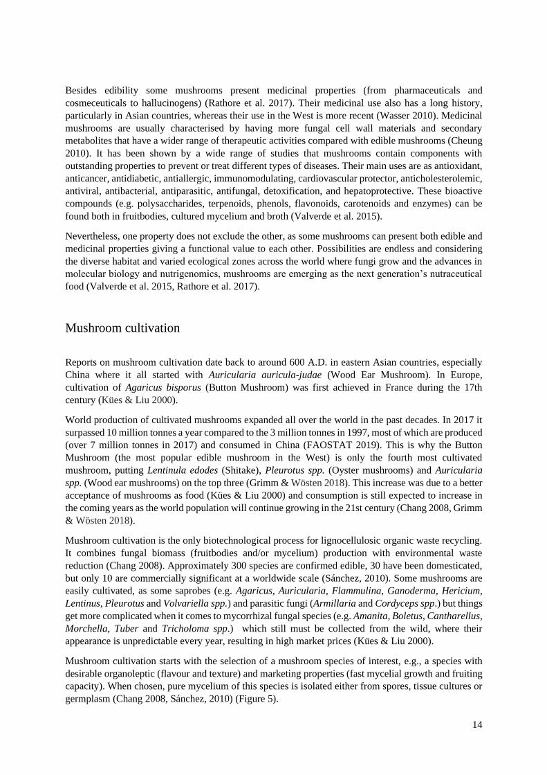
Mushroom cultivation
Reports on mushroom cultivation date back to around 600 A.D. in eastern Asian countries, especially
China where it all started with Auricularia auricula-judae (Wood Ear Mushroom). In Europe,
cultivation of Agaricus bisporus (Button Mushroom) was first achieved in France during the 17th
century (Kües & Liu 2000).
World production of cultivated mushrooms expanded all over the world in the past decades. In 2017 it
surpassed 10 million tonnes a year compared to the 3 million tonnes in 1997, most of which are produced
(over 7 million tonnes in 2017) and consumed in China (FAOSTAT 2019). This is why the Button
Mushroom (the most popular edible mushroom in the West) is only the fourth most cultivated
mushroom, putting Lentinula edodes (Shitake), Pleurotus spp. (Oyster mushrooms) and Auricularia
spp. (Wood ear mushrooms) on the top three (Grimm & Wösten 2018). This increase was due to a better
acceptance of mushrooms as food (Kües & Liu 2000) and consumption is still expected to increase in
the coming years as the world population will continue growing in the 21st century (Chang 2008, Grimm
& Wösten 2018).
Mushroom cultivation is the only biotechnological process for lignocellulosic organic waste recycling.
It combines fungal biomass (fruitbodies and/or mycelium) production with environmental waste
reduction (Chang 2008). Approximately 300 species are confirmed edible, 30 have been domesticated,
but only 10 are commercially significant at a worldwide scale (Sánchez, 2010). Some mushrooms are
easily cultivated, as some saprobes (e.g. Agaricus, Auricularia, Flammulina, Ganoderma, Hericium,
Lentinus, Pleurotus and Volvariella spp.) and parasitic fungi (Armillaria and Cordyceps spp.) but things
get more complicated when it comes to mycorrhizal fungal species (e.g. Amanita, Boletus, Cantharellus,
Morchella, Tuber and Tricholoma spp.) which still must be collected from the wild, where their
appearance is unpredictable every year, resulting in high market prices (Kües & Liu 2000).
Mushroom cultivation starts with the selection of a mushroom species of interest, e.g., a species with
desirable organoleptic (flavour and texture) and marketing properties (fast mycelial growth and fruiting
capacity). When chosen, pure mycelium of this species is isolated either from spores, tissue cultures or
germplasm (Chang 2008, Sánchez, 2010) (Figure 5).

15
Stock cultures should be made as they retain the mycelium’s initial genotype (Stamets 2011), since
strain degeneration (due to excessive asexual multiplication) if not considered, can lead to low yields
and malformed carpophores. Conservation methods range from keeping the cultures in the fridge (from
6-12 months), submerging in sterile water/mineral oil (for a few years), lyophilization and deep freezing
(over 25 years long). Subcultures are made from these stocks but eventually strains should be refreshed
either from the cultivated or new wild carpophores (Zied & Pardo-Giménez 2017).
Next, scaling up is needed in order to obtain inoculum. So, mycelium is used to be inoculated in
previously humidified and sterilized cereal grains (e.g. wheat, rye, millet, rice an d sorghum) that after
a period of incubation in the dark make the spawn (Figure 5) (liquid spawn is also a growing alternative)
(Zied & Pardo-Giménez 2017). Spawn works as the “seed” for the colonization of the final substrate. It
is a great source of nutrients that jumpstarts colonization and if accompanied with high inoculation rates,
results in faster colonization of the substrate (Chang 2008, Sánchez 2010).
Production substrates must be adapted to the mushroom species in question. Primary decomposers (e.g.
Pleurotus spp.) can degrade cellulose, hemicellulose, lignin, and other plant components (Sánchez 2010,
Stamets 2011) so they can be cultivated on a range of lignocellulosic wastes, usually considered
insignificant and of no commercial value. Options range from wood logs (traditional method) to artificial
logs (bagged substrate mixtures) and bottles filled mostly with some kind of lignocellulosic material
(e.g. cereal straw, wood sawdust, seed hulls, corn cobs, coffee pulp, paper products) (Figure 5), followed
by organic supplementation (usually a cereal bran), and a small percentage of inorganic supplementation
(gypsum/calcium sulphate) (Sánchez 2010, Stamets 2011, Grimm & Wösten 2018). Secondary
decomposers (e.g. Agaricus spp.) that colonize composted materials like manure, and tertiary
decomposers which are generally found in soils, but edible species are not exploited (Grimm & Wösten
2018).
Supplementation is implemented to boost yields. The lignocellulosic/compost base acts as the carbon
(C) source, organic supplements as nitrogen (N) sources and inorganic supplements provide calcium
and sulphur and prevent substrate aggregation. It should be noted that all substrate materials must be
free of toxic substances due to the bioaccumulation capacity of mushrooms, which can also be used to
enrich mushrooms with compounds at desired concentrations. After substrate composition, moisture is
one of the most important aspects of the substrate. It usually stands between 60-65%. Besides this, pH,
substrate aeration and gas exchanges with the surrounding environment should also be taken into
consideration (Stamets 2011).
Sterilization comes next at varying degrees (e.g. using hydrogen peroxide, pasteurization, autoclave
sterilization and microwave sterilization) depending on substrate, but the goal is always the same, which
is the elimination of competitive organisms (Chang 2008, Stamets 2011), since several pests are known
to attack mushrooms (e.g. other fungi, insects and virus) (Sánchez 2010).
The only thing missing after sterilization is spawning. Normal spawning rates are of 3-7%, but higher
rates are practicable. This results in an accelerated colonization of the substrate, which in turn narrows
down the window for contamination, boost yields and, thus, makes for a faster completion of the
production cycle (Chang 2008, Stamets 2011). The spawn running phase then takes place, during which
mycelium grows from the spawn into the substrate. Good mycelial growth is essential for mushroom
production and for that, the production substrates are incubated at in the dark until total colonization
(depending on species and strain) (Chang 2008).
Until this stage only vegetative growth is being observed since mycelium is growing and accumulating
nutritional stores (Moore et al. 2011). For mushroom formation to start, fruiting must be induced with a
combination of specific conditions for each mushroom species (mimicking wild conditions may be
helpful at this phase) (Sánchez 2010, Stamets 2011). Induction strategies (a.k.a “triggers”) intend to
disturb mycelial tips so that new hyphae are formed (as only they can be induced into fruiting) (Kües &

16
Liu 2000), and they include: diminishing the temperature (from 5 to 10 degrees Celsius); introduction
of a photoperiod and fresh air (in order to decrease CO2 levels); increasing air humidity (usually to 95%
or even submersion when a stronger shock is needed); and sometimes application of a mechanical shock
(dropping logs on the floor) (Oei & Nieuwenhuijzen 2005). After this switch, hyphae associate and
differentiate into primordia. Once established, cell division ceases and rapid cell elongation begins until
a fruitbody emerges, and cultivation is not over as fruiting can happen in consecutive cycles or “flushes”
(Kües & Liu 2000, Chang 2008).
For mycorrhizal fungi, artificial cultivation with a symbiotic plant species may lead to fruiting, but this
can also happen in saprophytic species like Agaricus spp. where microorganisms help by eliminating
inhibitory fruiting substances (Kües & Liu 2000).
At last, harvesting is carried out at different maturation stages depending upon the species, consumer
preferences and market value. Mushrooms can be kept fresh (but with a short shelf life) (Kalač 2013),
canned, dried and frozen (Stamets 2011).
Along with the production of mushrooms, their cultivation generates a massive amount of used
substrates which can also be a source of environmental pollution. In this way, uses for spent mushroom
substrates are being evaluated (Sánchez 2010), like: mushroom re-cultivation (the three categories of
decomposers represent a continuum in the metabolic transition from organic materials to soil so it is
possible to completely compost agricultural waste through the successive cultivation of mushrooms
from different stages in this continuum (Stamets 2011)); feed (with improved quality and digestibility);
high-quality compost (which works as soil conditioner/fertilizer); bioremediation (by degradation of
pollutants); biogas production; biomaterial production; and bioactive metabolite isolation (Grimm &
Wösten 2018).

17
Figure 5: Overview of several mushroom cultivation techniques (Stamets 2011).

18
Antioxidants
Lately, research has been focusing on antioxidant potential (scavenging effects and reducing power) due
to concerns with oxidative stress to the human body. Free radicals and reactive oxygen species (ROS)
are normal by-products of our body’s physiological processes but modern life practices and the
surrounding environment (Khatua et al. 2013) favour an imbalance of these molecules ultimately leading
to the destructions of tissues and cells (Rathore et al. 2017). Antioxidants are substances that, at low
concentrations, prevent or retard the oxidation of biomolecules (Ndhlala et al. 2010).
The antioxidant value of mushrooms is comparable with that of vegetables, and phenols are considered
as the main compound present with antioxidant potential, though synergistic effects with other present
antioxidants are presumed (Kalač 2013, Ndhlala et al. 2010).
Antioxidant activity screening assays can be divided into: 1) hydrogen atom transfer (HAT)-based
assays, which measure the capacity of an antioxidant to quench free radicals by donation of hydrogen
atoms (e.g. oxygen radical absorbance capacity (ORAC); total radical-trapping antioxidant parameter
(TRAP); total oxidant scavenging capacity (TOSC); chemiluminescence (CL); photo-
chemiluminescence (PCL); croton or β-carotene bleaching by LOO•; and low-density lipoprotein (LDL)
oxidation); 2) electron transfer (ET)-based assays, which measure the capability of an antioxidant to
transfer an electron in order to reduce any compound (e.g. 2,2’-azinobis-(3-ethylbenzothiazoline-6-
sulfonic acid) (ABTS) assays including Trolox equivalent antioxidant capacity (TEAC); 2,2-di(4-tert-
octylphenyl)-1-picrylhydrazyl (DPPH) assay; ferric reducing antioxidant power (FRAP) and cupric
reduction assay (CUPRAC)) (Ndhlala et al. 2010, Assunção et al. 2017).
As some assays focus on the antioxidant activity itself, others are focused on identifying and quantifying
specific antioxidant compounds (e.g. antibody techniques, fluorescence assays, Folin-Ciocalteu
spectrophotometric assay, gas chromatography (GC), high performance liquid chromatography (HPLC)
and light emission assays) (Carocho & Ferreira 2013).
For this work, especial importance was given to the ABTS, FRAP and Folin-C assays, which are all
colorimetric.
In this way, fungi seem to be a promising source of antioxidants, but since there is no single universal
method for measuring antioxidant capacity, standardised assays are lacking which makes it difficult to
compare results from different researches (Kalač 2013).
Laetiporus sulphureus
Laetiporus sulphureus (Bull.: Fr.) Murr. is a Basidiomycota of the Polyporaceae family, commonly
found growing as a parasite or saprobe on hardwoods throughout Europe, Asia and North America, still
there are reports of its worldwide presence. The typical mature basidiocarp of this fungus (Figure 6)
which can be found from summer to fall usually appears in clusters and is easily recognisable due to its
prominent (up to 90 cm wide) stemless fan-shaped overlapping “shelfs/brackets”, and vibrant colouring
ranging from the bright yellows of the porous hymenophore from which its name arises (Laetiporus:
“with bright pores” and sulphureus: “colour of sulphur”) to the strong oranges of the suede like cap
(Khatua et al. 2017, Kuo 2017). Laetiporus orangish mycelium is characterized for its cottony/powdered
appearance (Stamets 2011).
Nutrition-wise L. sulphureus has been considered a delicacy in various countries around the world. Its
taste and texture have been compared to that of the meat of chicken, hence it’s common name “Chicken

19
of the Woods”. Water content is lower than most mushrooms with 72-66% (fresh weight), carbohydrates
6.4-7.4%, crude protein 1-2.1%, fat 0.1-0.2% (dry weight) and energy 341-360 kcal/kg, which is
comparable to most mushrooms. Relating to the medicinal value, studies state that this species contains
several bioactive components with significant antioxidant, antibacterial, anticancer and anti–diabetic
activities (Kathua et al. 2017). What more, the submerged cultivation of this fungus mycelium allows
for the extraction of a pigment that could be used as food-colouring (Davoli et al. 2005).
The only documented case of cultivation of this fungus, was described by a polish research group. This
group made a large-scale experiment, where 2 out of 12 strains fruited, with both cold and humidity
shocks, high organic supplementation (45%) and low moisture (50%) (Pleszczyńska et al. 2013), paving
the way for a defined method of cultivation.
Figure 6: Mature fruiting of Laetiporus sulphureus (Bruno Simões).
20
Objectives and thesis layout
Nowadays mushroom market needs to expand beyond the classic mushrooms seen on sale, and
mycophobia must be fought as fungi could make for a great introduction in our lives. The potential to
benefit society regarding biodiversity/forest wellbeing, food shortage/malnutrition and medicine, is
immense and it can only happen by letting the community know of the benefits of fungi and how they
can help us.
The general aim of the present work is to bring noticeability and value to edible and medicinal fungi,
with special focus on Laetiporus sulphureus, by assessing different fungal biotechnological processes.
This thesis has three main objectives: the isolation of fungal species, the cultivation of Laetiporus
sulphureus and the assessment of the antioxidant activity of the same species.
The first objective provides insight on a conservation method, that works by submerging mycelium in
distilled water and keeping it at room temperature, for long periods of time. It also marks the beginning
of a new collection (MICOI), intended for regional fungal species with edible and medicinal properties,
which may not yet be well explored.
The second objective was to obtain carpophores of Laetiporus sulphureus in different cultivation
substrates, highlighting its edibility and organoleptic properties (resembling chicken), which could make
it a key mushroom, especially for vegetarians.
The third objective aimed to disclose the antioxidant potential of this species, since oxidation is also a
big concern these days. So, the ABTS, FRAP and Folin-C assays were performed in order to add
medicinal value to this species.

21
Materials and Methods
Establishment of mycelium cultures
Cultures were initiated from fresh carpophores and from conserved mycelium that had already been
isolated.
Carpophores collected from the field were cleaned superficially beforehand with a brush and/or cloth.
In the laminar flow hood, the fruitbody was torn apart by hand into smaller sized pieces (to facilitate
handling and so that contaminants would not be dragged into the inner flesh) and with the help of a
sterilized scalpel, pieces of trama were excised from all around the carpophore and distributed through
petri dishes filled with Potato Dextrose Agar (PDA, Sigma-Aldrich©) medium. After sealed and
identified, they were incubated at 25°C and followed daily, checking for both the growth of the desired
fungus or contaminations (such as moulds and/or yeasts, since these are very frequent in new isolates).
If contaminations were to appear, the desired mycelium would be transferred into fresh medium until a
pure culture was obtained. After which it was stored in the fridge at 4°C until further processing.
Molecular identification
For DNA extraction, REDExtract-N-Amp Plant PCR kit (Sygma-Aldrich©) was used. Approximately
5 mm3 of pure mycelium were placed in an Eppendorf to which 15 μl of the Extract solution were added
and after following a temperature cycle of 65°C for 10 minutes, 95°C for 13 minutes and 90°C for 10
minutes, 15 μl of the Dilution solution were added. This was our DNA template ready for use in
subsequent DNA amplifications.
DNA amplification was performed using NZYTaq 2x Green Master Mix (Nzytech©). The primers used
were the ITS1F and ITS4 in order to amplify the Internal Transcribed Spacer (ITS) region of the rDNA.
PCR began with an initial denaturation at 94°C for 3 min, 33 cycles of 94°C for 45 seconds of
denaturation, 54°C for 45 seconds for primer annealing and 72°C for 45 seconds for initial elongation,
followed by 72°C for 10 minutes for further elongation, using 1 μl of DNA template. After amplification,
samples were loaded on a 2% TBE agarose gel to check the efficacy of the amplification
Samples were then sent to Stabvida© for sequencing. The obtained sequences were analysed and edited
using Chromas© software (when needed) and Basic Local Alignment Search Tool (BLAST) was
performed with the sequences available in GenBank using sequences with >97% similarity for species
identification.
Conservation of mycelium cultures
In the laminar flow hood, 8mm mycelium discs were punched with a cork borer from pure cultures and
transferred into sealable test tubes filled to 2/3 of their volume with sterilised distilled water after which
they were sealed, identified (number, species name and date) and stored at room temperature in the dark
until needed. This collection was named, Mycological Collection of Coimbra (acronym MICOI).

22
Mycelium growth on PDA
Mycelium growth was evaluated for a 6-day period on Laetiporus sulphureus MICOI strain 07, 18, 19,
21, 22, 23 and 24 and Pleurotus ostreatus MICOI strain 05. Mycelium discs of 8mm of diameter were
punched with a cork borer from week old cultures and transferred onto fresh medium. Five replicates
were made for each strain. Colony growth was outlined with a marker on each petri dish on the 3rd and
6th day of growth, after which photographs were taken next to a coin (working as standard) and
processed using Photoshop© in order to obtain the area of the delineated colony in pixels.
The linear growth (LG) in cm was obtained using the formula bellow, where GP represents the colony
growth in pixels, SC the standard coin area and SP the measurement of the standard coin in pixels.
Growth rates (GR) were determined by calculation of the linear growth differences in consecutive
measures and dividing them by the days passed between them, also seen below.
Statistical analysis was performed using STATISTICA© 7. A one-way ANOVA was performed, to
compare the growth for each strain. Significant differences were identified with Tukey’s test and
accepted for a value of p0,05.
𝐿𝐺 =√(
𝐺𝑃 × 𝑆C𝑆𝑃 )
𝜋 𝐺𝑅 =
𝐿𝐺f − 𝐿𝐺i
days of growth
Figure 7: Formulas used to obtain the linear growth (LG, left) and growth rates (GR, right) of the isolates mycelia.
Cultivation trials
Grain spawn
Wheat grain was used for spawn production. Firstly, it was boiled in water for 15min in order to raise
its water content, the remaining water was discarded, and the grain was left to sit overnight. The boiled
grains were then used to fill filtered bags and sterilised for 1 hour at 121°C, after which each bag would
be inoculated and sealed, with a week-old petri dish of the desired Laetiporus strain (MICOI_23). The
inoculated grain bags were then incubated in the dark at 25°C for two weeks (by this time the grain
would be totally colonised) and ready to use as spawn.
Substrates
The formulation for the production substrates used comprised 50% lignocellulosic material (3
treatments: eucalyptus sawdust, poplar sawdust and wheat straw), 45% organic supplement (wheat bran)
and 5% inorganic supplement (gypsum), making up to 1kg of substrate per bag (Pleszczyńska et al.
2013). The substrates humidity was raised to 50% and then they were sterilized for 1h at 120°C, 3 days
in a row. On the 3rd day of sterilization and after the bag ranged room temperature, approximately 100gr
of grain spawn were used as inoculum for each bag and sealed. Colonization was carried in a climatic
chamber at 25°C, with 50/60% relative humidity (not ideal) and no light for two weeks (vegetative
phase).

23
Induction
After the two-week incubation period, a stimulus was introduced in order to induce the reproductive
phase of the mycelium’s development.
The stimuli used comprised temperature changes, besides the addition of a 14h:10h light/dark
photoperiod and increased aeration (Stamets 2011). On one of the experiments, the temperature was
decreased from 25°C to 17°C (the usual in mushroom cultivation) and it was maintained for the rest of
the experiment. In contrary, on the other experiment, the temperature was increased from 25°C to 33°C
(supported by meteorological data of when the mushroom was found) but only for two days, after which
it was back down to 25°C.
Mycelium growth on PDB
Growth experiments on PDB (Potato Dextrose Broth, Sigma-Aldrich©) had the duration of 26 days on
Laetiporus (MICOI_18) and Pleurotus (MICOI_05), after which a growth curve was obtained for each.
Homogenised and non-homogenised treatments were used.
For the homogenised treatment, mycelium from 3 fully colonized petri dishes was scraped and mixed
thoroughly with 250 ml of PDB after which 5 ml were used to inoculate 100 ml Erlenmeyer flasks filled
with 50 ml of PDB. As for the non-homogenised treatment, 5 mycelium discs were punched with a cork
borer and scraped to use as inoculum in 100 ml Erlenmeyer flasks filled with 50 ml of PDB. All flasks
were incubated in an orbital shaker at 25°C and 150 rpm.
In order to obtain the growth curves, 3 replicates of each strain and treatment were filtrated to previously
weighted Whatman filter disks at days 0, 2, 5, 8, 12, 15, 19, 22 and 26, dried at 60°C in a heater and
weighted again in order to obtain the mycelium’s dry weight at each day.
Statistical analysis was performed using STATISTICA© 7. A one-way ANOVA was performed, to
compare the growth at each day. Significant differences were identified with Tukey’s test and accepted
for a value of p0,05.
Antioxidant activity assays
In order to evaluate the antioxidant potential, biomass was firstly obtained. Starter cultures with a 1:1
culture/medium proportion were cultivated in 5L Erlenmeyer flasks for 12 and 22 days in a climatic
room at 23°C, 5,68 μmol m-2 s-1 of luminosity and a photoperiod of 16h:8h light/dark. After the stipulated
growth times, cultures were centrifuged, frozen, lyophilized and weighted.
For the extraction process 3 replicates of ≈500 mg were weighted and kept in 50 ml falcon tubes, then
the lyophilized biomass went through the following extraction process.
In a chilled mortar the biomass was macerated with liquid nitrogen. Then, divided by 1.5 ml Eppendorfs
with a 5 mm sphere and 1 ml of a 2:1 solution of dichloromethane/methanol and taken into the bead mill
for 10 minutes with a frequency of 30 s-1 for further cellular disruption. After this, the mixture of biomass
and solution was poured back in the falcon to which 20 ml of solution were added, taken to the vortex
for homogenization and centrifuged at 4500 rpm for 15 min. The resulting supernatant was recovered

24
to a flask, repeating this solution wash 10 times. The recovered supernatant was then evaporated in the
rotavapor.
After this, the extracts were resuspended with the minimal amount possible of
dichloromethane/methanol solution with the aid of ultrasounds and divided by previously weighted 2
ml Eppendorfs, followed by drying in the speed-vac and stored in the fridge until needed. Before being
used for the antioxidant assays, the dried extracts were resuspended in ethanol 50% to a concentration
of 5 mg/ml.
ABTS
ABTS•+ was prepared from two solutions dissolved in distilled water, ABTS (Sigma-Aldrich©) 7
mol/m3 and K2S8O2 (Merck©) 2.45 mol/m3 (Guedes et al. 2013). The solution was left for 16 hours at
room temperature and protected from light, so that the formation of the radical would be completed. In
order to obtain an absorbance reading at 734 nm, the solution was diluted. Relating to the experimental
trial, 350 μL of extract were added to 1 ml of the diluted ABTS•+ solution. After 6 minutes of reaction,
the absorbances at 734 nm were read and the averages calculated. Total antioxidant activity was
expressed in inhibition percentage, through the formula below, where ABTSi is the initial reading of the
ABTS solution and ABTSs is the sample reading.
𝐼𝑛ℎ𝑖𝑏𝑖𝑡𝑖𝑜𝑛 % = (𝐴𝐵𝑇𝑆𝑖 − 𝐴𝐵𝑇𝑆𝑠
𝐴𝐵𝑇𝑆𝑖) × 100
Figure 8: Formula used to obtain the inhibition percentage of the ABTS radical by the mycelia extracts.
FRAP
FRAP assays were preformed based on Goiris et al. (2012). The preparation of the FRAP reagent
comprises the preparation of 4 other solutions: a FeCl3 solution with a 20 mM, a HCl solution of 40
mM which is then added to the TPTZ solution of 10 mM and an acetate buffer with 0.3 mM at ph 3.6.
Calibration lines were made with Trolox solutions with 0.06, 0.04, 0.02, 0.01, 0.005, 0.003 and 0.001
g/L of concentration. For the testing, 100 μL of sample and 3 ml of FRAP reagent were incubated for
10 minutes at 37°C, followed by readings at 593 nm. Antioxidant activity was expressed in mmol
Trolox/kg dry weight.
Folin-C
The Folin-C method used was based on Goiris et al. (2012). First, a solution of sodium bicarbonate was
prepared by dissolving 6 g in 100 ml of distilled water. Before testing and prepared on the day of the
trial, 10 ml of Folin reagent were dissolved in 90 ml of distilled water. Calibration lines were made with
5 solutions with different concentrations (125, 100, 50, 25 and 5 mg/L) of Galic acid. As for the testing,
200 μL of extract were added to 1.5 ml of Folin reagent, homogenised and incubated for 5 minutes and
after that time 1.5 ml of the sodium bicarbonate solution were added as well. This time an incubation of

25
90 minutes is needed and after that the readings were done at 750 nm. Antioxidant activity was expressed
in mg GAE/g dry weight.
It should be noted that all assays were performed in low lighting, with control readings and with 3
replicates for each sample reading.
26
Results
Mycological Collection of Coimbra (MICOI)
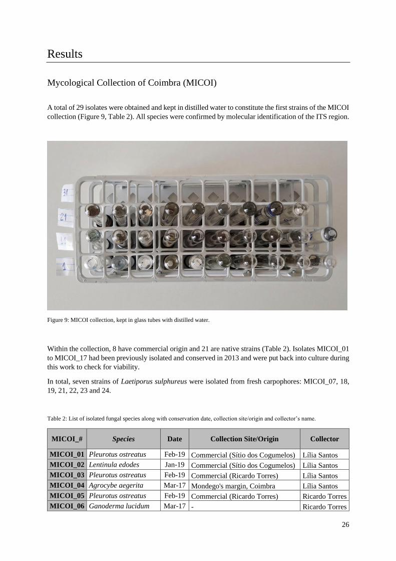
A total of 29 isolates were obtained and kept in distilled water to constitute the first strains of the MICOI
collection (Figure 9, Table 2). All species were confirmed by molecular identification of the ITS region.
Figure 9: MICOI collection, kept in glass tubes with distilled water.
Within the collection, 8 have commercial origin and 21 are native strains (Table 2). Isolates MICOI_01
to MICOI_17 had been previously isolated and conserved in 2013 and were put back into culture during
this work to check for viability.
In total, seven strains of Laetiporus sulphureus were isolated from fresh carpophores: MICOI_07, 18,
19, 21, 22, 23 and 24.
Table 2: List of isolated fungal species along with conservation date, collection site/origin and collector’s name.
MICOI_# Species Date Collection Site/Origin Collector
MICOI_01 Pleurotus ostreatus Feb-19 Commercial (Sítio dos Cogumelos) Lília Santos
MICOI_02 Lentinula edodes Jan-19 Commercial (Sítio dos Cogumelos) Lília Santos
MICOI_03 Pleurotus ostreatus Feb-19 Commercial (Ricardo Torres) Lília Santos
MICOI_04 Agrocybe aegerita Mar-17 Mondego's margin, Coimbra Lília Santos
MICOI_05 Pleurotus ostreatus Feb-19 Commercial (Ricardo Torres) Ricardo Torres
MICOI_06 Ganoderma lucidum Mar-17 - Ricardo Torres

27
MICOI_07 Laetiporus sulphureus Mar-17 - Ricardo Torres
MICOI_08 Agaricus bisporus Mar-17 Commercial (Continente) Lília Santos
MICOI_09 Pleurotus
citrinopileatus Jun-17
Commercial (Ricardo Torres) Ricardo Torres
MICOI_10 Agrocybe aegerita Jun-17 - Lília Santos
MICOI_11 Pleurotus
citrinopileatus Jan-19
Commercial (Marano) Lília Santos
MICOI_12 Pleurotus djamor Sep-17 Commercial (Marano) Lília Santos
MICOI_13 Lentinus tigrinus Jun-17 Rebolim, Coimbra Lília Santos
MICOI_14 Agrocybe aegerita Jun-17 Penedo da Saudade, Coimbra Lília Santos
MICOI_15 Suillus collinitus Sep-17 Copeira, Coimbra Lília Santos
MICOI_16 Fistulina hepatica Sep-17 Serra da Boa Viagem, Figueira da
Foz Lília Santos
MICOI_17 Agrocybe aegerita Jun-17 Condeixa Lília Santos
MICOI_18 Laetiporus sulphureus Sep-17 Choupal, Coimbra Bruno Simões
MICOI_19 Laetiporus sulphureus Sep-17 Choupal, Coimbra Bruno Simões
MICOI_20 Fomes fomentarius Jan-19 Portagem, Coimbra Bruno Simões
MICOI_21 Laetiporus sulphureus Sep-18 Choupal, Coimbra Bruno Simões
MICOI_22 Laetiporus sulphureus Jan-19 Águeda Joana Ferreira
MICOI_23 Laetiporus sulphureus Jan-19 Lousã's mountain, Lousã Bruno Simões
MICOI_24 Laetiporus sulphureus Jan-19 Meiral, Lousã Bruno Simões
MICOI_25 Macrolepiota procera Feb-19 Meiral, Lousã Bruno Simões
MICOI_26 Fistulina hepatica Feb-19 Góis Bruno Simões
MICOI_27 Licoperdon perlatum Feb-19 Góis Bruno Simões
MICOI_28 Calvatea gigantea Feb-19 - -
MICOI_29 Agrocybe aegerita Jan-19 JBUC, Coimbra Bruno Simões
Mycelium growth on PDA
Laetiporus sulphureus mycelium (Figure 10) linear growth on PDA was obtained from photographs of
colony area delineations, along with Pleurotus ostreatus (MICOI_05) a commercial strain used for
comparison.
Relating Laetiporus mycelium, young peripheral zones of growth were spread in a thin mycelial mat,
that became dense over time (Figure 10), in contrast to most species (e.g. Pleurotus) which have dense
mycelium from the beginning.

28
Figure 10: Common mycelium morphology of Laetiporus sulphureus.
The average linear growth rates of each of the eight strains were calculated (Figure 11).
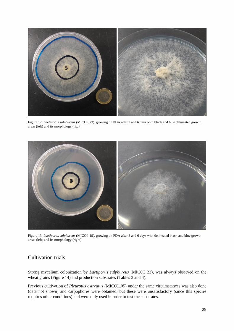
The fastest growth rate was exhibited by the strain MICOI_23 of L. sulphureus (Figures 11 and 12),
even higher than that of the commercial strain P. ostreatus (MICOI_05), with 0.92 and 0.90 cm/day,
respectively. L. sulphureus (MICOI_19) (Figures 11 and 13) and MICOI_24 showed the slowest rates,
with 0.51 and 0.56 cm/day, respectively.
Figure 11: Mycelial linear growth rate (cm/day) of several MICOI strains on PDA at 25°C. Columns with different superscript
letters are significantly different (p<0.05).
a
b b
c
bb
a
c
0.00
0.20
0.40
0.60
0.80
1.00
1.20
MICOI_05 MICOI_07 MICOI_18 MICOI_19 MICOI_21 MICOI_22 MICOI_23 MICOI_24
Lin
ear
gro
wth
rat
e (c
m/d
ay)
Strain
29
Figure 12: Laetiporus sulphureus (MICOI_23), growing on PDA after 3 and 6 days with black and blue delineated growth
areas (left) and its morphology (right).
Figure 13: Laetiporus sulphureus (MICOI_19), growing on PDA after 3 and 6 days with delineated black and blue growth
areas (left) and its morphology (right).
Cultivation trials
Strong mycelium colonization by Laetiporus sulphureus (MICOI_23), was always observed on the
wheat grains (Figure 14) and production substrates (Tables 3 and 4).
Previous cultivation of Pleurotus ostreatus (MICOI_05) under the same circumstances was also done
(data not shown) and carpophores were obtained, but these were unsatisfactory (since this species
requires other conditions) and were only used in order to test the substrates.

30
Figure 14: Spawn of Laetiporus sulphureus (MICOI_23) after two weeks of colonization.
After the substrate’s inoculation (Figure 15) and total colonization after two weeks, the “cottony”
mycelium would extend beyond the substrate and start climbing the walls of the bags (Figure 16).
Fructification was not obtained in any of the bags, regardless of substrate or induction treatment (Tables
3 and 4).
Figure 15: Cultivation bags right after inoculation with the spawn of MICOI_23. From left to right: straw, eucalyptus and
poplar substrates.

31
Figure 16: Substrates being colonized by MICOI_23. From left to right: straw, eucalyptus and poplar substrate.
Table 3: Colonization of grain and substrates by MICOI_23 and fruitification for the continuous cold shock. +: observed; -:
not observed and n.d.: not determined.
Grain Colonization Substrate Colonization Fruitification
Wheat +
Straw + -
Eucalyptus + n.d.
Poplar + n.d.
Table 4: Colonization of grain and substrates by MICOI_23 and fruitification for the periodic heat shock. +: observed; -: not
observed.
Grain Colonization Substrate Colonization Fruitification
Wheat +
Straw + -
Eucalyptus + -
Poplar + -
For the continuous cold shock induction, mycelium structures were observed after 3 weeks. Although,
they do not consist of a real carpophore, they had a carpophore-like concentric pattern (Figures 17 and
18).
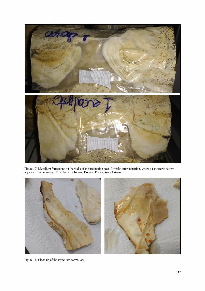
32
Figure 17: Mycelium formations on the walls of the production bags, 3 weeks after induction, where a concentric pattern
appears to be delineated. Top: Poplar substrate; Bottom: Eucalyptus substrate.
Figure 18: Close-up of the mycelium formations.

33
Mycelium growth on PDB
All Laetiporus sulphureus strains were grown on PDB (Figure 19), but only L. sulphureus (MICOI_18)
and P. ostreatus (MICOI_05, for comparison) had their mycelium growth monitored. This monitoring
was done with several weightings for a period of 26 days with homogenised and non-homogenised
treatments. The obtained growth curves can be seen in Figure 20.
Figure 19: Initial days of mycelium growth on PDB.
The homogenised Laetiporus growth curve shows a lag (latency) phase that goes from day 0 to day 2
(p˂0.05), followed by a log (exponential) phase from day 5 to 12, a stationary phase from day 15 to 22
(p˂0.05) and a decline at day 26. The non-homogenised Laetiporus growth curve shows a lag phase
from day 0 to 2 (p˂0.05), a transition phase from day 2 to 5 and an exponential phase from day 8 to 26.
The homogenised Pleurotus growth curve shows a lag phase from day 0 to 2 (p˂0.05), a log phase from
day 5 to 19, after which the stationary phase begins at day 22 until the end of the experiment (p˂0.05).
The non-homogenised Pleurotus growth curve shows a lag phase from day 0 to 2 (p˂0.05), a log phase
from day 5 to 15, a stationary phase from day 19 to 22 (p˂0.05) and a decline at day 26.
MICOI_05
MICOI_21
MICOI_07
MICOI_22
MICOI_18
MICOI_23
MICOI_19
MICOI_24

34
Figure 20: PDB growth curves of Pleurotus (blues) and Laetiporus (oranges) mycelium with homogenised and non-
homogenised treatments (L.s. – Laetiporus Sulphureus; P.o. – Pleurotus ostreatus)
On liquid medium, mycelium initially grows in the form of spherical pellets until biomass increases and
develops into a uniform mass (Figure 21, left). Also, in stationary cultures a hydrophobic mycelial mat
can be seen growing on top of the medium (Figure 21, right).
Figure 21: Left – mature culture of MICOI_23 growing on PDB after 22 days; Right – Mycelial mat formed on top of the PDB
medium of unshaken old cultures.
-0.05
0
0.05
0.1
0.15
0.2
0.25
0.3
0.35
0 5 10 15 20 25 30
g D
W/5
0 m
l
Days
L.s. Homogeniezed L.s. Non-Homogeniezed
P.o. Homogeniezed P.o. Non-Homogeniezed

35
Antioxidant activity assays
The obtained Laetiporus sulphureus (MICOI_18) growth curves were used as a reference to harvest the
biomass at 12 and 22 days of growth in order to prepare the extracts and perform the antioxidant assays
in all strains (Figure 22).
Figure 22: Resuspended Laetiporus sulphureus extracts in triplicate before being used for the antioxidant activity assays.
ABTS
ABTS radical inhibition percentage by strain can be seen in Figure 23. Except for MICOI_07 and 21
(which decrease), the antioxidant activity increases from day 12 to day 22 of growth.
The ABTS results at 12 days vary between 62.5% and 95.9% inhibition of the ABTS radical. MICOI_07
shows the highest inhibition at 95.9%. MICOI_23 and 24 show the lowest inhibition rates (62.5% and
62.8%, respectively).
At 22 days the ABTS inhibition percentage stands between 62.4% and 94.5%. MICOI_23 shows the
highest inhibition at 94.5%, but MICOI_18 and 22 also had high inhibition rates (87.3% and 86.9%,
respectively). The lowest inhibition rate (62.4%) belongs to MICOI_07.
MICOI_07 MICOI_18 MICOI_21 MICOI_22 MICOI_23 MICOI_24

36
Figure 23: Antioxidant activity determined by the ABTS assay (inhibition percentage of the ABTS radical) at 12 and 22 days
of growth.
FRAP
Antioxidant activity values expressed as equivalent mmol of Trolox per kilogram of mycelium dry
weight (DW) can be seen in Figure 24. Extracts of MICOI_07, 21, 22 and 24 showed a higher antioxidant
activity at 12 days of growth and MICOI_18 and 23 at 22 days of growth.
At 12 days MICOI_07 had the highest result with 5.5 mmol Trolox/kg DW and MICOI_23 the lowest
(2.7 mmol Trolox/kg DW).
At 22 days MICOI_18 had the highest antioxidant activity equivalent to 6 mmol Trolox/kg DW and
MICOI_24 the lowest (1.1 mmol Trolox/kg DW).
Figure 24: Antioxidant activity determined by the FRAP assay expressed as Trolox equivalents (mmol Trolox/kg DW) at 12
and 22 days of growth.
0
10
20
30
40
50
60
70
80
90
100
MICOI_07 MICOI_18 MICOI_21 MICOI_22 MICOI_23 MICOI_24
Inh
ibit
ion
%
Strain
12 days
22 days
0
1
2
3
4
5
6
7
MICOI_ 07 MICOI_18 MICOI_21 MICOI_ 22 MICOI_23 MICOI_ 24
mm
ol T
rolo
x/kg
DW
Strain
12 days
22 days

37
Folin-C
Total phenolic content was expressed as equivalent mg of gallic acid (GAE) per gram of mycelium dry
weight. MICOI_07, 21, 22 and 24 had higher results for 12-day cultures and MICOI_18 and 23 for 22-
day cultures (Figure 25).
Phenolic content at 12 days of growth revealed to be the highest for MICOI_21, with 1.1 mg GAE/g
DW and MICOI_07 (0.98 mg GAE/g DW), the lowest results belong to MICOI_23 (0.36 mg GAE/g
DW).
At 22 days of growth, MICOI_18 had the highest results with 1 mg GAE/g DW and MICOI_22 the
lowest (0.38 mg GAE/g DW).
Figure 25: Total content of phenolic compounds expressed in Gallic Acid Equivalents (mg GAE/g DW) at 12 and 22 days of
growth.
0
0.2
0.4
0.6
0.8
1
1.2
1.4
MICOI_07 MICOI_18 MICOI_21 MICOI_22 MICOI_23 MICOI_24
mg
GA
E/g
DW
Strain
12 days
22 days

38
Discussion
Mycological Collection of Coimbra (MICOI)
Compared to the other isolates (especially mycorrhizal), Laetiporus sulphureus was easy to isolate and
contaminations were few. The 7 strains found were a great start, because a higher number of collected
strains represents a higher probability to find a good cultivation candidate.
Conservation in distilled water proved to be effective for previously conserved strains, for a period of
at least 5 years, but sterile water storage may not be suitable for all groups of fungi. This was observed
in 1986 by Richter & Bruhn, where several basidiomycetes where stored in distilled water (from 3
months to 3 years in the refrigerator at 5°C). In that study saprophytes had higher survival rates than
mycorrhizal species. This was also confirmed by this work as almost all isolates are strictly saprophytic
and isolation/revival of mycorrhizal species, such as Hydnum repandum was proven difficult.
These results are promising, since this method of conservation is easy to execute and inexpensive, but
in the future, protocol changes (e.g. cold storage of some strains) or other conservation methods (e.g.
lyophilization) should be taken into consideration. Concerns with preservation are mainly due to
possible genotype and in turn phenotype changes, that may alter fungal traits, like growth rate, vigour
and cultivability (Richter & Bruhn 1986, Sigh et al. 2018, Zied & Pardo-Giménez 2017).
Mycelium growth on PDA
Growth rates of Laetiporus (MICOI_23) and the commercial strain of Pleurotus (MICOI_05) were the
highest in this experiment and the ones most in line with other studies. For Laetiporus, linear growth
was of 0.92 cm/day, which is comparable to the growth rate obtained by Luangharn in 2014 of 1.09
cm/day. For Pleurotus, linear growth was of 0.90 cm/day, but only one study was found to have similar
growth rates (Gibriel et al. 1996) that recorded a 1.23 cm/day growth.
Cultivation trials
The cultivation process involves many variables that must be carefully adjusted to each mushroom
species and strain, if one fails to be in accordance with the fungi preferences, cultivation may not be
achieved. (Stamets 2011, Zied & Pardo-Giménez 2017). So, for the most part of this work, the protocol
by Pleszczyńska et al. (2012) was used as a reference for the cultivation trials, since successful
cultivation of Laetiporus sulphureus was achieved.
Firstly, there is strain selection, which is rather complicated. Various criteria can be used, but mostly,
strains are selected by experimentation with wild strains and/or breeding (Zied & Pardo-Giménez 2017).
For this experiment, fast mycelial growth was used as the decisive criteria for selection of a candidate
for cultivation, and since Laetiporus (MICOI_23) linear growth was not significantly different from
MICOI_05 (a commercial Pleurotus strain), it was chosen as the one for cultivation. Pleszczyńska et al.
ran cultivation trials for 12 strains, of which only 2 revealed the potential for fructification.

39
Spawn colonization was strong and without any signs of contamination, and this is a great start to every
cultivation since strength and purity of spawn is extremely important (Stamets 2011, Zied & Pardo-
Giménez 2017).
Substrate related variables (e.g. composition, particle size, supplementation) may have been one of the
biggest steps back towards the successful cultivation of Laetiporus. Relating substrate composition,
eucalyptus and poplar sawdust were expected to show positive results since this mushroom tends to
appear in eucalyptus and other hardwoods, straw was expected to be unsuccessful, since only Pleurotus
species and the “Straw mushroom” (Volvariella volvacea) have shown the plasticity to grow in that
substrate (Stamets 2011). Particle size could have also been an impediment, since mycelium
colonization of the bags was strong in the peripheral zones of the substrate, but the interior seemed quite
unexplored by the mycelium, regarding the eucalyptus and poplar sawdust. When it comes to straw,
colonization was observed as well, but this time the lack of density of the substrate did not allow for the
appearance of the mycelial formations. A mix of the two (sawdust and straw), could be useful regarding
both composition and particle size. Supplementation was done accordingly to the successful case by
Pleszczyńska et al. (2012), but a simpler supplementation (in both organic and inorganic) was used since
a low cost and local approach regarding the use of waste products tried to be implemented.
Another big step for obtaining fruitbodies is the induction phase which also revealed to be inefficient.
Air humidity was difficult to maintain due to lack of humidity control by the growing chamber. This
was tackled with daily hand spraying of the chamber, but it did not allow for high humidity maintenance.
As for the induction treatments, the continuous cold shock was shown to be promissory (as it was for
Pleszczynska et al.), as differential development was observed in all replica of the eucalyptus and poplar
substrates. After noticing these formations, the bags were cut open to make its growth possible, but
development ceased. The mycelial formations had an overall brittle texture and for that reason were not
considered as primordia, but they were going that way, since regular mycelium is powdery/cottony and
has no consistency whatsoever. The periodic heat shock did not show any signs of differential
development and it was introduced regarding the seasonality of this mushroom (summer to fall),
observations from the wild showed that a week before primordia emerged high temperatures in the range
of the 30-40 degrees occurred. Aeration was always provided, but this could also be a problem since the
mycelial formations obtained strived for open air, so better oxygenation may be needed. Cutting the
bags prior to the appearance of these formations was not attempted since the polish group observed that
contaminations always occurred.
Mycelium growth on PDB
Liquid cultures are a great way of obtaining large amounts of biomass, whether for extraction of
compounds, use as spawn or as edible mycelium (like Quorn®) (Moore et al. 2011, Smith 2014, Zied
& Pardo-Giménez 2017).
From the statistical analysis it was possible to delineate the different phases of growth usually observed
in liquid cultures of filamentous fungi (Tay et al. 2011, Moore et al. 2011). Comparing with Laetiporus,
Pleurotus growth curves had the most biomass, suggesting that this species is better adapted to liquid
culture conditions.
The observed fluctuations in biomass towards the end of the experiment (for both species) were possibly
due to the formation of a mycelial mat on the flasks (of 100ml with 50ml of PDB). The small volume
probably allowed the mycelium to adhere to the walls of some flasks and a spike in growth was observed,
when the cultures were supposed to be in stationary culture.
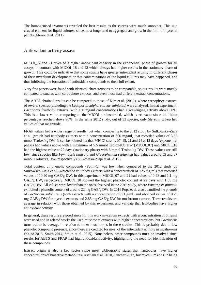
40
The homogenised treatments revealed the best results as the curves were much smoother. This is a
crucial element for liquid cultures, since most fungi tend to aggregate and grow in the form of mycelial
pellets (Moore et al. 2011).
Antioxidant activity assays
MICOI_07 and 21 revealed a higher antioxidant capacity in the exponential phase of growth for all
assays, in contrast with MICOI_18 and 23 which always had higher results in the stationary phase of
growth. This could be indicative that some strains have greater antioxidant activity in different phases
of their mycelium development or that contaminations of the liquid cultures may have happened, and
thus inhibiting the formation of antioxidant compounds to their full extent.
Very few papers were found with identical characteristics to be comparable, so our results were mostly
compared to studies with carpophore extracts, and even those had different extract concentrations.
The ABTS obtained results can be compared to those of Kim et al. (2012), where carpophore extracts
of several species (including the Laetiporus sulphureus var. miniatus) were analysed. In that experiment,
Laetiporus fruitbody extracts (with a 10mg/ml concentration) had a scavenging activity above 60%.
This is a lower value comparing to the MICOI strains tested, which is relevant, since inhibition
percentages reached above 90%. In the same 2012 study, out of 33 species, only Stereum ostrea had
values of that magnitude.
FRAP values had a wider range of results, but when comparing to the 2012 study by Sulkowska-Ziaja
et al. (which had fruitbody extracts with a concentration of 500 mg/ml) that recorded values of 3.53
mmol Trolox/kg DW. It can be pointed out that MICOI strains 07, 18, 21 and 24 at 12 days (exponential
phase) had values above with a maximum of 5.5 mmol Trolox/KG DW (MICOI_07) and MICOI_18
had the highest value at 22 days (stationary phase) with 6 mmol Trolox/kg DW. These values are still
low, since species like Fomitopsis pinicula and Gloeophyllum sepiarium had values around 55 and 87
mmol Trolox/kg DW, respectively (Sulkowska-Ziaja et al. 2012).
Total content of phenolic compounds (Folin-C) was low when compared to the 2012 study by
Sulkowska-Ziaja et al. (which had fruitbody extracts with a concentration of 125 mg/ml) that recorded
values of 10.40 mg GAE/g DW. In this experiment MICOI_07 and 21 had values of 0.98 and 1.1 mg
GAE/g DW, respectively. MICOI_18 showed the highest phenolic content at 22 days with 1.05 mg
GAE/g DW. All values were lower than the ones observed in the 2012 study, where Fomitopsis pinicola
exhibited a phenolic content of around 22 mg GAE/g DW. In 2016 Popa et al. also quantified the phenols
of Laetiporus sulphureus (with extracts with a concentration of 0.1 g/ml) and obtained values of 0.79
mg GAE/g DW for mycelia extracts and 2.83 mg GAE/g DW for mushroom extracts. These results are
average in relation with those obtained by this experiment and validate that fruitbodies have higher
antioxidant activity.
In general, these results are good since for this work mycelium extracts with a concentration of 5mg/ml
were used and in related works the used mushroom extracts with higher concentrations, but Laetiporus
turns out to be average in relation to other mushrooms in these studies. This is probably due to low
phenolic compound presence, since these are credited for most of the antioxidant activity in mushrooms
(Kalač 2013, Smith 2014, Smith et al. 2015). Nonetheless, other compounds must be involved since
results for ABTS and FRAP had high antioxidant activity, highlighting the need for identification of
these compounds.
Extract origin is also a key factor since most bibliography states that fruitbodies have higher
concentrations of bioactive metabolites (Asatiani et al. 2010, Sánchez 2017) but mycelium ends up being

41
faster to obtain so there is a clear advantage in using it as source of antioxidant and other metabolites
(Smith 2014).
Finally, antioxidant potential assays are difficult to compare since different researchers use different
protocols, controls, extracts (fruitbodies or mycelia), extract concentrations and measures of
presentation. Common ground should be defined so that results are significant.
Final remarks
This work proved insightful and it was a way of exploring the various processes used in fungal
biotechnology, with emphasis on the edible species Laetiporus sulphureus. Still, there is a lot of room
for improvement.
Regarding isolation, longer conservation trials should be performed as to evaluate for how long fungi
can be kept, other conservation techniques could also be explored, and new biotechnological interesting
species should keep being added to the collection.
In the cultivation trials a lot could be amended, mostly regarding substrate and abiotic conditions, but
cultivation is certain to be possible and would make a great addition to the shelfs of supermarkets.
Antioxidant activity was confirmed, but complementation with other assays and identification of the
exact compounds could be of interest. Furthermore, other medicinal aspects could also be explored.
42
Bibliography
Asatiani, M. D., Elisashvili, V., Songulashvili, G., Reznick, A. Z., & Wasser, S. P. (2010). Higher
basidiomycetes mushrooms as a source of antioxidants. In Progress in Mycology (pp. 311-326).
Springer, Dordrecht.
Assunção, M. F., Amaral, R., Martins, C. B., Ferreira, J. D., Ressurreição, S., Santos, S. D., ... & Santos,
L. M. (2017). Screening microalgae as potential sources of antioxidants. Journal of applied
phycology, 29(2), 865-877;
Bon, M., Wilkinson, J., & Ovenden, D. (2002). Mushrooms & Toadstools of Britain and North-Western
Europe. Collins, 3-19;
Carocho, M., & Ferreira, I. C. (2013). A review on antioxidants, prooxidants and related controversy:
natural and synthetic compounds, screening and analysis methodologies and future perspectives. Food
and chemical toxicology, 51, 15-25;
Chang, S. T. (2008). Overview of mushroom cultivation and utilization as functional foods. Mushrooms
as functional foods, 260;
Cheung, P. C. (2010). The nutritional and health benefits of mushrooms. Nutrition Bulletin, 35(4), 292-
299;
Davoli, P., Mucci, A., Schenetti, L., & Weber, R. W. (2005). Laetiporic acids, a family of non-carotenoid
polyene pigments from fruit-bodies and liquid cultures of Laetiporus sulphureus (Polyporales,
Fungi). Phytochemistry, 66(7), 817-823.
Deacon, J. W. (2013). Fungal biology. John Wiley & Sons, 1-15, 110-121, 213-236;
FAOSTAT. (2019). Fao.org. Retrieved from http://www.fao.org/faostat/en/?#data/QC;
Fischetti, M. (2016). The Circle of Life. Scientific American, 314(3), 76-76;
Gibriel, A. Y., Ahmed, M., Rasmy, N., Rizk, I., & Abdel-Rehem, N. S. (1996). Cultivation of oyster
mushrooms (Pleurotus spp.): Evaluations of different media and organic substrates. Mushroom Biology
and Mushroom Products, 415, 421.
Goiris, K., Muylaert, K., Fraeye, I., Foubert, I., De Brabanter, J., & De Cooman, L. (2012). Antioxidant
potential of microalgae in relation to their phenolic and carotenoid content. Journal of applied
phycology, 24(6), 1477-1486;
Grimm, D., & Wösten, H. A. (2018). Mushroom cultivation in the circular economy. Applied
microbiology and biotechnology, 102(18), 7795-7803;
Guedes, A. C., Amaro, H. M., Gião, M. S., & Malcata, F. X. (2013). Optimization of ABTS radical
cation assay specifically for determination of antioxidant capacity of intracellular extracts of microalgae
and cyanobacteria. Food chemistry, 138(1), 638-643;
Hawksworth, D. L., & Luecking, R. (2017). Fungal diversity revisited: 2.2 to 3.8 million
species. Microbiology spectrum, 5(4);
Kalač, P. (2013). A review of chemical composition and nutritional value of wild‐growing and cultivated
mushrooms. Journal of the Science of Food and Agriculture, 93(2), 209-218;
Khatua, S., Paul, S., & Acharya, K. (2013). Mushroom as the potential source of new generation of
antioxidant: a review. Research Journal of Pharmacy and Technology, 6(5), 3;

43
Khatua, S., Ghosh, S., & Acharya, K. (2017). Laetiporus sulphureus (Bull.: Fr.) Murr. as food as
medicine. Pharmacognosy Journal, 9(6s).
Kim, S. E., Lee, I. K., Jung, Y. A., Yeom, J. H., Ki, D. W., Lee, M. S., ... & Yun, B. S. (2012).
Mushrooms collected from Deogyu Mountain, Muju, Korea and their antioxidant
activity. Mycobiology, 40(2), 134-137.
Kües, U., & Liu, Y. (2000). Fruiting body production in basidiomycetes. Applied microbiology and
biotechnology, 54(2), 141-152;
Kuo, M. (2017). Laetiporus sulphureus. Retrieved from the MushroomExpert.Com website:
http://www.mushroomexpert.com/laetiporus_sulphureus.html;
Luangharn, T., Karunarathna, S. C., Hyde, K. D., & Chukeatirote, E. (2014). Optimal conditions of
mycelia growth of Laetiporus sulphureus sensu lato. Mycology, 5(4), 221-227. Moore, D., Robson, G. D., & Trinci, A. P. (2011). 21st century guidebook to fungi with CD. Cambridge
University Press, 1-15, 41-79, 266-280, 323-356, 449-506;
Ndhlala, A., Moyo, M., & Van Staden, J. (2010). Natural antioxidants: fascinating or mythical
biomolecules?. Molecules, 15(10), 6905-6930;
Oei, P., & Nieuwenhuijzen, B. V. (2005). Small scale mushroom cultivation. Agrodok, 40, 65-66;
Pleszczyńska, M., Wiater, A., Siwulski, M., & Szczodrak, J. (2013). Successful large-scale production
of fruiting bodies of Laetiporus sulphureus (Bull.: Fr.) Murrill on an artificial substrate. World Journal
of Microbiology and Biotechnology, 29(4), 753-758.
Popa, G., Cornea, C. P., Luta, G., Gherghina, E., Israel Roming, F., Bubueanu, C., & Toma, R. (2016).
Antioxidant and antimicrobial properties of Laetiporus sulphureus (Bull.) Murrill. AgroLife Scientific
Journal, 5(1), 168-173;
Raja, H. A., Miller, A. N., Pearce, C. J., & Oberlies, N. H. (2017). Fungal identification using molecular
tools: a primer for the natural products research community. Journal of natural products, 80(3), 756-
770;
Rathore, H., Prasad, S., & Sharma, S. (2017). Mushroom nutraceuticals for improved nutrition and better
human health: A review. PharmaNutrition, 5(2), 35-46;
Richter, D. L., & Bruhn, J. N. (1989). Revival of saprotrophic and mycorrhizal basidiomycete cultures
from cold storage in sterile water. Canadian journal of microbiology, 35(11), 1055-1060;
Sánchez, C. (2010). Cultivation of Pleurotus ostreatus and other edible mushrooms. Applied
microbiology and biotechnology, 85(5), 1321-1337;
Sánchez, C. (2017). Reactive oxygen species and antioxidant properties from mushrooms. Synthetic and
systems biotechnology, 2(1), 13-22.
Singh, S. K., Singh, P. N., Gaikwad, S. B., & Maurya, D. K. (2018). Conservation of Fungi: A Review
on Conventional Approaches. In Microbial Resource Conservation, 223-237;
Smith, H. A. (2014). Production of antimicrobials and antioxidants from filamentous fungi (Doctoral
dissertation, National University of Ireland Maynooth).
Smith, H., Doyle, S., & Murphy, R. (2015). Filamentous fungi as a source of natural antioxidants. Food
chemistry, 185, 389-397.
Stamets, P. (2011). Growing gourmet and medicinal mushrooms. Ten Speed Press, 47-204;

44
Sulkowska-Ziaja, K., Muszynska, B., Motyl, P., Pasko, P., & Ekiert, H. (2012). Phenolic compounds
and antioxidant activity in some species of polyporoid mushrooms from Poland. International journal of
medicinal mushrooms, 14(4).
Tay, C. C., Liew, H. H., Yin, C. Y., Abdul-Talib, S., Surif, S., Suhaimi, A. A., & Yong, S. K. (2011).
Biosorption of cadmium ions using Pleurotus ostreatus: Growth kinetics, isotherm study and
biosorption mechanism. Korean Journal of Chemical Engineering, 28(3), 825-830.
Valverde, M. E., Hernández-Pérez, T., & Paredes-López, O. (2015). Edible mushrooms: improving
human health and promoting quality life. International journal of microbiology, 2015;
Wasser, S. P. (2010). Medicinal mushroom science: history, current status, future trends, and unsolved
problems. International Journal of Medicinal Mushrooms, 12(1);
Watkinson, S. C., Boddy, L., & Money, N. (2015). The fungi. Academic Press, 1-66;
Willis, K. J. (2018). State of the World’s fungi 2018. Royal Botanic Gardens, Kew, 2-17;
Zied, D. C., & Pardo-Giménez, A. (Eds.). (2017). Edible and medicinal mushrooms: technology and
applications. John Wiley & Sons, 15-30, 89-128.