faculty of health sciences department of medical biology
TRANSCRIPT

Faculty of Health Sciences Department of Medical Biology
Determinants of Staphylococcus aureus Colonization and Infection “Exploring the role of Cell Wall Anchored Proteins in Adhesion and Immune Evasion” —Clement Olufemi Ajayi A dissertation for the degree of Philosophiae Doctor, November 2018

A dissertation for the degree of Philosophiae Doctor
Determinants of Staphylococcus aureus Colonization and
Infection
“Exploring the Role of Cell Wall Anchored Proteins in Adhesion
and Immune Evasion”
CLEMENT OLUFEMI AJAYI
November 2018
Research Group of Host-Microbe Interactions
Department of Medical Biology
Faculty of Health Sciences
UiT – The Arctic University of Norway

CONTENTS
ACKNOWLEDGMENT .......................................................................................................... i
ABBREVIATIONS ................................................................................................................ iii
LIST OF PAPERS .................................................................................................................. iv
SUMMARY .............................................................................................................................. v
INTRODUCTION .................................................................................................................... 1
1 STAPHYLOCOCCUS AUREUS ........................................................................................ 1
Clinical Significance .................................................................................................... 21.1
Molecular Typing of S. aureus .................................................................................... 41.2
1.2.1 Multilocus Sequence Typing (MLST) ............................................................. 4
1.2.2 Staphylococcus aureus Protein A (spa) Typing ............................................... 4
2 S. AUREUS COLONIZATION ........................................................................................ 5
Significance of Colonization ........................................................................................ 52.1
Sites of S. aureus Colonization .................................................................................... 62.2
Nasal Colonization by S. aureus .................................................................................. 62.3
2.3.1 Patterns of Nasal Colonization ........................................................................ 8
2.3.2 Structure and Components of Anterior Nares ................................................. 8
Intercellular Junctions of the Epidermis .................................................................... 102.4
3 S. AUREUS DETERMINANTS OF COLONIZATION AND/OR INFECTION ...... 13
S. aureus Secreted Factors ......................................................................................... 143.1
S. aureus Cell Surface Factors ................................................................................... 153.2
3.2.1 S. aureus Cell Wall Anchored Proteins ......................................................... 16
Expression variation in genes encoding S. aureus cell surface molecules ................ 193.3
Genetic diversity in S. aureus Cell Surface Molecules .............................................. 203.4
4 S. AUREUS AND HOST INTERACTION: HOST IMMUNITY ............................... 21
Innate Immunity ......................................................................................................... 214.1
4.1.1 Anatomical barriers: Immune properties of the Skin .................................... 22
4.1.2 Toll-like Receptors ........................................................................................ 23
4.1.3 The Complement System ............................................................................... 24
4.1.4 Neutrophils .................................................................................................... 25
S. aureus Immune Evasive Strategies ........................................................................ 264.2
4.2.1 Inhibition of Phagocytes ................................................................................ 26
4.2.2 S. aureus Resistance to Killing ...................................................................... 27

5 OBJECTIVES .................................................................................................................. 29
6 METHODOLOGY .......................................................................................................... 30
Host Protein-Pathogen Protein Interaction ................................................................ 306.1
Solid Phase Ligand Binding Assay ............................................................................ 306.2
Genetic manipulation of S. aureus ............................................................................. 316.3
Bioinformatic analysis ............................................................................................... 326.4
Host Model Systems to study functions of S. aureus Virulence Factors ................... 336.5
Recombinant protein expression ................................................................................ 346.6
7 SUMMARY OF RESULTS ............................................................................................ 35
Paper I: The interaction between Staphylococcus aureus SdrD and desmoglein 1 is
important for adhesion to host cells ................................................................................... 35
Paper II: Genetic variability in the sdrD gene in Staphylococcus aureus from healthy
nasal carriers ....................................................................................................................... 36
Paper III: Expression and Virulence properties of Staphylococcus aureus MSSA476
Surface protein G (SasG) ................................................................................................... 37
8 GENERAL DISCUSSION .............................................................................................. 38
S. aureus CWAs proteins: Interaction with Epithelial Cells ...................................... 388.1
8.1.1 Implication of SdrD-Dsg1 interaction in S. aureus colonization and/or
infection ..................................................................................................................... 40
S. aureus CWA genes: genetic diversity and expression ........................................... 418.2
S. aureus CWA proteins: evasion of host immune response ..................................... 438.3
9 CONCLUSION ................................................................................................................ 45
REFERENCES ....................................................................................................................... 46
PAPERS I-III
APPENDIX

i
ACKNOWLEDGMENT
The work presented in this thesis was performed at the Research Group of Host and Microbe
Interactions (HMI), Department of Medical Biology, Faculty of Health Sciences, UIT- The
Arctic University of Norway. I express my sincere gratitude to The Northern Norway
Regional Health Authority and Miljøstøtte MIL963–10 for providing the financial support for
this work.
The PhD journey is a highly exciting and challenging one. I appreciate everyone, who has
been involved in making my PhD successful and a reality.
I would like to express my utmost and sincere gratitude to my principal supervisor, Mona
Johannessen. Thanks for availing the opportunity to be part of your research team. I
appreciate your patience, support, encouragement and maintaining an open door throughout
my PhD. You have allowed me to pursue my ideas and also provided me with avenues to
become an independent researcher. I appreciate you for the belief you have reposed in me.
Thanks for all your contributions towards the writing and completion of the papers and this
thesis.
I would like to thank my co-supervisors Anne-Merethe Hanssen and Fatemeh Askarian for
their guidance and encouragement during this work. Thanks for all astute comments and
feedback for all my papers and during my thesis writing, Thanks, Anne-Merethe for those
wonderful discussions, continuous support and sharing your expertise. I appreciate you for
keeping your doors open for me during this work. Thanks, Fatemeh for the laboratory
training, helpful insight and discussion during the early days of my PhD.
I would like to thank all my colleagues at the HMI for providing a wonderful atmosphere to
carry out this work. Thanks, Theresa for the all the wonderful discussions during our PhD
journey and for providing those valuable feedbacks on my thesis. Thanks, Johanna Sollid for
all the discussions and feedbacks. I appreciate Alena, Kjersti, Ahmed and Runa for their
laboratory support. Sincere thanks to all my friends (Diana, Adrianna, Conny, Bishu,
Esmaeil, Jessin, Sabin) and all the wonderful people I have come across during my PhD.
Thanks to Ibrahim, for the needed laughs during those tense laboratory moments.

ii
I will also like to thank Joan Geoghegan for the opportunity to join your laboratory during
my research stay in the Trinity College, Dublin. Thanks for all your discussion and
suggestions for my manuscript.
Special thanks go to my family for their unrelenting love and support. Special gratitude goes
to my Dad for his crazy belief in me. You were always there encouraging me to keep
pursuing my dreams and setting the bar high. I would have given anything to have you
witness this dream turned reality, but heaven needed an Angel. The life lessons you have
taught me, provide the most needed succor these days. Thanks to my mom, Kemi and my
siblings Kayode, Tuyole and Busayo for your constant prayers, advice and crazy laughs. I
love you guys to Pluto and back. I could not have asked for a better family.
Last but not the least, I want to thank God for being with me through the darkest moments,
for his blessings and assurance of your continuous love.
Tromso, November 2018
Clement Olufemi Ajayi

iii
ABBREVIATIONS
SdrD Serine-aspartate repeats containing protein D
SasG S. aureus surface protein G
CWA Cell wall anchored
MRSA Methicillin resistant S. aureus
MSSA Methicillin sensitive S. aureus
PFGE Pulsed-field gel electrophoresis
MLST Multilocus sequence typing
SSTI Skin and soft tissue infections
SSSS Staphylococcal scalded skin syndrome
SCCmec Staphylococcal cassette chromosome mec
PVL Panton-Valentine leukocidin
spa Staphylococcus aureus Protein A
Dsg Desmoglein
Dsc Desmocollin
MSCRAMM Microbial surface components recognizing adhesive matrix molecule
SERAM Secretable expanded repertoire adhesive molecules
ADAM A disintegrin and metalloproteinase
PSM Phenol soluble modulin
TSST Toxic shock syndrome toxin
LTA Lipoteichoic acid
PIA Polysaccharide intracellular adhesion
WTA Wall teichoic acid
Clf Clumping factor
FnBP Fibronectin binding protein
Isd Iron regulated surface
AMPs Antimicrobial peptides
TLR Toll like receptor
ROS Reactive oxygen species
PSGL-1 P-selectin glycoprotein ligand 1
ICAM 1 Intercellular adhesion molecule 1
SCIN Staphyloccocal Complement Inhibitor
Y2H Yeast two-hybrid

iv
LIST OF PAPERS
Paper I
Fatemeh Askarian, Clement Ajayi, Anne-Merethe Hanssen, Nina M. van Sorge, Ingvild
Pettersen, Dzung Bao Diep, Johanna U.E Sollid, Mona Johannessen. 2016.
The interaction between Staphylococcus aureus SdrD and desmoglein 1 is important for
adhesion to host cells. Scientific Reports.6:22134.
Paper II
Clement Ajayi, Espen Åberg, Fatemeh Askarian, Johanna U.E Sollid, Mona Johannessen,
Anne-Merethe Hanssen. 2018.
Genetic variability in the sdrD gene in Staphylococcus aureus from healthy nasal
carriers. BMC Microbiology. 18:34.
Paper III
Clement Ajayi, Joan Geoghegan, Fatemeh Askarian, Mona Johannessen.
Expression and Virulence properties of Staphylococcus aureus MSSA476 Surface
protein G (SasG). Manuscript

v
SUMMARY
Staphylococcus aureus is an efficient human colonizer and pathogen. However, the molecular
mechanisms involved in the interaction of S. aureus with the host during colonization and
infection is not fully understood. Increasing incidences of antibiotics resistance by S. aureus
demand development of alternative strategies to combat S. aureus infections. However, this
requires an adequate understanding of the determinants involved in S. aureus colonization
and infection of its host. This thesis is aimed at understanding the role of two S. aureus cell
wall anchored proteins, Serine-aspartate repeats containing protein D (SdrD) and S. aureus
surface protein G (SasG) in the bacterial adhesion and immune evasion.
In Paper I, we identified desmoglein 1 (Dsg1) as the host ligand for SdrD using the yeast
two-hybrid assay. The interaction between SdrD and Dsg1 is specific as shown by the
concentration dependent binding of recombinant SdrD to immobilized recombinant Dsg1 in a
solid phase ligand-binding assay. Furthermore, using in vitro cell adhesion assay, we showed
that this interaction between SdrD and Dsg1 promotes adhesion of S. aureus to human
keratinocytes.
In Paper II, using multiple sequence alignment and phylogeny analyses, we showed the
genetic variability in the A region sequences of sdrD gene in S. aureus isolates from anterior
nares of healthy adults. We classified these variations into seven sdrD variants. In addition,
we showed that these genetic variations occurred within several regions of the SdrD protein.
However, the variations are concentrated on the N2-N3-B1 subdomains and R domain of
SdrD. In addition, the variations within the N2-N3-B1 subdomains were mostly surface
associated. Functional analyses using in vitro cell adhesion assay showed a significant
difference between two of the sdrD variants.
In Paper III, we showed that SasG expression promoted bacterial adhesion to human
keratinocytes. Furthermore, we showed that expression of the S. aureus sasG gene was
upregulated in human blood and that early expression of SasG in bacteriological medium is
induced by the presence of serum components. However, SasG did not promote the bacterial
survival in an ex vivo human blood model but promoted bacterial aggregation in the presence
of serum components.
Taken together, findings in this thesis indicate the complexities of the mechanisms involved
in S. aureus interaction with the host. S. aureus colonization and evasion of host immune
defense mechanisms is essential for subsequent development of infections. Additional studies
are required to further elucidate these S. aureus virulence factors


1
INTRODUCTION
The interaction between S. aureus and human has garnered lots of interest in recent times. S.
aureus persistently colonizes the squamous epithelium of the anterior nares of approximately
20-30% of the healthy adult human population, but can also be found in other body sites1,2.
Even though colonization is asymptomatic, S. aureus colonization is an important risk factor
for infection1,3,4.
S. aureus possesses a repertoire of virulence factors which aids its ability to survive and
cause infections in humans5,6. Despite advances made in healthcare treatment, S. aureus
remains a leading cause of nosocomial infections among hospital patients7,8. S. aureus is an
opportunistic pathogen and causes infections ranging from mild skin infections to severe
infections9. In addition, the development of antibiotic resistance by S. aureus has further
compounded S. aureus infections7,10.
Challenges posed by these and many more, necessitates the need for an adequate
understanding of the mechanisms deployed by S. aureus to successfully colonize and infect
its host. Improved understanding could lead to the development of alternative therapies to
combat S. aureus infections.
This study will increase the understanding of the determinants involved in S. aureus
colonization and infection of humans, with focus on the role of two specific S. aureus cell
wall anchored proteins.
1 STAPHYLOCOCCUS AUREUS
S. aureus was first described by Sir Alexander Ogston in the 1880s. He observed a grape-
like cluster of bacteria from slide preparations of pus from post-operative wounds and
abscess patients11,12. In 1884, Rosenbach was able to successfully isolate and grow the
bacteria on solid medium. He named the bacteria Staphylococcus aureus because of the
characteristic yellowish pigmentation of their colonies13. The yellow pigmentation of the
colonies is due to the production of carotenoids called staphyloxanthin14.

2
S. aureus belongs to the phylum Firmicutes, class Bacilli, order Bacillales, family
Staphylococcaceae, genus Staphylococcus. The genus comprises 53 species and 28
subspecies (http://www.bacterio.net/staphylococcus.html, accessed 23 July 2018). Aside
from S. aureus, it has other staphylococci including S. epidermidis, S. hemolyticus, S.
saprophyticus, S lugdunensis. The S. aureus coccus size is about 0.5-1.0 µm in diameter and
appears in pairs, short chains or grape-like clusters microscopically15. S. aureus is a
facultative anaerobe, Gram-positive, non-motile and non-spore forming microbe. Their cell
wall is made up of peptidoglycan, teichoic acid and other surface associated protein16–18.
Peptidoglycan forms the bulk of the cell wall and is composed of a matrix of disaccharide
chains cross-linked to one another19. Peptidoglycan of actively dividing cell is susceptible to
the endopeptidase lysostaphin20. Teichoic acid makes about 30-40% of S. aureus cell wall
weight and is linked with the peptidoglycan21. In addition, some S. aureus strains are also
coated with a polysaccharide layer called capsule, which envelops their cell surface22. S.
aureus expresses coagulase, an extracellular protein that binds to prothrombin and converts
fibrinogen to fibrin15. Furthermore, they are catalase-positive and cause haemolysis when
grown on blood agar plates15.
S. aureus is part of the normal microbial flora of humans. It can inhabit diverse ecological
niches within the human body, where it can thrive as an innocuous microbe or cause
infections9,15,23. The bacterium is commonly found in the anterior nares of healthy adults1,2. In
addition, S. aureus has also been indicated in animals such as dog, cat and pigs etc.24. S.
aureus also possesses the ability to grow in harsh conditions such as high salt (10 % NaCl) or
low pH conditions (≈ 4.0)15.
Clinical Significance 1.1
S. aureus is the most common human pathogen of the genus Staphylococcus and is the
etiological agent for several human diseases25. Infections caused by S. aureus can be
classified based on the site and mechanism of occurrence into (1) local infections, associated
with skin and soft tissue infections (SSTIs), (2) systemic infections such as bacteraemia,
sepsis, pneumonia etc., (3) invasive device entry infection associated with patients on
dialysis, intravascular catheters etc., and (4) toxin associated diseases such as toxic shock
syndrome and staphylococcal scalded skin syndrome (SSSS) etc.9,26,27.

3
Therapeutic interventions to combat S. aureus infections have been further compounded by
the development of resistance to most known antibiotics especially to methicillin and other
beta-lactam antibiotics10. Epidemiological studies have shown that S. aureus strains including
the methicillin-resistant S. aureus (MRSA) strains are responsible for about 30% of deaths in
USA28. To further give credence to its clinical significance to public health, S. aureus was
listed as one of the “ESKAPE pathogens”8, which are fundamentally a list of pathogens
recognized as leading causes of nosocomial infections and development of antimicrobial
resistance7,29.
MRSA was first identified as a nosocomial pathogen in the United Kingdom in 196130.
Initially considered confined to hospital settings, MRSA was identified within the community
in the USA in 198031. Since then, MRSA strains have been identified within the community
and hospitals in other parts of the world (reviewed in32,33). Despite increased awareness about
MRSA, the bacterium remains a main public health priority in most European countries34.
Infections caused by MRSA strains are generally classified into two groups: hospital-
associated MRSA (HA-MRSA) and community-associated MRSA (CA-MRSA). In order to
group these infections, different guidelines have been adopted. These guidelines include
evidence based on epidemiological data of the infection35, pulsed-field gel electrophoresis
(PFGE) profiles36 and the antibiotics susceptibility profiles of the isolated strain37. Overall,
these groupings are essential to determine the antibiotic regimen needed to combat MRSA
infection38. HA-MRSA infections need a more extensive and broad-spectrum based
antibiotics treatment compared with CA-MRSA infections38. It was originally thought that
HA-MRSA strains and CA-MRSA strains are epidemiologically distinct from each other39.
However, CA-MRSA strains such as USA300 has moved into the hospital and established
itself as a hospital associated strain40,41.
HA-MRSA strains cause invasive infections while CA-MRSA strains are largely responsible
for skin and soft tissue infections39,42. However, CA-MRSA strains have also been indicated
in more invasive infections43,44. S. aureus strains responsible for HA-MRSA and CA-MRSA
infections have certain attributes which make them unique from each other. The increased
susceptibility of CA-MRSA strains to antimicrobials other than β-lactam antibiotics is
because these strains contain one staphylococcal cassette chromosomes mec (SCCmec)
element (type IV)45. On the other hand, HA-MRSA strains contain type I, II and III

4
SCCmec46,47. In addition, CA-MRSA strains have genes encoding Panton-Valentine
leukocidin (PVL), which is not found in HA-MRSA strains39,48.
The success of S. aureus as an infectious microbe reflects its possession of an array of
abilities. These abilities enable to survive long on inanimate objects, effectively colonize and
exist as an asymptomatic microbe on its host. Furthermore, expression of virulence factors
implies it is able to enhance its virulence while damping effects of the host defence
system5,49,50.
Molecular Typing of S. aureus 1.2
Characterization of S. aureus isolates is important to determine their genetic relatedness and
develop intervention during investigation of epidemic spread especially for MRSA strains. A
number of molecular typing methods have been developed over the years and some of these
are expatiated below.
1.2.1 Multilocus Sequence Typing (MLST)
MLST is a molecular typing method based on assessing the genomic variation within
housekeeping genes. For S. aureus isolates characterization, MLST is based on the
sequencing of about 450-500 bp internal fragments of seven housekeeping genes, that is
carbamate kinase (arcC), shikimate dehydrogenase (aroE), glycerol kinase (glpF), guanylate
kinase (gmk), phosphatase acetyltransferase (pta), triosesphonate isomerase (tpi) and acetyl
coenzyme A acetyltransferase (yqiL)51. These sequences are submitted to the online S. aureus
MLST database (http://saureus.mlst.net). Based on the variations within each gene sequence,
an allelic identification number is assigned. The combination of allelic numbers for the seven
genes gives a unique allelic profile called the sequence type (ST) for each S. aureus isolate.
Further analysis to group related STs and assignment to MLST clonal complexes (MLST-
CC) is performed using eBURST (http://saureus.mlst.net/eburst)52.
1.2.2 Staphylococcus aureus Protein A (spa) Typing
spa typing is a molecular typing method based on variations within the spa gene of S. aureus
isolates53. Using spa typing, as a molecular typing method is quite attractive because of its
simplicity as it relies on amplification of sequences of a single gene. Furthermore, it is
inexpensive and less laborious compared to MLST. The spa gene encodes Staphylococcal

5
Protein A, made up of a signal sequence, IgG binding domains and polymorphic X region.
The X region consists of tandem repeats usually 24bp in length. Differences within these
repeats can be attributed to deletions, duplications and point mutations54. These genetic
differences generate unique spa profiles, which are used to characterize S. aureus isolates53.
Genetic relatedness of the spa-types is inferred by using the ‘based upon repeat pattern’
(BURP) algorithm, which clusters the spa types into spa-clonal complexes (spa-CCs)55.
2 S. AUREUS COLONIZATION
Significance of Colonization 2.1
Humans are constantly exposed to S. aureus in their environment and our body provides a
range of ecological niches for the S. aureus and other microbes to thrive56. However, not
every exposure to S. aureus will lead to successful colonization. S. aureus colonization of its
host involves a complex interplay of factors from the bacterium and its host57. Longitudinal
studies have shown that 20-30% of the healthy adult population is persistently colonized by
the S. aureus in their anterior nares1,2,58,59. S. aureus colonization requires that the bacterium
is able to adhere to the receptors present at the ecological niches. Furthermore, it must also be
able to thrive and not be eradicated by the host defence mechanisms or resident
microbiota60,61.
S. aureus colonization is an important and essential risk factor for subsequent development of
S. aureus infection and hospital acquired infections1,4,62,63. Danbolt established the first
correlation between nasal carriage and furunculosis skin infection in 1932 (reviewed in23). In
addition, correlation between S. aureus nasal carriage and infections such as continuous
peritoneal dialysis (CPD) related infections64, HIV65, post-operative infections66,67 and foot
ulcer68 have been observed. Studies have shown that the infecting strains are S. aureus strain,
which had colonized its carriers’ nares62,69,70. In addition, patients colonized with MRSA
before hospital admission, have a higher risk of developing MRSA infections71–73 and serve
as depots for transmission to other patients74.

6
Sites of S. aureus Colonization 2.2
The primary ecological niche for S. aureus colonization in human is the nose63,75. However,
S. aureus have been reported to colonize other sites within the human body including skin75,
perineum76, vagina77, axillae78, pharynx75,77, gastrointestinal tract75,79,80, urinary tract and
throat81,82. Exclusive S. aureus throat, intestinal and pharynx colonization without nasal
carriage have been reported79,81–83. In addition, some studies show higher incidences of S.
aureus prevalence in the throat and pharynx compared to the nasal carriage81,84. The ability of
S. aureus to survive in different ecological niches of the human body shows its versatility and
diversity in colonizing its host.
Nasal Colonization by S. aureus 2.3
S. aureus nasal carriage influences the bacterial colonization of other parts of the human
body85. This implies that S. aureus nasal carriage most likely serve as a repository for the
dispersal of S. aureus into environment or colonization of other body parts86. Habits such
as nose picking could be an avenue for transfer of S. aureus carried in the nose to other areas
of the human body87. In addition, patients and healthcare workers nasally colonized by S.
aureus can also spread the bacterium to non-colonized persons in hospital settings74,88.
Based on the risk posed by S. aureus nasal carriage, calls to develop effective nasal
decolonization strategies have increased89,90. Decolonization of S. aureus in the anterior nares
following courses of intranasal application of the antibiotics mupirocin has been reported91,92.
In addition, nasal decolonization treatment also eliminated S. aureus from the hands of health
workers93. Eradication of S. aureus in the anterior nares in the patients reduced the
occurrence of S. aureus infections94–96. Application of mupirocin has also been used to
eradicate MRSA carriage97. These observations further strengthen the notion that the nasal
environment provides a very viable environment for the colonization and subsequent
propagation of S. aureus. However, despite the success of mupirocin in eradicating S. aureus
nasal colonization, S. aureus has developed resistance to the antibiotic98.
In human nose, the main ecological niche of S. aureus is the moist squamous epithelium of
the anterior nares of healthy adults in a general population2,63,99. This has been further
supported by in vitro cell studies, which showed an increased adherence of S. aureus to
desquamated epithelial cells isolated from the anterior nares100,101. However, S. aureus also

7
colonize other regions nose as well, from mid region nares to the deeper regions of the
nose102. Interestingly, Kaspar et al., observed within the sampling population of their study
that the posterior region of the nose was consistently colonized compared to the anterior
nares103. The surface of the anterior nares is lined with a skin-like epithelium while the
middle and posterior region of the nose is lined with pseudostratified columnar ciliated
epithelium102,104,105. The role of these different surface cellular constituents on nasal
microbiota has been suggested102. However, in another study where the human nasal
microbiome evaluated, they concluded that the epithelium constituent does not affect the
nasal microbial diversity103, but a large proportion of the participants in the study had chronic
nasal inflammation.
The nasal cavity poses some obstacles which could make S. aureus nasal colonization
challenging. As an entrance into the olfactory and respiratory system, the nose serves as a
filter for air coming into the system105. Its production of mucus traps particulate molecules
including bacteria in its mucus blanket106. In addition, cells of the nasal epithelium are
constantly being shed, which further removes microbes from the nose. Aside from this, the
nasal environment contains antimicrobial compounds such as lysozyme, lactoferrin and
secretory IgA107. Interaction of the resident nasal microflora also influences S. aureus nasal
colonization and persistence102,108. Bacteria such as Corynebacterium pseudodiphtheriticum,
S. epidermidis and S. lugdunensis adversely influence S. aureus colonization while C.
accolens promotes S. aureus growth102,109,110.
For S. aureus to successfully colonize human nasal cavity, the bacteria should be able to
multiply and overcome the defence mechanisms encountered in the nose104,111. S. aureus
binds to the mucus components both in vitro and in vivo and probably could influence its
effective clearance from the nasal cavity112,113. Although, nasal secretions from S. aureus
carriers contain a higher concentration of α-defensins and β-defensins114, S. aureus survives
better in nasal fluids of S. aureus nasal carriers compared to non carriers99. In addition,
haemoglobin found in nasal secretions from S. aureus carriers promote surfaces colonization
by the bacterium115. Furthermore, there are increasing evidences of S. aureus being able to
persist within the cells of the nasal tissue116–118. Recurrent S. aureus infections such as
rhinosinusitis are due to the intracellular localization of S. aureus119,120.

8
2.3.1 Patterns of Nasal Colonization
Nasal colonization by S. aureus involves a complex array of factors (reviewed in23,104,121),
which are not fully understood yet. S. aureus nasal carriers within the healthy adult human
population have been classified into two categories; persistent carriers and non-persistent
carriers2. This classification replaced the traditional S. aureus nasal carriers types, which
were persistent carriers, intermittent carriers and non-carriers75. There have been questions
regarding the best definition for and/or criteria to use to classify a person as a persistent
carrier of S. aureus. However, a international guideline has been adopted and is based on the
“culture rule”59. Persistent carriers are defined as those who have at least two positive culture
from nasal sample taken one week apart while non-persistent carriers have one positive S.
aureus culture59.
Persistent carriers are observed to have higher loads of S. aureus2,102,122,123 and are more
prone to S. aureus infection62,64. In addition, persistent carriers can serve as reservoir for the
subsequent transmission of S. aureus to other members of the population124. This might be
due to the ability of S. aureus to survive longer in persistent nasal carriers compared to the
non-persistent carriers2,58,125. Persistent carriers are mainly colonized by a single strain of S.
aureus over a period of time while non-persistent carriers can be colonized by different S.
aureus strains throughout their life1,2,126,. Furthermore, antibody profile responses between
persistent carriers and non-persistent carriers also differs2. S. aureus carriers are reported to
have higher immunoglobulin G (IgG) titers and IgA against the bacteria compared to non-
persistent carriers127. It is also thought that the continuous presence of S. aureus in persistent
carriers provide a protective advantage for them4,128,129. This is logical since the infecting
strains are usually the endogenous strains carried by the host4,62. Interestingly, when
persistent S. aureus nasal carriers were artificially inoculated with mix of S. aureus inoculum,
they reacquired their endogenous strain from the mix2,125.
2.3.2 Structure and Components of Anterior Nares
The anterior nares surface is covered with stratified squamous epithelium continuous with of
the external skin118,130. The surface is made up of two layers, which are the epidermis, outer
layer and dermis, the inner layer (Figure 1). Interspacing these two layers are structures
including sweat glands, hair follicles and sebaceous gland131. The epidermis is a multilayered
structure resting on the basement membrane, which separates it from the dermis. The

9
epidermis is divided into four strata including the stratum basale, stratum spinosum, stratum
granulosum and stratum corneum from bottom to top. Overall, the different stratum work
together to make the nasal epithelium impenetrable for microbes and also withstand
environmental onslaught132.
The delineation of the epidermis into the respective strata begins at the basal layer via a
maturation process referred to as epidermal differentiation. Keratinocytes at the basal layer
are undifferentiated, attached to the basement membrane and continuously dividing131. As the
epidermis is continuously desquamated, the basal layer provides a continuous supply of new
cells to keep the maturation process and renewal of the skin ongoing133. At a point,
keratinocytes at the basal layer undergo transformation, detach from basement membrane,
stops dividing and start to differentiate. Thereafter, they migrate outwards, undergoing a
maturation process that gives rise to the distinct layers of the epidermis131,134.
Due to stratification of the epidermis, cells within each stratum have their own characteristic
cellular features and expressed proteins. Keratinocytes at the basal layer highly express
keratins 5, 14 and 15135,136. However, keratin 1 (K1) and 10 (K10) replaces these proteins as
the cells migrate through the spinosum136. Cells at the granular layer contain lamella bodies
(LBs). LBs contain lipids such as phospholipids, glucosylceramides, sphingomyelin, and
cholesterol137. During the transition of the cells to stratum corneum, LBs fuse with the plasma
membrane and release their content into the intercellular space133,137. At the stratum corneum,
the cytoplasmic membrane of cells is replaced by cornified envelope (CE). Proteins such as
filaggrin, involucrin, loricrin together with K1 and K10 make up the CE138. Lipids formed
from the contents of LBs become covalently attached to the cornified envelope, giving these
cells their characteristic features. Cells at the corneum layer are flattened, devoid of
organelles and tightly packed together132–134. This enables the stratum corneum to serve the
physical barrier functions of the skin132.
Aside from the keratinocytes present in the epidermis, other cells present are the Langerhans
cells and the melanocytes, which are involved in immune and ultraviolet protection
respectively (reviewed in139). The dermis is made up of connective tissue and other molecules
including elastin fibers and collagen. The dermis also provides residence for immune cells
including macrophage, dendritic cells and T helper cells139.

10
Figure 1. Structural components of the skin in the nasal anterior nares. The epidermis is composed of keratinocytes in different stages of differentiation. The epidermis is divided into strata including corneum, granulosum, spinosum and basale. The barrier function of the skin is provided by the stratum corneum. Cells at the stratum granulosum contain lamellar bodies, which releases its lipids content into the extracellular space to further strengthen the barrier. A layer of extracellular matrix called the basement membrane separates the dermis from the epidermis. Structures such as the hair follicles span the different layers of the skin. Immune cells such as the Langerhans cells are found in the epidermis while immune cells such as the mast cells, neutrophils, B cell, T cell and macrophage are found in the dermis. In addition, at the epidermis are the melanocytes, which are responsible for melanin production and ultraviolet protection. Based on139,140.
Intercellular Junctions of the Epidermis 2.4
Intercellular junctions including the adherens junctions and desmosomes facilitate cell-to-cell
adhesion within the epidermis, thus enabling it to serve as an effective physical barrier. Other
junctions include the tight and gap junctions (Figure 2). In addition, hemidesmosomes
facilitate adherence of cells within the basal layer to the basement membrane. Adherens
junction is associated with actin cytoskeleton while the desmosomes are associated with
keratin intermediate filament cytoskeleton. The intercellular junctions link the cytoskeleton to
the cell’s plasma membrane within a cell to that of the adjacent cell, creating a mesh network
that gives structure and integrity to the epidermis134,141.
Aside from the cells of the stratified epidermis, desmosomes are also found in tissues that
experience intense mechanical stress such as myocardium, hepatocytes and gastrointestinal
mucosa (reviewed in142–144). The corresponding effect of their mutations and other

11
autoimmune diseases that affect them on tissue integrity reflects desmosomes’ importance in
cell-to-cell adhesion145–147. Desmosomes are composed of two desmosomal cadherin
proteins: desmocollins (Dsc) and desmogleins (Dsg), which form the extracellular
transmembrane region of desmosomes. In addition, desmosomes cytoplasmic constituents
compose of armadillo proteins (plakoglobins and plakophilins) and plakins (desmoplakins)
(reviewed in143,148) (Figure 2).
Figure 2. Intercellular junctions of the epidermis. a. Cell to cell adhesion junctions of the skin’s epidermis are shown. Epidermal intercellular junctions include tight junctions, desmosomes, adherens junctions and gap junctions. Adhesion between keratinocytes at the stratum basale and the basement membrane is facilitated by the hemidesmosomes. Based on131,134. b. Proteins of the desmosome structure are shown. Desmogleins and desmocollins extend from the extracellular space across the plasma membrane into the intracellular space of the cell. Desmosomes’ intracellular components composed of the desmoplakins, plakophilins, and plakoglobins. Desmoplakins bind to the intermediate filament within the cell´s cytoplasm. Cell to cell adhesion is facilitated via interaction of Dsg or Dsc on neighbouring cells Adapted with permission from149.
12
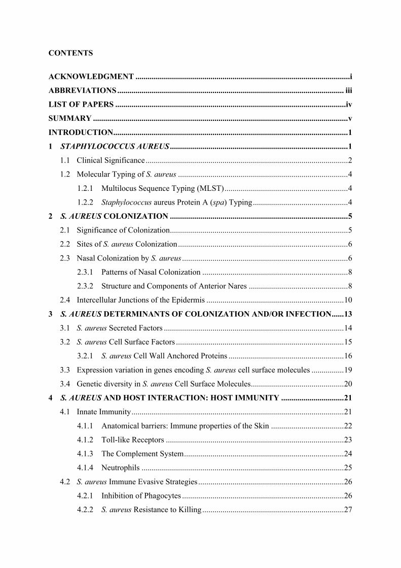
In human, there are different isoforms of the desmosomal cadherin proteins: three Dsc (Dsc1-
3) and four desmogleins (Dsg1- 4)150,151. Dsc and Dsg share similar structural features
(Figure 3). Their structure comprises of an extracellular cadherin domain (EC1- 4),
extracellular anchor (EA) followed by a single pass transmembrane region and an
intracellular anchor at the cytoplasmic side. However, Dsgs have additional motifs on their
intracellular region. The cadherin repeats are interspaced with calcium binding motifs and it
has been shown that calcium plays an important role in the structural integrity of
desmosomes during adhesion152. To facilitate adhesion, the desmosomal cadherin proteins
can engage in a homotypic or heterotypic interaction with each other148,153.
Desmoglein isoforms have varied expression patterns within the stratified epidermis154. This
differential expression is essential for epidermal maturation process and maintenance of
tissue homeostasis. Alterations in the expression patterns of the different isoforms result in
abnormal epidermal differentiation, reduction in barrier function and compromise in cell-to-
cell adhesion (reviewed in144,148). Within the epidermis, Dsg2 is expressed at the basal layer
while Dsg3 is expressed at basal and spinosum layers. Dsg1 is concentrated in the suprabasal
layers and Dsg4 expression is confined to the corneum and upper granular layers134 (Figure
3).

13
Figure 3. Structure and expression of the desmosomal cadherin proteins. a. Schematic representation of the different regions of desmogleins (Dsg) and desmocollins are shown. The desmosomal cadherins proteins contain four extracellular (EC) domains, an extracellular anchor (EA), transmembrane (TM) region, intracellular anchor (IA) and intracellular cadherin segment (ICS). Desmogleins contain additional regions including the intracellular proline rich linker (IPL), repeat unit domain (RUD) and desmoglein-specific terminal domain (DTD). Desmocollins isoforms have two splice variants “a” and “b”. Based on155. b. Dsg protein isoforms expression patterns within the epidermis. SC-Stratum corneum, SG-Stratum granulosum, SS-Stratum spinosum, SB-Stratum basale, BM-Basement membrane) Based on134.
3 S. AUREUS DETERMINANTS OF COLONIZATION AND/OR INFECTION
S. aureus can exist as a commensal or a pathogenic microbe within its human host. This
requires achieving a proper balance between efficient attachment at the colonized site and
withstanding the mechanical forces that aim to dislodge it from those niches. Furthermore, as
a pathogen, the bacteria should be able to survive and establish itself once the host defence
mechanisms are breached49. In addition, it should be able to cause tissue damage and spread
to other sites within the host body to establish infection.

14
S. aureus expresses a barrage of virulence factors that facilitate its ability to interact with host
tissue and the extracellular matrix components. Broadly, S. aureus virulence factors can be
classified into secreted factors and cell surface factors (Figure 4). Together, these factors
function to (1) adhere to the host cell surface and components, (2) spread bacteria through the
host, (3) evade host immune defence, and (4) produce toxins and other products, which can
cause damage to the host’s cells. Coupled with these factors, S. aureus also possesses
regulatory components and mechanisms, which ensures that the bacterium expresses these
factors only when needed (reviewed in5,6,156,157).
Figure 4. Schematic representation of localization of selected S. aureus virulence factors. S. aureus produces many factors which contribute to its colonization and/or infections. Examples of secreted factors: include Panton-Valentine leukocidin (PVL), phenol-soluble modulins (PSMs) toxic shock syndrome toxin (TSST) and Staphylokinase. Examples of cell surface factors include lipoteichoic acid (LTA), wall teichoic acid (WTA), polysaccharide intracellular adhesin (PIA), serine- aspartate repeat containing protein D (SdrD), surface protein G (SasG), clumping factor (Clf), fibronectin binding protein (FnBP), autolysin (Atl), extracellular matrix-binding protein homologue (Ebh), elastin binding proteins (Ebps), extracellular fibrinogen binding protein (Efb), extracellular matrix protein (Emp) and enolase. Based on 5,6,18,156,158,159.
S. aureus Secreted Factors 3.1
S. aureus produces many factors that are secreted into the extracellular milieu. These secreted
factors include enzymes, superantigens and membrane damaging toxins159,160. Superantigen
factors such as toxic shock syndrome toxin (TSST) activate the host’s T cells leading to their
excessive proliferation and production of cytokines, overall causing the fatal Staphylococcal
toxic shock syndrome159. Membrane damaging toxins bore into the cytoplasmic membrane of

15
the host cells leading to their lysis and escape of their intracellular contents159. Membrane
damaging toxins include proteins such as Hemolysin-α (α- toxin), Panton-Valentine
leukocidin (PVL), Phenol-soluble modulins (PSMs) and gamma-toxin (gamma-hemolysin,
HlgA, HlgB, HlgC)159,161. These proteins have different mechanisms of action. For example,
PVL binds to C5aR and C5L2 receptors on neutrophils162 while the effect on PSMs on host
cells is thought to be receptor independent159. In addition, α- toxin binds with A disintegrin
and metalloproteinase 10 (ADAM10)163. The interaction disrupts focal adhesion and degrades
E-cadherins, subsequently leading to loss of epithelial integrity163,164. S. aureus also secretes
enzymes such as Staphylokinase, Staphylocoagulase and Von Willebrand factor (vWF),
which further influence the bacterial virulence (reviewed in159). In addition, S. aureus
Exfoliative toxin (ET), has been indicated in the pathogenesis of staphylococcal scalded skin
syndrome (SSSS)165.
S. aureus Cell Surface Factors 3.2
The S. aureus cell surface is decorated with proteinaceous and non-proteinaceous
molecules18,156. The proteinaceous cell surface molecules include: (1) Cell wall anchored
proteins (CWA) which are covalently linked to the bacterial cell wall166, (2) Non covalently
attached cell wall associated proteins including proteins with specific cell wall-binding
domains e.g. autolysin (Atl), ‘secretable expanded repertoire adhesive molecules’ (SERAMs)
and cytoplasmic wall binding proteins, and (3) Membrane spanning proteins such as
extracellular matrix-binding protein homologue (Ebh) and elastin binding proteins
(Ebps)18,156. The non-proteinaceous S. aureus cell surface molecules include the Wall teichoic
acid (WTA), Lipoteichoic acid (LTA), Polysaccharide Intracellular adhesin (PIA) and other
polysaccharides18,156.
Although, there are ongoing investigations to further understand the contributions of these
cell surface factors in S. aureus colonization and/or virulence, the functions of some of these
cell surface factors has been described (reviewed in5,6,156,158). For example, WTA plays an
important role in the early stages of S. aureus nasal colonization167 and interacts with human
nasal epithelial cells via a type F scavenger receptor called SREC 1168. PIA and LTA are
involved in S. aureus biofilm formation169. Ebps binds elastin, a major component of the
extracellular matrix170. SERAMs proteins including extracellular adherence protein (Eap) and
extracellular matrix binding protein (Emp) bind to extracellular matrix molecules including
fibronectin, fibrinogen, collagen (reviewed in171).

16
3.2.1 S. aureus Cell Wall Anchored Proteins
CWA proteins are the main group of S. aureus cell surface factors. They meditate adhesion of
S. aureus to the host’s extracellular matrix and receptor(s) present on the host’s cell surface
(reviewed in5,6,154,164). They are involved in colonization, immune evasion, biofilm function
and other functions that contribute to S. aureus virulence (reviewed in5,6).
CWA proteins contain a signal sequence peptide at their amino terminal and a sorting signal
at their carboxyl terminal5. The signal sequence directs the translated product to sites within
the bacterial peptidoglycan cell wall172. The LPXTG motif in sorting signal at the carboxyl
terminal, facilitates the covalent anchorage of CWA proteins to the dividing peptidoglycan of
S. aureus cell wall173. The anchorage is facilitated via the action of the transpeptidase enzyme
called sortase A (SrtA)166. Interspacing the two terminals are different regions with diverse
functionality. Based on their structure and function, CWA proteins have been classified into
four groups (reviewed in5). These are (1) Microbial surface components recognizing adhesive
matrix molecules (MSCRAMMs) which include clumping factor A (ClfA) and ClfB, serine-
aspartate repeat containing protein (Sdr) C, D and E, bone sialo binding protein (Bbp),
collagen adhesion (CNA) and Fibronectin-binding protein A (FnBPA) and FnBPB, (2) Neat
motif family e.g. Iron-regulated surface (Isd) proteins, (3) Three helical bundle family e.g.
Protein A and (4) G5-E repeat family e.g. S. aureus surface protein G (SasG). Recently, a
review suggested two additional groups based on functional motifs without structural details
and the location of biological functions in a disordered region (reviewed in6). These are (1)
The legume lectin domain e.g. serine-rich adhesin of platelets (SraP) and (2) fibronectin
binding by tandem β-Zipper6.
Studies have shown the molecular mechanism behind CWA proteins involvement in S.
aureus virulence and their interaction with some host components5,6,156. CWA proteins are
involved in nasal colonization. For example, in vitro studies have shown that ClfB promotes
S. aureus binding to cytokeratin 10174 and loricrin101. The importance of ClfB-loricrin
interaction nasal colonization was emphasized by the reduced adherence of S. aureus in
loricrin deficient mouse101. Furthermore, ClfB promotes S. aureus nasal colonization and
persistence in humans artificially inoculated with ClfB expressing S. aureus175. In addition,
ClfB has been shown to bind to cytokeratin 8176. Other CWA proteins including as SasX,
SdrC and IsdA also promote adherence of S. aureus to human nasal epithelial cells100,177.
Deciphering CWA proteins functions are often complicated because S. aureus CWA proteins
17
are multifunctional and the proteins sometimes have redundant or complementary functions.
For example, CWA protein including FnBPA, FnBPB, ClfA, ClfB and IsdA all bind to
fibrinogen while IsdA, IsdB and IsdH bind to the haemoglobin component called haem5,6.
3.2.1.1 Serine-Aspartate Repeat Containing Protein D (SdrD)
S. aureus SdrD belongs to the MSCRAMMs group of CWA proteins. The sdrD open reading
frame (ORF) is encoded at the sdr locus in tandem with the ORFs of sdrC and sdrE178. The
prevalence of the sdrD gene within the genome of S. aureus strains varies179–181 and Trad et
al., observed a correlation between the presence of sdrD gene and bone infections182.
SdrD shares some structural similarities with S. aureus virulence factors ClfA and ClfB178
(Figure 5). Its structure comprises of a signal sequence and a sorting signal at its amino (N)
and carboxyl (C) terminus respectively. The N-terminal signal sequence is followed by the A
region, the B repeat and R domain (reviewed in5). SdrD A region is subdivided into N1, N2
and N3 domains and is responsible for ligand binding via a dock-lock- latch mechanism183.
SdrD B repeats compose of B1- B5 subdomains are composed of 110-113 amino acid
residues and functions as a spacer, extending the ligand binding A region further from the
cell wall121. The B1- B5 subdomains contains EF motifs, which bind calcium in a sequential
manner184,185. Furthermore, SdrD R domain is made up of serine aspartate repeats5,178.
Figure 5. Schematic representation of S. aureus Serine Aspartate repeats containing protein D (SdrD). The location of the S: Signal sequence, N1, N2, N3 subdomains of the SdrD A region, B1-B5 subdomains of the SdrD B repeat, SD-Repeats: Serine-Aspartate repeats of the SdrD R domain, W: wall spanning domain, M: membrane spanning domain, C: cytoplasmic domain, LPXTG: cell wall sorting signal are indicated. Based on5,186.
The function and molecular mechanism of SdrD in S. aureus virulence is still being
investigated. The sdrD gene expression is upregulated during nasal colonization187,188 and
SdrD S N1 N2 N3 SD-RepeatsB1 B2 B3 B5B4 W M C
LPXTG motif
N C
A region B repeat R domain

18
SdrD promote increased S. aureus adhesion to desquamated nasal epithelial cells100.
However, SdrD may also have a role during S. aureus infection, because its expression is
increased in human blood189 and it promotes S. aureus survival in human blood ex vivo190. In
addition, there is an increased level of Immunoglobulin G (IgG) against SdrD in serum of S.
aureus infected patients191. Moreover, SdrD is crucial in abscess formation following
invasive S. aureus infection192. Furthermore, mice immunized with a vaccine preparation
composed of SdrD, SdrE, IsdA and IsdB, showed an increased level of protection against S.
aureus infection193. These findings suggest that the SdrD protein could be important in S.
aureus colonization and infection of its host.
3.2.1.2 S.aureus Surface Protein G (SasG)
The SasG protein belongs to the G5E group of S. aureus CWA proteins. The protein has
some structural organization and sequence similarity with the Plasmin sensitive proteins (Pls)
and the Accumulation associated protein (Aap) of S. aureus and S. epidermidis
respectively194. The SasG protein consists of an A region and B repeat made up of tandem
repeats of G5 and E5,194 (Figure 6).
Figure 5. Schematic representation of S. aureus Surface protein G (SasG). S: Signal sequence, ligand binding A region, G5-E repeats of the SasG B repeat, W: wall spanning domain, M: membrane spanning domain, C: cytoplasmic domain, LPXTG: cell wall sorting signal are indicated. Based on194,195
The sasG gene is highly prevalent in clinical isolates compared to carriage isolates194. SasG is
involved in intercellular aggregation of SasG expressing S. aureus196,197. SasG also promotes
biofilm formation198 and Geoghegan et al. showed that the biofilm formation process is
mediated by the intercellular dimerization B repeat of neighbouring SasG expressing cells199.
In addition, it was shown that the intercellular dimerization of SasG B repeats occurs in a
zinc dependent manner197,199. Furthermore, SasG promotes adhesion of S. aureus to
S
A region
W M C
LPXTG motif
SasG G5 E
B repeat
G5 E G5 E G5 E G5 CN

19
desquamated nasal cells195,198 and the adhesion is mediated by the SasG A region195.
However, SasG does not promote adhesion to buccal epithelial cells or keratinocytes195. The
sasG gene is highly expressed in nasal samples from S.aureus nasal carriers188 and also high
levels of IgG against SasG have been observed in sera of infected patients194. This suggests
that SasG is relevant for S. aureus virulence. However, expression of SasG reduces
adherence of S. aureus to fibronectin and fibrinogen195. This was hypothesized to be the
effects of SasG masking other adhesins on S. aureus due to its B repeat extension from the
cell surface198.
Expression variation in genes encoding S. aureus cell surface molecules 3.3
The expression patterns of S. aureus virulence genes could suggest how and when the
expressed virulence factors are important during S. aureus colonization and/or infection.
Some studies have tried to delineate which S. aureus virulence factors are expressed during
nasal colonization200,201. For example, analysis of nasal samples from persistent S. aureus
carriers revealed an early upregulation of the WTA biosynthesis genes, tagO and tarK, during
the initial stages of nasal colonization200. Other CWA genes such as clfB, fnbA and isdA are
upregulated much later during colonization200. This suggests that WTA is important for
prompt S. aureus nasal colonization.
In S. aureus, about 24 different CWA proteins can be expressed5. However, the CWA
proteins expressed depends on strain202, the growth phase and conditions187,189,203. For
example, CWA genes such as isdA are highly expressed in iron-limiting conditions204 ,
others such as clfB and spa are expressed predominantly during the exponential growth
phase205,206 while clfA is expressed in the stationary growth phase207,208. In addition,
expression of CWA genes sasD and sdrH were highly upregulated in persistent S. aureus
nasal carriers compared to non-persistent carriers209. These differences are a result of the
regulatory factors in S. aureus including the accessory gene regulator (agr) locus, the
staphylococcal accessory regulator A (sarA), which direct expression of these factors in
response to cues within its environment such as bacterial density, available nutrients
etc.157,208,210.

20
Genetic diversity in S. aureus Cell Surface Molecules 3.4
Studies have also revealed genetic diversity within the sequences and region of genes
encoding virulence factors between S. aureus from diverse background202,211. Genetic
variations range from sequence variations within an individual gene212 to the absence or
presence of genes within the genome of different S. aureus strains202,213. For example, the A
domain of S. aureus virulence factor FnBPA exists as different isotypes214,215. These
variations were mainly concentrated in the N2-N3 subdomains of the A domain117. Though
variations within FnBPA A domain isotypes did not affect their ligand binding activity, it
affected their antigenicity214,215. This suggests that sequence variations within virulence genes
could have important implications on the virulence functions. Indeed, single nucleotide
polymorphism in fnbp genes have been shown to be associated with increased cardiovascular
devices infection216,217. Furthermore, sequence variations have been reported within other
S.aureus CWA genes such as fnbp218,219 and sdrD211 of S. aureus isolates from different host
origins.
A correlation between the presence of sdrD gene and bone infections have been
observed179,182. McCarthy and Lindsay reported that CWA genes such as fnbpA, isdA and
isdH were present in all the 58 S. aureus isolates studied while genes such as sdrC, sdrD and
sasG were absent from some of these isolates202. They also observed that the collagen
adhesion gene, cna was absent from the genome of the majority of these isolates202. Sabat et
al. found that the prevalence of sdrD gene was significantly higher in MRSA strains while
sdrC gene was limited to MSSA strains179. Furthermore, fnbpB gene was found to be more
prevalent among invasive isolates compared to carriage isolates219,220.
Overall, genetic and expression variation within virulence genes between S.aureus isolates
further indicate the complexity of identifying specific factors that account for how S. aureus
could be an effective colonizer or cause a wide range of diseases. What this implies is that the
dynamics of S. aureus interaction with humans cannot just be explained based on a single
bacterial virulence determinant.

21
4 S. AUREUS AND HOST INTERACTION: HOST IMMUNITY
The host immune system can recognize, resist and eliminate S. aureus (reviewed in 221,222). It
is divided into the innate immune system and adaptive immune system. The innate immune
responses are the first line of defences that are initiated immediately upon contact with
pathogens.. Innate immune responses are fast, non-specific but are able to discriminate
invading pathogens from self and other beneficial commensal flora. The adaptive immunity is
a delayed, specific response and is stimulated by components of the innate immune system.
The adaptive immune system develops immunological memory, which enables rapid
response to subsequent reinfection by the same pathogen. Adaptive immunity against S.
aureus infection begins later during the time course of infection. Responses by the adaptive
immunity lead to the activation of B and T cells, production of antibodies and also release of
cytokines. This can further modulate and/or amplify the initial response mounted by the
innate immunity222. Phagocytosis by the neutrophils is believed to be one of the main
clearance mechanisms for S. aureus infection221,224.
Innate Immunity 4.1
Innate immune system can be broadly grouped into anatomical barriers, toll-like receptors,
complement system and phagocytes223 (Figure 7)

22
Figure 7. Host Immune responses to S. aureus colonization and/or infection. S. aureus colonization of the anterior nares and skin surface is inhibited by the host immune defence mechanisms including antimicrobial peptides (AMPs) release, Toll-like receptor 2 (TLR2) recognition of conserved motifs on the bacterial surface, mucus production, the presence of resident microbes and low pH. Upon breaching the epidermis, components of the host immune defence including complement factors and Immunoglobulins (Ig) detect the bacteria. These components opsonize the bacteria surface leading to the activation of the complement cascade. This leads to production of complement factors C5a and C3a.These products initiate recruitment of circulating neutrophils from the blood. In addition, TLR activation induces chemokine production, which together with C5a and C3a form a chemotactic gradient that directs and guides the neutrophils to the infection site. Neutrophils recognize the opsonized bacteria via their Fc and complement receptors. Consequently, the bacteria are phagocytosed and killed by the neutrophils. Based on225,226.
4.1.1 Anatomical barriers: Immune properties of the Skin
The skin is the first barrier, which protects against onslaught of microbes present in the
environment139. The skin’s immune protection is ensured by tightly packed keratinocytes and
also the continuous desquamation of the epidermal cells132. In addition, filaggrin components
breakdown at the stratum corneum leads to the production of acidic components such as
urocanic acid (UCA) and pyrrolidone carboxylic acid (PCA)227. These components
contribute to the skin surface’s low pH and also inhibit expression of S. aureus CWA
proteins ClfB, FnbpA and protein A227. Commensal microbes of the skin also ensure
23
protection against S. aureus. For example, PSM and serine protease Esp produced by S.
epidermidis on the skin inhibits colonization by S. aureus109,228.
Furthermore, antimicrobial peptides such as β-defensins, RNase7, and cathelicidin expressed
by epidermal keratinocytes show inhibitory activity against S. aureus, thus preventing
successful colonization132,229. Human β-defensins are highly potent against S. aureus230.
Cathelicidins disrupt the S. aureus cell membrane by forming pores in them231 and has been
shown to be highly effective in killing extracellular and intracellular S. aureus232. In the skin,
RNase7 was found at the stratum corneum and inhibited colonization of skin explants by S.
aureus233,234. The production of these antimicrobial peptides can be induced by the presence
of S. aureus or components such as LTA235,236. Aside from these, antimicrobial peptides can
also induce cytokine release and recruitment of immune cells such as macrophages, dendritic
cells to the infection site237,238.
4.1.2 Toll-like Receptors
Cells within the nasal cavity, skin and other S. aureus colonization sites possess receptors
called pathogen recognition receptors (PRRs). These PRRs recognize conserved microbial
components referred to as pathogen associated molecular patterns (PAMPs) in S. aureus and
other pathogenic microbes239. S. aureus PAMPs include LTA, lipoproteins (LPP), teichoic
acid and other surface associated components (reviewed in240). An important group of PRRs
are the Toll-like receptors (TLRs). The TLRs are transmembrane proteins composed of an
extracellular domain, a transmembrane region and cytosolic Toll/IL-1 receptor (TIR)
domain240.
The important TLR responsible for recognition of S. aureus and its microbial component is
TLR2. Its importance in mitigating S. aureus infections has been demonstrated in mouse
lacking TLR2241,242. Furthermore, diminished TLR2 stimulation in atopic dermatitis patients
have been suggested to contribute in S. aureus skin infection243. To become functionally
activated, TLR2 forms heterodimer complex with either TLR6 or TLR1, via which it
interacts with LTAs and lipoproteins expressed on the surface of S. aureus240,244. Interaction
of TLR2 with its ligands, resulting in activation of intracellular signalling cascade that leads
to the activation of transcription factor nuclear factor- κB (NF-κB) which consequently leads
to the production of pro-inflammatory products such as chemokines and cytokines245,246. NF-
κB also promotes the expression of adhesion molecules such as E-selectin, Intercellular

24
adhesion molecule 1 (ICAM1) and vascular cell adhesion molecule 1 (VCAM1)247. These
adhesion molecules recruit circulating immune cells such as neutrophils from the blood.
Furthermore, TLR2 activation promote epidermal tight junction formation thus enhancing
skin’s barrier function248. In addition, activation of TLR2 by the skin’s commensal microbes
also enhances the production of antimicrobial peptides, which can inhibit S. aureus
colonization and infection249.
4.1.3 The Complement System
The complement system is made up of more than 30 protein found in blood and tissues.
Complement proteins are inactive until they are cleaved. After activation, they react with
each other, generating a sequence of events that helps to combat the pathogen. Complement
system can be activated via three different pathways, which are the classical pathway (CP),
the alternative pathway (AP) and the lectin pathway (LP). These pathways differ in the
molecules that can activate them. The classical pathway is activated either by direct binding
of C1q to the bacterial surface or C1q binding to antibody complexes (IgM or IgG) present
on bacterial surface. In contrast, binding of the spontaneously generated C3b on bacteria
activates alternative pathway. The lectin pathway is activated by mannose binding lectin or
ficolin to the mannose containing carbohydrates on the bacterial surface. Complement
activation irrespective of the pathways results in the production of C3 convertases (reviewed
in240,250,251).
Complement activation serves three purposes. First, the activated complement factors bind
the pathogen surfaces, opsonizing them thus making phagocytosis of the pathogen highly
efficient. Second, the effector proteins such as C5a and C3a generated during complement
activation, serve as chemoattractants for the recruitment of immune cells (phagocytes) from
circulation. Furthermore, activation of complement can also lead to the generation of
membrane attack complex (MAC) that lyse the pathogen’s membrane especially for Gram-
negative bacteria (reviewed in240,250,251). The importance of complements in combating S.
aureus has been demonstrated by the increased death observed in complement depleted
mouse after S. aureus bacteraemia252. Furthermore, it has also been shown that activation of
complements on S. aureus surfaces reduced their adherence to endothelial cell surfaces253.

25
4.1.4 Neutrophils
Neutrophils are the first set of phagocytes to migrate to the site of S. aureus infection
(reviewed in254). Their importance in combating S. aureus infection is demonstrated by the
increased predisposition of individuals with defective neutrophil functions to S. aureus
infections254,255. The primary role of neutrophils in combating infection is phagocytosis of the
pathogens recognized by the PRRs. They are also play an important role in abscess formation
upon S. aureus infection256.
Recruitment of circulating neutrophils to the infection site is facilitated by a gradient of
chemotactic signals including Interleukin-8 (IL-8), complement factors C3a and C5a254,257.
The recruitment process can be divided into four stages, which are rolling adhesion, integrin
activation, firm adhesion and transmigration258. Capturing of circulating neutrophils is
initiated by their attachment to adhesion molecules such as E- selectin, P-selectin,
Intracellular adhesion molecule (ICAM) etc. present on the endothelial cells259. Attachment
to these adhesion molecules is facilitated by receptors such as P-selectin glycoprotein ligand
1(PSGL-1) expressed by neutrophils260. Subsequently, they leave blood circulation and
transmigrate across the endothelial walls towards the infected tissue site261.
Efficient phagocytosis by neutrophils is enhanced by the presence of opsonins such as
complement factors and immunoglobulins on the pathogen’s surface254. Present on
neutrophils cell surface are receptors such as Fc and complement receptors, which interact
with these opsonins (reviewed in250,258,262). However, neutrophils mediated phagocytosis of
pathogens have also been observed to occur at a slower rate in absence of opsonization263.
These interaction leads to the phagocytosis of the pathogen and subsequently formation of
phagosomes. Phagosomes undergo series of maturation process, which eventually lead to
bacterial killing (reviewed in264). Reactive oxygen species (ROS), proteinases and AMPs etc.
produced by neutrophils ensure bacterial killing265. Furthermore, neutrophils can trap and kill
S. aureus via its neutrophil extracellular traps (NETs) covered with antimicrobials266.
26
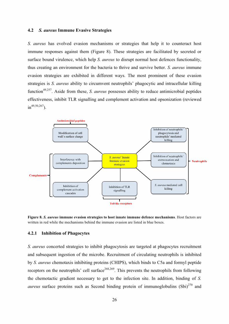
S. aureus Immune Evasive Strategies 4.2
S. aureus has evolved evasion mechanisms or strategies that help it to counteract host
immune responses against them (Figure 8). These strategies are facilitated by secreted or
surface bound virulence, which help S. aureus to disrupt normal host defences functionality,
thus creating an environment for the bacteria to thrive and survive better. S. aureus immune
evasion strategies are exhibited in different ways. The most prominent of these evasion
strategies is S. aureus ability to circumvent neutrophils’ phagocytic and intracellular killing
function49,257. Aside from these, S. aureus possesses ability to reduce antimicrobial peptides
effectiveness, inhibit TLR signalling and complement activation and opsonization (reviewed
in49,50,267).
Figure 8. S. aureus immune evasion strategies to host innate immune defence mechanisms. Host factors are written in red while the mechanisms behind the immune evasion are listed in blue boxes.
4.2.1 Inhibition of Phagocytes
S. aureus concerted strategies to inhibit phagocytosis are targeted at phagocytes recruitment
and subsequent ingestion of the microbe. Recruitment of circulating neutrophils is inhibited
by S. aureus chemotaxis inhibiting proteins (CHIPS), which binds to C5a and formyl peptide
receptors on the neutrophils’ cell surface268,269. This prevents the neutrophils from following
the chemotactic gradient necessary to get to the infection site. In addition, binding of S.
aureus surface proteins such as Second binding protein of immunoglobulins (Sbi)270 and

27
Staphyloccocal protein A (SpA) to the Fc of IgG, reorients antibodies in the wrong direction
preventing opsonization and phagocytosis49,258. Masking of bacterial surface receptors or
epitope by S. aureus capsule polysaccharide also inhibits phagocytosis271. Complement
mediated opsonization of the bacterial surface is affected by proteins such as extracellular
adherence proteins (Eap), Staphyloccocal Complement Inhibitor (SCIN) and extracellular
fibrinogen binding protein (Efb) 49,258. CWA proteins such as ClfA and IsdH also inhibit the
phagocytosis272,273.
4.2.2 S. aureus Resistance to Killing
When S. aureus is ingested, it can still survive within the phagocytes by inhibiting the
cytotoxic processes leading to bacterial degradation. S. aureus products such as
staphyloxanthin and superoxide dismutases protect the bacteria from effects of the reactive
oxygen species of neutrophils’ phagosomes49,274 .
S. aureus have also developed strategies to combat the effects of AMPs. S. aureus secreted
protein aureolysin degrades the LL37, a potent bactericidal agent275. It can also modify its
surface via the action of dtl operon, thus preventing the binding of AMPs276. Staphylokinase
also binds inhibits the activity of defensins on S. aureus by binding with them277. In addition,
S aureus produces toxins such as phenol soluble modulins (PSMs) which form pores on the
phagocytes and thus facilitating the escape of the ingested S. aureus278,279. CWA proteins
such as ClfA mediate survival of bacteria by promoting abscess formation. Others such as
SdrD, SdrE and SpA also contribute to S. aureus survival in blood 190,280,281.
Some of the molecules expressed by S. aureus to circumvent host immune responses are
listed in Table 1

28
Table 1: Examples of molecules used by S. aureus to evade or alter the host immune responses
Immune Response Evasion factor Abbreviation Function Effect Reference
Neutrophils
Staphyloccocal superantigen protein 5 SSL5 Binds P-selectin glycoprotein ligand-1
Disrupts neutrophils chemotaxis 282
Staphylococcal superantigen protein 11 SSL11 283 Chemotaxis inhibitory protein of S. aureus CHIPS
Binds to C5a receptors and formyl peptide receptor like-1
Disrupts neutrophils chemotaxis
268,269 Formly peptide receptor like-1 inhibitory protein FLIPr 284
Extracellular adherence protein Eap Binds to ICAM1 Blocks neutrophils adhesion to endothelial lining
285,286
Staphylococcal binder of Immunoglobin Sbi Binds IgG
Blocks antibody mediated opsonization and phagocytosis
270,287
S. aureus Protein A Spa Reviewed in33,49,50
Staphyloxanthin Carotenoid biosynthesis Protection against Reactive oxygen species (ROS) effects
288 Catalase and Superoxide dismutase Eliminate/ Inactivate ROS 289
Phenol Soluble modulin PSM
Bore pores in membrane of cells
Destroys neutrophils and other host immune cells
Reviewed in278
Panton- Valentine leukocidin PVL Reviewed in33,49,50 Leukocidin GH LukGH
TLR Staphylococcal superantigen 3 SSL3 Binds TLR2 ligand binding site Blocks TLR2 immune recognition 290
TIR containing protein TIRS Binds TLR2´s TIR domain Blocks TLR2 mediated NF-κB activation
291
Antimicrobial peptide
dlt operon WTA Modification of cell wall components Reduced antimicrobial peptide activity
276 Staphylokinase Sak Binds alpha defensins 277
Complement
Staphylococcal Complement Inhibitor SCIN Binds complement factor C3 and C3 convertases
Disrupts complement mediated opsonization of S. aureus and phagocytosis
292
Staphylococcal binder of Immunoglobin Sbi 293
Extracellular fibrinogen binding protein Efb 294,295
Staphylokinase Sak Converts S. aureus surface bound plasminogen into plasmin Removes opsonins on the
microbial cell surface
277,296
Serine aspartate repeat containing protein E SdrE Binds Factor H 280

29
5 OBJECTIVES
The main objective of this study was to investigate the molecular mechanisms underlying the
determinants involved in S. aureus colonization and/or infection of its host. We aimed to
provide increased knowledge regarding the role of the S. aureus CWA proteins SdrD and
SasG in bacterial adhesion to host cells and/or evasion of the host immune defence
mechanisms.
Our specific research questions were:
Paper I
S. aureus SdrD has been shown to promote bacterial adhesion to desquamated nasal epithelial
cells.
Does S. aureus SdrD interact with specific host ligand(s) in the epithelium? What
could be the role of such ligand interaction?
Paper II
Sequence diversity has been observed within S. aureus CWAs genes such as fnbp and clfA.
How conserved is the sdrD gene within S. aureus isolates from healthy
individuals? Could variation within the sdrD gene influence SdrD function?
Paper III
S. aureus SasG is a surface protein known to promote bacterial adhesion to desquamated
nasal epithelial cells.
Does S. aureus SasG contribute to host cells adhesion to human keratinocytes?
Under what conditions is SasG expressed? Does S. aureus SasG have immune
evasion properties?

30
6 METHODOLOGY
A detailed description of the experimental procedures performed in this thesis is listed and
explained in the articles and manuscript herein. An overview of some of the experimental
procedures performed is explained below.
Host Protein-Pathogen Protein Interaction 6.1
Protein-protein interaction detection assays have been employed to determine the interaction
between host proteins and S. aureus virulence factors. Elucidating these interactions is
particularly helpful in deciphering some of the molecular mechanism engaged by S. aureus in
colonizing humans. It has been suggested that this might form the basis for the development
of some anti-colonization strategies in future297. In Paper I, a yeast two-hybrid (Y2H)
assay298 was ordered from Hybrigenics to find the potential host partners for S. aureus SdrD
protein. Basically, Y2H assays are performed in yeast strains containing two proteins. One of
the proteins referred to as “bait” is fused with a DNA binding domain (DBD) while the other
protein referred to as “prey” is fused with an Activation domain (AD). Physical interaction of
the two proteins brings these two domains in close proximity and thus forming a functional
transcription factor. The transcription factor then activates the reporter gene, which can be
assayed by growing the yeast strain on a selective medium or observing for colorimetric
changes. In Paper I, the SdrD A region fused with GAL4DBD was the bait protein and this
was used to screen the human reconstituted skin libraries fused with GAL4AD (the “prey”).
We selected Y2H assay because it allowed for screening of large proteins libraries for SdrD
putative host partners. It is important to note that Y2H assay has some limitations, one of
which is the high false positive rates of potential candidates299. However, in Paper I, we
have employed complementary methods to validate the putative partners as indicated by the
Y2H.
Solid Phase Ligand Binding Assay 6.2
Solid phase ligand binding assay allows for further characterization and validation of protein-
protein interactions in vitro. The principle underlying this method is based on the enzyme
linked immunosorbent assay (ELISA). For this assay, the first protein (protein A) is
immobilized onto a solid phase such as wells of microtiter plate. Subsequently, the free in
solution second protein (protein B) is added to the wells containing immobilized protein A.

31
After washing, the binding of the two proteins is determined via colorimetric quantification.
It is expected that the colour intensity should be directly proportional to the amount of the
bound proteins. In Paper I, variants of this assay were adopted to confirm the interaction
between S. aureus SdrD and Dsg1, the Y2H assay detected putative interaction partner. For
example, purified recombinant SdrD A region protein were coated onto wells of microtiter
plates. Subsequently, recombinant Dsg1 protein was added. If there were any interaction, it
would be expected that the added Dsg1 would bind to the SdrD A region. This was assayed
by adding antibodies against Dsg1, followed by the secondary antibodies, followed by
quantification of binding. In addition, in Paper I, we also immobilized Dsg1 on wells of
microtiter plates. Wells coated with IgG1 were used as a control. Thereafter, we added S.
aureus NCTC 8325-4 or S. aureus NCTC 8325-4∆sdrD to the coated wells of microtiter
plates. We hypothesized that if SdrD binds with Dsg1, expression of SdrD in S. aureus
NCTC 8325-4 should increase binding to immobilized Dsg1. Subsequently, the plates were
washed, fixed and stained with crystal violet. The stained well were subsequently dissolved
with acetic acid. Semi quantitative measurement of the interaction was achieved by
measuring absorbance of the wells.
Genetic manipulation of S. aureus 6.3
Studying the biological functions of S. aureus virulence factors often involves a deletion or
heterologous expression of genes encoding these factors. Manipulations of S. aureus DNA
have been performed in this thesis.
In Paper I and III, isogenic mutants of S. aureus were generated using the allelic
replacement method300,301. This method relies on the replacement of DNA with a mutant
allele of the DNA via homologous recombination. To create a markless deletion of the target
gene, DNA sequences flanking up and down of the target gene are cloned into a S. aureus
shuttle plasmid such as pKOR1300 and pIMAY302. The replication origin for these plasmids is
temperature sensitive i.e. they can only replicate autonomously at certain permissive
temperature. Furthermore, the plasmid contains a selective marker that allows selection of
successful transformants. Thus, electroporation of the cloned plasmid into S. aureus and
growing the bacteria at the selective temperature allows for plasmid replication.
Subsequently, temperature shift is used to facilitate integration and excision of plasmid in and
out of the S. aureus genome. However, it is important that caution is taken during genetic
manipulations to ensure and verify that no other undesired modifications are introduced in

32
S. aureus genome. Thus, it is important to verify that other biological functions (such as
growth rate, hemolytic effect etc.) in the wild type and its isogenic mutant are comparable.
Genetic modifications of certain S. aureus strains are difficult due to the S. aureus restriction-
modification (RM) systems, which attacks foreign DNA (reviewed in303). S. aureus RM
systems methylate its DNA in specialized pattern, which are subsequently read and protected
from degradation. However, foreign DNA without the recognizable methylation pattern is
degraded when transformed into S. aureus. Type I and type IV RM systems are mainly
responsible for this in S. aureus. To bypass this, in Paper I, the cloned plasmid was
transformed into E. coli DC10B302,304. E.coli DC10B is deficient in Dcm methylase, which
methylates cytosine residues. Thus, plasmids transformed in it are not methylated on their
cytosine residues, thus bypassing the type IV RM system challenge. In Paper III, the cloned
plasmid was transformed in E. coli IM01B or SA08B301. Aside from being dcm deficient,
these E. coli strains have been engineered to methylate the adenine residues on the
transformed plasmid based on the methylation pattern of particular S. aureus clonal
complexes, this allows for bypass of Type I and type IV RM systems challenges in S. aureus.
Falkow explained Koch´s postulates as it relates to microbial pathogenicity investigation305.
He stipulated that it is important that expression of the gene in its isogenic mutant
(complementation) or expression in another model strain (heterologous expression) should
restore or confer virulence capacity in order to confirm that the gene is actually involved in
its pathogenicity. Therefore in Paper I, heterologous expression of SdrD was achieved by
cloning sdrD gene into pMG36e plasmid306 and which was then subsequently transformed
into Lactobacillus lactis. In Paper II, S. aureus NCTC 8325-4∆sdrD was complemented
with pMG36e plasmid containing variants of sdrD gene. This ensured that the sdrD variants
were expressed in a common genetic background, therefore eliminating contributory effects
of other factors not within the scope of the study. In Paper III, we made heterologous
expression and complementation constructs for SasG by transforming
pALC2072::SasGMSSA476 into S. aureus SH1000 and MSSA476∆sasG respectively.
Bioinformatic analysis 6.4
Bioinformatic tools and analyses have taken a front seat in providing more understanding
about S. aureus based on the sequences of their genes or genomes. In addition, relatedness of
S. aureus strains can also be inferred by comparing the sequences to each other and also with

33
other reference isolates which are present in the database. This has helped in prompt
resolution and typing of S. aureus responsible for infections.
In Paper II, genetic variation in sdrD gene between S. aureus isolates from healthy carriers
was investigated. Multiple sequence alignment (MSA) was performed in order to facilitate
comparison of the sdrD A region sequences from different S. aureus isolates. This will show
the sequence diversity of sdrD gene within S. aureus strains from different human hosts.
MSA can either be carried out either as global or local alignments. The major difference
between them is that global alignment tries to align the whole sequence while local alignment
aligns the sequence based on the region of highest similarity between the sequences. Multiple
Alignment Fast Fourier Transform (MAFFT)307 was used for the alignment. MAFFT has
been suggested to provide accurate MSA data compared to some other alignment tool such
CLUSTALW308. In addition, phylogenetic analyses were performed using Randomized
Axelerated Maximum Likelihood (RaxML)309 and Multilocus sequence typing (MLST)51.
Host Model Systems to study functions of S. aureus Virulence Factors 6.5
There is need to develop or use an appropriate in vitro or ex vivo model to investigate the
virulence effect of S. aureus proteins. Depending on the physiological conditions that are
being monitored, different technical methods can be used. The model system of choice
should mimic the conditions that the bacteria would normally be exposed to during
colonization or infection. However, the limitation of a host model system is that it is not
possible to include all human components and conditions that would normally be present
during S. aureus colonization or infection.
In Paper I, II and III, cell adhesion assays were performed to evaluate the contribution of the
studied virulence factor to S. aureus adhesion. As keratinocytes are the most abundant cells
types found in the skin and the anterior nares of the nose132,134, the model cell type chosen for
our in vitro studies was the immortalized human keratinocytes cell line (HaCaT)310 (Paper I,
II and III). However, in Paper I, we also used human embryonic kidney cells 293
(HEK293)311. Basically, in cell adhesion assay, the cells are seeded into tissue culture treated
wells and incubated to facilitate attachment to the well surface. Upon addition of S. aureus to
the seeded cells, S. aureus surface protein should interact with host surface expressed
proteins. As a control, a bacterium lacking the gene encoding the protein is used. The degree

34
of adherence can therefore be compared, in order to see if the protein contributes significantly
to S. aureus adhesion to the host cells.
As discussed previously, S. aureus virulence factors also contribute to its ability to evade and
survive host defence mechanisms. Human blood composes of components that aid to combat
bacterial infections. The whole blood assay has been used to study the expression and
immune evasion effect of S. aureus virulence genes189,190,287,312. Blood used for this assay is
usually freshly drawn blood from healthy participants. The blood is taken in tubes containing
hirudin, which serve as anticoagulant and also preserve the complement activity of the
blood313. Therefore, this assay provides a more accurate reflection of S. aureus response to
immune system components. In Paper III, the expression and contribution of S. aureus
MSSA476 sasG gene to survival was assayed in whole human blood assay.
Recombinant protein expression 6.6
Advances in the molecular biology techniques have made it possible to express protein
products of exogenous DNA within a heterologous expression system. Recombinant protein
production has helped to overcome the challenges of getting high amounts of the desired
protein. The recombinant protein process involves cloning the DNA sequences of the desired
protein into an expression vector such as pGEX-4T-1 and pRSETB. The cloned vector is
transformed into a heterologous expression vehicle such as bacteria, yeast or mammalian
cells. Thereafter, expression of the protein is induced and subsequently the recombinant
protein is isolated and purified.
In this study, we have expressed SdrD and SasG proteins and its subdomains using the E. coli
expression system. We used this system because E. coli is easy to culture, easy to handle and
has a fast growth rate. In addition, compared to other heterologous expression vehicles, E.
coli genetics is better understood and this system produces a high yield of the recombinant
protein. However, there are drawbacks associated with using E. coli as an expression vehicle
including likelihood of protein not being expressed in its natural form, production of
insoluble protein, and lack of post-translational modifications such as glycosylation.
Strategies including use of fusion tag and optimization of expression conditions such as
lowering of temperature and addition of glucose etc. have been suggested to overcome these
challenges (discussed in314,315).

35
7 SUMMARY OF RESULTS
Paper I: The interaction between Staphylococcus aureus SdrD and desmoglein 1 is
important for adhesion to host cells
• The expression of SdrD in S. aureus NCTC8325-4 promoted increased binding of the
bacteria to HaCaT cells compared to its isogenic sdrD mutant S. aureus NCTC8325-
4∆sdrD. Furthermore, the heterologous expression of SdrD in L. lactis transformed with
pMG36e::SdrDNCTC8325-4 increased bacterial adhesion to HaCaT cells compared to L.
lactis transformed with empty pMG36e.
• The Y2H assay identified Dsg1 as putative host interaction partner for S. aureus SdrD.
• S. aureus SdrD facilitated the binding of S. aureus to Dsg1. This was observed by the
increased adherence of S. aureus NCTC8325-4 to immobilized recombinant Dsg1
compared to its isogenic sdrD mutant S. aureus NCTC8325-4∆sdrD. The increased
adherence of S. aureus NCTC8325-4 occurred in a dose dependent manner.
• Preincubation of S. aureus NCTC8325-4 with recombinant Dsg1 reduced the adherence
of the bacteria to immobilized recombinant Dsg1. In addition, recombinant human Dsg1
binds to immobilized recombinant SdrD A region in a dose dependent manner.
• The interaction between S. aureus SdrD and Dsg1 is specific. Recombinant SdrD
interacted with HaCaT cells expressing Dsg1 but not with neutrophils, which does not
express Dsg1. Furthermore, transfection of HEK293 cells with plasmid expressing Dsg1
facilitated increased adhesion of S. aureus NCTC8325-4 compared to its isogenic sdrD
mutant S. aureus NCTC8325-4∆sdrD.

36
Paper II: Genetic variability in the sdrD gene in Staphylococcus aureus from
healthy nasal carriers
• The study revealed that the prevalence of sdrC, sdrD and sdrE genes varied
within S. aureus strains isolates from the anterior nares of healthy adult
participants. The sdrC gene was almost always present in the isolates, while the
sdrD gene was present in 29% of the isolates. None of the S. aureus isolates
carried the sdrD gene as a single gene.
• Sequence alignment of the sdrD A region from 48 S. aureus isolates from healthy
individuals together with those of 6 reference strains, showed diversity within
this region across the isolates. The diversity within the sdrD A region of S.
aureus strains analysed was classified into seven sdrD variants.
• The entire sdrD gene for the seven S. aureus isolates representing the seven sdrD
variants also showed sequence variation. The size of the SdrD polypeptides also
varied. The amino acid variations in SdrD were concentrated in the N2-N3
domains of the A region, B1 subdomain and R chain.
• Structural modelling revealed that the amino acid variations in the N2-N3-B1
domains were majorly concentrated in the N3 domain. Most of the amino acid
variations were surface associated. In addition, there were amino acid variations
within the groove formed between the N2-N3 domains.
• The cell adhesion assay revealed that the adhesion of the S. aureus expressing the
sdrD variants to HaCaT cells were comparable. Only two of the sdrD variants
differed significantly when compared to each other.
• Overall, our findings showed genetic variability in the sdrD gene from S. aureus
strains isolated from healthy adult individuals.

37
Paper III: Expression and Virulence properties of Staphylococcus aureus
MSSA476 Surface protein G (SasG)
• The presence of SasGMSSA476 promoted bacterial adhesion to HaCaT cells in a
heterologous SasGMSSA476 expression system compared to its empty vector
control.
• Pairwise comparison of amino acid sequences of SasGMSSA476 and SasGNCTC8325
revealed variations within the A region and B repeats of the two proteins.
• Expression of sasG gene in MSSA476 is upregulated upon exposed to human
blood. In addition, the presence of serum components induced early expression of
SasG protein in MSSA476 grown in bacteriological medium.
• The presence of SasG did not contribute to bacterial survival in human blood ex
vivo
• SasG mediated bacterial aggregation in the presence of FBS.

38
8 GENERAL DISCUSSION
Adequate understanding of the mechanisms of S. aureus colonization is vital in
developing alternatives strategies to combat its ability to cause infections and
diseases. Host colonization by S. aureus involves a complex interaction of diverse
factors, which are the pathogen, host and the environment23,316. In this thesis,
mechanisms of some of the bacterial determinants involved in S. aureus colonization
and infection were investigated. Particularly, we focused on the role of the S. aureus
CWA proteins SdrD and SasG in adhesion and immune evasion processes.
S. aureus CWAs proteins: Interaction with Epithelial Cells 8.1
Nasal colonization is an important risk for the subsequent development of S. aureus
infection4,72. This is facilitated by several adhesins such as CWA proteins expressed
by S. aureus strains5,6,101. In this study, we revealed the role of two S. aureus CWA
proteins, SdrD (Paper I and II) and SasG (Paper III) in promoting S. aureus
adherence to human keratinocytes. The importance of these proteins to S. aureus
adherence was shown by the significant impairment in adherence of their isogenic
mutant compared to the wildtype. Adhesion of the mutants to keratinocytes could not
be totally abolished due to the functional redundancy that exists within S. aureus
CWA proteins (reviewed in5,156). However, using complemented mutants and/or
heterologous expression construct, we confirmed SdrD and SasG importance in
promoting adherence to human keratinocytes (Paper I, II and III). Our findings
further reinforce the previous understanding regarding the multiple mechanism used
by S. aureus in its interaction with host cells and tissue5,6,156,171.
Previous in vitro studies have shown that S. aureus CWA proteins SdrD, SasG, SdrC,
SasX and ClfB promoted bacterial adherence to desquamated nasal epithelial
cells100,195,317. Furthermore, in vivo studies have shown ClfB and IsdA involvement in
nasal colonization175,318. The desquamated nasal epithelial cells are limited to the
stratum corneum layer of the skin epidermis133. On the other hand, keratinocytes are
abundant and present throughout the different layers of the skin epidermis and are the
most abundant cells present in the skin epidermis139. Therefore, it may not be
surprising that S. aureus expresses surface proteins that interact with the keratinocytes
(Paper I, II and III). The ability of S. aureus to interact with cells beyond the

39
superficial layers could provide an advantage in colonization and immune evasion.
Although, in murine model, SdrD did not facilitate nasal colonization319 but
expression of S. aureus sdrD and sasG genes are increased during nasal colonization
in human187,188. However, the involvement of SdrD and SasG proteins in establishing
successful nasal colonization is still unknown and requires further studies.
Our result showing S. aureus SasG protein mediated bacterial adherence to
keratinocytes (paper III) contradicted results of Roche et al195. However, this may be
due to differences in strains or experimental procedures. For example, in Paper III
the heterologous expression construct, S. aureus SH1000 transformed with
pALC2073::SasGMSSA476 was used while the previous study used L. lactis transformed
with pKS80::sasG195. Furthermore, the SasG mediated adherence to keratinocytes
could be influenced by or require the concerted and/or contributory effect of other S.
aureus surface proteins. Interestingly, SasG mediated bacterial adhesion was only
observed in FBS-deficient cell culture medium, the presence of FBS attenuated the
bacterial adhesion to keratinocytes (Paper III). Differences in adherence based on
media have been observed previously. The ability of ClfB expressing S. aureus to
adhere to cytokeratin 10 differed depending on whether the bacteria were grown in
either nutrient deficient or nutrient rich medium175. This further shows the complexity
and multifactorial nature underlying the molecular mechanism of CWA proteins
interaction with host cells5. SasG protein interaction with the host cells requires more
studies to further delineate its mechanism of interaction.
The interaction of S. aureus CWA proteins with host molecules mediates adherence
of S. aureus cells to host cells and it is also essential for nasal colonization101. Host
molecules such as loricrin and cytokeratin 10 are important host ligands for S. aureus
ClfB and IsdA101,320. In Paper I, we revealed that S. aureus SdrD interacts directly
with cell adhesion molecule Dsg1. SdrD A-domain has previously been shown to be
essential for the protein ligand binding function5,183. Here, we demonstrated that Dsg1
binds SdrD A-domain in dose dependent manner (Paper I). Furthermore, we showed
that the interaction between S. aureus SdrD and Dsg1 promoted bacterial adhesion to
human keratinocytes (Paper I). However, some interaction between the isogenic
sdrD mutant and Dsg1 was seen, suggesting other surface proteins might interact with
Dsg1. This is not surprising, taken into consideration the functional redundancy that

40
exist among S. aureus CWA proteins. For example CWA protein FnBPA, FnBPB and
IsdA could all bind to fibronectin while ClfA, ClfB FnBPA and FnBPB bind to
fibrinogen5. Therefore, further studies are required to investigate if there is other S.
aureus surface proteins that interact with Dsg1.
8.1.1 Implication of SdrD-Dsg1 interaction in S. aureus colonization and/or
infection
Dsg1 is a transmembrane desmosomal cadherins protein involved cell- cell adhesion
and maintenance of integrity of the epidermis134,148. Dsg1 has an extracellular region
which interacts with either Dsc1 or Dsg1 of its neighbouring cells, thus ensuring cell
cohesion143,148. It is concentrated in all the layers of the epidermis except the stratum
basale134,148. Our findings in Paper I, demonstrated S. aureus ability to use proteins
involved in host’s cell to cell adhesion as its receptor. One can speculate that SdrD-
Dsg1 interaction could be a means for S. aureus to breach the epithelial barrier and
disseminate further into the tissue. Initially thought to limited to the epidermal stratum
corneum, S. aureus cells have been found in the deeper layers of the epidermis in
samples from healthy S. aureus nasal carriers118. Bacteria such as Clostridium
botulinum hemagglutin bind to E-cadherin and causes disruption of the epithelial
barrier even without proteolytic cleavage of E-cadherin321. In addition, some
adenovirus serotypes bind with Dsg2, causing a transient opening of the epithelial
junction due to Dsg2 shedding322,323. Whether or not the SdrD-Dsg1 interaction
results in Dsg1 cleavage is still elusive and our assay data from preliminary
investigations were inconclusive (unpublished results).
Desquamation of the epithelium surface ensures the constant removal of bacteria from
the nose. Dsg1 mediate keratinocytes differentiation via several signalling pathway
such as suppression of mitogen-activated protein kinase (MAPK) pathway324
(reviewed in149). Furthermore, Dsg1 is part of the corneodesmosomes and its
proteolysis is required for efficient corneocytes desquamation325,326. Therefore, it is
possible that SdrD-Dsg1 interaction aid in the extracellular persistence of S. aureus on
the epithelial surface by interfering with the terminal differentiation of keratinocyte
and desquamation of nasal epithelial surface. Since, sdrD expression is upregulated
during nasal colonization187, SdrD- Dsg1 interaction might be important for sustained
nasal colonization. However, additional studies are required to confirm this.

41
Furthermore, bacteria can also target cell-cell junctions to facilitate the clinical
manifestations of their infections327,328. Staphylococcal Exfoliative toxin (ET) cleaves
Dsg1 resulting into loss of cell to cell adhesion between keratinocytes165,329. This is
responsible for the clinical manifestations observed in staphylococcal scalded skin
syndrome (SSSS)165,329. Whether the interaction between SdrD and Dsg1 contributes
to S. aureus infections or diseases is unknown. However, in human blood S. aureus
sdrD expression is upregulated189 and also promotes survival of the bacteria in
blood190. It is tempting to suggest that SdrD-Dsg1 binding and subsequent loss of cell-
cell adhesion caused by S. aureus ET could facilitate movement of S. aureus into
systemic circulation, where SdrD subsequently aids in the bacterial survival. Further
studies are required to understanding the subsequent events following SdrD-Dsg1
interaction.
S. aureus CWA genes: genetic diversity and expression 8.2
Previous studies have evaluated the prevalence of sdrC, sdrD and sdrE genes among
S. aureus isolates179,180. The sdrC, sdrD and sdrE genes are located in tandem
arrangement within the sdr locus186. In Paper II, we confirmed the presence of sdrC,
sdrD and sdrE genes in S. aureus isolated from the anterior nares of healthy adults.
However, the prevalence of the genes within these S. aureus isolates differs. In
agreement with another study179, we found that sdrC gene is almost always present in
within the strains. However, in Paper II, sdrD gene prevalence was lower than that
observed by Sabat et al.179. Josefsson et al. reported at least two sdr genes were
always present in all studied S. aureus isolates186. However, we found that sdrC and
sdrE can occur singly in S. aureus isolates (Paper II). This might be due to the
variations in the techniques adopted in our study and theirs. We have used a PCR
approach as used in Sabat et al.179 while Josefsson et al, have used a Southern blotting
approach186. Primers used in PCRs are highly specific, therefore, the absence of some
of these genes might be due to sequence variation in the genes.
Genetic variation in fnb, hla, spa etc. genes have previously been found214,330,331.
Using S. aureus NCTC8325-4 as our model strain, we found that SdrD is involved in
adherence to keratinocytes (Paper I). In Paper II, we revealed genetic variability
within sdrD gene in S. aureus strains. These variations were predominantly in the
SdrD A domain and R domain. The high sequence diversity observed within SdrD A

42
domain, is in agreement with previous studies202,211. In addition, we observed that
variations within sdrD were lower within lineages compared between lineages (Paper
II). This is in agreement with a study by McCarthy and Lindsay202. Variations within
the sequence of CWA proteins could have functional implications on S. aureus
colonization and/or infection. For example, a single nucleotide polymorphism (SNP)
in FnBPA has been found to be associated with cardiovascular devices infection
because of their increased binding to fibronectin216,332. In our study, there were
significant differences in adhesion of two S. aureus expressing the different sdrD
variants to human keratinocytes (Paper II). However, the functional effects of the
sdrD gene variation are not fully understood yet and thus require additional studies.
Expression of S. aureus virulence genes can be altered by the environmental or
growth conditions201,203. Analyses of virulence genes expression in conditions that
represent the host milieu could suggest the importance of the virulence factor in S.
aureus colonization and/or infection. For example, ClfB is expressed in nose and is
major determinant during in nasal colonization of humans101,175,201. In Paper III, sasG
expression is upregulated in human blood and SasG expression was induced by serum
components. In addition, the protein expression occurred in the stationary phase of the
bacterial growth. A previous study has shown that sasG is more prevalent in invasive
isolates compared to carriage isolates195. In our study, SasG did not promote bacterial
survival in human blood ex vivo (Paper III). This suggests that SasG might have
some other roles in S. aureus invasiveness and/or infection. Further studies are
required to delineate the molecular mechanism of SasG’s contribution to S. aureus
virulence.
A study showed that SasG expressing cells had fibrils structures on their surface198.
These structures have been suggested to mask other S. aureus surface adhesins,
causing reduced adherence of such cells to extracellular matrix components such as
fibronectin and fibrinogen198. This could be the reason why SasG expression is
delayed until stationary phase (Paper III). Perhaps ensuring that the other S. aureus
surface proteins could carry out their virulence functions before SasG is finally
expressed. However, this expression pattern might also be an indication of a yet to be
characterized role of SasG in S. aureus virulence. Further studies are needed to
properly understand this.

43
Understanding genetic and expression variation in S. aureus virulence genes is
complex and challenging. Allelic variation in bacterial virulence genes could be some
form of adaptation geared towards different host species specificity. This mechanism
has been observed in FimH, the type 1 fimbrial adhesin in Salmonella typhimurium
where certain SNPs drive specificity of the adhesin for either humans or animals
host333. Though, McCarthy and Lindsay202 reported no difference in sequences of S.
aureus surface proteins of animal or human lineages. It is likely that the genetic
variations within S. aureus virulence genes or lineages represent adaptation to
selective pressure such as host immune responses or antibiotic encountered at the
within different individuals. Successful S. aureus colonization of different hosts has
been suggested to be a result of a series of co-evolutionary processes involving the
host, pathogen and the environment57. Therefore, genetic and expression variations in
S. aureus CWA proteins as observed in Paper II and III, might be a means of
achieving this.
S. aureus CWA proteins: evasion of host immune response 8.3
S. aureus can evade the host immune response in several ways49. In Paper I, we
showed that S. aureus SdrD interacts with the host cell adhesion molecule Dsg1.
Internalization and cytoskeleton rearrangement have been indicated in Pemphigus, an
autoimmune skin disorder caused by antibodies directed against Dsg1 and Dsg3
ectodomains334,335. Moreover, interaction between the bacterial surface protein and
host adhesion molecules can facilitate their internalization by the host cells (reviewed
in328). Bacteria such as Listeria monocytogenes binds to E-cadherins via its surface
protein InIA and the interaction mediate its internalization by the epithelial cell336.
Intracellular localization protects the bacteria from the host immune sentinels and also
reduces the effect of antibiotics on them337. This can further aid in persistence of S.
aureus colonization or recurrent infection116,119. Thus, one can speculate that
interaction between SdrD and Dsg1 could lead to the endocytosis of the bacteria. S.
aureus internalization by keratinocytes via FnBP dependent or independent pathway
have been reported338. Our preliminary data indicated that compared to the isogenic
sdrD mutant, the presence of SdrD contributed to the internalization by HaCaT cells
(unpublished results). Further studies are required to if this is a result of SdrD-Dsg1
interaction.

44
We found that SasG promotes bacterial aggregation when grown in presence of serum
(Paper III). S. aureus ClfA mediated bacterial aggregation and protected the S.
aureus from clearance by phagocytes such as neutrophils and macrophages272,339.
Previous studies indicated that SasG does not contribute to sepsis340 or abscess
formation192. However, in mice, SasG contributed to bacterial deposition at the kidney
following intravenous inoculation with S. aureus192. SasG expression caused
formation of fibrils and reduced adherence to extracellular matrix198. Bacterial
aggregation could be a way of moving from the site of infection to other organs in the
host and also evading immune evasion mechanisms. Additional studies are ongoing to
properly understand this.
In Paper II, we reported variations within the sdrD gene from different S. aureus
isolates. In addition, we showed that variations within the SdrD A domain are surface
associated (Paper II). Microbes can also alter their surface protein via antigenic
variation to evade the host immune system341,342. For example, variations within S.
aureus FnBPA and FnBPB A domains have been observed to reduce host immuno-
cross reactivity within the different FnBP isotypes214,215. In addition, glycosylation of
serine aspartate (SD) repeats of CWA proteins protect the protein from neutrophil and
macrophages mediated proteolysis343. It contributed to ClfA virulence in a
bloodstream infection model344. It has been suggested that glycosylation increased
with the number of SD repeats present343,344. We found that variations were also high
with SdrD SD repeats (Paper II). Although the virulence capabilities of the different
sdrD variants is not fully understood, it is tempting to speculate that certain variations
within the SdrD functional domains of S. aureus isolates might contributes to evasion
of the host immune defence mechanisms. However, this requires further studies.

45
9 CONCLUSION
Though our understanding of S. aureus interaction with humans has improved greatly since
the discovery of the bacteria, there are still a lot of questions regarding the molecular
determinants involved. Despite this, S. aureus remains an efficient colonizer and an important
human pathogen. Host colonization and/or infection by S. aureus is both complex and
multifactorial. This study has characterized the role of S. aureus cell wall anchored proteins
in ensuring its adhesion and/or immune evasion.
We showed that CWA proteins SdrD and SasG mediate bacterial adhesion to host cells
(Paper I, II and III). Furthermore, we identified Dsg1 as the host ligand of SdrD and show
that this interaction promotes bacterial adhesion to host cells (Paper I). In addition, we found
that genetic variations within the sdrD gene is concentrated within its SdrD A domain and R
domain (Paper II). Furthermore, we revealed that expression of S. aureus SasG is
upregulated in human blood and in the presence of serum components (Paper III). However,
we found that SasG does not promote bacterial survival in human blood ex vivo (Paper III).
Our findings are consistent with the multifactorial nature of S. aureus-host interaction. We
have provided increased knowledge about the molecular mechanism S. aureus uses in its
interaction with the host. Additional studies into the subsequent events following this
interaction is needed in order to further clarify the mechanism used by S. aureus to colonize
and invade host cells.
In the future, our findings (Paper I, II and III) should help in providing molecular
mechanistic knowledge required in developing alternative therapeutics to combat S. aureus
colonization and/or infection.

REFERENCES
1. Kluytmans, J. et al. Nasal carriage of Staphylococcus aureus: epidemiology, underlying mechanisms, and associated risks. Clinical microbiology reviews 10, 505–20 (1997).
2. van Belkum, A. et al. Reclassification of Staphylococcus aureus Nasal Carriage Types. The Journal of Infectious Diseases 199, 1820–1826 (2009).
3. Wertheim, H. F. et al. The role of nasal carriage in Staphylococcus aureus infections. Lancet Infectious Diseases 5, 751–762 (2005).
4. Wertheim, H. F. et al. Risk and outcome of nosocomial Staphylococcus aureus bacteraemia in nasal carriers versus non-carriers. Lancet 364, 703–705 (2004).
5. Foster, T. J. et al. Adhesion, invasion and evasion: the many functions of the surface proteins of Staphylococcus aureus. Nature reviews. Microbiology 12, 49–62 (2014).
6. Geoghegan, J. A. & Foster, T. J. Cell wall-anchored surface proteins of Staphylococcus aureus: Many proteins, multiple functions. in Current Topics in Microbiology and Immunology 409, 95–120 (2017).
7. Pendleton, J. N. et al. Clinical relevance of the ESKAPE pathogens. Expert Review of Anti-infective Therapy 11, 297–308 (2013).
8. Rice, L. B. Federal Funding for the Study of Antimicrobial Resistance in Nosocomial Pathogens: No ESKAPE. The Journal of Infectious Diseases 197, 1079–1081 (2008).
9. Tong, S. Y. C. et al. Staphylococcus aureus infections: epidemiology, pathophysiology, clinical manifestations, and management. Clinical microbiology reviews 28, 603–61 (2015).
10. Chambers, H. F. & DeLeo, F. R. Waves of resistance: Staphylococcus aureus in the antibiotic era. Nature Reviews Microbiology 7, 629–641 (2009).
11. Ogston, A. Report upon micro-organisms in surgical diseases. British Medical Journal 1, 369–377 (1881).
12. Ogston, A. Micrococcus Poisoning. Journal of Anatomy and Physiology 17, 24–58 (1882).
13. Rosenbach, F. J. Mikro-organismen bei den Wund-Infections-Krankheiten des Menschen. (J.F. Bergmann, 1884).
14. Marshall, J. H. & Wilmoth, G. J. Pigments of Staphylococcus aureus, a series of triterpenoid carotenoids. Journal of Bacteriology 147, 900–913 (1981).
15. Foster, T. Staphylococcus. in Medical Microbiology.(4 th ed.) Galveston (TX): (ed. Baron S) 1–11 (University of Texas Medical Branch at Galveston, 1996).
16. Foster, T. J. & Höök, M. Surface protein adhesins of Staphylococcus aureus. Trends in Microbiology 6, 484–488 (1998).
17. Navarre, W. W. & Schneewind, O. Surface proteins of gram-positive bacteria and mechanisms of their targeting to the cell wall envelope. Microbiology and molecular biology reviews : MMBR 63, 174–229 (1999).

47
18. Dreisbach, A. et al. The cell surface proteome of Staphylococcus aureus. Proteomics 11, 3154–3168 (2011).
19. Ghuysen, J.-M. & Strominger, J. L. Structure of the Cell Wall of Staphylococcus aureus, Strain Copenhagen. II. Separation and Structure of Disaccharides. Biochemistry 2, 1119–1125 (1963).
20. Schindler, C. A. & Schuhardt, V. T. Lysostaphin: A New Bacteriolytic Agent for the Staphylococcus. Proceedings of the National Academy of Sciences of the United States of America 51, 414–421 (1964).
21. Knox, K. W. & Wicken, A. J. Immunological properties of teichoic acids. Bacteriological reviews 37, 215–57 (1973).
22. O’Riordan, K. & Lee, J. C. Staphylococcus aureus capsular polysaccharides. Clinical microbiology reviews 17, 218–34 (2004).
23. Sollid, J. U. E. et al. Staphylococcus aureus: Determinants of human carriage. Infection, Genetics and Evolution 21, 531–541 (2014).
24. Morgan, M. Methicillin-resistant Staphylococcus aureus and animals: Zoonosis or humanosis? Journal of Antimicrobial Chemotherapy 62, 1181–1187 (2008).
25. Lowy, F. D. Staphylococcus aureus Infections. New England Journal of Medicine 339, 520–532 (1998).
26. Liu, G. Y. Molecular Pathogenesis of Staphylococcus aureus Infection. Pediatric Research 65, 71R–77R (2009).
27. Kurlenda, J. & Grinholc, M. Alternative therapies in Staphylococcus aureus diseases. Acta Biochimica Polonica 59, 171–184 (2012).
28. Klevens, R. M. et al. Invasive Methicillin-Resistant Staphylococcus aureus Infections in the United States. Journal of the American Medical Association 298, 1763 (2007).
29. Santajit, S. & Indrawattana, N. Mechanisms of Antimicrobial Resistance in ESKAPE Pathogens. BioMed Research International 2016, 2475067 (2016).
30. Jevons, M. P. “Celbenin” -resistant Staphylococci. British Medical Journal 1, 124–125 (1961).
31. Saravolatz, L. D. et al. Methicillin-resistant Staphylococcus aureus. epidemiologic observations during a community-acquired outbreak. Annals of Internal Medicine 96, 11–16 (1982).
32. Uhlemann, A. C. et al. Evolution of community- and healthcare-associated methicillin-resistant Staphylococcus aureus. Infection, Genetics and Evolution 21, 563–574 (2014).
33. Otto, M. Basis of Virulence in Community-Associated Methicillin-Resistant Staphylococcus aureus. Annual Review of Microbiology 64, 143–162 (2010).
34. European Centre for Disease Prevention and Control. Antimicrobial resistance surveillance in Europe 2016. Annual Report of the European Antimicrobial Resistance Surveillance Network (EARS-Net). (2017).
35. Morrison, M. A. et al. Case definition for community-associated methicillin-resistant

48
Staphylococcus aureus. Journal of Hospital Infection 62, 241 (2006).
36. Roberts, R. B. et al. Molecular epidemiology of methicillin-resistant Staphylococcus aureus in 12 New York hospitals. MRSA Collaborative Study Group. J Infect Dis 178, 164–171 (1998).
37. David, M. Z. et al. What Is Community‐Associated Methicillin‐Resistant Staphylococcus aureus? The Journal of Infectious Diseases 197, 1235–1243 (2008).
38. Gastmeier, P. Healthcare-associated versus community-acquired infections: A new challenge for science and society. International Journal of Medical Microbiology 300, 342–345 (2010).
39. Naimi, T. S. et al. Comparison of Community- and Health Care–Associated Methicillin-Resistant Staphylococcus aureus Infection. JAMA 290, 2976 (2003).
40. Jenkins, T. C. et al. Epidemiology of Healthcare-Associated Bloodstream Infection Caused by USA300 Strains of Methicillin-Resistant Staphylococcus aureus in 3 Affiliated Hospitals. Infection Control & Hospital Epidemiology 30, 233–241 (2009).
41. Seybold, U. et al. Emergence of Community-Associated Methicillin-Resistant Staphylococcus aureus USA300 Genotype as a Major Cause of Health Care--Associated Blood Stream Infections. Clinical Infectious Diseases 42, 647–656 (2006).
42. Fridkin, S. K. et al. Methicillin-Resistant Staphylococcus aureus Disease in Three Communities. New England Journal of Medicine 352, 1436–1444 (2005).
43. Gonzalez, B. E. et al. Severe Staphylococcal Sepsis in Adolescents in the Era of Community-Acquired Methicillin-Resistant Staphylococcus aureus. PEDIATRICS 115, 642–648 (2005).
44. Pannaraj, P. S. et al. Infective Pyomyositis and Myositis in Children in the Era of Community‐Acquired, Methicillin‐Resistant Staphylococcus aureus Infection. Clinical Infectious Diseases 43, 953–960 (2006).
45. Xiao, X. M. et al. Novel type of staphylococcal cassette chromosome mec identified in community-acquired methicillin-resistant Staphylococcus aureus strains. Antimicrobial Agents and Chemotherapy 46, 1147–1152 (2002).
46. Moroney, S. M. et al. Staphylococcal cassette chromosome mec and Panton-Valentine leukocidin characterization of methicillin-resistant Staphylococcus aureus clones. Journal of clinical microbiology 45, 1019–21 (2007).
47. Ito, T. et al. Structural Comparison of Three Types of Staphylococcal Cassette Chromosome mec Integrated in the Chromosome in Methicillin-Resistant Staphylococcus aureus. Antimicrobial Agents and Chemotherapy 45, 1323–1336 (2001).
48. Vandenesch, F. et al. Community-Acquired Methicillin-Resistant Staphylococcus aureus Carrying Panton-Valentine Leukocidin Genes: Worldwide Emergence. Emerging Infectious Diseases 9, 978–984 (2003).
49. Foster, T. J. Immune evasion by staphylococci. Nature Reviews Microbiology 3, 948–958 (2005).
50. Rooijakkers, S. H. M. et al. Staphylococcal innate immune evasion. Trends in

49
Microbiology 13, 596–601 (2005).
51. Enright, M. C. et al. Multilocus sequence typing for characterization of methicillin-resistant and methicillin-susceptible clones of Staphylococcus aureus. Journal of clinical microbiology 38, 1008–15 (2000).
52. Feil, E. J. et al. eBURST: Inferring Patterns of Evolutionary Descent among Clusters of Related Bacterial Genotypes from Multilocus Sequence Typing Data. Journal of Bacteriology 186, 1518–1530 (2004).
53. Frénay, H. M. E. et al. Molecular typing of methicillin-resistant Staphylococcus aureus on the basis of protein A gene polymorphism. European Journal of Clinical Microbiology & Infectious Diseases 15, 60–64 (1996).
54. Brígido, M. de M. et al. Nucleotide sequence of a variant protein A of Staphylococcus aureus suggests molecular heterogeneity among strains. Journal of Basic Microbiology 31, 337–45 (1991).
55. Mellmann, A. et al. Based Upon Repeat Pattern (BURP): an algorithm to characterize the long-term evolution of Staphylococcus aureus populations based on spa polymorphisms. BMC Microbiology 7, 98 (2007).
56. Consortium, T. H. M. P. Structure, function and diversity of the healthy human microbiome. Nature 486, 207 (2012).
57. van Belkum, A. et al. Co-evolutionary aspects of human colonisation and infection by Staphylococcus aureus. Infection, Genetics and Evolution 9, 32–47 (2009).
58. Hu, L. et al. Typing of Staphylococcus aureus colonising human nasal carriers by pulsed-field gel electrophoresis. Journal of Medical Microbiology 42, 127–132 (1995).
59. Nouwen, J. L. et al. Predicting the Staphylococcus aureus Nasal Carrier State: Derivation and Validation of a ‘Culture Rule’. Clinical Infectious Diseases 39, 806–811 (2004).
60. Weidenmaier, C. et al. Staphylococcus aureus determinants for nasal colonization. Trends in Microbiology 20, 243–250 (2012).
61. Mulcahy, M. E. & Mcloughlin, R. M. Host – Bacterial Crosstalk Determines Staphylococcus aureus Nasal Colonization. Trends in Microbiology 1354, 1–15 (2016).
62. von Eiff, C. et al. Nasal Carriage as a Source of Staphylococcus aureus Bacteremia. New England Journal of Medicine 344, 11–16 (2001).
63. Kluytmans, J. A. J. W. & Wertheim, H. F. L. Nasal Carriage of Staphylococcus aureus and Prevention of Nosocomial Infections. Infection 33, 3–8 (2005).
64. Nouwen, J. L. et al. Persistent (not intermittent) nasal carriage of Staphylococcus aureus is the determinant of CPD-related infections. Kidney International 67, 1084–1092 (2005).
65. Weinke, T. et al. Association between Staphylococcus aureus nasopharyngeal colonization and septicemia in patients infected with the human immunodeficiency virus. European Journal of Clinical Microbiology & Infectious Diseases 11, 985–989 (1992).

50
66. Weinstein, H. J. The Relation between the Nasal-Staphylococcal-Carrier State and the Incidence of Postoperative Complications. New England Journal of Medicine 260, 1303–1308 (1959).
67. Muñoz, P. et al. Nasal carriage of S. aureus increases the risk of surgical site infection after major heart surgery. Journal of Hospital Infection 68, 25–31 (2008).
68. Stanaway, S. et al. Methicillin-resistant Staphyloccocus aureus (MRSA) isolation from diabetic foot ulcers correlates with nasal MRSA carriage. Diabetes Research and Clinical Practice 75, 47–50 (2007).
69. Nguyen, M. H. et al. Nasal Carriage of and Infection with Staphylococcus aureus in HIV-Infected Patients. Annals of Internal Medicine 130, 221 (1999).
70. Luzar, M. A. et al. Staphylococcus aureus Nasal Carriage and Infection in Patients on Continuous Ambulatory Peritoneal Dialysis. New England Journal of Medicine 322, 505–509 (1990).
71. Honda, H. et al. Staphylococcus aureus Nasal Colonization and Subsequent Infection in Intensive Care Unit Patients: Does Methicillin Resistance Matter? Infection Control & Hospital Epidemiology 31, 584–591 (2010).
72. Safdar, N. & Bradley, E. A. The Risk of Infection after Nasal Colonization with Staphylococcus Aureus. The American Journal of Medicine 121, 310–315 (2008).
73. Pujol, M. et al. Nosocomial Staphylococcus aureus bacteremia among nasal carriers of methicillin-resistant and methicillin-susceptible strains. The American Journal of Medicine 100, 509–516 (1996).
74. Girou, E. et al. Selective Screening of Carriers for Control of Methicillin-Resistant Staphylococcus aureus (MRSA) in High-Risk Hospital Areas with a High Level of Endemic MRSA. Clinical Infectious Diseases 27, 543–550 (1998).
75. Williams, R. E. Healthy carriage of Staphylococcus aureus: its prevalence and importance. Bacteriological reviews 27, 56–71 (1963).
76. Ridley, M. Perineal carriage of S. aureus. British Medical Journal 1, 270–273 (1959).
77. Guinan, M. E. et al. Vaginal colonization with Staphylococcus aureus in healthy women. A review of four studies. Annals of Internal Medicine 96, 944–947 (1982).
78. Dancer, S. J. & Noble, W. C. Nasal, axillary, and perineal carriage of Staphylococcus aureus among women: identification of strains producing epidermolytic toxin. Journal of clinical pathology 44, 681–4 (1991).
79. Acton, D. S. et al. Intestinal carriage of Staphylococcus aureus: how does its frequency compare with that of nasal carriage and what is its clinical impact? European Journal of Clinical Microbiology & Infectious Diseases 28, 115–127 (2009).
80. Rimland, D. & Roberson, B. Gastrointestinal carriage of methicillin-resistant Staphylococcus aureus. Journal of clinical microbiology 24, 137–8 (1986).
81. Nilsson, P. & Ripa, T. Staphylococcus aureus throat colonization is more frequent than colonization in the anterior nares. Journal of Clinical Microbiology 44, 3334–3339 (2006).

51
82. Mertz, D. et al. Exclusive Staphylococcus aureus throat carriage at-risk populations. Archives of Internal Medicine 169, 172–178 (2009).
83. Hamdan-Partida, A. et al. Characterization and persistence of Staphylococcus aureus strains isolated from the anterior nares and throats of healthy carriers in a Mexican community. Journal of Clinical Microbiology 48, 1701–1705 (2010).
84. Nakamura, M. M. et al. Higher prevalence of pharyngeal than nasal Staphylococcus aureus carriage in pediatric intensive care units. Journal of Clinical Microbiology 48, 2957–2959 (2010).
85. Mermel, L. A. et al. Methicillin-resistant Staphylococcus aureus colonization at different body sites: a prospective, quantitative analysis. Journal of clinical microbiology 49, 1119–21 (2011).
86. White, A. Increased Infection rates in heavy nasal carriers of coagulase positive Staphylococci. Antimicrobial agents and chemotherapy 161, 667–70 (1963).
87. Wertheim, H. F. L. et al. Nose Picking and Nasal Carriage of Staphylococcus aureus. Infection Control & Hospital Epidemiology 27, 863–867 (2006).
88. Blok, H. E. M. et al. Role of Healthcare Workers in Outbreaks of Methicillin-Resistant Staphylococcus aureus: A 10-Year Evaluation From a Dutch University Hospital. Infection Control & Hospital Epidemiology 24, 679–685 (2003).
89. Coia, J. E. et al. Guidelines for the control and prevention of meticillin-resistant Staphylococcus aureus (MRSA) in healthcare facilities. Journal of Hospital Infection 63, S1–S44 (2006).
90. Liu, C. et al. Clinical Practice Guidelines by the Infectious Diseases Society of America for the Treatment of Methicillin-Resistant Staphylococcus aureus Infections in Adults and Children. Clinical Infectious Diseases 52, e18–e55 (2011).
91. Doebbeling, B. N. et al. Elimination of Staphylococcus aureus Nasal Carriage in Health Care Workers: Analysis of Six Clinical Trials with Calcium Mupirocin Ointment. Clinical Infectious Diseases 17, 466–474 (1993).
92. Frank, U. et al. Nasal carriage of Staphylococcus aureus treated with topical mupirocin (pseudomonic acid) in a children’s hospital. Journal of Hospital Infection 13, 117–120 (1989).
93. Reagan, D. R. et al. Elimination of coincident Staphylococcus aureus Nasal and Hand Carriage with Intranasal Application of Mupirocin Calcium Ointment. Annals of Internal Medicine 114, 101 (1991).
94. Boelaert, J. R. et al. Nasal mupirocin ointment decreases the incidence of Staphylococcus aureus bacteraemias in haemodialysis patients. Nephrology Dialysis Transplantation 8, 235–239 (1993).
95. Perl, T. M. et al. Intranasal Mupirocin to Prevent Postoperative Staphylococcus aureus Infections. New England Journal of Medicine 346, 1871–1877 (2002).
96. Kluytmans, J. Reduction of surgical site infections in major surgery by elimination of nasal carriage of Staphylococcus aureus. in Journal of Hospital Infection 40, S25-9 (1998).

52
97. Casewell, M. W. & Hill, R. L. R. Minimal dose requirements for nasal mupirocin and its role in the control of epidemic MRSA. Journal of Hospital Infection 19, (1991).
98. Pérez-Fontán, M. et al. Mupirocin resistance after long-term use for Staphylococcus aureus colonization in patients undergoing chronic peritoneal dialysis. American journal of kidney diseases : the official journal of the National Kidney Foundation 39, 337–41 (2002).
99. Cole, A. M. et al. Determinants of Staphylococcus aureus nasal carriage. Clinical and diagnostic laboratory immunology 8, 1064–9 (2001).
100. Corrigan, R. M. et al. Surface proteins that promote adherence of Staphylococcus aureus to human desquamated nasal epithelial cells. BMC microbiology 9, 22 (2009).
101. Mulcahy, M. E. et al. Nasal Colonisation by Staphylococcus aureus Depends upon Clumping Factor B Binding to the Squamous Epithelial Cell Envelope Protein Loricrin. PLoS Pathogens 8, e1003092 (2012).
102. Yan, M. et al. Nasal Microenvironments and Interspecific Interactions Influence Nasal Microbiota Complexity and S. aureus Carriage. Cell Host & Microbe 14, 631–640 (2013).
103. Kaspar, U. et al. The culturome of the human nose habitats reveals individual bacterial fingerprint patterns. Environmental Microbiology 18, 2130–2142 (2016).
104. Weidenmaier, C. et al. Staphylococcus aureus determinants for nasal colonization. Trends in Microbiology 20, 243–250 (2012).
105. Geurkink, N. Nasal anatomy, physiology, and function. The Journal of allergy and clinical immunology 72, 123–8 (1983).
106. Cohen, N. A. Sinonasal mucociliary clearance in health and disease. The Annals of otology, rhinology & laryngology. Supplement 196, 20–6 (2006).
107. Ooi, E. H. et al. Innate immunity in the paranasal sinuses: A review of nasal host defenses. American Journal of Rhinology 22, 13–19 (2008).
108. Lina, G. et al. Bacterial Competition for Human Nasal Cavity Colonization: Role of Staphylococcal agr Alleles. Applied and Environmental Microbiology 69, 18–23 (2003).
109. Iwase, T. et al. Staphylococcus epidermidis Esp inhibits Staphylococcus aureus biofilm formation and nasal colonization. Nature 465, 346–349 (2010).
110. Zipperer, A. et al. Human commensals producing a novel antibiotic impair pathogen colonization. Nature 535, 511–516 (2016).
111. Edwards, A. M. et al. Molecular mechanisms of Staphylococcus aureus nasopharyngeal colonization. Molecular Oral Microbiology 27, 1–10 (2012).
112. Sanford, B. A. et al. Binding of staphylococci to mucus in vivo and in vitro. Infection and immunity 57, 3735–42 (1989).
113. Shuter, J. et al. Staphylococcus aureus binding to human nasal mucin. Infection and immunity 64, 310–8 (1996).
114. van Belkum, A. et al. The role of human innate immune factors in nasal colonization

53
by Staphylococcus aureus. Microbes and Infection 9, 1471–1477 (2007).
115. Pynnonen, M. et al. Hemoglobin Promotes Staphylococcus aureus Nasal Colonization. PLoS Pathogens 7, e1002104 (2011).
116. Clement, S. et al. Evidence of an intracellular reservoir in the nasal mucosa of patients with recurrent Staphylococcus aureus rhinosinusitis. The Journal of infectious diseases 192, 1023–8 (2005).
117. Garzoni, C. & Kelley, W. L. Staphylococcus aureus: new evidence for intracellular persistence. Trends in Microbiology 17, 59–65 (2009).
118. Hanssen, A.-M. et al. Localization of Staphylococcus aureus in tissue from the nasal vestibule in healthy carriers. BMC microbiology 17, 89 (2017).
119. Plouin-Gaudon, I. et al. Intracellular residency is frequently associated with recurrent Staphylococcus aureus rhinosinusitis. Rhinology 44, 249–254 (2006).
120. Sinha, B. & Fraunholz, M. Staphylococcus aureus host cell invasion and post-invasion events. International Journal of Medical Microbiology 300, 170–175 (2010).
121. Peacock, S. J. et al. What determines nasal carriage of Staphylococcus aureus? Trends in Microbiology 9, 605–610 (2001).
122. Eriksen, N. H. et al. Carriage of Staphylococcus aureus among 104 healthy persons during a 19-month period. Epidemiology and infection 115, 51–60 (1995).
123. VandenBergh, M. F. et al. Follow-up of Staphylococcus aureus nasal carriage after 8 years: redefining the persistent carrier state. Journal of clinical microbiology 37, 3133–40 (1999).
124. White, A. Relation between quantitative nasal cultures and dissemination of Staphylococci. J Lab Clin Med (1961).
125. Nouwen, J. et al. Human factor in Staphylococcus aureus nasal carriage. Infection and immunity 72, 6685–8 (2004).
126. White, A. Quantitative studies of nasal carriers of Staphylococci among hospitalized patients. The Journal of clinical investigation 40, 23–30 (1961).
127. Verkaik, N. J. et al. Anti-Staphylococcal Humoral Immune Response in Persistent Nasal Carriers and Noncarriers of Staphylococcus aureus. Journal of Infectious Diseases 199, 625–632 (2009).
128. Ritz, H. L. et al. Association of high levels of serum antibody to staphylococcal toxic shock antigen with nasal carriage of toxic shock antigen-producing strains of Staphylococcus aureus. Infection and immunity 43, 954–8 (1984).
129. Kolata, J. et al. Distinctive patterns in the human antibody response to Staphylococcus aureus bacteremia in carriers and non-carriers. PROTEOMICS 11, 3914–3927 (2011).
130. Harkema, J. R. et al. Nose, Sinus, Pharynx, and Larynx. in Comparative Anatomy and Histology 71–94 (Academic Press, 2012). doi:10.1016/B978-0-12-381361-9.00006-8
131. Fuchs, E. & Raghavan, S. Getting under the skin of epidermal morphogenesis. Nature Reviews Genetics 3, 199–209 (2002).
132. Proksch, E. et al. The skin: an indispensable barrier. Experimental Dermatology 17,

54
1063–1072 (2008).
133. Candi, E. et al. The cornified envelope: a model of cell death in the skin. Nature Reviews Molecular Cell Biology 6, 328–340 (2005).
134. Simpson, C. L. et al. Deconstructing the skin: cytoarchitectural determinants of epidermal morphogenesis. Nature Reviews Molecular Cell Biology 12, 565–580 (2011).
135. Fuchs, E. & Green, H. Changes in keratin gene expression during terminal differentiation of the keratinocyte. Cell 19, 1033–42 (1980).
136. Porter, R. M. & Birgitte Lane, E. Phenotypes, genotypes and their contribution to understanding keratin function. Trends in Genetics 19, 278–285 (2003).
137. Feingold, K. R. Lamellar bodies: The key to cutaneous barrier function. Journal of Investigative Dermatology 132, 1951–1953 (2012).
138. Steinert, P. M. & Marekov, L. N. The proteins elafin, filaggrin, keratin intermediate filaments, loricrin, and small proline-rich proteins 1 and 2 are isodipeptide cross-linked components of the human epidermal cornified cell envelope. The Journal of biological chemistry 270, 17702–11 (1995).
139. Nestle, F. O. et al. Skin immune sentinels in health and disease. Nat Rev Immunol 9, 679–691 (2009).
140. Krishna, S. & Miller, L. S. Host–pathogen interactions between the skin and Staphylococcus aureus. Current Opinion in Microbiology 15, 28–35 (2012).
141. Owen, G. R. & Stokes, D. L. Exploring the Nature of Desmosomal Cadherin Associations in 3D. Dermatology Research and Practice 2010, 1–12 (2010).
142. Holthöfer, B. et al. Structure and Function of Desmosomes. International Review of Cytology 264, 65–163 (2007).
143. Green, K. J. & Simpson, C. L. Desmosomes: New Perspectives on a Classic. J Invest Dermatol 127, 2499–2515 (2007).
144. Delva, E. et al. The desmosome. Cold Spring Harbor perspectives in biology 1, a002543 (2009).
145. Shimizu, A. et al. IgG Binds to Desmoglein 3 in Desmosomes and Causes a Desmosomal Split Without Keratin Retraction in a Pemphigus Mouse Model. Journal of Investigative Dermatology 122, 1145–1153 (2004).
146. McGrath, J. A. et al. Mutations in the plakophilin 1 gene result in ectodermal dysplasia/skin fragility syndrome. Nature Genetics 17, 240–244 (1997).
147. Cheng, X. & Koch, P. J. In vivo function of desmosomes. The Journal of dermatology 31, 171–87 (2004).
148. Garrod, D. & Chidgey, M. Desmosome structure, composition and function. Biochimica et Biophysica Acta (BBA) - Biomembranes 1778, 572–587 (2008).
149. Broussard, J. A. et al. Desmosome regulation and signaling in disease. Cell and Tissue Research 360, 501–512 (2015).
150. Nollet, F. et al. Phylogenetic analysis of the cadherin superfamily allows identification

55
of six major subfamilies besides several solitary members. Journal of Molecular Biology 299, 551–572 (2000).
151. Mahoney, M. G. et al. Delineation of diversified desmoglein distribution in stratified squamous epithelia: implications in diseases. Experimental Dermatology 15, 101–109 (2006).
152. Windoffer, R. et al. Desmosomes: interconnected calcium-dependent structures of remarkable stability with significant integral membrane protein turnover. Journal of cell science 115, 1717–1732 (2002).
153. Dusek, R. L. et al. Discriminating roles of desmosomal cadherins: Beyond desmosomal adhesion. Journal of Dermatological Science 45, 7–21 (2007).
154. Johnson, J. L. et al. Desmosomes: Regulators of cellular signaling and adhesion in epidermal health and disease. Cold Spring Harbor Perspectives in Medicine 4, a015297 (2014).
155. Nekrasova, O. & Green, K. J. Desmosome assembly and dynamics. Trends in Cell Biology 23, 537–546 (2013).
156. Heilmann, C. Adhesion mechanisms of Staphylococci. Advances in Experimental Medicine and Biology 715, 105–123 (2011).
157. Cheung, A. L. et al. Regulation of virulence determinants in vitro and in vivo in Staphylococcus aureus. FEMS Immunology and Medical Microbiology 40, 1–9 (2004).
158. Clarke, S. R. & Foster, S. J. Surface Adhesins of Staphylococcus aureus. Advances in Microbial Physiology 51, 187–224 (2006).
159. Otto, M. Staphylococcus aureus toxins. Current Opinion in Microbiology 17, 32–37 (2014).
160. Costa, A. R. et al. Staphylococcus aureus virulence factors and disease. Microbial pathogens and strategies for combating them: science, technology and education 702–710 (2013).
161. Grumann, D. et al. Staphylococcus aureus toxins - Their functions and genetics. Infection, Genetics and Evolution 21, 583–592 (2014).
162. Spaan, A. N. et al. The staphylococcal toxin Panton Valentine leukocidin targets human C5a receptors. Cell Host and Microbe 13, 584–594 (2013).
163. Wilke, G. A. & Wardenburg, J. B. Role of a disintegrin and metalloprotease 10 in Staphylococcus aureus α-hemolysin-mediated cellular injury. Proceedings of the National Academy of Sciences 107, 13473–13478 (2010).
164. Inoshima, I. et al. A Staphylococcus aureus pore-forming toxin subverts the activity of ADAM10 to cause lethal infection in mice. Nature Medicine 17, 1310–1314 (2011).
165. Nishifuji, K. et al. Staphylococcal exfoliative toxins: ‘Molecular scissors’ of bacteria that attack the cutaneous defense barrier in mammals. Journal of Dermatological Science 49, 21–31 (2008).
166. Mazmanian, S. K. et al. Sortase-catalysed anchoring of surface proteins to the cell wall of Staphylococcus aureus. Molecular Microbiology 40, 1049–1057 (2001).

56
167. Weidenmaier, C. et al. Differential roles of sortase-anchored surface proteins and wall teichoic acid in Staphylococcus aureus nasal colonization. International Journal of Medical Microbiology 298, 505–513 (2008).
168. Baur, S. et al. A Nasal Epithelial Receptor for Staphylococcus aureus WTA Governs Adhesion to Epithelial Cells and Modulates Nasal Colonization. PLoS Pathogens 10, e1004089 (2014).
169. Fedtke, I. et al. A Staphylococcus aureus ypfP mutant with strongly reduced lipoteichoic acid (LTA) content: LTA governs bacterial surface properties and autolysin activity. Molecular Microbiology 65, 1078–1091 (2007).
170. Downer, R. et al. The elastin-binding protein of Staphylococcus aureus (EbpS) is expressed at the cell surface as an integral membrane protein and not as a cell wall-associated protein. Journal of Biological Chemistry 277, 243–250 (2002).
171. Chavakis, T. et al. Staphylococcus aureus interactions with the endothelium. The role of bacterial “Secretable Expanded Repertoire Adhesive Molecules” (SERAM) in disturbing host defense systems. Thrombosis and Haemostasis 94, 278–285 (2005).
172. DeDent, A. et al. Signal peptides direct surface proteins to two distinct envelope locations of Staphylococcus aureus. The EMBO journal 27, 2656–68 (2008).
173. Schneewind, O. et al. Cell wall sorting signals in surface proteins of gram-positive bacteria. The EMBO journal 12, 4803–11 (1993).
174. O’Brien, M. M. et al. Staphylococcus aureus clumping factor B (ClfB) promotes adherence to human type I cytokeratin 10: Implications for nasal colonization. Cellular Microbiology 4, 759–770 (2002).
175. Wertheim, H. F. L. et al. Key Role for Clumping Factor B in Staphylococcus aureus Nasal Colonization of Humans. PLoS Medicine 5, e17 (2008).
176. Haim, M. et al. Cytokeratin 8 interacts with clumping factor B: a new possible virulence factor target. Microbiology 156, 3710–3721 (2010).
177. Liu, Q. et al. Targeting surface protein SasX by active and passive vaccination to reduce Staphylococcus aureus colonization and infection. Infection and immunity 83, 2168–74 (2015).
178. Josefsson, E. et al. Three new members of the serine-aspartate repeat protein multigene family of Staphylococcus aureus. Microbiology 144, 3387–3395 (1998).
179. Sabat, A. et al. Distribution of the serine-aspartate repeat protein-encoding sdr genes among nasal-carriage and invasive Staphylococcus aureus strains. Journal of Clinical Microbiology 44, 1135–1138 (2006).
180. Peacock, S. J. et al. Virulent combinations of adhesin and toxin genes in natural populations of Staphylococcus aureus. Infection and Immunity 70, 4987–4996 (2002).
181. Liu, H. et al. The carriage of the serine-aspartate repeat protein-encoding sdr genes among Staphylococcus aureus lineages. Brazilian Journal of Infectious Diseases 19, 498–502 (2015).
182. Trad, S. et al. DNA macroarray for identification and typing of Staphylococcus aureus isolates. Journal of clinical microbiology 42, 2054–64 (2004).

57
183. Wang, X. et al. Structures of SdrD from Staphylococcus aureus reveal the molecular mechanism of how the cell surface receptors recognize their ligands. Protein and Cell 4, 277–285 (2013).
184. Josefsson, E. et al. The binding of calcium to the B-repeat segment of SdrD, a cell surface protein of Staphylococcus aureus. Journal of Biological Chemistry 273, 31145–31152 (1998).
185. Roman, A. Y. et al. Sequential binding of calcium ions to the B-repeat domain of SdrD from Staphylococcus aureus. Canadian journal of microbiology 62, 123–9 (2016).
186. Josefsson, E. et al. Three new members of the serine-aspartate repeat protein multigene family of Staphylococcus aureus. Microbiology 144, 3387–3395 (1998).
187. Jenkins, A. et al. Differential expression and roles of Staphylococcus aureus virulence determinants during colonization and disease. mBio 6, (2015).
188. Chaves-Moreno, D. et al. Exploring the transcriptome of Staphylococcus aureus in its natural niche. Scientific Reports 6, (2016).
189. Sitkiewicz, I. et al. Characterization of transcription within sdr region of Staphylococcus aureus. Antonie van Leeuwenhoek 99, 409–16 (2011).
190. Askarian, F. et al. Serine-aspartate repeat protein D increases Staphylococcus aureus virulence and survival in blood. Infection and Immunity 85, IAI.00559-16 (2016).
191. Dryla, A. et al. Comparison of Antibody Repertoires against Staphylococcus aureus in Healthy Individuals and in Acutely Infected Patients. Clinical and Vaccine Immunology 12, 387–398 (2005).
192. Cheng, A. G. et al. Genetic requirements for Staphylococcus aureus abscess formation and persistence in host tissues. The FASEB Journal 23, 3393–3404 (2009).
193. Stranger-Jones, Y. K. et al. Vaccine assembly from surface proteins of Staphylococcus aureus. Proceedings of the National Academy of Sciences of the United States of America 103, 16942–7 (2006).
194. Roche, F. M. et al. Characterization of novel LPXTG-containing proteins of Staphylococcus aureus identified from genome sequences. Microbiology 149, 643–654 (2003).
195. Roche, F. M. et al. The Staphylococcus aureus surface protein SasG and its homologues promote bacterial adherence to human desquamated nasal epithelial cells. Microbiology 149, 2759–2767 (2003).
196. Kuroda, M. et al. Staphylococcus aureus surface protein SasG contributes to intercellular autoaggregation of Staphylococcus aureus. Biochemical and Biophysical Research Communications 377, 1102–1106 (2008).
197. Formosa-Dague, C. et al. Zinc-dependent mechanical properties of Staphylococcus aureus biofilm-forming surface protein SasG. Proceedings of the National Academy of Sciences of the United States of America 113, 410–5 (2016).
198. Corrigan, R. M. et al. The role of Staphylococcus aureus surface protein SasG in adherence and biofilm formation. Microbiology (Reading, England) 153, 2435–46 (2007).

58
199. Geoghegan, J. A. et al. Role of surface protein SasG in biofilm formation by Staphylococcus aureus. Journal of Bacteriology 192, 5663–5673 (2010).
200. Burian, M. et al. Regulatory adaptation of Staphylococcus aureus during nasal colonization of humans. PLoS ONE 5, (2010).
201. Burian, M. et al. Temporal Expression of Adhesion Factors and Activity of Global Regulators during Establishment of Staphylococcus aureus Nasal Colonization. The Journal of Infectious Diseases 201, 1414–1421 (2010).
202. McCarthy, A. J. & Lindsay, J. A. Genetic variation in Staphylococcus aureus surface and immune evasion genes is lineage associated: implications for vaccine design and host-pathogen interactions. BMC.Microbiol. 10, 173 (2010).
203. Oogai, Y. et al. Expression of virulence factors by Staphylococcus aureus grown in serum. Applied and environmental microbiology 77, 8097–105 (2011).
204. Hammer, N. D. & Skaar, E. P. Molecular mechanisms of Staphylococcus aureus iron acquisition. Annual review of microbiology 65, 129–47 (2011).
205. Ní Eidhin, D. et al. Clumping factor B (ClfB), a new surface-located fibrinogen-binding adhesin of Staphylococcus aureus. Molecular Microbiology 30, 245–257 (1998).
206. McAleese, F. M. et al. Loss of Clumping Factor B Fibrinogen Binding Activity by Staphylococcus aureus Involves Cessation of Transcription, Shedding and Cleavage by Metalloprotease. Journal of Biological Chemistry 276, 29969–29978 (2001).
207. Entenza, J. M. et al. Role of sigmaB in the expression of Staphylococcus aureus cell wall adhesins ClfA and FnbA and contribution to infectivity in a rat model of experimental endocarditis. Infection and Immunity 73, 990–998 (2005).
208. Bischoff, M. et al. Microarray-based analysis of the Staphylococcus aureus sigmaB regulon. Journal of bacteriology 186, 4085–99 (2004).
209. Muthukrishnan, G. et al. Exoproteome of Staphylococcus aureus reveals putative determinants of nasal carriage. Journal of Proteome Research 10, 2064–2078 (2011).
210. Wang, B. & Muir, T. W. Regulation of Virulence in Staphylococcus aureus: Molecular Mechanisms and Remaining Puzzles. Cell Chemical Biology 23, 214–224 (2016).
211. Xue, H. et al. Sequence diversities of serine-aspartate repeat genes among Staphylococcus aureus isolates from different hosts presumably by horizontal gene transfer. PLoS ONE 6, e20332 (2011).
212. Wolter, D. J. et al. Allelic variation in genes encoding Panton–Valentine leukocidin from community-associated Staphylococcus aureus. Clinical Microbiology and Infection 13, 827–830 (2007).
213. Rice, K. et al. Variance in fibronectin binding and fnb locus polymorphisms in Staphylococcus aureus: identification of antigenic variation in a fibronectin binding protein adhesin of the epidemic CMRSA-1 strain of methicillin-resistant S. aureus. Infection and immunity 69, 3791–9 (2001).
214. Loughman, A. et al. Sequence diversity in the A domain of Staphylococcus aureus fibronectin-binding protein A. BMC microbiology 8, 74 (2008).

59
215. Burke, F. M. et al. Fibronectin-binding protein B variation in Staphylococcus aureus. BMC Microbiology 10, 160 (2010).
216. Lower, S. K. et al. Polymorphisms in fibronectin binding protein A of Staphylococcus aureus are associated with infection of cardiovascular devices. Proceedings of the National Academy of Sciences 108, 18372–18377 (2011).
217. Hos, N. J. et al. Amino acid alterations in fibronectin binding protein A (FnBPA) and bacterial genotype are associated with cardiac device related infection in Staphylococcus aureus bacteraemia. Journal of Infection 70, 153–159 (2015).
218. Murai, M. et al. Variation and association of fibronectin-binding protein genes fnbA and fnbB in Staphylococcus aureus Japanese isolates. Microbiology and Immunology 60, 312–325 (2016).
219. Peacock, S. J. et al. Clinical isolates of Staphylococcus aureus exhibit diversity in fnb genes and adhesion to human fibronectin. Journal of Infection 41, 23–31 (2000).
220. Rasmussen, G. et al. Prevalence of Clonal Complexes and Virulence Genes among Commensal and Invasive Staphylococcus aureus Isolates in Sweden. PLoS ONE 8, e77477 (2013).
221. Bekeredjian-Ding, I. et al. The Innate Immune Response Against Staphylococcus aureus. in 385–418 (Springer, Cham, 2015).
222. Karauzum, H. & Datta, S. K. Adaptive immunity against Staphylococcus aureus. in Current Topics in Microbiology and Immunology 409, 419–439 (2017).
223. Janeway, C. A. & Medzhitov, R. Innate Immune Recognition. Annual Review of Immunology 20, 197–216 (2002).
224. Schmaler, M. et al. T and B Cells Are Not Required for Clearing Staphylococcus aureus in Systemic Infection Despite a Strong TLR2-MyD88-Dependent T Cell Activation. The Journal of Immunology 186, 443–452 (2011).
225. Miller, L. S. & Cho, J. S. Immunity against Staphylococcus aureus cutaneous infections. Nature Reviews Immunology 11, 505–518 (2011).
226. McCarthy, A. J. & Lindsay, J. A. Staphylococcus aureus innate immune evasion is lineage-specific: A bioinfomatics study. Infection, Genetics and Evolution 19, 7–14 (2013).
227. Miajlovic, H. et al. Effect of filaggrin breakdown products on growth of and protein expression by Staphylococcus aureus. Journal of Allergy and Clinical Immunology 126, 1184–1190.e3 (2010).
228. Cogen, A. L. et al. Selective Antimicrobial Action Is Provided by Phenol-Soluble Modulins Derived from Staphylococcus epidermidis, a Normal Resident of the Skin. Journal of Investigative Dermatology 130, 192–200 (2010).
229. Schauber, J. & Gallo, R. L. Antimicrobial peptides and the skin immune defense system. Journal of Allergy and Clinical Immunology 124, R13–R18 (2009).
230. Kisich, K. O. et al. The Constitutive Capacity of Human Keratinocytes to Kill Staphylococcus aureus Is Dependent on β-Defensin 3. Journal of Investigative Dermatology 127, 2368–2380 (2007).

60
231. Xhindoli, D. et al. The human cathelicidin LL-37 — A pore-forming antibacterial peptide and host-cell modulator. Biochimica et Biophysica Acta (BBA) - Biomembranes 1858, 546–566 (2016).
232. Noore, J. et al. Cationic Antimicrobial Peptide LL-37 Is Effective against both Extra- and Intracellular Staphylococcus aureus. Antimicrobial Agents and Chemotherapy 57, 1283–1290 (2013).
233. Ryu, S. et al. Colonization and infection of the skin by S. aureus: immune system evasion and the response to cationic antimicrobial peptides. International journal of molecular sciences 15, 8753–72 (2014).
234. Simanski, M. et al. RNase 7 Protects Healthy Skin from Staphylococcus aureus Colonization. Journal of Investigative Dermatology 130, 2836–2838 (2010).
235. Menzies, B. E. & Kenoyer, A. Staphylococcus aureus Infection of Epidermal Keratinocytes Promotes Expression of Innate Antimicrobial Peptides. Infection and Immunity 73, 5241–5244 (2005).
236. Sumikawa, Y. et al. Induction of β-defensin 3 in keratinocytes stimulated by bacterial lipopeptides through toll-like receptor 2. Microbes and Infection 8, 1513–1521 (2006).
237. Yang, D. et al. Participation of mammalian defensins and cathelicidins in anti-microbial immunity: receptors and activities of human defensins and cathelicidin (LL-37). Journal of leukocyte biology 69, 691–7 (2001).
238. Brogden, K. A. et al. Oral inflammation, a role for antimicrobial peptide modulation of cytokine and chemokine responses. Expert Review of Anti-infective Therapy 11, 1097–1113 (2013).
239. Janeway, C. A. Approaching the asymptote? Evolution and revolution in immunology. Cold Spring Harbor symposia on quantitative biology 54 Pt 1, 1–13 (1989).
240. Kawai, T. & Akira, S. Toll-like Receptors and Their Crosstalk with Other Innate Receptors in Infection and Immunity. Immunity 34, 637–650 (2011).
241. Yimin et al. Contribution of Toll-Like Receptor 2 to the Innate Response against Staphylococcus aureus Infection in Mice. PLoS ONE 8, e74287 (2013).
242. Takeuchi, O. et al. Cutting edge: TLR2-deficient and MyD88-deficient mice are highly susceptible to Staphylococcus aureus infection. Journal of immunology (Baltimore, Md. : 1950) 165, 5392–6 (2000).
243. Niebuhr, M. et al. Intrinsic alterations of pro-inflammatory mediators in unstimulated and TLR-2 stimulated keratinocytes from atopic dermatitis patients. Experimental Dermatology 20, 468–472 (2011).
244. Hashimoto, M. et al. Lipoprotein is a predominant toll-like receptor 2 ligand in Staphylococcus aureus cell wall components. International Immunology 18, 355–362 (2006).
245. Niebuhr, M. et al. TLR-2-mediated cytokine and chemokine secretion in human keratinocytes. Experimental Dermatology 19, 873–877 (2010).
246. Fournier, B. & Philpott, D. J. Recognition of Staphylococcus aureus by the Innate Immune System. Clinical Microbiology Reviews 18, 521–540 (2005).

61
247. Barnes, P. J. Nuclear factor-kappa B. The international journal of biochemistry & cell biology 29, 867–70 (1997).
248. Kuo, I.-H. et al. Activation of Epidermal Toll-Like Receptor 2 Enhances Tight Junction Function: Implications for Atopic Dermatitis and Skin Barrier Repair. Journal of Investigative Dermatology 133, 988–998 (2013).
249. Wanke, I. et al. Skin Commensals Amplify the Innate Immune Response to Pathogens by Activation of Distinct Signaling Pathways. Journal of Investigative Dermatology 131, 382–390 (2011).
250. Brown, E. J. Complement receptors and phagocytosis. Current Opinion in Immunology 3, 76–82 (1991).
251. Noris, M. & Remuzzi, G. Overview of complement activation and regulation. Seminars in nephrology 33, 479–92 (2013).
252. Cunnion, K. M. et al. Capsule production and growth phase influence binding of complement to Staphylococcus aureus. Infection and immunity 69, 6796–803 (2001).
253. Cunnion, K. M. & Frank, M. M. Complement activation influences Staphylococcus aureus adherence to endothelial cells. Infection and immunity 71, 1321–7 (2003).
254. Rigby, K. M. & DeLeo, F. R. Neutrophils in innate host defense against Staphylococcus aureus infections. Seminars in Immunopathology 34, 237–259 (2012).
255. Lekstrom-Himes, J. A. & Gallin, J. I. Immunodeficiency Diseases Caused by Defects in Phagocytes. New England Journal of Medicine 343, 1703–1714 (2000).
256. Kobayashi, S. D. et al. Pathogenesis of Staphylococcus aureus abscesses. American Journal of Pathology 185, 1518–1527 (2015).
257. Spaan, A. N. et al. Neutrophils Versus Staphylococcus aureus : A Biological Tug of War. Annual Review of Microbiology 67, 629–650 (2013).
258. McGuinness, W. et al. Evasion of Neutrophil Killing by Staphylococcus aureus. Pathogens 5, 32 (2016).
259. McEver, R. P. & Cummings, R. D. Perspectives series: cell adhesion in vascular biology. Role of PSGL-1 binding to selectins in leukocyte recruitment. Journal of Clinical Investigation 100, 485–491 (1997).
260. Hidalgo, A. et al. Complete Identification of E-Selectin Ligands on Neutrophils Reveals Distinct Functions of PSGL-1, ESL-1, and CD44. Immunity 26, 477–489 (2007).
261. Ley, K. et al. Getting to the site of inflammation: the leukocyte adhesion cascade updated. Nature Reviews Immunology 7, 678–689 (2007).
262. Futosi, K. et al. Neutrophil cell surface receptors and their intracellular signal transduction pathways. International immunopharmacology 17, 638–50 (2013).
263. Vandenbroucke-Grauls, C. M. et al. Interaction between human polymorphonuclear leucocytes and Staphylococcus aureus in the presence and absence of opsonins. Immunology 52, 427–35 (1984).
264. Lee, W. L. et al. Phagocytosis by neutrophils. Microbes and Infection 5, 1299–1306

62
(2003).
265. Segal, A. W. How Neutrohils kill microbes. Annual Review of Immunology 23, 197–223 (2005).
266. Brinkmann, V. et al. Neutrophil Extracellular Traps Kill Bacteria. Science 303, 1532–1535 (2004).
267. Askarian, F. et al. Staphylococcus aureus modulation of innate immune responses through Toll-like (TLR), (NOD)-like (NLR) and C-type lectin (CLR) receptors. FEMS Microbiology Reviews (2018).
268. de Haas, C. J. C. et al. Chemotaxis Inhibitory Protein of Staphylococcus aureus , a Bacterial Antiinflammatory Agent. The Journal of Experimental Medicine 199, 687–695 (2004).
269. Postma, B. et al. Chemotaxis inhibitory protein of Staphylococcus aureus binds specifically to the C5a and formylated peptide receptor. Journal of immunology (Baltimore, Md. : 1950) 172, 6994–7001 (2004).
270. Zhang, L. et al. A second IgG-binding protein in Staphylococcus aureus. Microbiology 144, 985–991 (1998).
271. Thakker, M. et al. Staphylococcus aureus serotype 5 capsular polysaccharide is antiphagocytic and enhances bacterial virulence in a murine bacteremia model. Infection and Immunity 66, 5183–5189 (1998).
272. Higgins, J. et al. Clumping factor A of Staphylococcus aureus inhibits phagocytosis by human polymorphonuclear leucocytes. FEMS Microbiology Letters 258, 290–296 (2006).
273. Visai, L. et al. Immune evasion by Staphylococcus aureus conferred by iron-regulated surface determinant protein IsdH. Microbiology 155, 667–679 (2009).
274. Guerra, F. E. et al. Epic Immune Battles of History: Neutrophils vs. Staphylococcus aureus. Frontiers in Cellular and Infection Microbiology 7, 286 (2017).
275. Sieprawska-Lupa, M. et al. Degradation of Human Antimicrobial Peptide LL-37 by Staphylococcus aureus-Derived Proteinases. Antimicrobial Agents and Chemotherapy 48, 4673–4679 (2004).
276. Collins, L. V. et al. Staphylococcus aureus Strains Lacking d ‐Alanine Modifications of Teichoic Acids Are Highly Susceptible to Human Neutrophil Killing and Are Virulence Attenuated in Mice. The Journal of Infectious Diseases 186, 214–219 (2002).
277. Jin, T. et al. Staphylococcus aureus Resists Human Defensins by Production of Staphylokinase, a Novel Bacterial Evasion Mechanism. The Journal of Immunology 172, 1169–1176 (2004).
278. Peschel, A. & Otto, M. Phenol-soluble modulins and staphylococcal infection. Nature reviews. Microbiology 11, 667–73 (2013).
279. Wang, R. et al. Identification of novel cytolytic peptides as key virulence determinants for community-associated MRSA. Nature Medicine 13, 1510–1514 (2007).
280. Sharp, J. A. et al. Staphylococcus aureus surface protein SdrE binds complement

63
regulator factor H as an immune evasion tactic. PloS one 7, e38407 (2012).
281. Clarke, S. R. et al. The Staphylococcus aureus Surface Protein IsdA Mediates Resistance to Innate Defenses of Human Skin. Cell Host & Microbe 1, 199–212 (2007).
282. Bestebroer, J. et al. Staphylococcal SSL5 inhibits leukocyte activation by chemokines and anaphylatoxins. Blood 113, 328–337 (2008).
283. Chung, M. C. et al. The crystal structure of staphylococcal superantigen-like protein 11 in complex with sialyl Lewis X reveals the mechanism for cell binding and immune inhibition. Molecular Microbiology 66, 1342–1355 (2007).
284. Prat, C. et al. A New Staphylococcal Anti-Inflammatory Protein That Antagonizes the Formyl Peptide Receptor-Like 1. The Journal of Immunology 177, 8017–8026 (2006).
285. Athanasopoulos, A. N. et al. The extracellular adherence protein (Eap) of Staphylococcus aureus inhibits wound healing by interfering with host defense and repair mechanisms. Blood 107, 2720–2727 (2006).
286. Chavakis, T. et al. Staphylococcus aureus extracellular adherence protein serves as anti-inflammatory factor by inhibiting the recruitment of host leukocytes. Nature Medicine 8, 687–693 (2002).
287. Smith, E. J. et al. The Sbi protein is a multifunctional immune evasion factor of Staphylococcus aureus. Infection and immunity 79, 3801–9 (2011).
288. Liu, G. Y. et al. Staphylococcus aureus golden pigment impairs neutrophil killing and promotes virulence through its antioxidant activity. The Journal of Experimental Medicine 202, 209–215 (2005).
289. Karavolos, M. H. et al. Role and regulation of the superoxide dismutases of Staphylococcus aureus. Microbiology 149, 2749–2758 (2003).
290. Koymans, K. J. et al. Structural basis for inhibition of TLR2 by staphylococcal superantigen-like protein 3 (SSL3). Proceedings of the National Academy of Sciences 112, 11018–11023 (2015).
291. Askarian, F. et al. A Staphylococcus aureus TIR domain protein virulence factor blocks TLR2-mediated NF-κB signaling. Journal of Innate Immunity 6, 485–498 (2014).
292. Rooijakkers, S. H. M. et al. Immune evasion by a staphylococcal complement inhibitor that acts on C3 convertases. Nature Immunology 6, 920–927 (2005).
293. Burman, J. D. et al. Interaction of human complement with Sbi, a staphylococcal immunoglobulin-binding protein: indications of a novel mechanism of complement evasion by Staphylococcus aureus. The Journal of biological chemistry 283, 17579–93 (2008).
294. Lee, L. Y. L. et al. Inhibition of Complement Activation by a Secreted Staphylococcus aureus Protein. The Journal of Infectious Diseases 190, 571–579 (2004).
295. Lee, L. Y. L. et al. Identification and Characterization of the C3 Binding Domain of the Staphylococcus aureus Extracellular Fibrinogen-binding Protein (Efb). Journal of Biological Chemistry 279, 50710–50716 (2004).

64
296. Rooijakkers, S. H. M. et al. Anti-opsonic properties of staphylokinase. Microbes and Infection 7, 476–484 (2005).
297. Krachler, A. M. & Orth, K. Targeting the bacteria-host interface strategies in anti-adhesion therapy. Virulence 4, 284–294 (2013).
298. Fields, S. & Song, O. A novel genetic system to detect protein–protein interactions. Nature 340, 245–246 (1989).
299. Hamdi, A. & Colas, P. Yeast two-hybrid methods and their applications in drug discovery. Trends in Pharmacological Sciences 33, 109–118 (2012).
300. Bae, T. & Schneewind, O. Allelic replacement in Staphylococcus aureus with inducible counter-selection. Plasmid 55, 58–63 (2006).
301. Monk, I. R. et al. Complete Bypass of Restriction Systems for Major Staphylococcus aureus Lineages. mBio 6, e00308-15 (2015).
302. Monk, I. R. et al. Transforming the untransformable: application of direct transformation to manipulate genetically Staphylococcus aureus and Staphylococcus epidermidis. mBio 3, e00277-11- (2012).
303. Sadykov, M. R. Restriction-Modification Systems as a Barrier for Genetic Manipulation of Staphylococcus aureus. Methods in molecular biology (Clifton, N.J.) 1373, 9–23 (2016).
304. Monk, I. R. Genetic manipulation of Staphylococci—breaking through the barrier. Frontiers in Cellular and Infection Microbiology 2, 49 (2012).
305. Falkow, S. Molecular Koch’s postulates applied to microbial pathogenicity. Reviews of infectious diseases 10 Suppl 2, S274-6
306. van de Guchte, M. et al. Construction of a lactococcal expression vector: expression of hen egg white lysozyme in Lactococcus lactis subsp. lactis. Applied and environmental microbiology 55, 224–8 (1989).
307. Katoh, K. & Standley, D. M. MAFFT multiple sequence alignment software version 7: Improvements in performance and usability. Molecular Biology and Evolution 30, 772–780 (2013).
308. Pais, F. S.-M. et al. Assessing the efficiency of multiple sequence alignment programs. Algorithms for Molecular Biology 9, 4 (2014).
309. Stamatakis, A. RAxML version 8: A tool for phylogenetic analysis and post-analysis of large phylogenies. Bioinformatics 30, 1312–1313 (2014).
310. Boukamp, P. et al. Normal keratinization in a spontaneously immortalized aneuploid human keratinocyte cell line. The Journal of Cell Biology 106, 761–771 (1988).
311. Graham, F. L. et al. Characteristics of a human cell line transformed by DNA from human adenovirus type 5. Journal of General Virology 36, 59–72 (1977).
312. Liu, G. Y. et al. Staphylococcus aureus golden pigment impairs neutrophil killing and promotes virulence through its antioxidant activity. The Journal of Experimental Medicine 202, 209–215 (2005).
313. Bexborn, F. et al. Hirudin versus heparin for use in whole blood in vitro

65
biocompatibility models. Journal of Biomedical Materials Research Part A 89A, 951–959 (2009).
314. Correa, A. & Oppezzo, P. Overcoming the solubility problem in E. coli: Available approaches for recombinant protein production. in Insoluble Proteins: Methods and Protocols 27–44 (Humana Press, New York, NY, 2014).
315. Rosano, G. L. & Ceccarelli, E. A. Recombinant protein expression in Escherichia coli: advances and challenges. Frontiers in microbiology 5, 172 (2014).
316. Johannessen, M. et al. Host- and microbe determinants that may influence the success of S. aureus colonization. Front.Cell.Infect.Microbiol. 2, 56 (2012).
317. Li, M. et al. MRSA epidemic linked to a quickly spreading colonization and virulence determinant. Nature Medicine 18, 816–819 (2012).
318. Clarke, S. R. et al. Identification of In Vivo–Expressed Antigens of Staphylococcus aureus and Their Use in Vaccinations for Protection against Nasal Carriage. The Journal of Infectious Diseases 193, 1098–1108 (2006).
319. Schaffer, A. C. et al. Immunization with Staphylococcus aureus clumping factor B, a major determinant in nasal carriage, reduces nasal colonization in a murine model. Infection and Immunity 74, 2145–2153 (2006).
320. Walsh, E. J. et al. Clumping factor B, a fibrinogen-binding MSCRAMM (microbial surface components recognizing adhesive matrix molecules) adhesin of Staphylococcus aureus, also binds to the tail region of type I cytokeratin 10. The Journal of biological chemistry 279, 50691–9 (2004).
321. Sugawara, Y. et al. Botulinum hemagglutinin disrupts the intercellular epithelial barrier by directly binding E-cadherin. Journal of Cell Biology 189, 691–700 (2010).
322. Wang, H. et al. Intracellular Signaling and Desmoglein 2 Shedding Triggered by Human Adenoviruses Ad3, Ad14, and Ad14P1. Journal of Virology 89, 10841–10859 (2015).
323. Wang, H. et al. Desmoglein 2 is a receptor for adenovirus serotypes 3, 7, 11 and 14. Nature Medicine 17, 96–104 (2011).
324. Harmon, R. M. et al. Desmoglein-1/erbin interaction suppresses erk activation to support epidermal differentiation. Journal of Clinical Investigation 123, 1556–1570 (2013).
325. Lundstrom, A. & Egelrud, T. Cell shedding from human plantar skin in vitro: evidence that two different types of protein structures are degraded by a chymotrypsin-like enzyme. Arch.Dermatol.Res. 282, 234–237 (1990).
326. Caubet, C. et al. Degradation of corneodesmosome proteins by two serine proteases of the kallikrein family, SCTE/KLK5/hK5 and SCCE/KLK7/hK7. Journal of Investigative Dermatology 122, 1235–1244 (2004).
327. Costa, A. M. et al. Adherens junctions as targets of microorganisms: A focus on Helicobacter pylori. FEBS Letters 587, 259–265 (2013).
328. Bonazzi, M. & Cossart, P. Impenetrable barriers or entry portals? The role of cell-cell adhesion during infection. Journal of Cell Biology 195, 349–358 (2011).

66
329. Amagai, M. et al. Staphylococcal exfoliative toxin B specifically cleaves desmoglein 1. Journal of Investigative Dermatology 118, 845–850 (2002).
330. Xiao, M. et al. Genotypic Diversity of Staphylococcus aureus α-Hemolysin Gene (hla) and Its Association with Clonal Background: Implications for Vaccine Development. PloS one 11, e0149112 (2016).
331. Hos, N. J. et al. Amino acid alterations in fibronectin binding protein A (FnBPA) and bacterial genotype are associated with cardiac device related infection in Staphylococcus aureus bacteraemia. Journal of Infection 70, 153–159 (2015).
332. Yue, M. et al. Allelic variation contributes to bacterial host specificity. Nature Communications 6, 8754 (2015).
333. Jolly, P. S. et al. p38MAPK signaling and desmoglein-3 internalization are linked events in pemphigus acantholysis. Journal of Biological Chemistry 285, 8936–8941 (2010).
334. Spindler, V. & Waschke, J. Role of Rho GTPases in desmosomal adhesion and pemphigus pathogenesis. Annals of Anatomy 193, 177–180 (2011).
335. Bonazzi, M. et al. Listeria monocytogenes internalin and E-cadherin: From structure to pathogenesis. Cellular Microbiology 11, 693–702 (2009).
336. Alexander, E. H. & Hudson, M. C. Factors influencing the internalization of Staphylococcus aureus and impacts on the course of infections in humans. Applied Microbiology and Biotechnology 56, 361–366 (2001).
337. Kintarak, S. et al. Internalization of Staphylococcus aureus by human keratinocytes. Infection and immunity 72, 5668–75 (2004).
338. Palmqvist, N. et al. Expression of staphylococcal clumping factor A impedes macrophage phagocytosis. Microbes and Infection 6, 188–195 (2004).
339. McAdow, M. et al. Preventing Staphylococcus aureus Sepsis through the Inhibition of Its Agglutination in Blood. PLoS Pathogens 7, e1002307 (2011).
340. Deitsch, K. W. et al. Common strategies for antigenic variation by bacterial, fungal and protozoan pathogens. Nature Reviews Microbiology 7, 493–503 (2009).
341. Van Der Woude, M. W. & Bäumler, A. J. Phase and antigenic variation in bacteria. Clinical Microbiology Reviews 17, 581–611 (2004).
342. Hazenbos, W. L. W. et al. Novel staphylococcal glycosyltransferases SdgA and SdgB mediate immunogenicity and protection of virulence-associated cell wall proteins. PLoS pathogens 9, e1003653 (2013).
343. Thomer, L. et al. N-Acetylglucosaminylation of Serine-Aspartate repeat Proteins Promotes Staphylococcus aureus Bloodstream infection. Journal of Biological Chemistry 289, 3478–3486 (2014).