familial diffuse progressive encephalopathy · familial diffuse progressive encephalopathy by m. c....
TRANSCRIPT

FAMILIAL DIFFUSE PROGRESSIVE ENCEPHALOPATHY
BY
M. C. LIU and PETER E. SYLVESTERFrom the Fountain Hospital, London
(RECEIVED FOR PUBLICATION OCTOBER 21, 1959)
The purpose of this paper is to present the clinicaland neuropathological findings in two idiotbrothers in whom the mode and time of onset andthe pattern of brain anomalies were remarkablysimilar. Clinically there were similarities withwell-known syndromes such as Tay Sachs andSchilder's disease. However, the ultimate diagnosiswas to emphasize the frequent difficulties encoun-tered in determining the aetiology of mental defi-ciency with progressive neurological features.
Family HistoryThe parents, each an only child, were English and in
good health. There was no history of consanguinity,mental illness, epilepsy, ataxia, blindness, skeletaldeformity or other neurological disease in three genera-tions of the family.
Case HistoriesCase 1. This child, the first, was born when the
father was 29 years old and the mother 28. The motherattended an antenatal clinic and, after seven monthsgestation, developed hydramnios. Pregnancy was other-wise normal as far as is known and the baby was bornat full term. The presentation was occipito-posterior,labour was prolonged, and the child was delivered byforceps. No asphyxia was noted at birth but the child'shead was bruised. The birth weight was 9 lb. 3 oz.
(4,160 g.). He was regarded as normal until 3 weeks old,when epileptiform movements were noticed on severaloccasions. At 6 weeks these movements started again.They consisted of a spasmodic twitching of the neck tothe right, and of the left arm and leg, and increased inseverity,to involve the whole body, 'which became straightand rigid with the head and eyes deviated to the left andthe face becoming red. To begin with he had betweentwo and four such attacks a day but this became in-creasingly more frequent until he was having betweenseven and- 10 or more, despite treatment with pheno-barbitone.
Initially, he was breast fed but at 7 weeks increasingdifficulty with sucking made it necessary to substitutespoon feeding. Up to this time he had been considereda normal child mentally and physically, apart from the
seizures. From then on he deteriorated and at the age of3 months he was described as lying helpless and barelyable to move his limbs. He never smiled and appearedto be blind. By the age of 3 years and 2 months he wasdescribed as inert, silent, helpless and drowsy all day.He was unable to sit up and could move only his rightleg slightly. He failed to respond to any stimuli exceptpainful ones which provoked a monotonous scream.He was deaf and speech had not developed. Feedingwas difficult because of the extreme slowness in swallow-ing. He suffered from severe constipation. Mentationand behaviour were such that both were described as'less than that of a new born child'. He had no graspreflex and his developmental quotient was zero.
CLINICAL FINDINGS. On admission to the FountainHospital he weighed 10-6 kg., height 84-2 cm. (normalfor age and sex is 14- 61 kg., height 96- 7 cm.).At 3 months old his head was markedly asymmetrical,
though its circumference (45-1 cm.) was slightly largerfor age (normal: 40 9 cm.), At 3 years 2 months thecircumference (49 5 cm.) was still within normal limits(normal: 50 4 cm.), but the asymmetry was more marked.The left frontal region had become very prominent andthere was left occipital bossing. His teeth were yellowish.A spastic paralysis was more marked on the left side,especially the leg. There was a right facial weakness, theright palpebral fissure being wider than the left and themovements of the muscles of the forehead and the rightside of the mouth being diminished. Contractures ofthe wrist flexors were present, the hand being flexed atthe metacarpophalangeal and extended at the inter-phalangeal joints. A severe thoracolumbar scoliosis andtalipes equino-valus was observed. Babinski's sign waspositive on both sides and the knee and other tendonjerks were exaggerated while the abdominal reflexes wereabsent. A light bilateral rotary nystagmus was alsonoted. The optic discs were atrophic, the edges beingclear, cut. Vessels of the fundi and the maculae werenormal.On the ninth day after admission to the Fountain
Hospital he developed bronchopneumonia. He died16 days later, at the age of 3 years and 2 months.
INVESTIGATIONS. C.S.F. examinations showed noabnormality and W.R. and Kahn tests were negative.An air encephalogram revealed internal hydrocephalus.PATHOLOGICAL FINDINGS. The necropsy, on the day
345
copyright. on 5 A
ugust 2019 by guest. Protected by
http://adc.bmj.com
/A
rch Dis C
hild: first published as 10.1136/adc.35.182.345 on 1 August 1960. D
ownloaded from

ARCHIVES OF DISEASE IN CHILDHOOD
after death, was done by the Coroner's pathologist.The cause of death was acute suppurative broncho-pneumonia.The fixed brain, asymmetrical due to the right hemi-
sphere being slightly larger than the left, with the cere-bellum and brain stem, weighed 915 g. (average normalfor age is 1,141 g.). Radiographically, the ventricleswere moderately distended; there was no evidence ofcalcification. The meninges over the frontal lobes andtips of the temporal lobes were opaque and congested.The superior and middle frontal, and the superiorprecentral sulci, were widened. The surface of the uncuswas finely granular; the entire corpus callosum was verythin, being only I mm. thick in its thinnest part. Thebasal ganglia looked normal. There were widespreadfine ependymal granulations of the lateral ventricularwalls. The optic nerves were small and at the chiasmawere 2-5 x 1-5 mm. (normal 4-7 mm. (Duke-Elder,1932)), but other cranial nerves and the vessels at thebase of the brain were normal.HISTOLOGICAL FINDINGS. Coronal sections of the
frontal, parietal and occipital lobes, the basal gangliawith temporal lobes, the mid brain, pons, medulla andcerebellum were embedded in celloidin and sectionsstained by the usual neuropathological procedures-Nissl, Heidenhain, H.V.G., Mallory's P.T.A.H. andHolzer. Paraffin and frozen sections were used whennecessary for the Holzer, Kultschitzky-Pal, Bielchowsky,Tumbull and Scarlet R methods.There were conspicuous structural changes in the
cerebral hemispheres. Nerve cells were sparse in all
-i_
FIG. la.-Case 1, paucity of cells in the substantia nigra of one halfof the mid-brain. H.V.G. x 6.
regions of the cortex, being most marked in layer III.Surviving cells appeared normal in size and shape.Status spongiosus affected some cortical areas at thelevel of layer lII. Perigyral gliosis was marked in thefrontal, parietal and occipital lobes and to a much lessextent around the uncus. Apart from shallow undula-tions of the surface of the uncus and this slight gliosis,there was no histological explanation such as granularatrophy to account for the curious naked eye granularappearances of the part.The white matter in the parietal, temporal and occipital
lobes was abnormally pale in sections stained by myelintechniques. Higher magnifications revealed variousstages of degeneration of the myelin sheaths and break-down such as tortuosity, focal ballooning, varicosity,uneven staining and beading. Astrocytes were increasedthroughout the white matter and there was a fine reticularfibrous gliosis in the corona radiata and the central coreof white matter of the gyri of the frontal, parietal, tem-poral and occipital regions; it was densest in the tapetum.Lipid phagocytes were confined locally in a scantyfashion to the vicinity of blood vessels. There were noareas of localized softenings, tissue breakdown and cystformation in the white matter. The arcuate fibres wereinvolved to an equal extent with other white fibre tracts.The thalamus, striatum and pallidum appeared to have
a normal number of healthy looking cells and a normalamount of white matter. Fibrous gliosis was absent.Similarly the corpus Luysii and red nucleus were healthy.Comparison of the mid brain section at the level of the
superior corpora quadrigemina with a similar section
FIG. lb.-Case 2, absence of cells in the substantia nigra.H.V.G. x 6.
346
copyright. on 5 A
ugust 2019 by guest. Protected by
http://adc.bmj.com
/A
rch Dis C
hild: first published as 10.1136/adc.35.182.345 on 1 August 1960. D
ownloaded from

FAMILIAL DIFFUSE PROGRESSIVE ENCEPHALOPATHY
from a control brain (a child aged 18 months) revealeddefinite sparseness of nerve cells in the substantia nigra.Those cells present lacked pigment, a normal finding atthis age, and were in three or four scatteied groups
': .. (Fig. Ia). Status spongiosus affected the area. In otherparts of the brain stem there was a normal complementof healthy looking cells in the olives pontine and cranial
.~~ ~ ~~~~~~~~~~~~~~~~~~~~~~~~~~~~~~~ ....2; nnerve nuclei. The pyramids were small and pale.
Fibrous gliosis was present in the inferior olives and intheir central core of white matter, hila and white fibres
-'' | | L ;around their convolutions. A fine diffuse network ofglial fibres was present throughout the reticular formation.
Periventricular gliosis was increased around thedilated ventricles. The ependyma was absent in placesand replaced by ependymal granulations-a featuremost marked in the fourth ventricle.
xX ....Cellular components of the cerebellum were fewer inFic;. 2. numbers than normal. This was most obvious in the
dentate nucleus but Purkinje and granular cells were alsoreduced in numbers. Pallor of the myelin was present
.- .in the central core of white matter of the dentate nucleus,and medullary white matter of the cerebellum and thecentral core of the folia and leaflets (Fig. 2). There werelarge globules up to 70 [L in diameter around severalblood vessels which stained with the myelin stain. Allparts of the cerebellum exhibited fibrous gliosis with anincrease of glial cells; for instance, there were reticulatedmasses of fibres in the medullary white matter and folia(Fig. 3), and in the molecular, Purkinje and granularcell layers there were focal areas of perpendiculargliosis (Fig. 4).The optic nerves were demyelinated and composed
of a network of glial fibres surrounding lacunae fromwhich the myelin sheaths had disappeared.
FIGl 3.
lXi(. 4.
FIG. 2.-Case 1, pallor of the cer-ebellar white matter. Kultschitzky-
Pal x 21. .-.
FIG. 3.-Case 1, gliosis of the cere-bellar white matter. Holzer x 2j.
FIG. 4.-Case I, foci of perpendi- f..4cular gliosis in the molecular,Purkinje cell and granular cell layercaused puckering of the surface of ~,.. 2t~the folia. Mallory's P.T.A.H. x 100..-'v;~j$ ,... ..-
347
copyright. on 5 A
ugust 2019 by guest. Protected by
http://adc.bmj.com
/A
rch Dis C
hild: first published as 10.1136/adc.35.182.345 on 1 August 1960. D
ownloaded from

ARCHIVES OF DISEASE IN CHILDHOODPia arachnoid vessels were congested. A fine picro-
philic deposit was present under the pia in places. Thepia arachnoid was thickened and fibrosed over thefrontal region, but in other cases the membranes werenormal.
Clear lacunae were present around many bloodvessels, especially those in the grey matter and brainstem. In several instances similar spaces were filledby a fine picrophilic substance.There was no evidence of lipoidosis, abnormal pig-
mentation or calcification.
Case 2.-Pregnancy was apparently normal, the childbeing deliviered by caesarean section just before labourbegan. This operation was performed at the mother'srequest to avoid a possible birth injury to which theelder brother's condition had been attributed. Thebirth weight was 8 lb. 15 oz. (4,040 g.). Discharge fromthe hospital was on the 21st day, normal progressapparently having occurred. At 7 weeks, he wasadmitted to hospital because he had started havingepileptic seizures. They were tonic in type beginningwith suddi-n jerks of the limbs after which the wholebody stiffened. The face became cyanosed and frothingof the mouth occurred. These seizures persisted withincreasing frequency and were triggered off by the slighteststimuli, especially during feeds. As in his brother, pheno-barbitone seemed to have no effect on the course of thefits. Feeding difficulties were also present, and hesuffered from severe constipation.He was regarded as a normal baby up to 7 weeks old
when the fits started. After that mental deficiencygradually became more and more obvious and by thetime he was admitted to the Fountain Hospital at15 months, he was a gross idiot. He lay inert in bed,in a sleep-like state most of the time. He appeared tobe blind and was probably deaf, though he was thoughtto respond to the sound of a bell by momentarily wideninghis eyes and blinking. He failed to respond to anystimuli except painful ones to which he reacted bycrying in a faint monotone. He was unable to suck,having to be fed slowly with a spoon; he had no graspreflex and his developmental quotient was scored as lessthan one month, i.e. a little better than that of the elderbrother.
CLINICAL FINDINGS. On admission to the FountainHospital he weighed 10 kg., height 75 cm. (normal forage and sex is 10 75 kg. and height 78 5 cm.). Thehead circumference was 45 6 cm. (normal for age andsex is 48 cm.).At 7 weeks his head was noticed to be asymmetrical.
The right side of the skull had a steeper contour thanthe left and the anterior fontanelle had closed by 15months. The nose was flat and the eyes were slit-like.No teeth had erupted. His extremities resembled thoseof a young infant. The thumbs were flexed firmlywithin the palms of the hands. He was unable to holdup his head, to sit up or perform any other coordinatedmovements. The muscle tone was slightly increased.The Babinski sign was present bilaterally, though allthe tendon jerks and abdominal reflexes were reported
absent. His testicles were undescended (as was thecase with the elder brother).On the eighth day after admission to the Fountain
Hospital, he suddenly collapsed with severe dyspnoea,with a high fever and signs of bronchopneumonia. Hedied two hours later, at the age of 16 months.
INVESTIGATIONS. At 7 weeks skull radiograph wasnormal. Subdural explorations revealed no haematoma.The optic discs were pale. C.S.F., serum chlorides andW.R. were all normal.PATHOLOGICAL FINDINGS. The necropsy on the third
day after death was performed by the Coroner's patho-logist. Death was due to bronchopneumonia.The fixed brain, asymmetrical due to the right hemi-
sphere being slightly larger than the left, with the cere-bellum and brain stem weighed 807 g. (average normalfor age is 944 g.). Radiographically, the lateral ventricleswere moderately distended; there was no evidence ofcalcification. The meninges were congested but notopaque. The superior frontal, superior precentral andcingulate sulci were widened. The corpus callosumwas thinner than normal throughout its length; itsmid part was 1 mm. thick. The surface of the uncus,as in the brother, was finely granular (Fig. 5). The basal
.. . b.............- 212 3 4 5 6 7 8 4 .oQII
FIG. 5.-Case 2, the granular appearance of the surface of eachuncus accentuated by a network of vessels.
ganglia appeared to be normal size. The ependyma wassmooth. The optic nerves were small and at theirapproach to the chiasma were 2-75 x 2-0 mm. (normal4 to 7 mm. (Duke-Elder, 1932)). The other cranial
348
copyright. on 5 A
ugust 2019 by guest. Protected by
http://adc.bmj.com
/A
rch Dis C
hild: first published as 10.1136/adc.35.182.345 on 1 August 1960. D
ownloaded from

FAMILIAL DIFFUSE PROGRESSIVE ENCEPHALOPATHYnerves were normal and vessels at the base of the brainwere normal.
HISTOLOGICAL FINDINGS. Coronal sections wereprepared and stained as for Case 1. The findings in thecerebrum, brain stem, cerebellum and optic nerveswere very similar to those of the brother, although therewere some differences, notably in the extent of damagein the two cases.The substantia nigra of Case 2 was more severely
affected than Case 1 (Fig. lb). Nerve cells were almostcompletely absent and those which remained weredegenerated ghost forms. Unlike Case 1, similarchanges to those in the substantia nigra were presentin the corpus luysii. There was a condensation of denseglial tissue around the putamen of Case 2 (Fig. 6) inwhose brain stem all parts were more gliosed than in thebrother. Gliosis was particularly dense in the trapezoidbodies, the grey part of the reticular formation, the XIInerve nucleus, and in and around the inferior olives.Cerebellar involvement was less than in Case 1, forinstance, the molecular, Purkinje and granular cell layershad escaped gliosis, although their numbers were reduced.The dentate nucleus and white matter were diseased
FIG. 6.-Case 2, glial tissue around the putamen. Holzer x 5.
in a similar manner to Case 1. Case 2 had no ependymalgranulations, although periventricular gliosis was in-creased. The meninges were nowhere thickened. Therewas apparently less histological evidence to explain the
curious naked eye appearances of the granularity to thesurface of the uncus since perigyral gliosis was muchless than in Case 1. The surface was definitely moreundulant than usual and the hollows were occupied bycongested ineningeal vessels, otherwise the architectonicswere quite normal.
DiscussionCases bearing some similarity to these brothers
have been described by Alpers (1931) and called'diffuse progressive degeneration of the grey matterof the cerebrum'; Christensen and Krabbe (1949)who called their case 'poliodystrophia cerebriprogressiva (infantalis)', and Ford, Livingston andPryles (1951) who called the condition 'familialdegeneration of the cerebral grey matter in child-hood'. Wolf and Cowen (1956) included a caseunder the heading 'diffuse progressive cerebralcortical atrophy'. Their principal findings arelisted in the Table.
Convulsions, the earliest clinical manifestationsin our cases, commenced at 3 weeks (Case 1) and7 weeks (Case 2) and were earlier in onset than thoseof other observers. It was possible that the illnessoccurred prenatally in Case 1 since hydramnios wasassociated with intra-uterine life from the seventhmonth. This accords with a view expressed byChristensen and Krabbe (1949) who thought thatthe disorganization of the cortical cellular layersin their case was congenital in origin although theycould not exclude the possibility that it may havebeen a consequence of later degeneration.
Ataxia and choreo-athetoid movements present insome of the reported cases (see Table) were absentin the brothers. This was possibly due to thewidespread lesions masking the expected clinicalfeatures.
Despite the patients' failure to pass any develop-mental milestones, there were features that thedisease was progressive, borne out by the fitsbecoming more frequent and severe, increasingdrowsiness and increasing paralysis of the limbs.This conformed with the findings of Christensenand Krabbe (1949) who found a series of featuresindicating postnatal progressive processes.
Birth trauma was an unlikely cause of the con-dition, especially in view of the extra care taken toprevent this in Case 2, and the lack of supportingpathological features such as ulegyria (corticalatrophy) and etat marbre (marbling) the cause ofwhich is often attributed to birth injury. Slightmeningeal thickening and some ependymal granu-lations in Case 1 suggested an infective cause, butboth these features were absent in the other child.Since the lesions in the two brains were so similar inother respects, it was felt that the basic cause of the
349
copyright. on 5 A
ugust 2019 by guest. Protected by
http://adc.bmj.com
/A
rch Dis C
hild: first published as 10.1136/adc.35.182.345 on 1 August 1960. D
ownloaded from
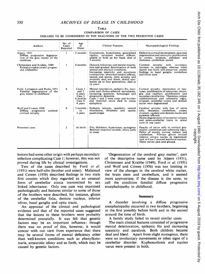
ARCHIVES OF DISEASE IN CHILDHOODTABLE
COMPARISON OF CASESDISEASES TO BE CONSIDERED IN THE DIAGNOSIS OF THE TWO PRESENTED CASES
Authors
Alpers, 1931Diffuse progiessive degenera-tion of the grey matter of thecerebrum
Christensen and Krabbe, 1949Poliodystrophia cerebri progres-sive (infantilis)
Ford, Livingston and Pryles, 1951Familial degeneration of thegrey matter in childhood
Wolf and Cowen, 1956Diffuse progressive cerebralcortical atrophy
Presented cases
No. of AgeCases at
Reported OnsetClinical Features
3 months Convulsions, hyperkinesis, generalizedrigidity, increased tendon reflexes,unable to hold up her head, died at5 months
Neuropathological Findings
Defective cortical development, neuronaldegeneration and astrocytic proliferationin cortex, striatum, pallidum andthalamus, cerebellum spared
8 months Delayed milestones and mental retarda- Cerebral atrophy with secondarytion and gradual deterioration of both increase in microglia, whereas whitemental and physical conditions matter was fairly well preserved, similarIncreasing spasticity and myoclonic findings in basal ganglia, cerebellumcontractions. abnormal tendon reflexes, and brain stemparesis and ataxia, optic atrophy andprobably deaf and dumb, dental ano-malies up to four generations, died at2- years
4
81
8
2
Cases 1 Mental retardation, epileptic fits, myo-and 2 tonic and choreo-athetoid movements,6 years increasing spasticity, hemiplegia andCase 3 progressive dementia
6 months Cases I and 2 were sister and brotherCase 4 and maternal uncle died in status4 years epilepticus
3 months Epileptic seizures, spasticity, mentalto retardation, blindness and spastic
2 years quadriplegia
At birth Fits, blindness, hypertonus, nystagmusBabinski response variable, idiocy earlyin onset
Cortical atrophy, destruction of neu-rones, proliferation of astrocytes, micro-glia and capillary proliferation andsecondary myelin loss. Thalamus, corpussubthalamicum and substantia nigra,striatum, cerebellar cortex and dentatenuclei were degenerated
Cerebral atrophy with loss of nervecells; thalamus, cerebellum, corpusstriatum, caudate and dentate nuclei andputamen affected(Some degeneration of posterior-columnand spino-cerebellar tracts in spinalcord in one case)
Paucity of nerve cells in cortical greymatter, cerebellum and substantia nigra.Pallor of myelin corona radiata andcerebellum. Fibrous gliosis aroundinferior olivary nuclei, in tegmentumand in Case 2 around lentiform nucleus.Optic nerves pale and gliosed.
lesions had some other origin with perhaps secondaryinfection complicating Case 1; however, this was notproved during life by clinical investigations.Two of the cases described by Ford et al.
(1951) were half-sibs (brother and sister). Malamudand Cowen (1958) described findings in two malefirst cousins which they regarded as an unusualform of cerebellar ataxia transmitted by sex
linked inheritance. Only one case was examinedpathologically and features similar to some of thoseof the brothers were described, for instance, gliosisof the cerebellar folia, dentate nucleus, inferiorolives, basal ganglia and optic tracts.An appraisal of the clinical and pathological
evidence and that of the reported cases suggestedthat the lesions in these brothers were probablydetermined prenatally. It was felt that geneticfactors may be an important influence; thoughthere was no proof of this, however, it wouldconcur with our view from experience that theremay be several forms of mental deficiency otherthan well-known conditions such as phenylketo-nuria, amaurotic idiocy and so forth, which may becaused by genetic factors.
'Degeneration of the cerebral grey matter', partof the descriptive name used by Alpers (1931),Christensen and Krabbe (1949), Ford et al. (1951)and Wolf and Cowen (1956) was too limiting inview of the changes in the cerebral white matter,the brain stem and cerebellum, and it seemedmore appropriate, if the disease is the same, tocall the condition familial diffuse progressiveencephalopathy in childhood.
SummaryA disorder involving a diffuse progressive
encephalopathy occurred in two brothers, beginningin the first possibly before birth and in the secondaround the time of birth.A family study failed to reveal similar cases.The main clinical features consisted of progressive
mental deterioration, epileptic fits and increasingspasticity and paralysis. Both children becamedeaf and blind. Apart from slight nystagmus, therewere no involuntary movements or other signs of a
cerebellar disorder. Kyphoscoliosis and equinovarus were present in both.
350
copyright. on 5 A
ugust 2019 by guest. Protected by
http://adc.bmj.com
/A
rch Dis C
hild: first published as 10.1136/adc.35.182.345 on 1 August 1960. D
ownloaded from

FAMILIAL DIFFUSE PROGRESSIVE ENCEPHALOPATHY 351
The principal neuropathological features werepaucity of nerve cells in cortical grey matter, cere-bellum and substantia nigra. There was pallor ofmyelin in the corona radiata and the cerebellum.Fibrous gliosis was present around the inferiorolivary nuclei, in the tegmentum and in one casearound the lentiform nucleus. The optic nerveswere pale and gliosed.
Norms of weight, height and head circumference arequoted from Studies of Child Health and Development,Harvard School of Public Health, in Nelson's 'Textbookof Pediatrics', Philadelphia, 1959.We are indebted to Drs. L. T. Hilliard and L. Crome,
and many other colleagues at the Fountain Hospital fortheir help and advice.
REFERENCES
Alpers, B. J. (1931). Diffuse progressive degeneration of the graymatter of the cerebrum. Arch. Neurol. Psychiat. (Chicago),25, 469.
Christensen, E. and Krabbe, K. H. (1949). Poliodystrophia cerebriprogressiva (infantalis). Ibid., 61, 28.
Duke-Elder, W. S. (1932). Textbook of Ophthalmology, Vol. 1,p. 102. Kimpton, London.
Ford, F. R., Livingston, S. and Pryles, C. V. (1951). Familialdegeneration of the cerebral gray matter in childhood. J.Pediat., 39, 33.
Malamud, N. and Cohen, P. (1958). Unusual form of cerebellarataxia with sex-linked inheritance. Neurology, 8, 261.
Wolf, A. and Cowen, D. (1956). The cerebral atrophies and en-cephalomalacias of infancy and childhood. In Neurology andPsychiatry in Childhood, Res. Publ. Ass. nerv. ment. Dis., Vol. 34,p. 199.
copyright. on 5 A
ugust 2019 by guest. Protected by
http://adc.bmj.com
/A
rch Dis C
hild: first published as 10.1136/adc.35.182.345 on 1 August 1960. D
ownloaded from