fannp 27th national nnp symposium: clinical … review of common... · sos: review of common...
TRANSCRIPT
SOS: Review of Common Neonatal GI Conditions Jacqui Hoffman, DNP, ARNP, NNP-BC NNP Track Coordinator, College of Nursing University of Florida, Gainesville, FL Neonatal Nurse Practitioner Pediatrix Medical Group, Tampa, FL
The speaker has signed a disclosure form and indicated she has no significant financial interest or relationship with the companies or the manufacturer(s) of any commercial product and/or service that will be discussed as part of this presentation.
Session Summary This lecture provides a general overview of common neonatal GI problems to help prepare for certification exams.
Session Objectives Upon completion of this presentation, the participant will be able to:
identify the three parts of the primordial gut and the common structures/organs arising from each;
contrast the difference between the infant with a gastroschisis and the infant with an omphalocele;
discuss clinical presentation, diagnostic evaluation, and management of the infant with a suspected abdominal obstruction.
Test Questions 1. A previously well full-term infant presents with bilious vomiting. What is the first disease
process that the infant should be evaluated for?
a. Pyloric stenosis b. Sepsis c. Malrotation with midgut volvulus
2. Omphalocele and Gastroschisis can best be differentiated by:
a. Assessing involvement of the umbilicus b Identifying the organs exposed by the defect c. Noting the presence of a membranous covering
3. Which of the following gastrointestinal conditions is associated with a high incidence of associated malformation?
a. Gastroschisis b. Omphalocele c. Jejunoileal atresia
B7 FANNP 27th National NNP Symposium: Clinical Update and Review
B7: SOS: Review of Common Neonatal GI Conditions Page 1 of 31
4. A term male neonate at 50 hours of age has abdominal distention and episodes of vomiting. No meconium has been passed since birth except during your physical when a rectal examination is done. An abdominal X-ray is non-specific; a contrast study depicts areas of dilatation and constriction in the sigmoid colon. The most likely diagnosis is:
a. Meconium ileus b. Malrotation with volvulus c. Hirschsprung’s disease
5. Almost all infants pass meconium by:
a. 12 hours of life b. 24 hours of life c. 48 hours of life
6. A 4 week old male infant presented with projectile vomiting of nonbilious emesis. The physical exam reveals a small “olive-shaped” mass in the abdomen. The most likely diagnosis is:
a. Meconium plug b. Pyloric stenosis c. Necrotizing enterocolitis
References Bevacqua, J. (2009). Umbilical hernias in infants and children. The Nurse Practitioner, 34, 12-13.
Casaccia, G., Trucchi, A., Spirydakis, I., Giorlandino, C., et al. (2006). Congenital intestinal anomalies, neonatal short bowel syndrome, and prenatal/neonatal counseling. Journal of Pediatric Surgery, 41, 804-807.
Choi, Y. (2014). Necrotizing enterocolitis in newborns: Update in pathophysiology and newly emerging therapeutic strategies. Korean Journal of Pediatrics, 57(12), 505-13.
Christison-Lagay, E., Kelleher, C., & Langer, J. (2011). Neonatal abdominal wall defects. Seminars in Fetal & Neonatal Medicine, 16(3), 164-72.
Cloherty, J., Eichenwald, E., Hansen, A., & Stark, A. (2012). Manual of neonatal care (7th ed.). Philadelphia, PA: Wolters Kluwer/Lippincott Williams & Wilkins.
De Silva, N., Young, J., & Wales, P. (2006). Understanding neonatal bowel obstruction: Building knowledge to advanced practice. Neonatal Network, 25, 303-318.
Gordon, P., Clark, R., Swanson, J., & Spitzer, A. (2014). Can a national dataset generate a nomogram for necrotizing enterocolitis onset? Journal of Perinatology, 34, 732-35.
Juang, D. & Snyder, C. (2012). Neonatal bowel obstruction. Surgical Clinics of North America, 92(3), 685-711.
Ledbetter, D. (2012). Congenital abdominal wall defects and reconstruction in pediatric surgery: Gastroschisis and omphalocele. Surgical Clinics of North America, 92(3), 713-27.
Martin, C. & Fishman, S. (2003). Gastrointestinal disorders part 1: Gastroschisis. In A. Hansen & M. Puder (Eds., pp. 203-212), Manual of neonatal surgical intensive care. Ontario, Canada: BC Decker, Inc. Hamilton.
Martin, C. & Fishman, S. (2003). Gastrointestinal disorders part 2: Omphalocele. In A. Hansen & M. Puder (Eds., pp. 213-220), Manual of neonatal surgical intensive care. Ontario, Canada: BC Decker, Inc. Hamilton.
Martin, C. & Fishman, S. (2003). Gastrointestinal disorders part 8: Umbilical hernia. In A. Hansen & M. Puder (Eds., pp. 300-302), Manual of neonatal surgical intensive care. Ontario, Canada: BC Decker, Inc. Hamilton.
FANNP 27th National NNP Symposium: Clinical Update and Review
B7: SOS: Review of Common Neonatal GI Conditions Page 2 of 31
Morgan, J., Young, L., & McGuire, W. (2011). Pathogenesis and prevention of NEC. Current Opinion in Infectious Diseases, 24, 183-89.
Neu, J. & Walker, W. (2011). Necrotizing enterocolitis. New England Journal of Medicine, 364(3), 255-264.
Pursley, D., Hansen, A., & Puder M. (2003). Gastrointestinal disorders part 4: Obstruction. In A. Hansen & M. Puder (Eds., pp. 237-259), Manual of neonatal surgical intensive care. Ontario, Canada: BC Decker, Inc. Hamilton.
Sharma, R. & Hudak, M. l. (2013). A clinical perspective of necrotizing enterocolitis: Past, present and future. Clinics in Perinatology, 40(1), 27-51.
Tappero, E. & Witt, C. (2007). Neonatal gastrointestinal surgical conditions. In D. Longobucco & V. Ruth (Eds., pp 141-), Neonatal surgical procedures: A guide for care and management. Santa Rosa, CA: NICU Ink Book Publishers.
Torrazza, R. & Neu, J. (2013). The altered gut microbiome and necrotizing enterocolitis. Clinics in Perinatology, 40, 93-108.
Trotter, C. W. (Ed.) (2012). Abnormalities of the gastrointestinal tract. In Neonatal radiology basics (2nd ed., chapter 4), Neonatal Network.
Veyrac, C., Baud, C., Prodhomme, O., et al. (2012). US assessment of neonatal bowel (necrotizing enterocolitis excluded). Pediatric Radiology, 42(Suppl 1), S107-14.
Vinocur, D., Lee, E. & Eisenberg, R. (2012). Neonatal intestinal obstruction. AJR American Journal of Roentgenology, 198(1), W1-10.
Yee, W., Soraisham, A., Shah, V., et al (2012). Incidence and timing of presentation of necrotizing enterocolitis in preterm infants. Pediatrics, 129, e298-e304.
FANNP 27th National NNP Symposium: Clinical Update and Review
B7: SOS: Review of Common Neonatal GI Conditions Page 3 of 31
SOS: REVIEW OF COMMON GI CONDITIONS
Jacqui Hoffman, DNP, ARNP, NNP‐BCNNP‐DNP Track Coordinator, University of Florida
NNP Pediatrix Medical Group, Tampa
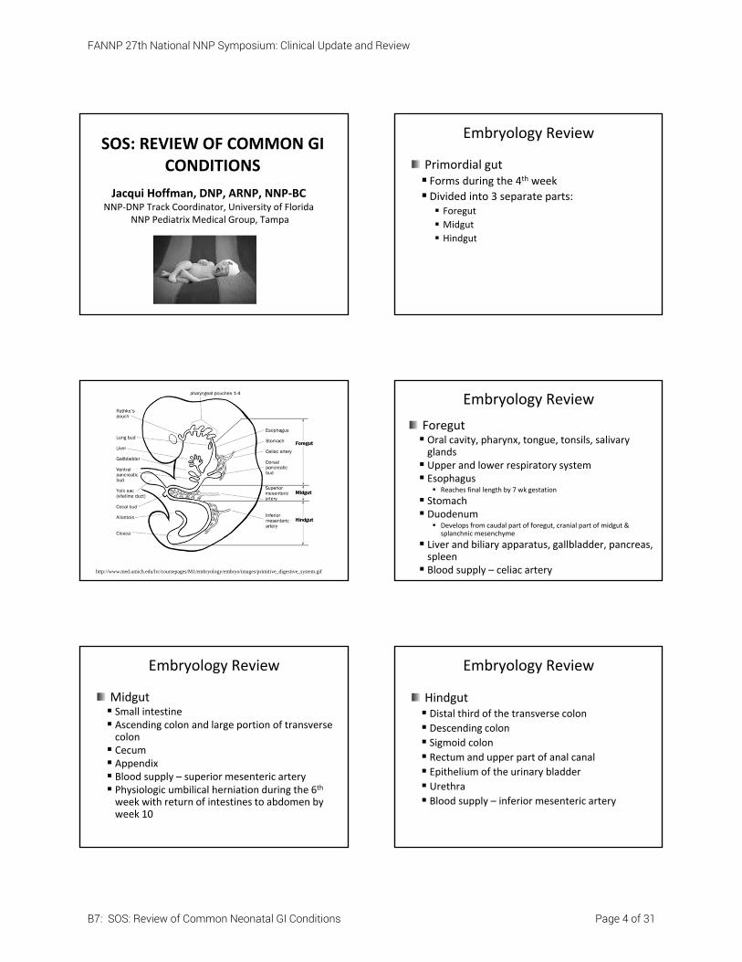
Embryology Review
Primordial gut Forms during the 4th week
Divided into 3 separate parts: Foregut Midgut
Hindgut
http://www.med.umich.edu/lrc/coursepages/M1/embryology/embryo/images/primitive_digestive_system.gif
Embryology Review
Foregut Oral cavity, pharynx, tongue, tonsils, salivary glands Upper and lower respiratory system Esophagus
Reaches final length by 7 wk gestation
Stomach Duodenum
Develops from caudal part of foregut, cranial part of midgut & splanchnic mesenchyme
Liver and biliary apparatus, gallbladder, pancreas, spleen Blood supply – celiac artery
Embryology Review
Midgut Small intestine Ascending colon and large portion of transverse colon Cecum Appendix Blood supply – superior mesenteric artery Physiologic umbilical herniation during the 6th
week with return of intestines to abdomen by week 10
Embryology Review
Hindgut Distal third of the transverse colon Descending colon Sigmoid colon
Rectum and upper part of anal canal
Epithelium of the urinary bladder
Urethra Blood supply – inferior mesenteric artery
FANNP 27th National NNP Symposium: Clinical Update and Review
B7: SOS: Review of Common Neonatal GI Conditions Page 4 of 31
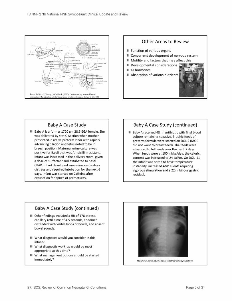
From: de Silva N, Young J, & Wales P. (2006). Understanding neonatal bowel obstruction: Building knowledge to advance practice. Neonatal Network, 25: 304
Other Areas to Review
Function of various organs
Concurrent development of nervous system
Motility and factors that may affect this
Developmental considerations
GI hormones
Absorption of various nutrients
Baby A Case StudyBaby A is a former 1720 gm 28.5 EGA female. She was delivered by stat C‐Section when mother presented in active preterm labor with rapidly advancing dilation and fetus noted to be in breech position. Maternal urine culture was positive for E.coli that was Ampicillin resistant. Infant was intubated in the delivery room, given a dose of surfactant and extubated to nasal CPAP. Infant developed worsening respiratory distress and required intubation for the next 6 days. Infant was started on Caffeine after extubation for apnea of prematurity.
Baby A Case Study (continued)
Baby A received 48 hr antibiotic with final blood culture remaining negative. Trophic feeds of preterm formula were started on DOL 2 (MOB did not want to breast feed). The feeds were advanced to full feeds over the next 7 days. When feeds were at 100 ml/kg/day, the caloric content was increased to 24 cal/oz. On DOL 11 the infant was noted to have temperature instability, increased A&B events requiring vigorous stimulation and a 22ml bilious gastric residual.
Baby A Case Study (continued)
Other findings included a HR of 178 at rest, capillary refill time of 4‐5 seconds, abdomen distended with visible loops of bowel, and absent bowel sounds.
What diagnoses would you consider in this infant?
What diagnostic work‐up would be most appropriate at this time?
What management options should be started immediately? http://www.hawaii.edu/medicine/pediatrics/pemxray/v6c14.html
FANNP 27th National NNP Symposium: Clinical Update and Review
B7: SOS: Review of Common Neonatal GI Conditions Page 5 of 31
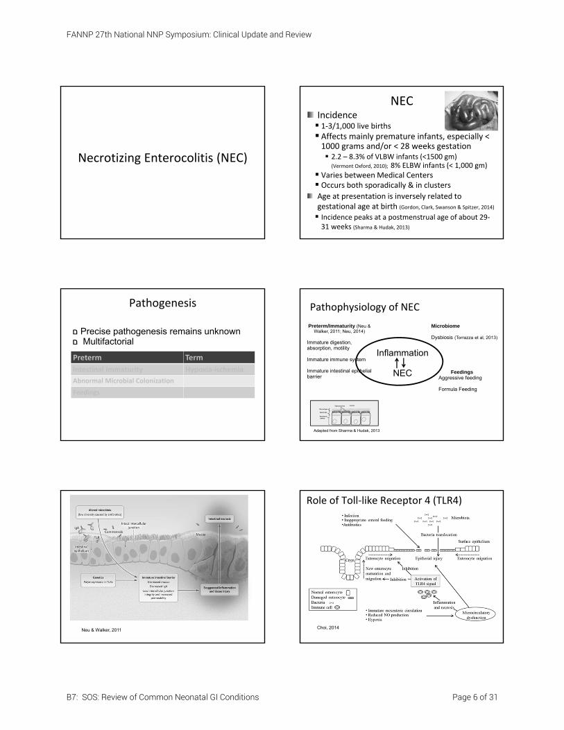
Necrotizing Enterocolitis (NEC)
NECIncidence 1‐3/1,000 live births Affects mainly premature infants, especially < 1000 grams and/or < 28 weeks gestation 2.2 – 8.3% of VLBW infants (<1500 gm)
(Vermont Oxford, 2010); 8% ELBW infants (< 1,000 gm) Varies between Medical Centers Occurs both sporadically & in clustersAge at presentation is inversely related to gestational age at birth (Gordon, Clark, Swanson & Spitzer, 2014) Incidence peaks at a postmenstrual age of about 29‐31 weeks (Sharma & Hudak, 2013)
Pathogenesis
Preterm Term
Intestinal Immaturity Hypoxia‐ischemia
Abnormal Microbial Colonization
Feedings
ם Precise pathogenesis remains unknownם Multifactorial
Pathophysiology of NEC
Preterm/Immaturity (Neu & Walker, 2011; Neu, 2014)
Immature digestion, absorption, motility
Immature immune system
Immature intestinal epithelial barrier
Microbiome
Dysbiosis (Torrazza et al, 2013)
FeedingsAggressive feeding
Formula Feeding
Inflammation
NEC
Adapted from Sharma & Hudak, 2013
Neu & Walker, 2011
Role of Toll‐like Receptor 4 (TLR4)
Choi, 2014
FANNP 27th National NNP Symposium: Clinical Update and Review
B7: SOS: Review of Common Neonatal GI Conditions Page 6 of 31

Clinical Presentation
Varies from non‐specific to a fulminant onset
Gastrointestinal symptoms Abdominal distention
Feeding intolerance Emesis (may or may not be bilious)
Bloody stools Abdominal wall erythema or bluish discoloration
Neu, 2014
Clinical Presentation
Systemic instability mimicking sepsis Increased apnea & bradycardia Worsening of respiratory function Lethargy Poor perfusion, pallor, hypotension, Temperature and/or glucose instability
Differential Diagnosis (DDx)
Mucosal Inflammation NEC, allergic colitis, gastritis/stress ulcer
Infection Systemic infection, infectious gastroenteritis, pseudomembranous colitis
Congenital abnormalities and malformations Intestinal stenosis/atresia, imperforate anus, meconium ileus/plug, Hirschsprungs, Malrotation/volvulus
Vascular accidents Intestinal thromboembolic infarct
Other Intussusception, gastritis/gastric ulcer/perforation, swallowed maternal blood, pneumothorax → pneumoperitoneum
Diagnostic Evaluation
History and physical findings
Laboratory (poor sensitivity/specificity)Neutropenia, left‐shift of neutrophils Thrombocytopenia
Coagulation disturbancesHemolytic anemia
Metabolic acidosis
Glucose instabilityHyponatremia
Diagnostic Evaluation
Radiographic findings Ileus Pneumatosis intestinalis (intramural air)
Dilated loops Thickened bowel wall Pneumoperitoneum
Portal venous gas
FANNP 27th National NNP Symposium: Clinical Update and Review
B7: SOS: Review of Common Neonatal GI Conditions Page 7 of 31
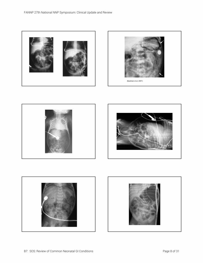
(Epelman et al, 2007)
FANNP 27th National NNP Symposium: Clinical Update and Review
B7: SOS: Review of Common Neonatal GI Conditions Page 8 of 31
Other Diagnostic Modalities
Abdominal Ultrasound Can identify even small volumes of gas
Preferred for visualizing abdominal fluid and ascites
Doppler study can be used to assess arterial perfusion to the bowel wall
Portal venous gas can be detected easier vs x‐ray
www.ultrasound cases.info
Bell Staging Criteria for NEC
Stage I – Suspected NEC Temperature instability, apnea & bradycardia, ↑ gastric residuals, mild abdominal distention, occult blood in stool, normal or mild ileus on X‐ray
Bell Staging Criteria for NEC
Definite NEC – Stage IIStage IIA – Mild NEC Same as Stage I plus prominent abd distention +/‐ tenderness, absent bowel sounds, grossly bloody stools, ileus or dilated bowel loops with focal pneumatosis on X‐rayStage IIB – Moderate NECMild acidosis & thrombocytopenia, abd wall edema & tenderness +/‐ palpable mass, extensive pneumatosis +/‐ portal venous gas and early ascites on X‐ray
Bell Staging Criteria for NEC
Stage IIIA – Advanced NEC Resp & metabolic acidosis, mechanical ventilation, hypotension, oliguria, DIC, worsening wall edema & erythema with induration, prominent ascites with persistent bowel loop but no free air on X‐rayStage IIIB – Advanced NEC Vital sign & laboratory evidence of deterioration, shock, evidence of perforation, and pneumoperitoneum on X‐ray
Medical Management
Surgery consult
Bowel rest (NPO), hyperalimentation
Gastrointestinal decompression
Blood culture & broad‐spectrum antibiotics
Serial abdominal girth measurements
Serial abdominal X‐rays, CBC, coagulation studies, electrolytes & blood gases based on clinical condition
Supportive therapies based on clinical presentation (don’t forget pain management)
Surgical Management
Absolute indications Pneumoperitoneum
Clinical deterioration despite maximal medical treatment
Abdominal mass with persistent intestinal obstruction or sepsis
Development of intestinal stricture
FANNP 27th National NNP Symposium: Clinical Update and Review
B7: SOS: Review of Common Neonatal GI Conditions Page 9 of 31
Surgical Management
Peritoneal drainageAlternative to laparotomy – definitive treatment or temporizing measure
Exploratory laparotomy with resection of diseased bowel, enterostomy & stoma formation
Management Postoperatively
NPO, IVF (central line)
Replogle to low, intermittent suction
Pain management
Broad‐spectrum antibiotics; ?Clindamycin
Ostomy care
Prevention of NECPrevent preterm delivery
Evidence of efficacy and safety Human milk
Feeding protocols using careful advancement
Measures that may be beneficial Trophic feeds safer alternative to NPO Each day NPO, increases risk by 8% (Kirtsman et al, 2015)
Withholding feeds during transfusions or while on Indomethacin
Acidification of formula Restrict use of H2‐blockers
Pre/probiotics
Morbidity and Mortality
20‐30% overall mortalityMedical 21%, surgical (35‐50%) (Hull et al, 2014)
Complications (increase LOS/HC costs) Intestinal strictures (affects up to 40%) Bloody stools, FTT, feeding abnormalities, diarrhea
Feeding intolerance Short bowel syndrome
Parenteral nutrition‐induced cholestasis Neurodevelopmental delay
Spontaneous Ileal Perforation (SIP)
Cause unknown
Occurs more frequently in VLBW & ELBW
Risk Factors Postnatal steroid, Indocin, and vasopressors use
Some studies show association with chorioamnionitis
Most commonly perforation occurs in terminal ileum
FANNP 27th National NNP Symposium: Clinical Update and Review
B7: SOS: Review of Common Neonatal GI Conditions Page 10 of 31
Pathogenesis
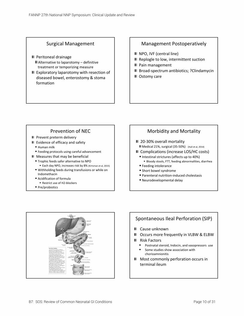
Medications or other exposures lead to mucosal hyperplasia, submucosal thinning and smooth muscle necrosis
These occurrences lead to bowel wall fragility and depletion of endothelial nitric oxide
Gordon, Peds Res, 2009
Spontaneous Ileal Perforation (SIP)
Clinical presentation Sudden onset, typically in the first two
weeks of life
May have few symptoms
Lack of infectious symptoms
Pneumoperitoneum on x‐ray
Spontaneous Ileal Perforation (SIP)
Treatment NPO
Replogle to low, intermittent suction
Surgery consult
Antibiotics
Supportive care
Morbidity and Mortality Decreased mortality and neurodevelopmental
impairment compared to infants with NEC
Baby B Case Study
A 17 y/o G1P0 white female presented to the ER with complaint of abdominal pain. She was diagnosed with active labor and transferred immediately to L&D where she precipitously delivered in the bed. A 3200gm male infant, estimated to be 38 weeks was given Apgar scores of 9/9. Physical exam demonstrated an abdominal wall defect with exposed intestinal contents.
What is your DDx? Is there other information you would like?
Abdominal Wall Defects
Umbilical Hernia
Protrusion of tissue or viscera through the umbilical fascial ring
www.yoursurgery.com
FANNP 27th National NNP Symposium: Clinical Update and Review
B7: SOS: Review of Common Neonatal GI Conditions Page 11 of 31
Umbilical Hernia
IncidenceUnknown Estimated to be 18% in white infants and as high as 42% in black infants
Increased incidence in preterm infants and low birth weight infants
Can be associated with certain syndromes and disease processes (Trisomy 21, congenital hypothyroidism, Beckwith‐Wiedemann syndrome)
Umbilical Hernia
Clinical Presentation Protrusion of the umbilicus especially when crying or straining
Fascial defect is usually < 2 cm in diameter
Redundant umbilical skin
DDx Small omphalocele
Umbilical Hernia
Diagnostic work‐upDiagnosed by physical examManagementMajority spontaneously close if defect is small by 3 years of age
Surgery recommended if hernia persists after 4‐5 years of age Infraumbilical or intraumbilical incision
Hernia sac excised and fascial defect is sutured
Gastroschisis
Abdominal wall defect with herniation of abdominal contents lateral to the umbilical cord
Etiology unknown Vascular accident during embryogenesis
Incidence: 1/4,000 to 1/20,000 births Association with teen pregnancies and low socioeconomic status
Malrotation is almost universal
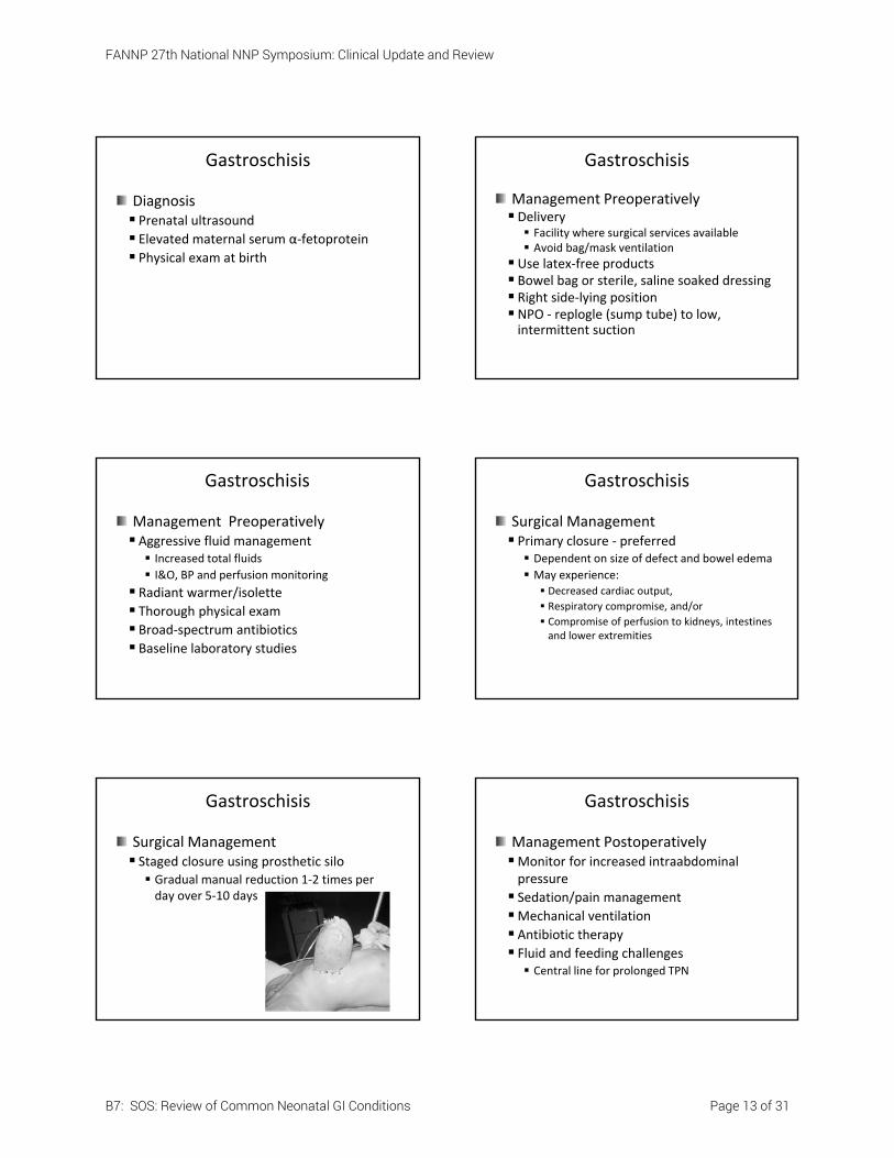
Gastroschisis
Clinical Presentation Herniated bowel that may be edematous or even matted protruding through an abdominal wall defect located lateral to an intact umbilical cord Occasionally liver herniated No peritoneal sac Usually isolated defect without other non‐GI anomalies
Differential diagnosis (DDx) Ruptured omphalocele, cloacal exstrophy
FANNP 27th National NNP Symposium: Clinical Update and Review
B7: SOS: Review of Common Neonatal GI Conditions Page 12 of 31
Gastroschisis
Diagnosis Prenatal ultrasound Elevated maternal serum α‐fetoprotein
Physical exam at birth
Gastroschisis
Management PreoperativelyDelivery Facility where surgical services available Avoid bag/mask ventilation
Use latex‐free products Bowel bag or sterile, saline soaked dressing Right side‐lying positionNPO ‐ replogle (sump tube) to low, intermittent suction
Gastroschisis
Management Preoperatively Aggressive fluid management Increased total fluids I&O, BP and perfusion monitoring
Radiant warmer/isolette
Thorough physical exam Broad‐spectrum antibiotics
Baseline laboratory studies
Gastroschisis
Surgical Management Primary closure ‐ preferred Dependent on size of defect and bowel edema
May experience: Decreased cardiac output, Respiratory compromise, and/or
Compromise of perfusion to kidneys, intestines and lower extremities
Gastroschisis
Surgical Management Staged closure using prosthetic silo Gradual manual reduction 1‐2 times per day over 5‐10 days
Gastroschisis
Management PostoperativelyMonitor for increased intraabdominal pressure
Sedation/pain management
Mechanical ventilation
Antibiotic therapy Fluid and feeding challenges Central line for prolonged TPN
FANNP 27th National NNP Symposium: Clinical Update and Review
B7: SOS: Review of Common Neonatal GI Conditions Page 13 of 31
Gastroschisis
Morbidity and Mortality Sepsis Prolonged ileusNecrotizing enterocolitis (NEC) Complications from prolonged TPN
10% will have intestinal atresiaMortality < 5%
Omphalocele
Abdominal wall defect with herniation of abdominal contents into the umbilical cordEtiology Incomplete return of bowel into abdomen or incomplete closure of anterior abdominal wall
Incidence: 1‐3/10,000 live births 3:1 male‐to‐female predominance; more common in older maternal age 50‐70% will have associated anomalies; Beckwith‐Wiedemann should be considered
Omphalocele
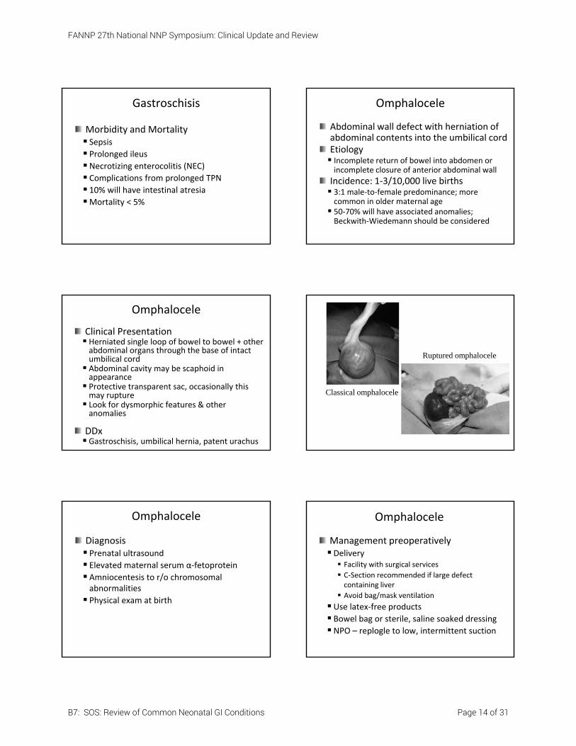
Clinical Presentation Herniated single loop of bowel to bowel + other abdominal organs through the base of intact umbilical cord Abdominal cavity may be scaphoid in appearance Protective transparent sac, occasionally this may rupture Look for dysmorphic features & other anomalies
DDx Gastroschisis, umbilical hernia, patent urachus
Classical omphalocele
Ruptured omphalocele
Omphalocele
Diagnosis Prenatal ultrasound Elevated maternal serum α‐fetoprotein
Amniocentesis to r/o chromosomal abnormalities
Physical exam at birth
Omphalocele
Management preoperativelyDelivery Facility with surgical services C‐Section recommended if large defect containing liver
Avoid bag/mask ventilation
Use latex‐free products Bowel bag or sterile, saline soaked dressingNPO – replogle to low, intermittent suction
FANNP 27th National NNP Symposium: Clinical Update and Review
B7: SOS: Review of Common Neonatal GI Conditions Page 14 of 31
Omphalocele
Management preoperatively Intravenous fluids Radiant warmer/isolette
Thorough physical exam Screening echocardiogram Obtain chromosomes
Consider radiographic evaluation or other evaluation as deemed necessary
Broad‐spectrum antibiotics
Omphalocele
Surgical Management Primary closure Dependent on size of defect and abdominal cavity size
Monitor for cardiac, respiratory, renal and even liver compromise
Staged closure using prosthetic silo Gradual manual reduction 1‐2 times per day over 5‐10 days
Omphalocele
Management postoperativelyMonitor for increased intraabdominal pressure Monitor LFTs
Sedation/pain management
Mechanical ventilation
Antibiotic therapy Fluid and feeding challenges Central line for maximum nutrition
Omphalocele
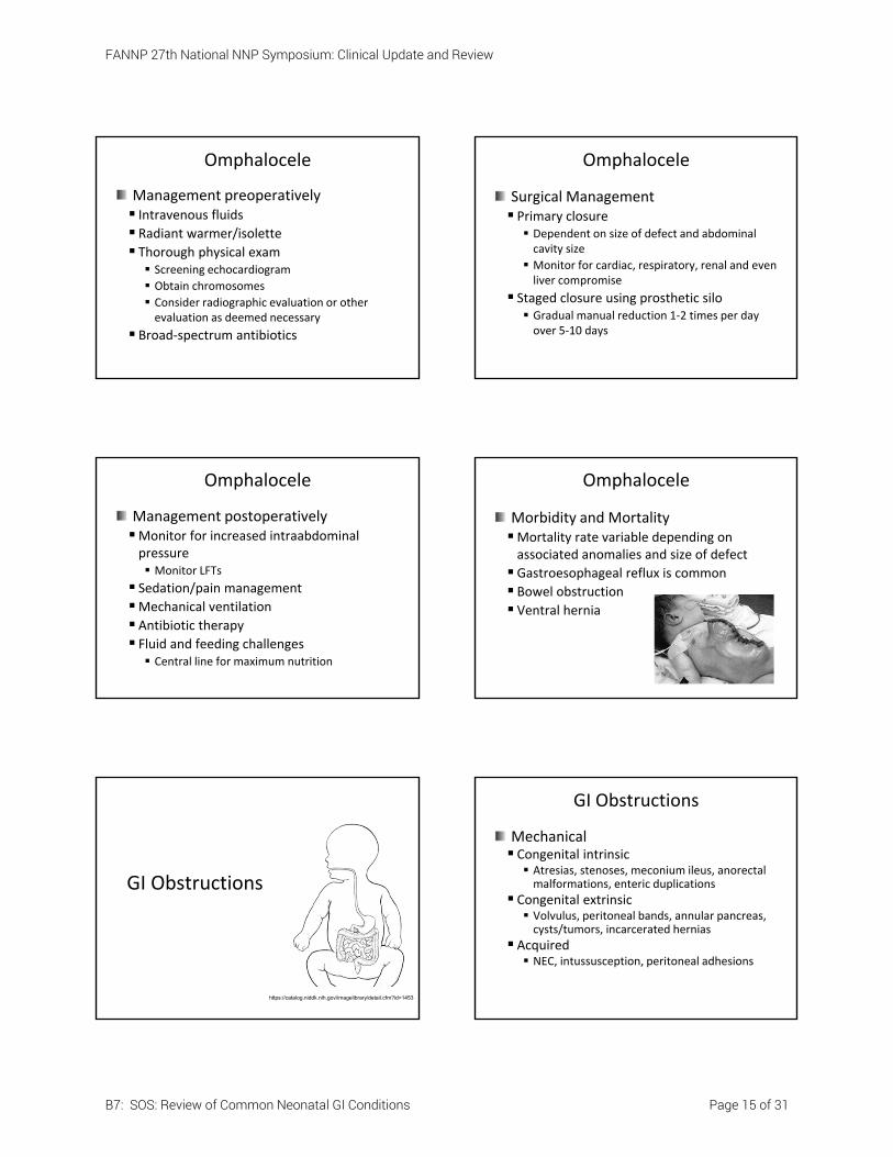
Morbidity and MortalityMortality rate variable depending on associated anomalies and size of defect
Gastroesophageal reflux is common
Bowel obstruction Ventral hernia
GI Obstructions
https://catalog.niddk.nih.gov/imagelibrary/detail.cfm?id=1453
GI Obstructions
Mechanical Congenital intrinsic Atresias, stenoses, meconium ileus, anorectal malformations, enteric duplications
Congenital extrinsic Volvulus, peritoneal bands, annular pancreas, cysts/tumors, incarcerated hernias
Acquired NEC, intussusception, peritoneal adhesions
FANNP 27th National NNP Symposium: Clinical Update and Review
B7: SOS: Review of Common Neonatal GI Conditions Page 15 of 31
GI Obstructions
Functional Intrinsic Hirschsprung disease, meconium plug syndrome, ileus, peritonitis
Extrinsic Intestinal pseudo‐obstruction syndrome
GI Obstruction Pearls
Polyhydramnios More common in proximal obstructions
Abdominal distention More common in distal obstructions (and TEF)
Emesis Bilious ‐more common when obstruction is distal to the ampulla of Vater
Early onset indicates high obstruction; late ‐ low
Normal meconium patterns 94% pass mec by 24 hr of age; 99.8% by 48 hr
GI Obstruction Generalizations
Management Replogle to low, intermittent suction
NPO, IVF Abdominal x‐ray and/or contrast study
Consult pediatric surgeon
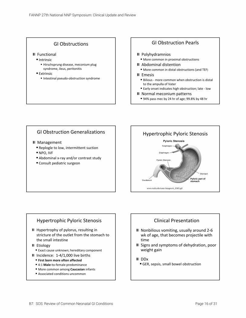
Hypertrophic Pyloric Stenosis
Pyloric part of stomach
www.rush.edu/rumc/images/ei_0365.gif
Hypertrophic Pyloric Stenosis
Hypertrophy of pylorus, resulting in stricture of the outlet from the stomach to the small intestine
Etiology Exact cause unknown; hereditary component
Incidence: 1‐4/1,000 live births First born more often affected
4:1 Male‐to‐female predominance
More common among Caucasian infants
Associated conditions uncommon
Clinical Presentation
Nonbilious vomiting, usually around 2‐6 wk of age, that becomes projectile with timeSigns and symptoms of dehydration, poor weight gain
DDxGER, sepsis, small bowel obstruction
FANNP 27th National NNP Symposium: Clinical Update and Review
B7: SOS: Review of Common Neonatal GI Conditions Page 16 of 31

Diagnostic Evaluation
History and Physical exam
Abdominal ultrasound
Upper GI contrast study“String sign”
“Double track sign”
Preoperative Management
Baseline laboratory studies
Correct electrolyte and acid‐base imbalances
Fluid resuscitation may be necessary
Replogle to low, intermittent suction
Surgical Management
Pyloromyotomy Laparotomy
Laparoscopy
Postoperative Management
Pain management
Fluid and feeding challengesMonitor serum electrolytes, I&O, weight
Feedings started 6‐8hr post‐op Never place gastric tube post‐op
Genetics consult?
Prognosis
Generally, complete recovery with no residual effects
Surgery corrects stenosis and stenosis generally does not reoccur
Persistent vomiting first few days post‐op, resolves quickly
Baby C Case Study
Baby C is a 3675 gram male infant delivered by spontaneous vaginal delivery at 39.1 wk EGA. The mother had breast fed the infant twice with no reported issues. At the next feeding , the mother was back in the operating room postpartum hemorrhage, therefore, the infant was fed a small amount of term formula. The infant was noted to have approximately 20ml bilious emesis. An OGT is placed and while awaiting X‐ray, additional bilious aspirate is noted.
FANNP 27th National NNP Symposium: Clinical Update and Review
B7: SOS: Review of Common Neonatal GI Conditions Page 17 of 31
Baby C Case Study (continued)
What diagnoses would you consider in this infant?
What diagnostic work‐up would be most appropriate at this time?
What management options should be started immediately?
Baby D Case Study
Baby D is a 3150 gram term female infant delivered by scheduled repeat C‐Section. Mother’s past medical and this pregnancy history were unremarkable. The infant has been breast feeding ad lib demand for the past 3 days with good voiding and stooling pattern. Just prior to discharge, the infant was noted to have a small bilious emesis. An abdominal X‐ray was non‐specific and a barium enema was read as the cecum somewhat high‐riding otherwise normal; further correlation with infant’s clinical condition recommended.
Baby D Case Study (continued)
Feedings were resumed and the infant’s discharge was cancelled. The infant remained in the nursery overnight for further observation. The infant continued to breast feed well overnight. The following afternoon just before the pedi came in to do evening discharge rounds, the abdomen was noted to be significantly distended. An OGT was placed and approximately 20ml bilious aspirate was noted. The pedi requested a neonatology consult.
Baby D Case Study (continued)
What diagnoses would you consider in this infant?
What diagnostic work‐up would be most appropriate at this time?
What management options should be started immediately?
Baby E Case Study
Baby E is a 3620 gram 36.5 wk EGA late‐preterm male infant delivered by spontaneous vaginal delivery. Maternal GBS status was not on the chart; mother reported she thought it was negative. SROM x 19 hours with maternal temperature of 100.9 noted just prior to delivery. A screening CBC (nl) and blood culture were drawn on the infant per hospital policy. The infant was breast fed on demand every 1‐3 hours with good latch scores.
Baby E Case Study (continued)
At 28 hours of age, the infant developed increasingly poorer latch scores. Over the next several hours, the infant was noted to have increasingly abdominal distention with the girth up 3 cm. After the last feeding at approximately 37 hours of age, the infant had a large bilious emesis . In reviewing the chart, vital signs have been stable, infant has been voiding, no stool has been documented since birth.
FANNP 27th National NNP Symposium: Clinical Update and Review
B7: SOS: Review of Common Neonatal GI Conditions Page 18 of 31
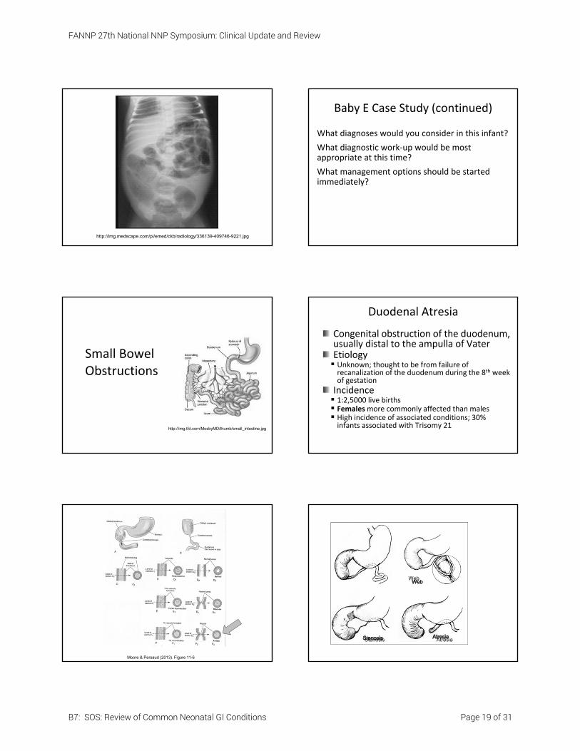
http://img.medscape.com/pi/emed/ckb/radiology/336139-409746-9221.jpg
Baby E Case Study (continued)
What diagnoses would you consider in this infant?
What diagnostic work‐up would be most appropriate at this time?
What management options should be started immediately?
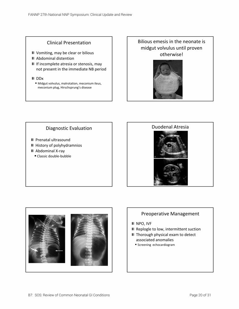
Small Bowel Obstructions
http://img.tfd.com/MosbyMD/thumb/small_intestine.jpg
Duodenal Atresia
Congenital obstruction of the duodenum, usually distal to the ampulla of VaterEtiology Unknown; thought to be from failure of recanalization of the duodenum during the 8th week of gestation
Incidence 1:2,5000 live births Femalesmore commonly affected than males High incidence of associated conditions; 30% infants associated with Trisomy 21
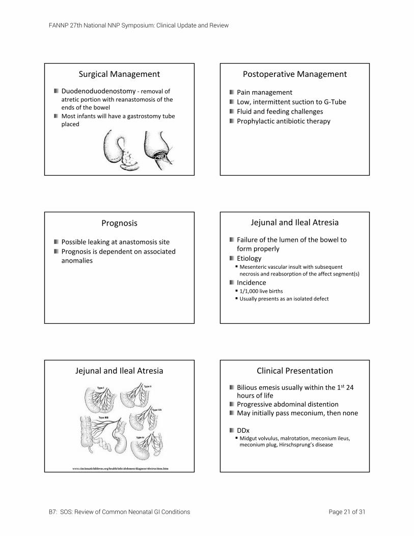
Moore & Persaud (2013). Figure 11-6
AtresiaStenosis
Web
FANNP 27th National NNP Symposium: Clinical Update and Review
B7: SOS: Review of Common Neonatal GI Conditions Page 19 of 31
Clinical Presentation
Vomiting, may be clear or bilious
Abdominal distention
If incomplete atresia or stenosis, may not present in the immediate NB period
DDx Midgut volvulus, malrotation, meconium ileus, meconium plug, Hirschsprung’s disease
Bilious emesis in the neonate is midgut volvulus until proven
otherwise!
Diagnostic Evaluation
Prenatal ultrasound
History of polyhydramnios
Abdominal X‐ray Classic double‐bubble
Duodenal Atresia
Preoperative Management
NPO, IVF
Replogle to low, intermittent suction
Thorough physical exam to detect associated anomalies Screening echocardiogram
FANNP 27th National NNP Symposium: Clinical Update and Review
B7: SOS: Review of Common Neonatal GI Conditions Page 20 of 31
Surgical Management
Duodenoduodenostomy ‐ removal of atretic portion with reanastomosis of the ends of the bowel
Most infants will have a gastrostomy tube placed
Postoperative Management
Pain management
Low, intermittent suction to G‐Tube
Fluid and feeding challenges
Prophylactic antibiotic therapy
Prognosis
Possible leaking at anastomosis site
Prognosis is dependent on associated anomalies
Jejunal and Ileal Atresia
Failure of the lumen of the bowel to form properly
Etiology Mesenteric vascular insult with subsequent necrosis and reabsorption of the affect segment(s)
Incidence 1/1,000 live births Usually presents as an isolated defect
Jejunal and Ileal Atresia
www.cincinnatichildrens.org/health/info/abdomen/diagnose/obstructions.htm
Clinical Presentation
Bilious emesis usually within the 1st 24 hours of lifeProgressive abdominal distentionMay initially pass meconium, then none
DDx Midgut volvulus, malrotation, meconium ileus, meconium plug, Hirschsprung’s disease
FANNP 27th National NNP Symposium: Clinical Update and Review
B7: SOS: Review of Common Neonatal GI Conditions Page 21 of 31
Diagnostic Evaluation
Presence of symptoms
? History of polyhydramnios
Prenatal ultrasound
Abdominal X‐ray Gas or fluid‐filled dilated loops of bowel with scant amounts of gas distal to the obstruction
“Triple‐bubble” ‐ proximal jejunal atresia
Contrast enema
Triple Bubble
Preoperative Management
Replogle to low, intermittent suction
NPO, IVF
Correct any electrolyte imbalances
Surgical Management
Surgical procedure dependent on amount of intestine involved End‐to‐end or end‐to‐oblique‐side anastomosis
Externalization of the proximal and distal ends
Postoperative Management
Pain management
Replogle to low, intermittent suction
Fluid and feeding challenges
Prophylactic antibiotic therapy
Prognosis
Ileus
Peritonitis, if perforation occurred
Short bowel syndrome (SBS)
Strictures or adhesions
Leak at anastomosis site
Decreased survival in neonates with multiple atresias
FANNP 27th National NNP Symposium: Clinical Update and Review
B7: SOS: Review of Common Neonatal GI Conditions Page 22 of 31
Meconium Ileus
Mechanical obstruction of the distal lumen due to meconium
Etiology Unknown; due to hyposecretion of pancreatic enzymes or abnl viscid secretions from the mucous glands of the sm. intestine
Incidence Majority of cases are associated with cystic fibrosis
Clinical Presentation
Abdominal distention with thickened bowel loops often visibleBilious emesisFailure to pass meconiumBowel perforation with peritonitis (will have tenderness and/or erythema)
DDx Meconium plug, small bowel atresia, Hirschsprung’s Disease
Diagnostic Evaluation
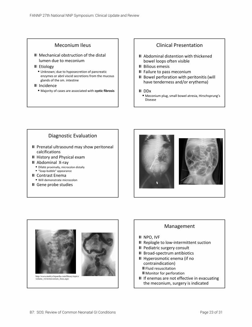
Prenatal ultrasound may show peritoneal calcificationsHistory and Physical examAbdominal X‐ray Dilate proximally, microcolon distally “Soap‐bubble” appearance
Contrast Enema Will demonstrate microcolon
Gene probe studies
http://www.medcyclopaedia.com/library/topics/volume_vii/m/meconium_ileus.aspx
Management
NPO, IVFReplogle to low‐intermittent suctionPediatric surgery consultBroad‐spectrum antibioticsHyperosmotic enema (if no contraindication)Fluid resuscitationMonitor for perforation
If enemas are not effective in evacuating the meconium, surgery is indicated
FANNP 27th National NNP Symposium: Clinical Update and Review
B7: SOS: Review of Common Neonatal GI Conditions Page 23 of 31
Surgical Management
If prenatal/postnatal perforation is present or if enemas were not effective in evacuating the meconium, surgery is indicated
Laparotomy with end‐to‐end anastomosis or creation of stoma
Postoperative Management
Pain management
Replogle to low, intermittent suction
Fluid and feeding challenges Need pancreatic enzymes
Antibiotic therapy
Genetic counseling DNA Sweat chloridePulmonary toilet CPT, aerosols, humidity
Prognosis
Post‐operatively Volvulus, perforation with peritonitis, infection
Long‐term If CF diagnosed, will need careful f/uMorbidity and mortality due to complications of CF
Meconium Plug Syndrome
Mechanical obstruction of the distal colon/rectum due to thick, inspissated meconium
Etiology Unknown
Incidence 1:100 newborns Associated with pre‐eclampsia with Mag therapy, IDM, prematurity, hypothyroidism, hypotonia and sepsis
Clinical Presentation
Failure to pass meconium
Abdominal distention
Visible loops of bowel
Bilious emesis (late finding)
DDx Meconium ileus, Hirschsprung’s Disease
Diagnostic Evaluation
DiagnosisHistory and Physical exam Abdominal X‐ray Dilated loops of bowel and few air fluid levels
Water‐soluble contrast enema Diagnostic and therapeutic
FANNP 27th National NNP Symposium: Clinical Update and Review
B7: SOS: Review of Common Neonatal GI Conditions Page 24 of 31
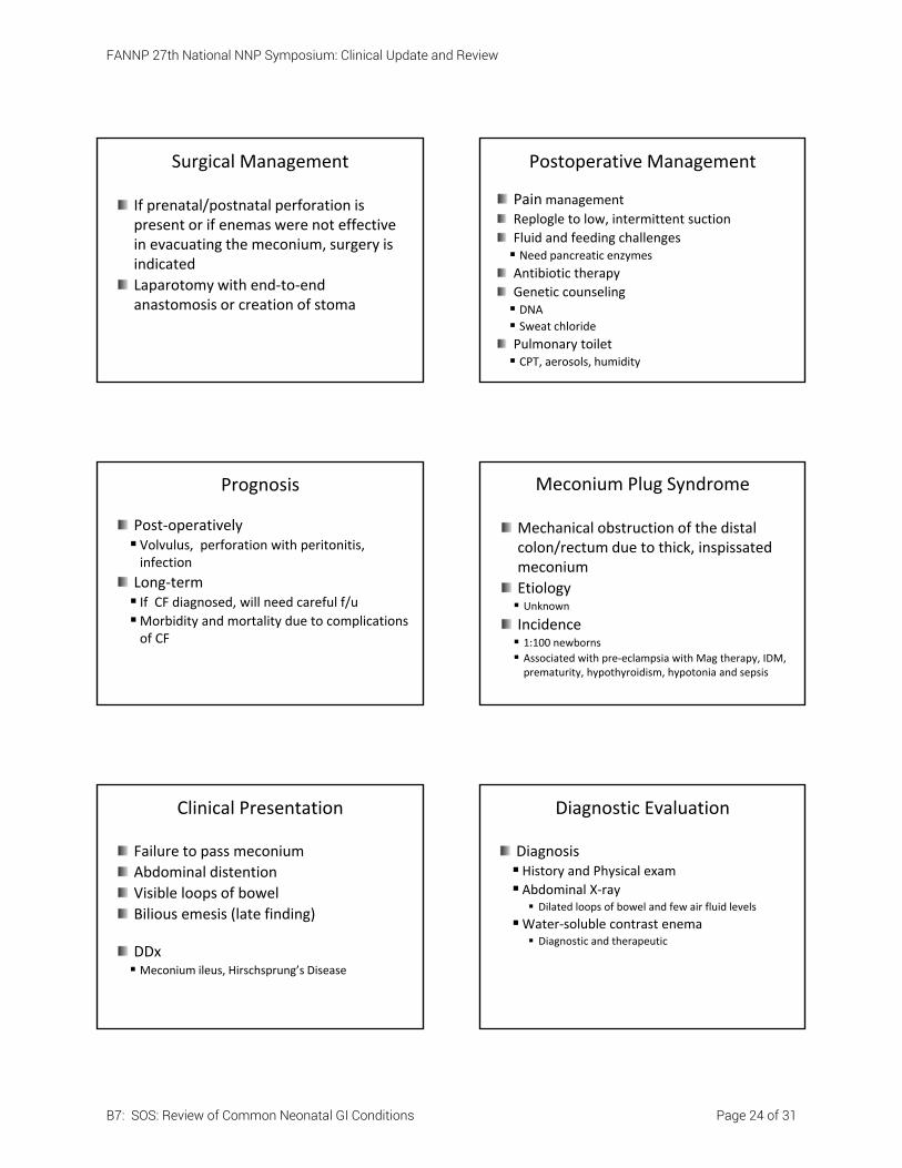
http://www.medcyclopaedia.com/library/topics/volume_vii/m/meconium_plug_syndrome.aspx
Management
NPO, IVF
Replogle to low‐intermittent suction
? digital rectal exam
Contrast enema
Further work‐up?
Malrotation/Volvulus
Abnormal rotation and fixation of intestines during 7th to 12th week of gestationEtiology Unknown; occurs when the intestines do not rotate and/or the mesentery does not fixate during embryologic development
Incidence 1:6,000 live births Associated with abdominal wall defects, intestinal atresia, imperforate anus, cardiac anomalies, and Trisomy 21
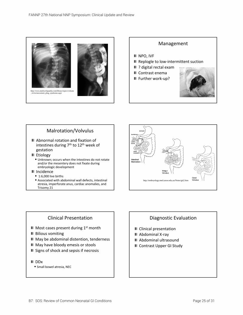
http://embryology.med.unsw.edu.au/Notes/git2.htm
Clinical Presentation
Most cases present during 1st month
Bilious vomiting
May be abdominal distention, tenderness
May have bloody emesis or stools
Signs of shock and sepsis if necrosis
DDx Small bowel atresia, NEC
Diagnostic Evaluation
Clinical presentation
Abdominal X‐ray
Abdominal ultrasound
Contrast Upper GI Study
FANNP 27th National NNP Symposium: Clinical Update and Review
B7: SOS: Review of Common Neonatal GI Conditions Page 25 of 31
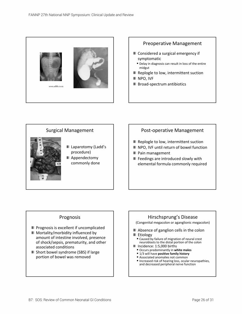
www.adhb.co.nz
Preoperative Management
Considered a surgical emergency if symptomatic Delay in diagnosis can result in loss of the entire midgut
Replogle to low, intermittent suction
NPO, IVF
Broad‐spectrum antibiotics
Surgical Management
Laparotomy (Ladd’s procedure)
Appendectomy commonly done
Post‐operative Management
Replogle to low, intermittent suction
NPO, IVF until return of bowel function
Pain management
Feedings are introduced slowly with elemental formula commonly required
Prognosis
Prognosis is excellent if uncomplicatedMortality/morbidity influenced by amount of intestine involved, presence of shock/sepsis, prematurity, and other associated conditionsShort bowel syndrome (SBS) if large portion of bowel was removed
Hirschsprung’s Disease(Congenital megacolon or aganglionic megacolon)
Absence of ganglion cells in the colonEtiology Caused by failure of migration of neural crest neuroblasts to the distal portion of the colonIncidence: 1:5,000 births Occurs predominantly in white males 1/3 will have positive family history Associated anomalies not common Increased risk of hearing loss, ocular neuropathies, and decreased peripheral nerve function
FANNP 27th National NNP Symposium: Clinical Update and Review
B7: SOS: Review of Common Neonatal GI Conditions Page 26 of 31
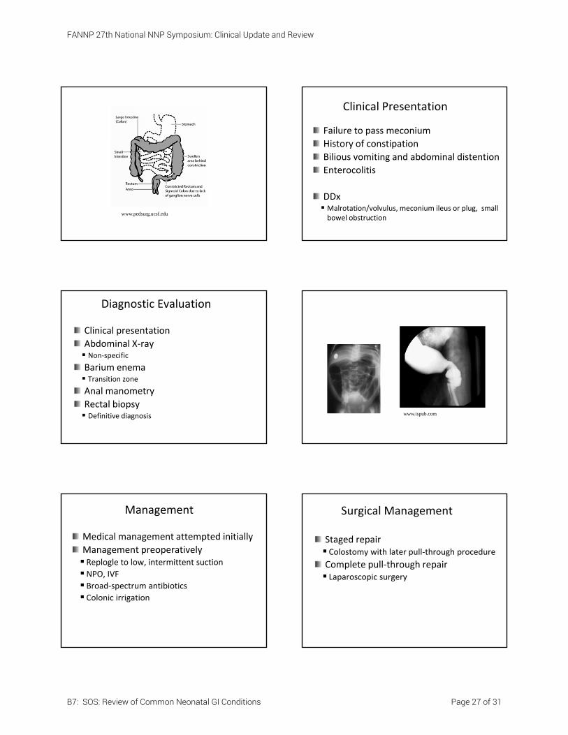
www.pedsurg.ucsf.edu
Clinical Presentation
Failure to pass meconium
History of constipation
Bilious vomiting and abdominal distention
Enterocolitis
DDx Malrotation/volvulus, meconium ileus or plug, small bowel obstruction
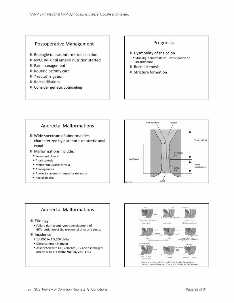
Diagnostic Evaluation
Clinical presentation
Abdominal X‐ray Non‐specific
Barium enema Transition zone
Anal manometry
Rectal biopsy Definitive diagnosis www.ispub.com
Management
Medical management attempted initially
Management preoperatively Replogle to low, intermittent suction
NPO, IVF Broad‐spectrum antibiotics
Colonic irrigation
Surgical Management
Staged repair Colostomy with later pull‐through procedure
Complete pull‐through repair Laparoscopic surgery
FANNP 27th National NNP Symposium: Clinical Update and Review
B7: SOS: Review of Common Neonatal GI Conditions Page 27 of 31
Postoperative Management
Replogle to low, intermittent suction
NPO, IVF until enteral nutrition started
Pain management
Routine ostomy care
? rectal irrigation
Rectal dilations
Consider genetic counseling
Prognosis
Dysmotility of the colon Stooling abnormalities – constipation or incontinence
Rectal stenosis
Stricture formation
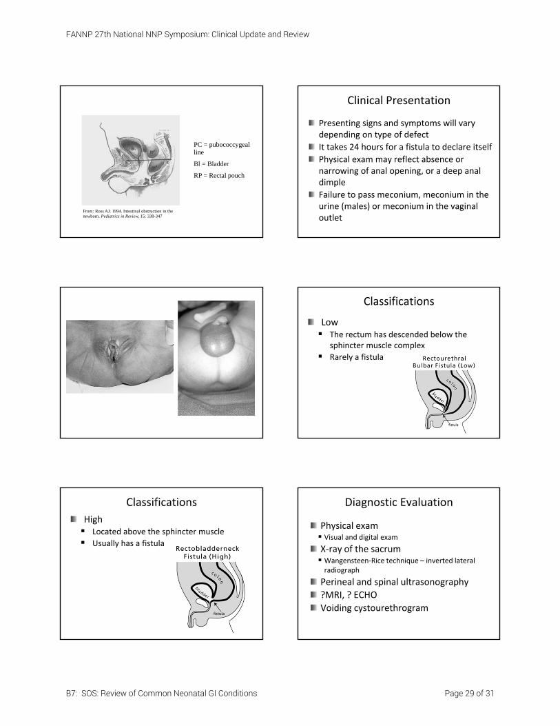
Anorectal Malformations
Wide spectrum of abnormalities characterized by a stenotic or atretic anal canal
Malformations include: Persistent cloaca Anal stenosis Membranous anal atresia
Anal agenesis Anorectal agenesis (imperforate anus)
Rectal atresia
Anal canal
Anus
Anal columns Rectum
From hindgut
Pectinateline
White line
From proctodeum
Slide 12.44
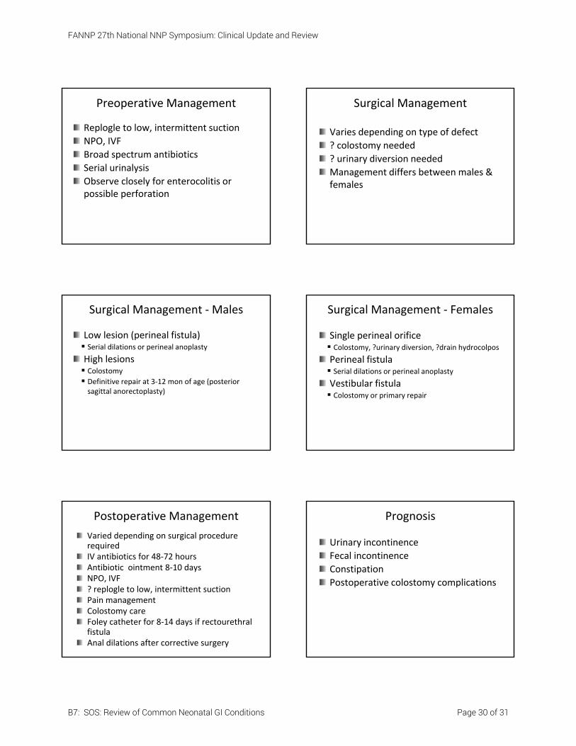
Anorectal Malformations
Etiology Failure during embryonic development of differentiation of the urogenital sinus and cloaca
Incidence 1:4,000 to 1:5,000 births More common in males
Associated with GU, vertebral, CV and esophageal atresia with TEF (think VATER/VACTERL)
Adapted from: Moore KL & Persaud T. 1998. The Developing Human: Clinically Oriented Embryology, 6th ed, p. 299. Philadelphia: WB Saunders.
Persistent cloaca Anal Stenosis Membranous anal atresia
Anal agenesis with a perineal fistula Anorectal agenesis + rectovaginal fistula
Anorectal agenesis + rectourethral fistula
Rectal atresia
FANNP 27th National NNP Symposium: Clinical Update and Review
B7: SOS: Review of Common Neonatal GI Conditions Page 28 of 31
From: Ross AJ. 1994. Intestinal obstruction in the newborn. Pediatrics in Review, 15: 338-347
PC = pubococcygeal line
Bl = Bladder
RP = Rectal pouch
Clinical Presentation
Presenting signs and symptoms will vary depending on type of defect
It takes 24 hours for a fistula to declare itself
Physical exam may reflect absence or narrowing of anal opening, or a deep anal dimple
Failure to pass meconium, meconium in the urine (males) or meconium in the vaginal outlet
Classifications
Low The rectum has descended below the
sphincter muscle complex
Rarely a fistula
Classifications
High Located above the sphincter muscle
Usually has a fistula
Diagnostic Evaluation
Physical exam Visual and digital exam
X‐ray of the sacrum Wangensteen‐Rice technique – inverted lateral radiograph
Perineal and spinal ultrasonography
?MRI, ? ECHO
Voiding cystourethrogram
FANNP 27th National NNP Symposium: Clinical Update and Review
B7: SOS: Review of Common Neonatal GI Conditions Page 29 of 31
Preoperative Management
Replogle to low, intermittent suction
NPO, IVF
Broad spectrum antibiotics
Serial urinalysis
Observe closely for enterocolitis or possible perforation
Surgical Management
Varies depending on type of defect
? colostomy needed
? urinary diversion needed
Management differs between males & females
Surgical Management ‐Males
Low lesion (perineal fistula) Serial dilations or perineal anoplasty
High lesions Colostomy
Definitive repair at 3‐12 mon of age (posterior sagittal anorectoplasty)
Surgical Management ‐ Females
Single perineal orifice Colostomy, ?urinary diversion, ?drain hydrocolpos
Perineal fistula Serial dilations or perineal anoplasty
Vestibular fistula Colostomy or primary repair
Postoperative Management
Varied depending on surgical procedure requiredIV antibiotics for 48‐72 hoursAntibiotic ointment 8‐10 daysNPO, IVF? replogle to low, intermittent suctionPain managementColostomy careFoley catheter for 8‐14 days if rectourethral fistulaAnal dilations after corrective surgery
Prognosis
Urinary incontinence
Fecal incontinence
Constipation
Postoperative colostomy complications
FANNP 27th National NNP Symposium: Clinical Update and Review
B7: SOS: Review of Common Neonatal GI Conditions Page 30 of 31
Other Miscellaneous Conditions to Review
Small left colon syndrome
Inguinal hernia
Annular pancreas
GI bleeding
Malabsorption/maldigestion
Gastroesophageal reflux
Short bowel syndrome
Biliary atresia
Cleft lip/palate
Questions?
FANNP 27th National NNP Symposium: Clinical Update and Review
B7: SOS: Review of Common Neonatal GI Conditions Page 31 of 31