faraday discussions mazza,ab avnish patel,a ramon pons,c cyrill bussyab and kostas kostarelos*ab...
TRANSCRIPT

Faraday DiscussionsCite this: Faraday Discuss., 2013, 166, 181
PAPER
Publ
ishe
d on
17
July
201
3. D
ownl
oade
d by
The
Uni
vers
ity o
f M
anch
este
r L
ibra
ry o
n 28
/01/
2014
12:
44:4
5.
View Article OnlineView Journal | View Issue
Peptide nanofibres as molecular transporters:from self-assembly to in vivo degradation
Mariarosa Mazza,ab Avnish Patel,a Ramon Pons,c Cyrill Bussyab
and Kostas Kostarelos*ab
Received 17th June 2013, Accepted 15th July 2013
DOI: 10.1039/c3fd00100h
Peptide nanofibres (PNFs) have gained increasing interest as engineered biomaterials for
drug delivery and tissue repair because of the versatility in design they offer through the
self-assembly of amphiphilic peptide molecules. Their self-assembly is governed by
hydrophobic interactions and hydrogen bonds between peptide sequences able to
form b-sheets. In this report, we describe the self-assembly of PNFs by using palmitoyl-
peptide molecules containing two different cationic amino acid sequences and offer a
description of the nanofiber physicochemical characteristics. The structural degradation
of these PNFs in physiologically-relevant media was evaluated experimentally and two
mechanisms are proposed. We also piloted the tracking of PNFs intracellularly in vitro,
upon interaction with primary neuronal cultures, and intracranially in vivo, after
stereotactic administration deep within the brain using two types of fluorescent
labelled PNFs. Overall, the self-assembled PNFs were seen to internalise within neurons
and be removed or degrade in the brain. Further work is needed to determine the
utility of such PNFs as molecular transporters within neuronal tissue.
Introduction
Peptide nanobres (PNFs) are high axial ratio nanosized structures mimickingthe size and shape of various naturally occurring nanoparticulate objects, i.e.viral capsids, actin cytoskeletons, and amyloid brils. PNFs can be formedby the self-assembly of peptides and peptide amphiphiles as self-assembly isa thermodynamically favourable event in aqueous environments. PNFsare versatile supramolecular architectures with potential as moleculartransporters. Their building blocks can be designed to possess differentfunctionalities resulting in chemical and physical properties tailored tospecic needs.
aUCL School of Pharmacy, University College London, Brunswick Square, London WC1N 1AX, UK. E-mail:
[email protected] of Medicine & National Graphene Institute, Faculty of Medical & Human Sciences, The University of
Manchester, AV Hill Building, Manchester M13 9NT, UKcInstitut de Quımica Avançada de Catalunya, IQAC-CSIC, Jordi Girona 18–26, 08034 Barcelona, Spain
This journal is ª The Royal Society of Chemistry 2013 Faraday Discuss., 2013, 166, 181–194 | 181
Faraday Discussions PaperPu
blis
hed
on 1
7 Ju
ly 2
013.
Dow
nloa
ded
by T
he U
nive
rsity
of
Man
ches
ter
Lib
rary
on
28/0
1/20
14 1
2:44
:45.
View Article Online
The rational design of PNFs has to take into account several parameters thatwill inuence the formation of high axial ratio nanostructures. Stupp and co-workers have designed many peptide amphiphiles having a palmitic moietyattached to the N-terminus of the peptide sequence conferring a surfactant-likearchitecture to the molecules. The same strategy has been adopted in this workenabling peptide self-assembly into nanobres. Similar nanobres have found awide range of applications as biomaterials for tissue engineering.1,2 Zhang and co-workers have developed peptide amphiphiles that are made of amino acids byalternating alanine residues in such a way that they pack with inter-digitatedhydrophobic interactions in a similar fashion as occurs for alanine sequences insilk broin.3,4 Other classes of peptide amphiphiles have been synthesized byvarying the length of the alkyl chain.5–7
At the molecular level, parameters that need ne tuning in order to achievethe formation of PNFs include the hydrophobic domain, the hydrophilic blockand a linker region of b-sheet forming peptides. The hydrophobic block can berepresented by hydrophobic amino acids or alternatively a hydrophobic tail,oen being represented by long acyl chains. The hydrophilic block is asequence of basic or/and acid amino acids, oen embedded within a biolog-ically active peptide sequence. The linker region of b-sheet forming peptidescontributes to the formation of elongated structures versus spherical micellesthat most surfactant would form by self-assembling. Furthermore, the pres-ence of aromatic side chain amino acids can determine the formation ofcylindrical, ribbon like, twisted single or multiple bres via the formationof p–p stacking. All of these parameters can give rise to a combination offactors that will inuence the physicochemical characteristics of the resultingbres as well as the interactions between nanobers and other biologicalconstituents.
In this work we report the formation of PNFs by two peptide molecules withemphasis on the structural disassembly of the nanobres in physiological envi-ronments, as a result of the interaction between nanobers and enzymes withpossible degradation activities. We observed the self-assembly and characterizedthe PNFs formed. Their internalization within primary neuron cultures wasdetermined by uorescence microscopy. The in vivo localisation and structuralstability of the PNFs were investigated following stereotactic administration in thebrain parenchyma non-invasively using an IVIS camera.
Materials and methodsPreparation of PNFs
Peptides (palmitoyl-GGGAAAR and palmitoyl-GGGAAAKRK) were custom-made by Peptide Synthetics (Cambridge, UK) as freeze-dried powders at 95%purity. PNFs were prepared from a dispersion of 1 mg mL�1 peptide amphi-phile in 5% dextrose (pH ¼ 6.4) ltered through a 0.22 micron Milliporemembrane lter. The nal pH of the samples was 3.9–4.0, as both K and R arebasic residues (R pKa ¼ 12; K pKa ¼ 10.4)8 and thus bear a positive charge onthe ionisable groups of the side chain. The dispersions were subject to 5 min ofprobe sonication at 20% of the maximum amplitude alternated with pulsedsonication of 20 s each.
182 | Faraday Discuss., 2013, 166, 181–194 This journal is ª The Royal Society of Chemistry 2013

Paper Faraday DiscussionsPu
blis
hed
on 1
7 Ju
ly 2
013.
Dow
nloa
ded
by T
he U
nive
rsity
of
Man
ches
ter
Lib
rary
on
28/0
1/20
14 1
2:44
:45.
View Article Online
Preparation of uorescent labelled PNFs
PNF conjugation with uorescent probe VivoTag 680. VivoTag 680 XL (absor-bance at 688 � 5 nm; Perkin Elmer) was reconstituted with 1 mL DMSO. A 1 mgmL�1 suspension was made using the freeze-dried peptide and phosphate buff-ered saline. This suspension was transferred into an amber Eppendorf tube toprotect it from light. 50 mL of 1 M sodium bicarbonate and 2 mL of VivoTag 680 XLin DMSO were added. The tubes were maintained under constant agitation in aTissuelyser (Qiagen) and set to oscillate 15 times per second for 2 h. Thesuspension was then centrifuged at 12 000 rpm for 4 min. The supernatant whichcontained unreacted dye was removed and discarded. The pellet was resuspendedin 500 mL of distilled water which was lter sterilised by a 0.22 mm celluloseacetate syringe lter. The tubes were then centrifuged at 12 000 rpm for 4 minaer which the supernatant was removed along with any remaining unreacteddye. The pellet was nally resuspended in lter sterilised dextrose 5% (w/v) givinga nal concentration of 1 mg mL�1. The prepared samples were then bath soni-cated for 5 min using the pulse function, 15 s on and 5 s off. The samples werestored at 4 �C until needed.
Labelling PNFs non-covalently with Nile Red. An ethanol solution of Nile Red(1 mg mL�1) was added dropwise to PNFs (1 mg mL�1) in 5% dextrose during theprobe sonication step to obtain a nal v/v ratio of 1 : 9. The excess ethanol waseliminated by solvent evaporation while stirring the suspension overnight.
Transmission electron microscopy
TEM was performed using a Phillips Biotwin CM 210 electron microscope. 1%uranil acetate was used to counterstain the PNFs. For the degradation experi-ments 90 mL of either MEM, 0.05% trypsin–EDTA or rat plasma–PBS (1 : 1) wasvortex mixed with 10 mL of PNFs in 5% dextrose. The samples were placed in ashaking incubator at 37 �C. Each sample was imaged at the following time points:0, 0.5, 24, 48 h and aer two weeks of incubation. The samples were prepared bytransferring 10 mL onto a 300 grid copper mesh, and the drop was allowed toadsorb on top of the mesh for 10 s, aer which the excess was wicked off using theedge of a lter disc. Uranyl acetate 1% (w/v) was used as a negative contrast agent,and was applied to the mesh, allowed to stand for approximately 5 s and thenremoved using the edge of a lter disc. The copper grid was then loaded forimaging. PNF lengths were measured for each time point using Image J toperform the semiquantitative image analysis.
Small angle X-ray scattering
Small-angle X-ray scattering (SAXS) measurements were carried out using aS3-MICRO (Hecus X-ray systems GMBH Graz, Austria) coupled to a GENIX-Fox 3DX-ray source (Xenocs, Grenoble), which provided a detector focused X-ray beamwith a l ¼ 0.1542 nm Cu Ka-line with more than 97% purity and less than 0.3%Ka. Scattering of the transmission was detected using a PSD 50 Hecus. Temper-ature was controlled by means of a Peltier TCCS-3 Hecus. The samples wereinserted in a ow-through glass capillary 1 mm diameter with 20 mm wall thick-ness. The SAXS scattering curves are shown as a function of the scattering vectormodulus,
This journal is ª The Royal Society of Chemistry 2013 Faraday Discuss., 2013, 166, 181–194 | 183
Faraday Discussions PaperPu
blis
hed
on 1
7 Ju
ly 2
013.
Dow
nloa
ded
by T
he U
nive
rsity
of
Man
ches
ter
Lib
rary
on
28/0
1/20
14 1
2:44
:45.
View Article Online
q ¼ 4p
l$sin
q
2(eq. 1)
where q is the scattering angle. The q values with our setup ranged from0.08 nm�1 to 6.0 nm�1. The system scattering vector was calibrated by measuringa standard silver behenate sample. Because of the use of a detector focused smallbeam (300 � 400 mm full width at half maximum) the scattering curves weremainly smeared by the detector width. This mainly produced a widening of thepeaks without a noticeable effect on the peak position. The scattering curves forliquid samples have been background subtracted and put onto an absolute scaleby comparison with the scattering of a water sample.9,10
The instrumentally smeared experimental SAXS curves were tted to numeri-cally smeared models for the beam size and detector width effects. A least squaresroutine based on the Levenberg–Marquardt scheme was used. Spherical andcylindrical models were tted using two shell models.11,12
Degradation in plasma measured using UV-vis
Fluorescently labelled KRK PNFs were incubated in rat plasma–PBS (1 : 1), incu-bated at 37 �C under constant agitation in a water bath. UV absorbance measuredusing a Cary Bio 50 at different time points (0, 30 min, 1 h, 3 h, 6 h, 24 h, 2, 4, 7and 10 days). The UV-vis spectrophotometer was set to measure the absorbance oflight between 500 and 1000 nm, with data captured at 5 nm intervals. A blank wasused to set a baseline before measuring the samples. Aer time 0 all samples,including blanks, were placed into a shaking water bath at 37 �C. All measure-ments were performed in a black 6Q quartz cuvette (Starna Scientic Ltd) with apath length of 1 cm. KRK PNFs were quantied by a mean calibration curve. Allsamples were measured in triplicate.
Cell uptake of PNFs
Primary neuronal cell cultures. Cell culture reagents (PBS, HBSS, trypsin, foetalbovine serum, DMEM:F12, neurobasal, B27, antibiotics) were purchased fromGibco (Life Technologies, UK). Neuron enriched cell cultures were prepared withhippocampus extracted from E16–E18 Wistar foetal rat brains (standard Witschistages 33–34). Hippocampal tissue pieces were dissociated to single cell suspen-sions by trypsinization followed by mechanical trituration in Ca2+/Mg2+ free HBSSsolution. Aer determination of the number of live cells, 50 000 cells per well wereplated onto a poly-D-lysine (50 mg ml�1) coated glass bottom 8 well chamber slide(Millicell EZ SLIDE, Merck-Millipore) with DMEM:F12 medium completed with12% heat inactivated foetal bovine serum and incubated at 37 �C in a humidied5% CO2 incubator. Aer 12 h, the medium was changed to serum free NeurobasalMedium supplemented with B27, glutamine (0.5 mM), penicillin (100 U), andstreptomycin (100 mg). B27 supplement was used as a means to reduce glial cellsgrowth and to obtain a nearly pure neuronal cell culture. Cultures were incubatedin a humidied 37 �C/5% CO2 incubator for 10 days before experimentation. Halfof the medium was changed every 3 days. Primary neurons were exposed to thePNF–Nile Red formulations for 24 hours and the uorescence was recorded usinga Zeiss Axiovision Fluorescence Microscope.
184 | Faraday Discuss., 2013, 166, 181–194 This journal is ª The Royal Society of Chemistry 2013

Paper Faraday DiscussionsPu
blis
hed
on 1
7 Ju
ly 2
013.
Dow
nloa
ded
by T
he U
nive
rsity
of
Man
ches
ter
Lib
rary
on
28/0
1/20
14 1
2:44
:45.
View Article Online
Intracranial injection in mice
All experiments were performed in accordance with the approved recommenda-tions and policies of the UK Home Office (Animal Scientic Procedures Act 1986,UK). Athymic nude mice (8 weeks old, Harlan, UK) were used for the in vivoexperiments. Anaesthesia was induced by inhalation of isourane. 1 mL of uo-rescently labelled KRK PNFs (1 mg mL�1) was injected intracranially using astereotactic frame. The stereotactic coordinates were: 0.1 mm posterior to thebregma, 2.3 mm to the right of the midline and 3 mm below the surface of thebrain to target the caudate–putamen area of the brain. During surgical proce-dures, the animals were oxygenated and heated to ensure a 37 �C rectal temper-ature. Aer recovery, the animals were returned to their cages.
IVIS imaging
The uorescence signal from the KRK PNFs labelled with VivoTag 680 XL in thebrain was measured using an IVIS Lumina uorescent imager (Caliper LifeSciences) with excitation at 675 nm and emission lter sets 700–840 nm.
Results and discussion
PNFs form as a result of the self-assembly of amphiphilic peptides. Although self-assembly is a thermodynamically favourable process, the time for the formationof these supramolecular structures has been shown to vary, from days to weeks forsimilar designer peptides and the nanobril formation can take days or weeks toget to completion. It was recently reported that upon dissolution in ultrapurewater of a hexapeptide amphiphile bearing aromatic side chain groups, shorttwisted bilayer ribbon segments were observed to be the dominant morphologyaer only 30 s, which then elongate on the time scale of minutes, and nallytransform into helical ribbons over the course of weeks.13,14 Under the sameconditions, a similar peptide lacking aromatic side chains was shown to formcylindrical nanoparticles and not undergo any transition over a period of weeks.14
Thus self-assembly may not only occur over a relatively long time, resulting innanobre formation, but there may also be a transition through metastablenanostructures before maturation into the equilibrated morphology. For suchreasons, here we used a fast bottom approach to self-assembly for the preparationof PNFs. By employing probe sonication we reduced to a few minutes the timeneeded for the self-assembly process to take place. As previously reported byMazza et al.,15 this strategy can be employed to prepare an aqueous dispersion ofshort nanobres below one micron (length range). PNFs were prepared by usingtwo designer peptide amphiphiles in isotonic media (5% dextrose) at a concen-tration of 1 mg mL�1 and as a result of sonication we could observe by TEM theformation of cylindrical nanobres as depicted in Fig.1A, C and D, F.
For both peptides under investigation in the present work we chose a palmitoylchain as the hydrophobic block. Peptide and protein palmitoylation has beenpreviously exploited to synthesize lipid derivatives16 intended to increasemembrane permeation. Unmodied peptides and proteins are generally poorlytransported across biological membranes due to their hydrophilic nature, theirhigh molecular weight and their instability to enzymatic degradation. Such pal-mitoylation strategies have been employed to increase the biological half-life of
This journal is ª The Royal Society of Chemistry 2013 Faraday Discuss., 2013, 166, 181–194 | 185

Fig. 1 (A) Molecular structure of peptide amphiphiles KRK. (B) SAXS pattern for KRK PNFs with the bestfit of the core–shell cylindrical model. The error bars of the points at the right hand side have not beenplotted for clarity. The scattering is smeared by the detector width and beam size. (C) Transmissionelectron micrograph of KRK PNFs (1 mg mL�1) in 5% dextrose; the inset shows the physical appearance(clear solution) of the formulation after probe sonication. (D) Molecular structure of peptide amphiphilesR. (E) SAXS pattern for R PNFs with the best fit of the core–shell cylindrical model. The error bars of thepoints at the right hand side have not been plotted for clarity. The scattering is smeared by the detectorwidth and beam size. (F) Transmission electron micrograph of R PNFs (1 mg mL�1) in 5% dextrose; theinset shows the physical appearance (Tyndall effect) of the formulation after probe sonication.
Faraday Discussions PaperPu
blis
hed
on 1
7 Ju
ly 2
013.
Dow
nloa
ded
by T
he U
nive
rsity
of
Man
ches
ter
Lib
rary
on
28/0
1/20
14 1
2:44
:45.
View Article Online
therapeutic peptide molecules.17 The covalent linkage of the C16 chain wasdesigned to achieve a change in the physical properties of the peptide, enablingits self-assembly into high axial ratio nanostructures, as reported in the litera-ture.2,18,19 We then selected a short sequence of 6 amino acids able to form ab-sheet as a linker. Several reports have suggested that ve is the minimumnumber of residues to include in a sequence in order to form a sequence capableof forming b-sheets for palmitoylated peptide amphiphiles.20,21 Nevertheless, it isalso known from the literature that b-sheet forming structures can arise fromsequences as short as 2 amino acids, as in the case of Fmoc–diphenylalaninepeptide self-assembly.22,23
The hydrophilic positively-charged block is the only segment that was differentbetween the two structures and we believe the critical parameter determining thephysico-chemical differences between the two PNF systems. The hydrophilicblock in both peptide amphiphile structures was designed to be positivelycharged by the inclusion of basic amino acids. Charged PNFs can repel each otherminimizing the aggregation in the system. Roberts et al.24 have shown that forb-sheet forming peptides the net charge of the system, or charge modulus, has tobe greater than 1 so that the electrostatic repulsion can prevent ber aggregation,
186 | Faraday Discuss., 2013, 166, 181–194 This journal is ª The Royal Society of Chemistry 2013

Paper Faraday DiscussionsPu
blis
hed
on 1
7 Ju
ly 2
013.
Dow
nloa
ded
by T
he U
nive
rsity
of
Man
ches
ter
Lib
rary
on
28/0
1/20
14 1
2:44
:45.
View Article Online
independent of the sign of the charge. The choice of making the system positivelycharged rather than negatively charged was based on the consideration that mostof the nanoparticles designed for intracellular delivery purposes should bepositively charged, to favour the interaction with the moderately negativelycharged cell surfaces.
One of the two peptides was assigned a positive charge by incorporating anarginine (R) at the C-terminus, while the other peptide was engineered byincorporating three peptides, lysine, arginine and lysine (KRK). The molecularstructures of the two peptides are shown schematically in Fig. 1(A and D) and theywill be referred to throughout the text as peptides R and KRK, respectively. In thecase of peptide R, nal PNFs with a modest positive charge were aimed for, whilein the case of peptide KRK, highly positively charged PNFs were studied. Bothpeptides included in their sequence the highly basic residue arginine, that isoen found in dense protein patches and is able to form bidentate hydrogenbonds as a consequence of the charge delocalization on the guanidinium group.25
Oligopeptides incorporating an arginine headgroup have been reported toundergo self-assembly into nanosheets26 and nanobrils.27
Aer sonication we could observe that the PNF dispersion of the less positivepeptide amphiphile (peptide R) was translucent, due to the Tyndall effect (inset inFig. 1F). The PNF dispersion prepared with the KRK peptide was transparent(inset in Fig. 1C). We then studied the morphology of the PNFs by SAXS tocharacterize the shape and the dimensions of the nanostructures. Fig. 1B showsthe scattering pattern of the KRK PNFs in 5% dextrose at 25 �C together with thebest t of a core–shell cylindrical model. There is a medium range shouldercentred at 0.9 nm�1 and an increase of intensity at small q.
The tting of the core–shell model adequately captures the essence of theexperimental scattering. The parameters of the ts correspond to 5.2 � 0.6 nmfor the outer radius and 4.4 � 0.4 nm for the inner radius. According to thedetermined electron densities of the core and shell (306 e nm�3 for the core and346 e nm�3 for the shell), there should be signicant presence of high electrondensity atoms (nitrogen and oxygen as compared to the low electron densityhydrocarbon chains) in the cores of the cylinders. The whole picture isconsistent because, if only hydrocarbon chains were in the core, this shouldhave a maximum radius of about 2.0 nm based on the palmitoyl chain lengthand the electron density of the core should be approx. 280 e nm�3. If weaccount for the maximum length of the hydrophobic core as corresponding tothe palmitoyl chain plus the hydrophobic amino acid chain, this correspondsto approx. 5.6 nm which is enough to be accommodated within the determined4.4 nm. The length of the cylinders lies out of our SAXS window and we can onlyestimate a minimum length of 100 nm.
The Guinier analysis in the small q regime corresponds to only 7 nm radius forglobular aggregates and the cylinder form of the Guinier analysis produces a3.2 nm cylinder radius and 38 nm cylinder length. This could explain part of thedifference with the R peptide amphiphile whose charge corresponds only to theterminal arginine group which at neutral pH should bear both positive andnegative charges. The SAXS result is shown in Fig. 1E. In this case the intensitygrows faster at small q and also the shoulder at intermediate q is more evidentthan for the case of KRK. This is partially due to stronger segregation of thehydrophilic and hydrophobic moieties of the peptide amphiphile. The cylinder
This journal is ª The Royal Society of Chemistry 2013 Faraday Discuss., 2013, 166, 181–194 | 187

Faraday Discussions PaperPu
blis
hed
on 1
7 Ju
ly 2
013.
Dow
nloa
ded
by T
he U
nive
rsity
of
Man
ches
ter
Lib
rary
on
28/0
1/20
14 1
2:44
:45.
View Article Online
core radius corresponds to 0.8� 0.2 nm while the outer radius is similar to that ofKRK, 5.2 � 0.4 nm. The core electron density is 299 e nm�3 while the shellelectron density is 342 e nm�3. The cylinder length is also out of the observationwindow and can be estimated as more than 100 nm. In this case, the Guinieranalysis produces an estimated globular radius of 32 nm (clearly excessive) andcylindrical estimates of 9 nm for the radius and 64 nm for the length of the PNFs,in agreement with the tted values.28
Similar peptide amphiphile solutions have also been studied previously usingSAXS. Long bril-forming systems, as determined by optical techniques, tend toshow q�1 behaviour at small q values while the presence or absence of shouldersat intermediate q values is determined by the specic contrast variation across thecylinder section. The q�1 behaviour at small q can also be obscured by the speciccontrast variation, as shown by the tting obtained for KRK or by interparticleeffects.29 The tendency to form cylindrical aggregates at low concentrations can beconsidered a property of peptide amphiphiles and is probably promoted by thepresence of intermolecular hydrogen bonding. Indeed, the formation of micellesat low concentration of peptide amphiphiles has been demonstrated in bothsingle chain30 and gemini lipoaminoacid surfactants.10
The structure analysis performed using transmission electron microscopyshows that peptide R (Fig. 1F), could assemble into short PNFs with a rod-like stiffstructure and a formulation resulting in a highly light-scattering suspension.Peptide KRK formedmore exible elongated nanobres (Fig. 1C). Taken together,this information supports the hypothesis that surface charge is an importantfactor to be considered for the fabrication of PNFs that could be designed for thetransport of various molecules.
Peptide nanobres are commonly considered biodegradable because theyconsist of amphiphilic peptides and peptides that can be enzymatically degradedby endogenous peptidases via hydrolysis of the amidic bond formed betweenamino acids. Nevertheless, adjacent peptide amphiphiles can form H-bonds atthe level of their b-sheet forming sequences.
We postulate that enzymatic degradation may occur in two different ways: a)the enzyme can dissociate the PNFs into smaller fragments, or b) the disassemblymay occur from the two ends towards the centre, as the single peptide units arecleaved to result in the shortening of the PNFs (Fig. 4). In both cases, the enzymescan hydrolyse the peptidic bonds between two amino acids and break down eachpeptide amphiphile monomer into its amino acid constituents. Peptidases cancleave the amino acids either by hydrolysis of the amide bond starting from theN-terminus (aminopeptidases) or by hydrolysis from the C-terminus (carboxy-peptidases). In the two peptide amphiphiles the N-terminus was chemicallymodied via the attachment of the palmitoyl chain and these hydrophobic chainswere packed to form the lipophilic core of these nanostructures, making this sitenot readily available to aminopeptidases. Furthermore the b-sheet formingpeptides are involved in the formation of the H-bonds, also inaccessible toenzymes. The C-terminus will be available to enzymes, since it is part of thehydrophilic head group, exposed towards the aqueous environment. Carboxy-peptidases should be able to interact with the C-terminus and act to hydrolyse theamino acids.
To test this hypothesis we decided to study the PNF degradation as a functionof UV absorbance by incorporation of a probe. We modied the peptides using
188 | Faraday Discuss., 2013, 166, 181–194 This journal is ª The Royal Society of Chemistry 2013

Paper Faraday DiscussionsPu
blis
hed
on 1
7 Ju
ly 2
013.
Dow
nloa
ded
by T
he U
nive
rsity
of
Man
ches
ter
Lib
rary
on
28/0
1/20
14 1
2:44
:45.
View Article Online
VivoTag 680 XL by the formation of an ester link between the amino group on thepeptide side chains (K and R) of the basic amino acids and the N-hydrox-ysuccinimide of the probe molecule. The reaction with peptide R resulted in theformation of a highly hydrophobic peptide that precipitated into an insolubleproduct. This was due to the chemical modication of the basic R leading to theneutralization of its positive charge. Modication of peptide KRK was successfuland the conjugate was well dispersed in 5% dextrose, thus PNFs were readilyprepared using the sonication protocol.
The uorescently labelled PNFs were incubated with plasma (plasma : PBS ¼1 : 1) and the UV-vis absorbancemeasured to estimate the structural integrity overten days (Fig. 2). There was an intensity decrease of about 10–15% in the rst 6days, and it was indicated that the structural integrity of the PNFs was reduced by50% in 7 days and it reached about 33% aer 14 days of incubation. At 21 days theabsorbance was below the limit of detection, thus the value measured for theabsorbance was not considered reliable. These data suggest that circulatingpeptidases present in plasma were able to metabolize the PNFs.
Fig. 2 UV-vis spectroscopy of KRK PNFs. Labelled KRK PNFs were incubated in rat plasma–PBS (1 : 1)and measured by UV absorbance. The percentage of intact fiber was measured by means of a calibrationcurve (inset).
This journal is ª The Royal Society of Chemistry 2013 Faraday Discuss., 2013, 166, 181–194 | 189
Faraday Discussions PaperPu
blis
hed
on 1
7 Ju
ly 2
013.
Dow
nloa
ded
by T
he U
nive
rsity
of
Man
ches
ter
Lib
rary
on
28/0
1/20
14 1
2:44
:45.
View Article Online
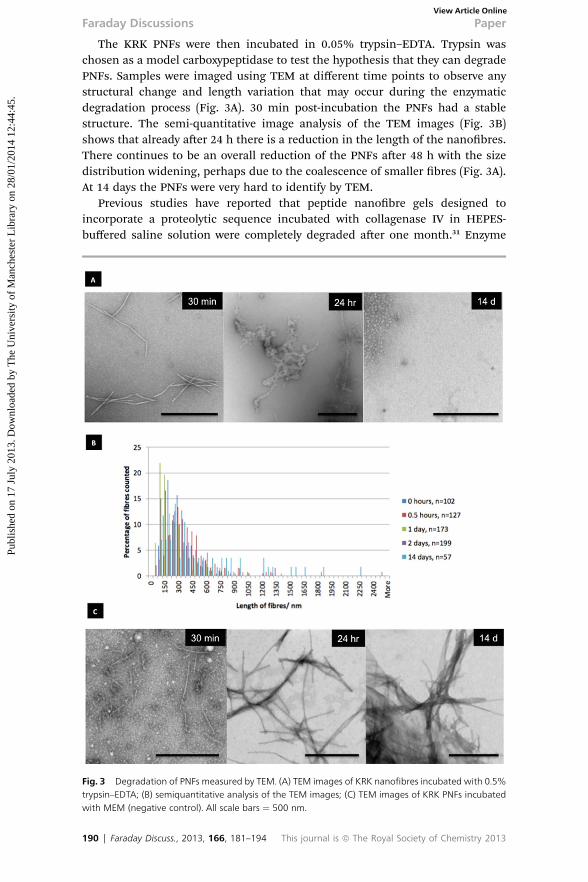
The KRK PNFs were then incubated in 0.05% trypsin–EDTA. Trypsin waschosen as a model carboxypeptidase to test the hypothesis that they can degradePNFs. Samples were imaged using TEM at different time points to observe anystructural change and length variation that may occur during the enzymaticdegradation process (Fig. 3A). 30 min post-incubation the PNFs had a stablestructure. The semi-quantitative image analysis of the TEM images (Fig. 3B)shows that already aer 24 h there is a reduction in the length of the nanobres.There continues to be an overall reduction of the PNFs aer 48 h with the sizedistribution widening, perhaps due to the coalescence of smaller bres (Fig. 3A).At 14 days the PNFs were very hard to identify by TEM.
Previous studies have reported that peptide nanobre gels designed toincorporate a proteolytic sequence incubated with collagenase IV in HEPES-buffered saline solution were completely degraded aer one month.31 Enzyme
Fig. 3 Degradation of PNFs measured by TEM. (A) TEM images of KRK nanofibres incubated with 0.5%trypsin–EDTA; (B) semiquantitative analysis of the TEM images; (C) TEM images of KRK PNFs incubatedwith MEM (negative control). All scale bars ¼ 500 nm.
190 | Faraday Discuss., 2013, 166, 181–194 This journal is ª The Royal Society of Chemistry 2013
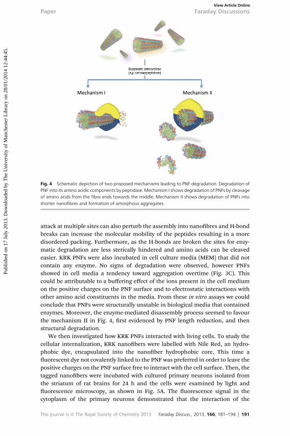
Fig. 4 Schematic depiction of two proposed mechanisms leading to PNF degradation. Degradation ofPNF into its amino acidic components by peptidase. Mechanism I shows degradation of PNFs by cleavageof amino acids from the fibre ends towards the middle. Mechanism II shows degradation of PNFs intoshorter nanofibres and formation of amorphous aggregates.
Paper Faraday DiscussionsPu
blis
hed
on 1
7 Ju
ly 2
013.
Dow
nloa
ded
by T
he U
nive
rsity
of
Man
ches
ter
Lib
rary
on
28/0
1/20
14 1
2:44
:45.
View Article Online
attack at multiple sites can also perturb the assembly into nanobres and H-bondbreaks can increase the molecular mobility of the peptides resulting in a moredisordered packing. Furthermore, as the H-bonds are broken the sites for enzy-matic degradation are less sterically hindered and amino acids can be cleavedeasier. KRK PNFs were also incubated in cell culture media (MEM) that did notcontain any enzyme. No signs of degradation were observed, however PNFsshowed in cell media a tendency toward aggregation overtime (Fig. 3C). Thiscould be attributable to a buffering effect of the ions present in the cell mediumon the positive charges on the PNF surface and to electrostatic interactions withother amino acid constituents in the media. From these in vitro assays we couldconclude that PNFs were structurally unstable in biological media that containedenzymes. Moreover, the enzyme-mediated disassembly process seemed to favourthe mechanism II in Fig. 4, rst evidenced by PNF length reduction, and thenstructural degradation.
We then investigated how KRK PNFs interacted with living cells. To study thecellular internalization, KRK nanobers were labelled with Nile Red, an hydro-phobic dye, encapsulated into the nanober hydrophobic core. This time auorescent dye not covalently linked to the PNF was preferred in order to leave thepositive charges on the PNF surface free to interact with the cell surface. Then, thetagged nanobers were incubated with cultured primary neurons isolated fromthe striatum of rat brains for 24 h and the cells were examined by light anduorescence microscopy, as shown in Fig. 5A. The uorescence signal in thecytoplasm of the primary neurons demonstrated that the interaction of the
This journal is ª The Royal Society of Chemistry 2013 Faraday Discuss., 2013, 166, 181–194 | 191

Fig. 5 (A) Cell internalization of KRK PNFs encapsulating Nile Red by primary neurons isolated from ratbrain (scale bar ¼ 50 mm); (B) residence time (days) of KRK PNFs fluorescently labelled with VivoTag 680XL injected intracranially in the right brain hemisphere (caudate–putamen) in athymic nude mice,imaged with an IVIS Lumina camera at 675 nm.
Faraday Discussions PaperPu
blis
hed
on 1
7 Ju
ly 2
013.
Dow
nloa
ded
by T
he U
nive
rsity
of
Man
ches
ter
Lib
rary
on
28/0
1/20
14 1
2:44
:45.
View Article Online
positively charged PNFs with the cell surface results in internalization of thePNFs. The ber shaped PNFs may offer an additional cell entrance gate by energy-independent internalisation mechanisms, similar to other high axial rationanostructures that have been previously described to pierce the plasmamembrane.32
In this context, PNFs may offer the ideal platform as a nanocarrier because ofthe versatility with which they can be engineered. They can be employed ascarriers for small hydrophobic molecules,33 and at the same time potentially showmore versatility as a ber-shaped transporter. PNFs also seem to be degradable inbiological environments, so we decided to perform a pilot in vivo degradabilitystudy. PNFs have been shown to be biocompatible scaffolds for neuronal differ-entiation34 and have been investigated as carriers of bioactive molecules to theCNS.15 However, PNFs structurally resemble in shape amyloid brils, misfolded
192 | Faraday Discuss., 2013, 166, 181–194 This journal is ª The Royal Society of Chemistry 2013

Paper Faraday DiscussionsPu
blis
hed
on 1
7 Ju
ly 2
013.
Dow
nloa
ded
by T
he U
nive
rsity
of
Man
ches
ter
Lib
rary
on
28/0
1/20
14 1
2:44
:45.
View Article Online
protein aggregates that are involved in a number of neuropathologies associatedwith neurodegeneration.35
For these reasons we decided to study the residence time of the PNFs injecteddirectly in the brain parenchyma, as an initial step towards assessment of thesafety of this approach for CNS therapeutic delivery. A variety of peptidases havebeen detected in the tissues of the CNS, some of these are membrane bound andothers are soluble and exist in the highest concentration in the neural cellcytosol.36 Carboxypeptidases are present in the brain and CSF37 and are respon-sible for the enzymatic degradation of peptides starting from the C-terminus. Westudied the residence time of the VivoTag 680 XL uorescently labelled KRK PNFsaer intracranial injection in the caudate–putamen of athymic nude mice.
The residence time of the PNFs in living animals (under anaesthesia) wasmeasured by following the uorescent signal using an IVIS uorescence imagingcamera. A strong uorescent signal was observed for up to 1 week upon intra-cranial injection. Furthermore, it is worth noting that the signal remained locatedin the right hemisphere, in proximity to the site of injection. Aer 10 days the PNFuorescent signal exhibited a considerable decrease and was further weakenedaer 15 days. This data suggests that the PNFs were removed or degraded in thebrain parenchyma. Much further work should be performed in continuation ofthis pilot in vivo study.
Conclusions
This study has demonstrated that PNFs in suspension can be internalized by cells,and degraded overtime in enzyme containing biological uids and possibly byenzymes expressed in vivo. We provided here some initial evidence that nano-bres may have a reduced risk of accumulation and prompt further exploration astransporters of therapeutic and diagnostic agents to the CNS.
Acknowledgements
This work was partially funded by the European Commission research pro-gramme FP7-NANOSOLUTIONS (FP7-NMP-2012-309329). RP acknowledgesnancial support from Generalitat de Catalunya (2009SGR1331), MINECO(CTQ2010-14897) and J. Caelles for the SAXS-WAXS service at IQAC, and technicalsupport on the SAXS measurements.
References1 H. Cui, M. J. Webber and S. I. Stupp, Biopolymers, 2010, 94, 1.2 J. D. Hartgerink, E. Beniash and S. I. Stupp, Science, 2001, 294, 1684.3 S. Vauthey, S. Santoso, H. Gong, N. Watson and S. Zhang, Proc. Natl. Acad. Sci. U. S. A.,2002, 99, 5355.
4 S. Zhang, D. M. Marini, W. Hwang and S. Santoso, Curr. Opin. Chem. Biol., 2002, 6, 865.5 P. Forns, J. L. Lauer-Fields, S. Gao and G. B. Fields, Biopolymers, 2000, 54, 531.6 Y.-C. Yu, M. Tirrell and G. B. Fields, J. Am. Chem. Soc., 1998, 120, 9979.7 X.-D. Xu, Y. Jin, Y. Liu, X.-Z. Zhang and R.-X. Zhuo, Colloids Surf., B, 2010, 81, 329.8 R. L. Thurlkill, G. R. Grimsley, J. M. Scholtz and C. N. Pace, Protein Sci., 2006, 15, 1214.9 D. Orthaber, A. Bergmann and O. Glatter, J. Appl. Crystallogr., 2000, 33, 218.10 L. Perez, A. Pinazo, M. R. Infante and R. Pons, J. Phys. Chem. B, 2007, 111, 11379.11 R. Pons, M. Valiente and G. Montalvo, Langmuir, 2010, 26, 2256.12 J. S. Pedersen, Adv. Colloid Interface Sci., 1997, 70, 171.13 E. T. Pashuck, H. Cui and S. I. Stupp, J. Am. Chem. Soc., 2010, 132, 6041.
This journal is ª The Royal Society of Chemistry 2013 Faraday Discuss., 2013, 166, 181–194 | 193

Faraday Discussions PaperPu
blis
hed
on 1
7 Ju
ly 2
013.
Dow
nloa
ded
by T
he U
nive
rsity
of
Man
ches
ter
Lib
rary
on
28/0
1/20
14 1
2:44
:45.
View Article Online
14 E. T. Pashuck and S. I. Stupp, J. Am. Chem. Soc., 2010, 132, 8819.15 M. Mazza, R. Notman, J. Anwar, A. Rodger, M. Hicks, G. Parkinson, D. McCarthy,
T. Daviter, J. Moger, N. Garrett, T. Mead, M. Briggs, A. G. Schatzlein andI. F. Uchegbu, ACS Nano, 2013, 7, 1016.
16 M. Foldvari, S. Attah-Poku, J. Hu, Q. Li, H. Hughes, A. B. Lorne and S. Kruger, J. Pharm.Sci., 1998, 87, 1203.
17 L. Yuan, J. Wang and W. C. Shen, Eur. J. Pharm. Biopharm., 2008, 70, 615.18 Z. Yang, W. T. Huck, S. M. Clarke, A. R. Tajbakhsh and E. M. Terentjev, Nat. Mater., 2005,
4, 486.19 I. W. Hamley, A. Dehsorkhi and V. Castelletto, Langmuir, 2013, 29, 5050.20 V. Castelletto, I. W. Hamley, J. Perez, L. Abezgauz and D. Danino, Chem. Commun., 2010,
46, 9185.21 A. Dehsorkhi, I. W. Hamley, J. Seitsonen and J. Ruokolainen, Langmuir, 2013, 29, 6665–
6672.22 C. Tang, A. M. Smith, R. F. Collins, R. V. Ulijn and A. Saiani, Langmuir, 2009, 25, 9447.23 A. M. Smith, R. J. Williams, C. Tang, P. Coppo, R. F. Collins, M. L. Turner, A. Saiani and
R. V. Ulijn, Adv. Mater., 2008, 20, 37.24 D. Roberts, C. Rochas, A. Saiani and A. F. Miller, Langmuir, 2012, 28, 16196.25 K. Zhao, U.-J. Choe, D. T. Kamei and G. C. L. Wong, So Matter, 2012, 8, 6430.26 I. W. Hamley, A. Dehsorkhi and V. Castelletto, Chem. Commun., 2013, 49, 1850.27 I. W. Hamley, A. Dehsorkhi, V. Castelletto, J. Seitsonen, J. Ruokolainen andH. Iatrou, So
Matter, 2013, 9, 4794.28 A. Pinazo, L. Perez, M. R. Infante and R. Pons, Phys. Chem. Chem. Phys., 2004, 6, 1475.29 C. L. Pizzey, W. C. Pomerantz, B.-J. Sung, V. M. Yuwono, S. H. Gellman, J. D. Hartgerink,
A. Yethiraj and N. L. Abbott, J. Chem. Phys., 2008, 129, 095103.30 T. Imae, N. Hayashi, T. Matsumoto, T. Tada and M. Furusaka, J. Colloid Interface Sci.,
2000, 225, 285.31 H. W. Jun, V. Yuwono, S. E. Paramonov and J. D. Hartgerink, Adv. Mater., 2005, 17, 2612.32 L. Lacerda, J. Russier, G. Pastorin, M. A. Herrero, E. Venturelli, H. Dumortier, K. T. Al-
Jamal, M. Prato, K. Kostarelos and A. Bianco, Biomaterials, 2012, 33, 3334.33 S. Soukasene, D. J. To, T. J. Moyer, H. Lu, H. K. Lee, S. M. Standley, V. L. Cryns and
S. I. Stupp, ACS Nano, 2011, 5, 9113.34 G. A. Silva, C. Czeisler, K. L. Niece, E. Beniash, D. A. Harrington, J. A. Kessler and
S. I. Stupp, Science, 2004, 303, 1352.35 E. H. Koo, P. T. Lansbury, Jr and J. W. Kelly, Proc. Natl. Acad. Sci. U. S. A., 1999, 96, 9989.36 A. Turner, in Neuropeptides and their peptidases, ed. A. Turner, Ellis Horwood, Chichester
and Verlagsgesellscha, 1987.37 M. B. Segal, Barriers and uids of the eye and brain, Macmillan, 1992.
194 | Faraday Discuss., 2013, 166, 181–194 This journal is ª The Royal Society of Chemistry 2013