fat suppression imaging
TRANSCRIPT

Fat suppression
imaging
By
Roshan Shah
B.Sc. MIT 3rd year
3rd batch

Introduction
Fat saturation is an MRI technique used to suppress the signal from normal adipose tissue.
To suppress the fat signal for a given MR sequence a fat suppression module is typically inserted at the beginning of an otherwise normal MRI sequence.

Indication
It is used in MRI for mainly two purpose.To suppress the signal from normal
adipose tissue to reduce chemical shift artifact or improve visualization of uptake of contrast material.
Tissue characterization, particularly in adrenal gland tumors, bone marrow infiltration, fatty tumors, etc.

Physics for fat suppression
(FAT SAT) A specialized technique that selectively saturates fat protons prior to acquiring data as in standard sequence, so that they produce a negligible signal.
This technique requires a homogeneous magnetic field and homogeneous volume of tissue.
To prepare this type of sequence, the following properties should be used.
Fat and water have different resonant frequencies
They have different Larmor precession frequencies
They have different T1 relaxation times.
Advantages
This method is reliable for contrast material enhanced T1 weighted imaging.
It is useful in tissue characterization particularly in area with a large amount of fat.
It also useful for avoiding chemical shift misregistration artifact.
Allows good visualization of small anatomical details.
Disadvantage
Inhomogeneities of the static magnetic field.
Inhomogeneities In the radio-frequency field.
Inhomogeneities in volume of tissue.

Methods
Fat suppression can be achieved in a number of different ways. Short tau inversion recovery (STIR)
Chemical shift Selective (CHESS) Fat-Sat.
Spectral Pre-saturation with Inversion Recovery (SPIR)
Spectral Attenuated Inversion Recovery (SPAIR)
Water excitation
DIXON-based

STIR
It is an inversion recovery pulse sequence with specific timing so as to suppress the signal from fat.

Cont.…
Advantages:
1. It suppresses whole of the adipose tissue including water fraction
2. This is only method which can be used even in magnetic field inhomogeneities.
3. It can be used with low magnetic field strength.
Disadvantages:
1. Beginning at TI null most of the proton have not completely relaxation, and are therefore still partially saturated, in this situation will overall produce signal loss and SNR ratio will decrease.
2. It cannot be used post gadolinium to demonstrate contrast enhancement.
3. Long acquisition time
4. Tissue contrast is affected. SNR is reduced.

MRI image appearance Fluids normally appear bright and fat appear very
dark in a STIR images. Pathological processes normally increase the water
content in tissues. Due to the added water component this results in a signal increase on STIR images. Consequently pathological processes are usually bright on STIR images.

STIR sagittal sequence used in C spine imaging and knee imaging
CHESS
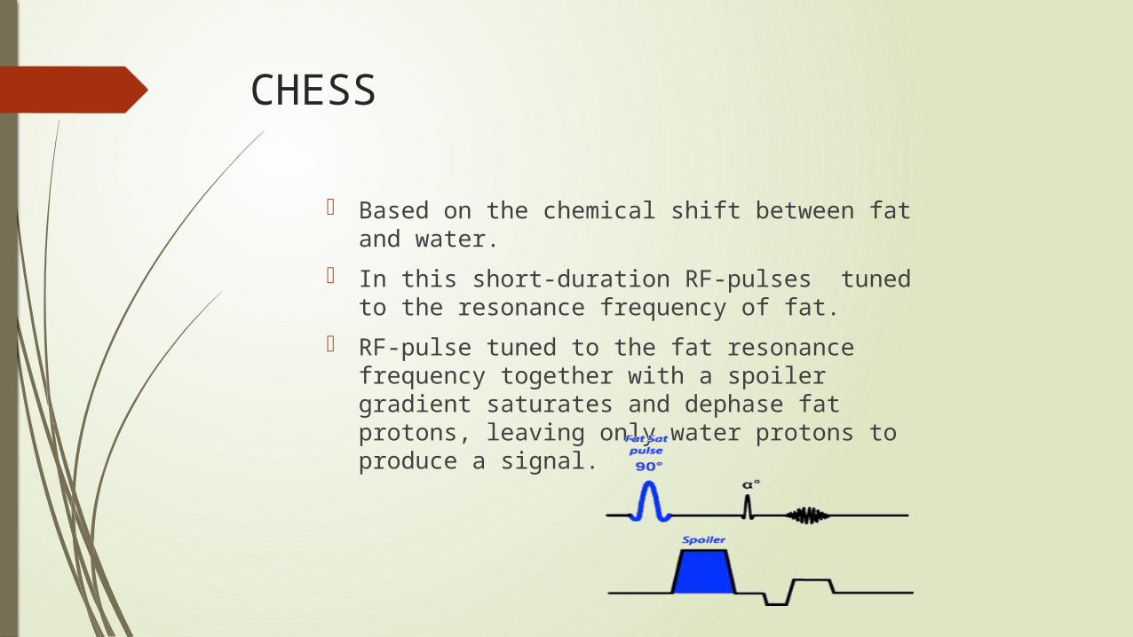
Based on the chemical shift between fat and water.
In this short-duration RF-pulses tuned to the resonance frequency of fat.
RF-pulse tuned to the fat resonance frequency together with a spoiler gradient saturates and dephase fat protons, leaving only water protons to produce a signal.

Cont.…
Advantages
It can be added to any pulse sequence.
Can be used for post contrast imaging.
Disadvantages
It cannot be used at low field strength.
It also cannot be use in Inhomogeneities of the static magnetic field.
Can be used in post contrast MR arthrography.
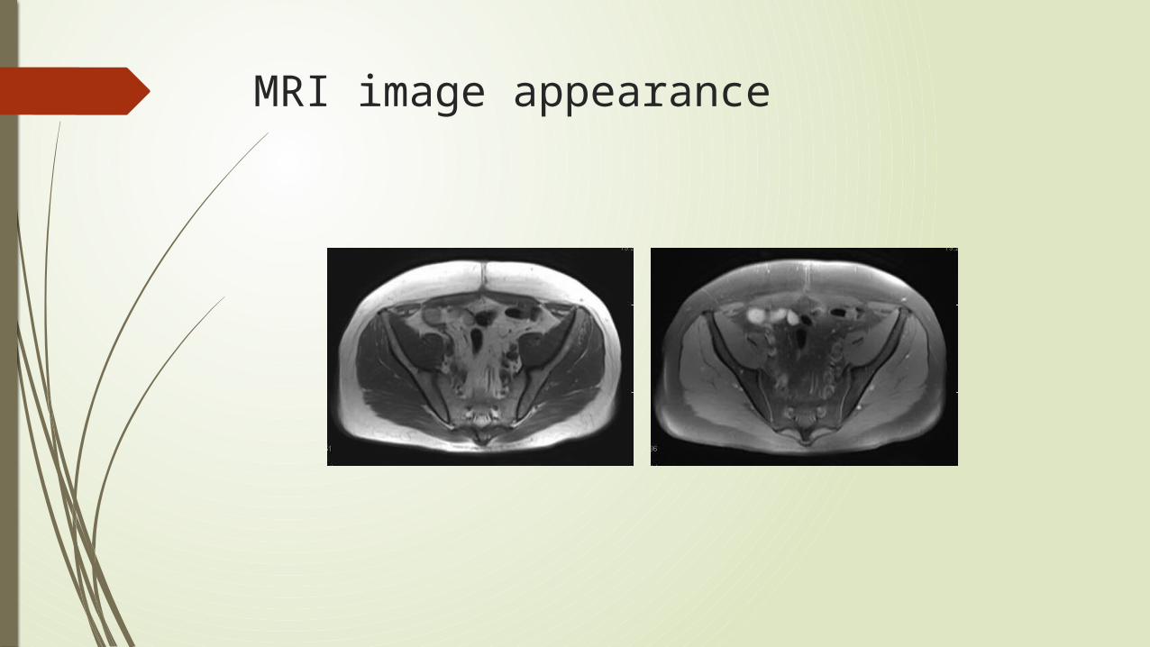
MRI image appearance

SPIR
It is the combination of the spectral saturation and STIR routines.

MRI image appearance

SPAIR
It is a hybrid technique combining features of both CHESS and STIR.
SPAIR uses an adiabatic pulses.

Cont.…
AdvantagesSPAIR provide better and more homogeneous fat
suppression than SPIRTissue contrast is not affected.
DisadvantagesThe inversion time is longer.Reduce number of slice for a given TR.

MRI image appearance

Water excitation
This technique is based on the chemical shift.
Instead of suppressing fat, these techniques use a short series of RF pulses(binomial pulse) to selectively excite water protons.
No spoilers are needed.


Cont.…
Advantages No additional preparation pulse is
necessary.Reduced sensitivity to B1
Inhomogeneities Disadvantages
Increased total measurement timeReduced maximum number of slices.
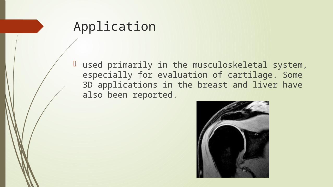
Application
used primarily in the musculoskeletal system, especially for evaluation of cartilage. Some 3D applications in the breast and liver have also been reported.

DIXON-based
This technique is based on the chemical shift. It uses ‘In phase’ and ‘Out phase’ cycling of
fat and water. Acquiring 2, 3 or more echoes at different TE's,
"water only" and "fat only" images can be extracted

In-phase
water
Out-phase
fat

Cont.…
Advantages Insensitive to B0 and B1
Inhomogeneities.4 contrasts delivered in one
measurement. Disadvantages
Increases minimal TR because in- and opposed phase data must be acquired.
Application
The Dixon technique is widely used in abdominal imaging, imaging of the extremities, and the spine.
The opposed phased imaging is useful for detection of small amounts of fat. Ex- adrenal gland tumors or steatosis.

Thank
you