fatal mastitis of dairy cows: a retrospective study
TRANSCRIPT

Fatal Mastitis of Dairy Cows: A Retrospective Study
M.J. Hazlett, P.B. Little, M.G. Maxie and D.A. Barnum*
ABSTRACT
The necropsy records of dairy cowswith mastitis were reviewed from theprovincial veterinary laboratory inGuelph (44 cases of mastitis in nineyears) and from the Ontario Veteri-nary College (168 cases in 14 years).Mastitis was considered to be theprimary cause of death in 167 of 212cows (79%). Of these 167 cases of mas-titis, Escherichia coli was involved in107 (64%), Klebsiella sp. in 12 (7%)and Staphylococcus aureus in 11 (7%).Bacteriology was not reported in 22cases.
Coliform mastitis, the most com-monly identified type of fatal mastitis,was characterized histologically by thepresence of infarcted areas in affectedglands and by the lack of demonstra-ble bacteria, and was thus easily identi-fied from fatal mastitis caused by S.aureus.
Key words: Bovine mastitis, coliform,bacteria, fatal mastitis.
RESUME
Les auteurs ont realise, au labora-toire veterinaire provincial de Guelphet au College Veterinaire de l'Ontario,une etude retrospective des rapportsde necropsie de vaches laitieresatteintes de mammite. Au premierendroit, ils en retracerent 44, eche-lonnes sur une periode de neuf ans, etau deuxieme, 168, echelonnes sur une
periode de 14 ans. uls attribuerent a lamammite la mort de 167, ou 79% deces 212 vaches. Des 167 cas de mam-mite, 107, ou 64%, etaient imputablesa Escherichia coli; 12, ou 7%, a Kleb-siella sp., et 11, ou 7%, a Staphylococ-
cus aureus. La bacteriologie de 22 cas
n'avait pas ete rapportee.La mammite a coliformes, la variete
fatale la plus frequente, se caracterisaitpar des lesions microscopiques d'in-farction du tissu mammaire et par l'ab-sence de bacteries demontrables; ces
particularites permirent de la differen-cier facilement de la mammite fataledue a S. aureus.
Mots cles: mammite bovine, coli-forme, bacteries, mammite fatale.
INTRODUCTION
Mastitis is an economically impor-tant disease of dairy cows. Most masti-tis research has concentrated on var-
ious aspects of nonfatal disease,however losses do occur from thedeath of cows, sometimes after a very
short clinical course (1,2).The relative prevalence of patho-
gens involved in naturally-occurringfatal cases of bovine mastitis is poorlydocumented, as are the associated his-topathological findings.We, therefore, conducted a retro-
spective study of the necropsy recordsof our provincial and university vete-rinary diagnostic laboratories in orderto determine if historical, histopatho-logical and/ or bacteriological corre-
lates existed in a large sample of casesof fatal bovine mastitis.
MATERIALS AND METHODS
Necropsy records of dairy cows withmastitis were reviewed from theGuelph Veterinary Laboratory Servi-ces Branch (VLS) of the Ontario Min-
istry of Agriculture and Food for theyears 1974 to 1982 inclusive. Recordsof both mailed-in tissue specimens andfull necropsies were examined. Avail-able pertinent history (age, stage oflactation, number of quarters in-volved), etiological diagnosis and thecause of death were recorded.
Necropsy records were reviewedfrom mastitic cows diagnosed at theOntario Veterinary College (OVC),University of Guelph between January1969 and May 1983. History, etiologi-cal diagnosis and cause of death were
recorded. Cows euthanized because ofmastitis were included in the fatal mas-titis group. Where available, histologi-cal sections were reexamined fromOVC cases when one of Escherichiacoli, Klebsiella sp., Corynebacteriumpyogenes or Staphylococcus aureus
was the only pathogen isolated in thefatal mastitis cases. The quarter oforigin of the sections was usuallyunknown, but was obviously theaffected gland in most cases. Sectionswere assessed without knowledge ofthe etiological diagnosis. The sectionswere evaluated for infarction, an
infarcted gland being characterized as
having an edematous area of coagula-tion necrosis surrounded by a zone ofinflammatory cells (Fig. 1). Sectionswere then graded from zero to fivewith respect to severity of edema,hemorrhage, vascular thrombosis,lymphatic dilation/ thrombosis, neu-
trophil infiltrate, macrophage infil-trate and presence of bacteria. Infarc-ted areas were scored for bacteria, butnot for the other parameters. A zero
signified not present. A score of one
indicated equivocal thrombosis,equivocal presence of edema or bacte-ria, small numbers of neutrophils or
macrophages or small quantities of
Can J. Comp Med 1984; 48: 125-129.
*Department of Pathology (Hazlett and Little), Department of Veterinary Microbiology and Immunology (Barnum), Ontario Veterinary College,University of Guelph, Guelph, Ontario NIG 2WI and Veterinary Laboratory Services Branch (Maxie), Ontario Ministry of Agriculture and Food,Guelph, Ontario N 1H 6R8.
Submitted September 13, 1983.
125

Fig. 1. Section from a fatal case of E. coli mastitis. An inflammatory cell boundary (arrows)separates the area of coagulation necrosis (bottom right) from the more normal glandular paren-chyma. H & E. X50.
hemorrhage. A score of five signifiedthat the feature was very prominent.Only hematoxylin and eosin (H & E)stained sections were examined.
RESULTS
Veterinary Laboratory Services andOVC records when combined listed212 cases of mastitis. In 167 cases mas-titis was considered as the primarycause of death (Table I). Escherichiacoli was isolated in pure culture in47.3% of the fatal mastitis cases. WhenE. coli was isolated in pure culturefrom a mammary gland, it was felt tobe the cause of death in 91% of cases.Fatal mastitis caused by E. coliinvolved 1.8 quarters per cow, with50% of the infections occurring in thefirst week postpartum and 5% of casesin dry or prepartum cows. The meanage of affected cows was 7.1 years(range 2 to 14 yr). Several cows in thisgroup had concurrent endometritis. Inaddition, E. coli was involved in 28fatal mastitis cases in which more thanone pathogen was recovered (TableIII).
Klebsiella sp. was isolated only inpure culture and was the second mostcommon cause of fatal mastitis. In all12 cases listed, the mastitis caused bythis bacteria was considered the prim-
ary cause of death. In these animals,mastitis was present in 2.1 quarters percow. Only six of the 12 cases had thestage of lactation recorded. Two werein dry cows, and none were recorded asinvolving cows in their first week post-partum. The mean age of cows in thisgroup was 7.3 years (range 4 to 12 yr).
Staphylococcus aureus was reco-vered in pure culture in seven fatalcases of mastitis. Two other cases werediagnosed at necropsy but were notconsidered to be the cause of death. Infatal cases, 2.4 quarters per cow wereinvolved. The mean age of affectedcows was 4.8 yr. Six of the seven fatalcases occurred in the first week post-partum.
Corynebacterium pyogenes was thecause of fatal mastitis in five cows.Two of these cows did not die natu-rally, but were euthanized because ofmastitis.
Streptococci involved in two mas-
TABLE I. Bovine Mastitis Cases Diagnosed at Necropsy at the Ontario Veterinary College (1969-1983) and the Veterinary Services Laboratory (Guelph), Ontario Ministry of Agriculture and Food(1973-1982)
Etiological Diagnosis Number Diagnosed (%) Number Cause of Death (%)
E. colia 87 (41.0) 79 (47.3)Klebsiella sp.a 12 (5.7) 12 (7.2)S. aureusa 9 (4.2) 7 (4.2)C. pyogenesa 16 (7.5) 5 (3.0)Streptococcia 4 (1.9) 2 (1.2)Others (1 pathogen)a 14 (6.6) 12 (7.2)Mixed (2 pathogens) 47 (22.2) 38 (22.8)No etiological diagnosis 23 (10.8) 12 (7.2)
Total 212 167
alsolated in pure culture
TABLE II. Miscellaneous Pathogens Isolated in Pure Culture from Fatal Bovine Mastitis Cases atthe Ontario Veterinary College (1969-1983) and the Veterinary Services Laboratory (Guelph),Ontario Ministry of Agriculture and Food (1973-1982)
Stage of# Quarters Lactation
Pathogen # Cows Age (Yr) Affected (Days)
Aspergillus sp. I Ma 4 21ilb 6 4 42
Bacillus sp. 1 4 1 4Clostridium perfringens 1 8 3 3Clostridium septicum 1 9 3 DryCorynebacterium pyogenes 1 5 1 -
Mycoplasma bovis ib 3 4 30Pasteurella hemolytica I M 1 4Pseudomonas aeruginosa 1 6 4 3
8 1 7Proteus sp. 1 8 1 28Mycoticc -_ _
aMaturebEuthanized due to mastitiscNot further identified
126

titic deaths were typed only as an alphaand a nonhemolytic Streptococcus sp.
Other pathogens isolated in pureculture are listed (Table II).Mixed infections were common, in
particular involving E. coli (Table III).Mixed infections not including E. coliare listed (Table IV).Nonspore forming obligate anae-
robic bacterial infections were diag-nosed in two cases (one Bacteroides sp.and one unspecified).The review of histological sections
of OVC cases in which E. coli, Kleb-siella sp., S. aureus and C. pyogeneswere recovered revealed that infarc-tion only occurred in the coliformgroups (E. coli and Klebsiella sp.) (Fig.1). Infarction was not noted in grossdescription of the cases, but was easilyidentified in histological sections aslarge areas of coagulation necrosis
Fig. 2. Fatal Corynebaderium pyogenes mastitis with prominent infiltration of macrophages andplasma cells in the interstitial tissue and masses ofdegenerate leukocytes with bacteria in alveoli. H &E. X150.
TABLE III. Mixed Infections Involving E. coli from Fatal Cases of Bovine Mastitis at the OntarioVeterinary College (1969-1983) and the Veterinary Services Laboratories (Guelph), Ontario Minis-try of Agriculture and Food (1973-1982)
Pathogens Isolated Number of Cases
E. coli Alcaligenesfaecalis IE. coli Anaerobesa IE. coli Bacillus sp. IE. coli Bacteroides sp. IE. coli Clostridium perfringens 3E. coli Corynebacterium pyogenes 8E. coli Proteus sp. 3E. coli Pseudomonas aeruginosa 2E. coli Pseudomonas sp., Streptococcib 2E. coli Staphylococcus aureus IE. coli Streptococci alpha hemolytic 2E. coli Streptococci beta hemolytic IE. coli Streptococci nonhemolytic IE. coli Streptococcia I
Total 28
aNot further identifiedbNonagalactiae
TABLE IV. Fatal Mixed Mammary Pathogens not Involving Coliform Bacteria Diagnosed at theOntario Veterinary College (1969-1983) and the Veterinary Services Laboratories (Guelph), OntarioMinistry of Agriculture and Food (1973-1982)
Etiological Agents Number of Cases
Clostridium perfringens, Pseudomonas aeruginosa IClostridium perfringens, Staphylococcus aureus 2Clostridium perfringens, Streptococcia IClostridium septicum, Pasteurella hemoli'tica IClostridium septicum, Pasteurella multocida ICorynebacterium pyogenes, Mvcoplasma bovis ICorynebacterium pyogenes, Streptococcib iStaphylococcus aureus, Streptococcic IYeastd, Pseudomonas Sp. I
Total 10
aNonhemolyticbHemolytic, type unknowncBeta hemolysisdNot further identifiedeNonaeruginosa
surrounded by an inflammatory cellboundary. These cells were difficult toidentify morphologically, most beingdegenerate, however they wereassumed to be leukocytes because oftheir characteristic location in ahypercellular border around an area ofbland coagulation necrosis. Edema innoninfarcted mammary parenchymawas most prominent in S. aureus mas-titis. Corynebacterium pyogenes mas-titis tended to have fewer neutrophilsand more macrophages than did othertypes (Fig. 2). Bacteria were very diffi-cult to see in coliform mastitis, butformed large readily visible clumps inS. aureus mastitis, with closer exami-nation revealing clusters of large cocci(Fig. 3). In C. pyogenes mastitis bacte-ria appeared as basophilic stipplingamongst amorphous eosinophilicdebris, with these focal to coalescingareas surrounded predominantly bymacrophages and lymphocytes.
DISCUSSION
This retrospective study illustratesthe importance of coliform bacteria,particularly E. coli, as a cause of fatalbovine mastitis. The word "coliform"in mastitis literature, refers in generalto an association with E. coli, Kleb-siella sp. and Enterobacter sp. Some ofthe cows in our study also had coli-form endometritis and death may have
127
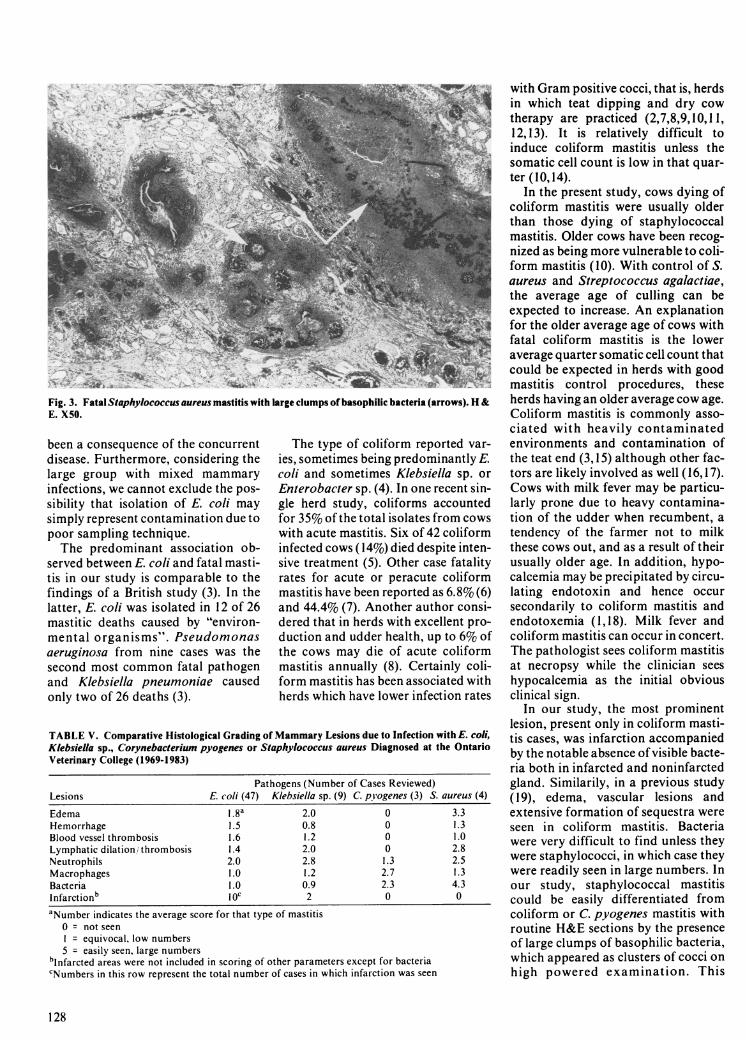
Fig. 3. Fatal Staphylococcus aureus mastitis with large clumps of basophilic bacteria (arrows). H &E. X50.
been a consequence of the concurrentdisease. Furthermore, considering thelarge group with mixed mammaryinfections, we cannot exclude the pos-sibility that isolation of E. coli maysimply represent contamination due topoor sampling technique.The predominant association ob-
served between E. coli and fatal masti-tis in our study is comparable to thefindings of a British study (3). In thelatter, E. coli was isolated in 12 of 26mastitic deaths caused by "environ-mental organisms". Pseudomonasaeruginosa from nine cases was thesecond most common fatal pathogenand Klebsiella pneumoniae causedonly two of 26 deaths (3).
The type of coliform reported var-ies, sometimes being predominantly E.coli and sometimes Klebsiella sp. orEnterobacter sp. (4). In one recent sin-gle herd study, coliforms accountedfor 35% of the total isolates from cowswith acute mastitis. Six of 42 coliforminfected cows (14%) died despite inten-sive treatment (5). Other case fatalityrates for acute or peracute coliformmastitis have been reported as 6.8% (6)and 44.4% (7). Another author consi-dered that in herds with excellent pro-duction and udder health, up to 6% ofthe cows may die of acute coliformmastitis annually (8). Certainly coli-form mastitis has been associated withherds which have lower infection rates
TABLE V. Comparative Histological Grading of Mammary Lesions due to Infection with E. coli,Kiebsiella sp., Corynebacterium pyogenes or Staphylococcus aureus Diagnosed at the OntarioVeterinary College (1969-1983)
Pathogens (Number of Cases Reviewed)Lesions E. coli (47) Klebsiella sp. (9) C. p'ogenes (3) S. aureus (4)
Edema 1.8a 2.0 0 3.3Hemorrhage 1.5 0.8 0 1.3Blood vessel thrombosis 1.6 1.2 0 1.0Lymphatic dilation/ thrombosis 1.4 2.0 0 2.8Neutrophils 2.0 2.8 1.3 2.5Macrophages 1.0 1.2 2.7 1.3Bacteria 1.0 0.9 2.3 4.3Infarctionb I Oc 2 0 0
aNumber indicates the average score for that type of mastitis0 = not seenI = equivocal, low numbers5 = easily seen, large numbers
blnfarcted areas were not included in scoring of other parameters except for bacteriacNumbers in this row represent the total number of cases in which infarction was seen
with Gram positive cocci, that is, herdsin which teat dipping and dry cowtherapy are practiced (2,7,8,9,10,11,12,13). It is relatively difficult toinduce coliform mastitis unless thesomatic cell count is low in that quar-ter (10,14).
In the present study, cows dying ofcoliform mastitis were usually olderthan those dying of staphylococcalmastitis. Older cows have been recog-nized as being more vulnerable to coli-form mastitis (10). With control of S.aureus and Streptococcus agalactiae,the average age of culling can beexpected to increase. An explanationfor the older average age of cows withfatal coliform mastitis is the loweraverage quarter somatic cell count thatcould be expected in herds with goodmastitis control procedures, theseherds having an older average cow age.Coliform mastitis is commonly asso-ciated with heavily contaminatedenvironments and contamination ofthe teat end (3,15) although other fac-tors are likely involved as well (16,17).Cows with milk fever may be particu-larly prone due to heavy contamina-tion of the udder when recumbent, atendency of the farmer not to milkthese cows out, and as a result of theirusually older age. In addition, hypo-calcemia may be precipitated by circu-lating endotoxin and hence occursecondarily to coliform mastitis andendotoxemia (1,18). Milk fever andcoliform mastitis can occur in concert.The pathologist sees coliform mastitisat necropsy while the clinician seeshypocalcemia as the initial obviousclinical sign.
In our study, the most prominentlesion, present only in coliform masti-tis cases, was infarction accompaniedby the notable absence of visible bacte-ria both in infarcted and noninfarctedgland. Similarily, in a previous study(19), edema, vascular lesions andextensive formation of sequestra wereseen in coliform mastitis. Bacteriawere very difficult to find unless theywere staphylococci, in which case theywere readily seen in large numbers. Inour study, staphylococcal mastitiscould be easily differentiated fromcoliform or C. pyogenes mastitis withroutine H&E sections by the presenceof large clumps of basophilic bacteria,which appeared as clusters of cocci onhigh powered examination. This
128
1- .."Emp"""t, ;.-, .A -. '..
information should be of some assist-ance in the diagnosis of mastitis whenonly formalin fixed tissue is availableto the pathologist.
Nonspore forming obligate anae-robic bacterial infections were diag-nosed in two of 133 OVC cases (oneBacteroides sp. and one unspecified).Routine microbiological methodspresently used probably make this agross underestimation of the impor-tance of these organisms in mastitis.Recent work has shown nonsporeforming anaerobes to be present in12% of lactating mastitic cows (20),and mastitis has been reproducedexperimentally using these organisms(21).Our study demonstrates that coli-
form mastitis was the most commoncause of mastitic death in dairy cowsexamined at these laboratories. Masti-tis tended to occur in older cows and inmultiple quarters. Staphylococcalmastitis could be differentiated histo-logically from coliform mastitis by thevisible presence of bacteria in theformer, and the frequent presence ofinfarcted gland in the latter.
REFERENCES
1. BLOOD DC, HENDERSON JA, RADO-STITS OM. Mastitis. In: Veterinary medi-cine, 5th ed. London: Bailliere Tindall,1979: 363-405.
2. RADOSTITS OM. The clinical aspects ofcoliform mastitis in cattle. Proc V I nt ConfCattle Dis 1970: 67-74.
3. HOWELL D. Survey on mastitis caused byenvironmental bacteria. Vet Rec 1972; 90:654-657.
4. EBERHART RJ. Coliform mastitis. J AmVet Med Assoc 1977; 170: 1160-1163.
5. ANDERSON KL, SMITH AR, GUS-TAFSSON BK, SPAHR SL, WHIT-MORE HL. Diagnosis and treatment ofacute mastitis in a large dairy herd. J AmVet Med Assoc 1982; 181: 690-693.
6. GOLODETZ CL, WHITE ME. Prognosisfor cows with severe clinical coliform masti-tis. Vet Rec 1983; 112: 402-403.
7. ARMSTRONG KR. Clinical experienceswith dry cow therapy and coliform mastitis.Bovine Practitioner 1976; 12: 85-87.
8. BUSCHNELL RB. Where are we on coli-form mastitis? Proc 13th Ann Meet NatlMast Counc 1974; 62-69.
9. SCHALM OW, CARROLL EJ, JAIN NC.Bovine mastitis. Lea & Febiger, 1971:254-256.
10. SCHALM OW. The bovine leukocytes partI1: The neutrophil as a barrier to coliformmastitis. Bovine Practice 1981; 2: 28-31.
11. JASPER DE. The coliform mastitisenigma. Bovine Practitioner 1980; 15:39-43.
12. JASPER DE. Coliform mastitis - a causefor concern. Proc Ann Meet Nati MastCounc 1973; 59-68.
13. JOHNSON LW, SIDDIQUE IH. A herdproblem of acute coliform mastitis. VetMed Small Anim Clin 1965; 60: 940-942.
14. BRAMLEY AJ. Variations in the suscepti-bility of lactating and non-lactating bovineudders to infection when infused withEscherichia coli. J Dairy Res 1976; 43:205-211.
15. BRAMLEY AJ, GODINHO KS, GRIN-DAL RJ. Evidence of penetration of thebovine teat duct by Escherichia coli in theinterval between milkings. J Dairy Res1981; 48: 379-386.
16. RENDOS JJ, EBERHART RJ, KESLEREM. Microbial population of teat ends ofdairy cows and bedding materials. J DairySci 1975; 58: 1492-1500.
17. NATZKE RP, LeCLAIR BJ. Coliformcontaminated bedding and new infections. JDairy Sci 1976; 59: 2152-2154.
18. GRIEL LC JR, ZARKOWER A, EBER-HART RJ. Clinical and clinico-patho-logical effects of Escherichia coli endotoxinin mature cattle. Can J Comp Med 1975; 39:1-6.
19. RENK W. Mastitiden bei infectionen mitEscherichia co/i, Aerobacter aerogenes undKlebsiella. Zentralbl Veterinaermed 1962;9: 264-281.
20. PREEZ JH DU, GREEFF AS, EKSTEENN. Isolation and significance of anaerobicbacteria isolated from cases of bovine masti-tis. Onderstepoort J Vet Res 1981; 48: 123-126.
21. PREEZ JH DU, GREEFF AS, BOTHAWS. Pathology of the bovine udder paren-chyma caused by asporogenous obligateanaerobic bacteria isolated from cases ofbovine mastitis. J S Afr Vet Assoc 1982; 53:157-159.
129