fatty acid uptake, transport and storage in placenta in
TRANSCRIPT

Fatty acid uptake, transport and storage in placenta
in normal and dyslipidemic pregnancies
Guro Mørk Johnsen
Department of Obstetrics and Gynaecology
Oslo University Hospital, Ullevål
Faculty of Medicine
University of Oslo
Norway
January 2010

Supervisors: Anne Cathrine Staff Asim K. Duttaroy Kari Anne Risan Tobin Adjudicative committee: Fiona Lyall Trine Ranheim Svein Olav Kolset © Guro Mørk Johnsen, 2010 Series of dissertations submitted to the Faculty of Medicine, University of Oslo No. 937 ISBN 978-82-8072-618-6 All rights reserved. No part of this publication may be reproduced or transmitted, in any form or by any means, without permission. Cover: Inger Sandved Anfinsen. Printed in Norway: AiT e-dit AS. Produced in co-operation with Unipub. The thesis is produced by Unipub merely in connection with the thesis defence. Kindly direct all inquiries regarding the thesis to the copyright holder or the unit which grants the doctorate.

3
Contents
Acknowledgements..................................................................................................................... 5 List of Papers............................................................................................................................... 7 Abbreviations .............................................................................................................................. 9 1. Introduction .......................................................................................................................... 11
1.1. Placenta........................................................................................................................... 11 Placentation ....................................................................................................................... 11
1.2. Fatty acids and their importance in fetal nutrition.................................................... 15 Essential fatty acids .......................................................................................................... 16 Physiological functions of LCPUFAs............................................................................ 17 Importance of LCPUFAs in fetal nutrition .................................................................. 18 Fatty acid metabolism in the placenta............................................................................ 18 Cholesterol......................................................................................................................... 19
1.3. Fatty acid transport in the placenta............................................................................. 19 Preferential uptake of LCPUFAs ................................................................................... 20 Fatty acid binding and transport proteins ..................................................................... 21 Long chain acyl-CoA synthetases (ACSLs)................................................................... 23 Lipid droplets and associated proteins .......................................................................... 23 ADRP (Adipose Differentiation Related Protein) ....................................................... 24
1.4. Nuclear receptors .......................................................................................................... 25 Peroxisome Proliferator Activated Receptors (PPARs).............................................. 25 Liver X Receptors (LXRs)............................................................................................... 26 PPARs and LXRs in placenta ......................................................................................... 27
1.5. Preeclampsia................................................................................................................... 28 Definitions......................................................................................................................... 28 Pathophysiology................................................................................................................ 29 Risk factors and treatment .............................................................................................. 30 Inflammation..................................................................................................................... 31 Oxidative stress................................................................................................................. 32 Dyslipidemia...................................................................................................................... 33 Lipotoxicity........................................................................................................................ 34 Acute atherosis.................................................................................................................. 34 Diabetes mellitus in pregnancy ....................................................................................... 35
2. Aims of present study .......................................................................................................... 36 3. Summary of Papers .............................................................................................................. 37
Paper 1 ............................................................................................................................... 37 Paper 2 ............................................................................................................................... 38 Paper 3 ............................................................................................................................... 39 Paper 4 ............................................................................................................................... 40
4. Methodological considerations ........................................................................................... 41 4.1. Advantages of combining clinical research and in vitro experiments .................... 41 4.2. Cell system...................................................................................................................... 41 4.3. Patient selection............................................................................................................. 42 4.4. Delivery mode................................................................................................................ 43 4.5. Gestational age............................................................................................................... 45

4
4.6. Protein expression in placenta samples ...................................................................... 45 4.7. Quantitative real-time PCR.......................................................................................... 46
Selection of genes for gene expression analysis ........................................................... 46 Calculation of gene expression ....................................................................................... 47 Relative versus absolute quantification of gene expression........................................ 49
5. General discussion................................................................................................................ 50 5.1. Transport and uptake of LCPUFAs in placenta ....................................................... 50 5.2. Role of ADRP and lipid droplets in placenta............................................................ 52 5.3. Role of ADRP and lipid droplets in preeclampsia.................................................... 53
Dyslipidemia and lipotoxicity.......................................................................................... 54 Oxidative stress and inflammation................................................................................. 56 Fibrinoid tissue ................................................................................................................. 58
5.4. Dysregulation of FA transport genes in preeclampsia ............................................. 59 6. Conclusions ........................................................................................................................... 62 7. Future perspectives............................................................................................................... 64 8. References.............................................................................................................................. 66
5
Acknowledgements
The work presented in this thesis was performed at the Department of
Obstetrics and Gynaecology, Oslo University Hospital, Ullevål, and the Department of
Nutrition, Institute of Basic Medical Sciences, University of Oslo. My work was
supported by a Ph.D. scholarship from Helse Sør-Øst and additional financial support
was given by VIRUUS, the Women and Children’s Divison at Ullevål, and the Throne
Holst foundation.
First of all I want to thank my supervisors. Thank you, Annetine Staff, for your
support the last four years. Thank you for your enthusiasm and optimism, for sharing
your knowledge of the placenta and for introducing me to all your interesting
international scientist friends in the field. Thank you, Asim Duttaroy, for welcoming me
to the Department of Nutrition. Thank you for sharing your knowledge of the lipid field
and for help with discussing and writing papers. Thank you, Kari Anne Tobin, for
teaching me the secrets of lab work and how to think scientifically, and for being a
supporting friend.
I especially want to thank my co-author and fellow Ph.D. student Susanne
Weedon-Fekjaer. You have been a great support for me! The way you always helped me
in a most sincere and thorough way even though you were in the final stage of your own
thesis writing and dissertation has really meant a lot to me.
I am grateful to everyone in Annetine’s group at Ullevål. Thanks for all the work
you have done collecting material to the Biobank. Special thanks to Meryam for good
cooperation and friendship. Thanks to Tone and Lise for always being helpful with
practical problems, and for your interest and encouragement.
Thanks to my colleagues at the Department of Nutrition. Special thanks to
Kirsten Holven for “adopting” me. Aud, -thanks for all your practical help in the lab
and for always making time to answer my questions. Thanks to Tirill and Marit for cosy
lunch and coffee breaks and for company in the lab. Thanks to my great Spanish
friends, Amaia and Yolanda. And a special thanks to my dinner and quiz friends Sverre,
Trude and Ingvild! Thanks to my office mates Ingunn, Astri and Trine for putting up
with me these last months and for creating a good working environment.

6
Thanks to my colleagues in Oxford for making my stay there unforgettable. I am
grateful to Chris Redman and Ian Sargent for inviting me to their lab. Thank you for a
wonderful time! Liz and Gavin, thanks for all your helpful advice and interesting
discussions during coffee breaks. Thanks to May, my lovely roommate, and to everyone
else at NDOG.
I want to thank my co-author Harvey Kliman for a memorable visit in his home
in Connecticut and for teaching me a lot about placental pathology.
Thanks to my great friends in Oslo and Stockholm for keeping me sane in this
most interesting, fun, challenging, inspiring, stressful, lonely and crazy period of my life!
Thanks to my sister Kaja for drawing the beautiful illustrations in my thesis and
for being my best friend. I want to thank my parents for always being there for me.
Christian,-thank you for encouraging me and for making me believe that I can to
anything I set my mind to.
Oslo, January 2010
Guro Mørk Johnsen
7
List of Papers
Paper 1:
Tobin KAR, Johnsen GM, Staff AC, Duttaroy AK.
Long-chain polyunsaturated fatty acid transport across human placental
choriocarcinoma (BeWo) cells.
Placenta. 2009 Jan; 30(1):41-7. Epub 2008 Nov 17.
Paper 2:
Johnsen GM, Weedon-Fekjær MS, Tobin KAR, Staff AC, Duttaroy AK.
Long-chain polyunsaturated fatty acids stimulate cellular fatty acid uptake in human
placental choriocarcinoma (BeWo) cells.
Placenta. 2009 Dec; 30(12):1037-1044. Epub 2009 Oct 31.
Paper 3:
Weedon-Fekjær MS, Johnsen GM, Sugulle M, Anthonisen EH, Nebb HI, Duttaroy AK,
Staff AC.
Expression of liver X receptors in pregnancies complicated by PE.
In revision.
Paper 4:
Johnsen GM, Weedon-Fekjær MS, Tobin KAR, Sugulle M, Kliman HJ, Duttaroy AK,
Staff AC.
Increased Adipose Differentiation Related Protein (ADRP) expression in preeclamptic
placenta.
Submitted.

8
9
Abbreviations
AA Arachidonic acid ACS Acyl-CoA synthetase ACSBG Acyl-CoA synthetase, bubblegum ACSL Acyl-CoA synthetase, long-chain ADRP Adipose differentiation-related protein ALA �-linolenic acid APOE Apolipoprotein E CAV1 Caveolin-1 CD36/FAT CD 36 molecule (thrombospondin receptor)/fatty acid translocase cDNA Complementary DNA ChREBP Carbohydrate responsive element-binding protein CoA Coenzyme A DHA Docosahexaenoic acid DM Diabetes mellitus EPA Eicosapentaenoic acid FABP Fatty acid binding protein FABPpm Plasma membrane-associated FABP FATP Fatty acid transport protein FFA Free fatty acid FGR Fetal growth restriction GAPDH Glyceraldehyde-3-phosphate dehydrogenase hCG Human chorionic gonadotropin HDL High density lipoprotein HELLP Hemolysis, elevated liver enzymes, and low platelet count IL-6 Interleukin 6 IUGR Intrauterine growth restriction LA Linoleic acid LCPUFA Long chain polyunsaturated fatty acid LDA Low density array LDL Low density lipoprotein LDLR LDL receptor LPIN1 Lipin-1 LPL Lipoprotein lipase LSDP5/PLIN5 Lipid storage droplet protein 5/perilipin 5 LXR Liver X receptor NR Nuclear receptor OA Oleic acid oxLDL Oxidized low density lipoprotein PA Palmitic acid PCR Polymerase chain reaction pFABPpm Placenta FABPpm PLIN1 Perilipin 1
10
PPAR Peroxisome proliferator activated receptor PUFA Poly unsaturated fatty acid qRT-PCR Quantitative real-time reverse transcriptase PCR ROS Reactive oxygen species RXR Retinoid X receptor S3-12/PLIN4 S3-12/perilipin 4 SGA Small for gestational age SPE Superimposed preeclampsia on diabetes mellitus SREBP Sterol regulatory element binding protein TAG Triacylglyceride TBP TATA binding protein TIP47/PLIN3 Tail-interacting protein, 47 kDa/perilipin 3 TNF-� Tumor necrosis factor alpha VLDL Very low density lipoprotein YWHAZ Tyrosine 3/tryptophan 5 -monooxygenase activation protein, zeta polypeptide 18S 18S ribosomal RNA

11
1. Introduction
1.1. Placenta
The placenta is a transient organ that supports the growth and development of
the fetus (Figure 1). It provides exchange of oxygen, nutrients and waste products
between mother and fetus. It functions as a substitute for the lungs, intestines and
kidneys of the fetus until these organs are fully developed and can perform these
functions on their own. The nutrients transported across the placenta to the fetus
include amino acids, carbohydrates, lipids, vitamins, minerals and water. The waste
products transported from the fetus to the mother are carbon dioxide and urea [1].
Placenta is also an important endocrine gland responsible for the production of many
hormones important for the maintenance of pregnancy. Other placental functions
include energy metabolism to support the placentas own needs, modification of
nutrients destined for the fetus, maintenance of a immunological barrier, transfer of
heat and detoxification of xenobiotics.
Placentation
After conception the fertilized egg develops into a blastocyst. The inner cell mass
of the blastocyst develops to become the fetus while the outer cell mass becomes the
placenta and the fetal membranes. The outer cell layer consists of highly specialized
placental cells called trophoblasts (Figure 2). The trophoblasts invade the endometrium
in a tightly controlled manner that is important for the implantation and placentation
process. Failure to control the invasion process results in a very aggressive cancer,
choriocarcinoma [2].
Placentation occurs from about weeks 6 to 18 of pregnancy [4]. The maternal
and fetal circulations are separated by the chorionic villi. The chorionic villi are finger-
like structures covered with an outer layer of syncytiotrophoblast, surrounding a cell
layer of cytotrophoblasts. Underneath the cytotrophoblasts there are stromal cells and
fetal endothelial cells that line the fetal vessels in the chorionic villi. The villi are divided
in floating and anchoring villi; the floating villi are bathed in maternal blood and
function as the place of exchange of gases and nutrients between mother and fetus,
whereas the anchoring villi attach the placenta to the endometrium.

12
Figure 1: Schematic overview of the placenta, the placental structure and a placental terminal villus.
Modified from Benirschke & Kaufmann [3].

13
Figure 2: Implantation of the blastocyst. Within 4-5 days after fertilization the embryo develops into
a blastocyst, a spherical structure composed on the outside of trophoblasts and on the inside the inner
cell mass. The inner cell mass will develop into the fetus and the trophoblasts will develop into the
placenta and the fetal membranes. At about day 6 the blastocyst will attach to the uterine wall
(endometrium) and the trophoblast cells will start invading the endometrium and by that the process of
placentation begins.
In the first trimester of gestation the cytotrophoblasts within the floating villi
proliferate and differentiate into the multinucleate syncytiotrophoblast by fusion. The
syncytiotrophoblast is subject to continuous renewal and the aged nuclei form syncytial
knots that buds out from the villi and can be released into the maternal circulation as
cellular debris. The cytotrophoblasts within the anchoring villi can fuse to form
syncytiotrophoblast or they form columns of extravillous trophoblasts. The extravillous
trophoblasts invade the decidua (the gestationally altered maternal uterine endometrium
during pregnancy) and are believed to migrate either interstitially (through the decidual
tissue) or retrogradely (through the spiral arteries) into the maternal decidua. They
transform the narrow spiral arteries into wide, thin-walled and dilated vessels, which
transport the arterial blood from the maternal side to the placenta (Figure 3).

14
Figure 3: Trophoblast invasion. The villous cytotrophoblasts are trophoblast stem cells that can
differentiate into two major cell lineages; the syncytiotrophoblast and the invasive extravillous
trophoblasts. In the first trimester the cytotrophoblasts proliferate and form cell columns that anchor
the placenta to the uterus. Two types of extravillous trophoblasts are derived from the cell columns; the
interstitial and the endovascular invasive trophoblasts. The interstitial invasive trophoblast migrates
through and invades the uterine tissue, whereas the endovascular invasive trophoblast migrates to the
maternal uterine spiral arteries. There the trophoblast displaces and replaces the endothelial cell lining of
the spiral arteries and plays a role in the degradation of the muscle and elastic coat which is replaced
with fibrinoid tissue. The trophoblasts migrate deep into the uterine myometrium where they fuse to
form giant cells. Modified from Moffet-King [5].
The blood flow to the placenta changes dramatically during early pregnancy. In
the first trimester the spiral arteries are in the process of being transformed and are
essentially blocked by a column of trophoblast cells so the maternal blood flow to the
placenta is at a minimum. Ultrasound measurements show that the uteroplacental blood
flow increase significantly at week 12 and reaches maximum at week 14 of gestation [6].
Burton et al found by studying hysterectomy samples that the maternal spiral arteries are
blocked by trophoblasts at week 6-8 and that this blockage gradually disappears between
week 8-12 of gestation so that a substantial blood flow to the placenta is not established
until week 12 [7]. The oxygen tension in the intervillous space increases gradually from

15
week 8-12, and also coincides with an increase in anti-oxidant systems in placental tissue
[8].
This means that before week 12 the invading cytotrophoblasts are subjected to
an oxygen gradient, with increasing oxygen tension from the intervillous space to the
maternal blood in the myometrium. This gradient could be important in the regulation
of trophoblast invasion [9]. At the end of the first trimester with full onset of the
uteroplacental circulation, the oxygen concentration in the placenta rises three-fold and
poses new challenges for the trophoblast cells [10]. The syncytiotrophoblast
mitochondria are particularly sensitive to high oxygen levels, as they have low levels of
antioxidants [10]. It has been found that excessive levels of antioxidants in
cytotrophoblasts inhibit syncytialization and therefore the oxygen level is also important
for the differentiation of cytotrophoblasts [10].
The area of the placenta that is available for exchange of nutrients is increasing
rapidly until week 26 of gestation, when the villous surface area exceeds 4 m2 [3]. At the
end of the second trimester the mature intermediate villi appear and a few weeks later
the terminal villi appear and increase rapidly in numbers from this point onwards. The
fetal blood flow to the placenta also increases exponentially with gestational age and the
increase in exchange area. Increased blood flow from the maternal side and the
appearance of terminal villi coincide with a marked increase in fetal fat deposition.
1.2. Fatty acids and their importance in fetal nutrition
Placental uptake of maternal fatty acids (FAs) is essential for growth and
development of the feto-placental unit. During the last trimester of pregnancy the fetus
accumulates large amounts of fat, and free fatty acids (FFAs) are the main class of
naturally occurring lipids transferred across the placenta [11]. The fetal circulation is
enriched with long-chained polyunsaturated fatty acids (LCPUFAs) compared to the
maternal circulation.
Fatty acids
FAs are a normal constituent of the human diet and they are derived from animal
or vegetable fats. They serve as building blocks of phospholipids and glycolipids, they

16
are fuel molecules stored intracellularly as triacylglycerides (TAG) (predominantly in
adipocytes), and they are precursors for hormones and intracellular messenger
molecules. FAs are carboxylic acids with hydrocarbon chains of varying lengths and a
carboxyl group at the terminal end. Natural FAs normally have between 4 to 28 carbons
in their chain, and most have an even number of carbon atoms because their
biosynthesis involves acetyl-CoA, a coenzyme carrying a two-carbon-atom group. The
FAs are classified by the numbers of double bonds in their hydrocarbon chain or the
degree of saturation. A saturated FA has no double bonds; a mono saturated FA has
one double bond, while polyunsaturated FAs (PUFAs) have 2 or more double bonds.
The location of the first double bond in the hydrocarbon chain relative to the methyl
end (termed n or �) is of importance for the physiological function of the FA (Figure
4).
Figure 4: Structure of the n-3 long-chain polyunsaturated fatty acid docosahexaenoic acid (DHA,
22:6n-3), with 22 carbon atoms in the hydrocarbon chain and 6 double bonds. The first double bond
relative to the methyl (H3C) end is located in the n-3 position.
Essential fatty acids
The FAs linoleic acid (LA, 18:2n-6) and �-linolenic acid (ALA, 18:3n-3) are called
essential FAs because they cannot be synthesized by the body itself, because humans
lack the enzymes necessary for introducing a double bond below the n-9 position.
Consequently these FAs must be derived from the diet. The primary producers of these
FAs are plants and marine microalgae, and together with oils from vegetables, seeds and
nuts they are the main source for the essentials FAs in our diet. The long chained n-3
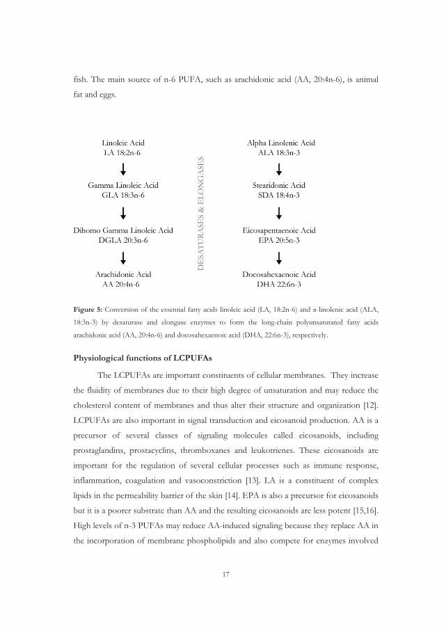
and n-6 PUFAs may be synthesized in the body from the essential FAs (Figure 5),
however this conversion is not very efficient and therefore it is important to obtain
these from the diet as well. The main dietary source of n-3 PUFAs, such as
eicosapentaenoic acid (EPA, 20:5n-3) and docosahexaenoic acid (DHA, 22:6n-3), is oily
17
fish. The main source of n-6 PUFA, such as arachidonic acid (AA, 20:4n-6), is animal
fat and eggs.
Figure 5: Conversion of the essential fatty acids linoleic acid (LA, 18:2n-6) and �-linolenic acid (ALA,
18:3n-3) by desaturase and elongase enzymes to form the long-chain polyunsaturated fatty acids
arachidonic acid (AA, 20:4n-6) and docosahexaenoic acid (DHA, 22:6n-3), respectively.
Physiological functions of LCPUFAs
The LCPUFAs are important constituents of cellular membranes. They increase
the fluidity of membranes due to their high degree of unsaturation and may reduce the
cholesterol content of membranes and thus alter their structure and organization [12].
LCPUFAs are also important in signal transduction and eicosanoid production. AA is a
precursor of several classes of signaling molecules called eicosanoids, including
prostaglandins, prostacyclins, thromboxanes and leukotrienes. These eicosanoids are
important for the regulation of several cellular processes such as immune response,
inflammation, coagulation and vasoconstriction [13]. LA is a constituent of complex
lipids in the permeability barrier of the skin [14]. EPA is also a precursor for eicosanoids
but it is a poorer substrate than AA and the resulting eicosanoids are less potent [15,16].
High levels of n-3 PUFAs may reduce AA-induced signaling because they replace AA in
the incorporation of membrane phospholipids and also compete for enzymes involved

18
in the eicosanoid synthesis, and thereby contribute to an anti-inflammatory effect [17].
DHA is the most abundant n-3 PUFA in tissues, especially in brain and retina [14] and
it is important in fetal development.
The PUFAs may also regulate gene expression due to their effect on several
transcription factors. Both n-3 and n-6 PUFAs are natural ligands for the transcription
factors PPARs and SREBP-1 which control various genes of inflammatory signaling
and lipid metabolism [17].
Importance of LCPUFAs in fetal nutrition
The fetus depends on maternal supply of essential FAs and LCPUFAs. Even if
the essential FAs (LA and ALA) are obtained from the maternal circulation, they must
be elongated and desaturated to be converted into LCPUFAs. Basal expression of �-5
and -6 desaturase and elongase have been reported both in fetal liver and in the placenta
[18,19], however their enzyme activities are low. Both AA and DHA are important
structural components of the nervous system and adequate supply of these could be
critical at the time of embryonic organogenesis as well as during the growth of the fetal
brain, which is at its peak in the last trimester [20].
Supplementation of pregnant women with ALA did not lead to increased DHA
levels in the umbilical cord [21]. On the other hand, several studies report that intake of
EPA and DHA by pregnant women raises the content of these FAs in fetal tissues [22].
Increased consumption of n-3 LCPUFAs from fish and fish oils during pregnancy has
also been suggested to be beneficial to the fetus and to lower the risk of PE [23,24].
Hence, an adequate maternal dietary intake of LCPUFAs and subsequent adequate
transport across the placenta is critical for the development for the fetus.
Fatty acid metabolism in the placenta
During the first trimester of pregnancy there is an accumulation of maternal
body fat that allows accumulation of LCPUFAs in adipose tissue, which can be
mobilized in the latter half of the pregnancy and transferred to the fetus. FAs are mainly
stored in the body as TAGs. In order to be utilized by the cells as fuel, the FAs are
processed in three steps. First the TAGs are degraded to FAs and glycerol by lipase
enzymes in a process called lipolysis, which takes place on the outer plasma membrane
19
of the cell. The FFAs are then bound to serum albumin which transports them across
the membrane. Inside the cell, on the outer membrane of the mitochondria, the FAs
must be activated by the attachment of coenzyme A (CoA) before they can be
transported into the mitochondria where they are degraded. This activation is catalyzed
by the enzyme acyl-CoA synthetase (ACS). In the degradation process the FA is
oxidized to introduce a double bond. This double bond is subsequently hydrated to
introduce an oxygen atom and thereby the FA is converted to an alcohol. The alcohol is
then oxidized to a ketone and finally a two-carbon unit is cleaved off by CoA to yield
acyl-CoA and a FA, which is two carbon atoms shorter than the original FA. This
process can be repeated until the FA is completely converted in to acyl-CoA. FA
degradation and synthesis are reverse processes regulated in response to diet by a host
of different hormones and enzymes.
Cholesterol
Cholesterol is an important component of structural membranes and a precursor
of steroid hormones and oxysterols. Cholesterol is distributed in cells as free cholesterol
in the plasma membrane and internal membranes and as cholesteryl esters stored in
lipid droplets [25,26]. The plasma membrane is highly enriched in cholesterol which
constitutes about 30 mole percent of the lipids [27] and contributes to the rigidity of the
membrane and the organization of specialized membrane domains called lipid rafts.
Cells need a constant supply of cholesterol in order to maintain their membranes [28],
but on the other hand accumulation of excessive free cholesterol is toxic to the cell
[26,29]. Correlation between maternal hypercholesterolemia and fatty streak formation
in fetal aorta [30] suggests the existence of a placental cholesterol transport system.
1.3. Fatty acid transport in the placenta
Fetal lipid deposition increases exponentially during gestation and 90% of the
fat is deposited in the last 10 weeks of pregnancy (reaching 7 g/day) [31]. The fetus is
capable of synthesizing FAs, however it obtains most of its FAs from the maternal
circulation via the placenta [32]. The barrier between the maternal and fetal circulation
consists of trophoblast cells connective tissue and fetal endothelial cells. All these cell

20
layers may contribute to the transport across the placenta; in addition the placental
metabolism also contributes to transfer of nutrients from the maternal to the fetal side.
There are different mechanisms involved in placental transport such as simple
diffusion, facilitated diffusion and active transport [1].
Most maternal FAs are transported to the fetus as TAGs in lipoprotein particles
[33]. TAGs cannot directly cross the placental barrier and consequently a complex
system of placental transport has developed. This system involves several receptors and
enzymes such as LDL receptor, VLDL/apoprotein E receptors, placental lipoprotein
lipase, placental phospholipase A2 and intracellular lipases [33,34,35,36,37,38,39,40].
Alterations in placental LPL activity and LDL receptor protein expression are
associated with IUGR [41,42,43,44], and imply that the placental lipid transport system
is important for adequate fetal growth.
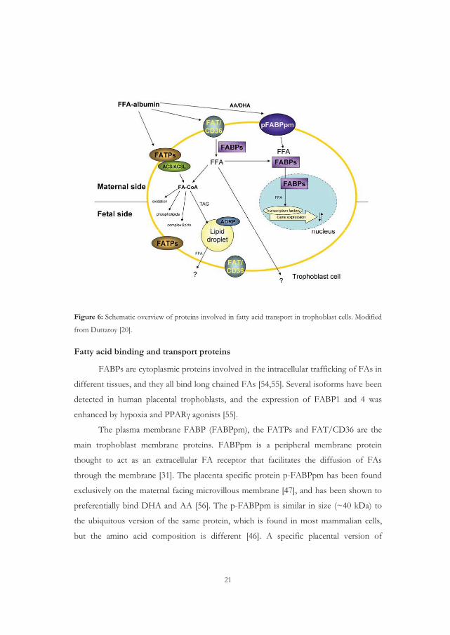
Maternal FAs, as well as FAs newly synthesized in the placenta, are transported
across the trophoblasts by diffusion or by active transport [45,46]. The existence of a
complex FA transport system comprising multiple membrane and cytoplasmic proteins
responsible for FA transport and metabolism in human placenta has been demonstrated
[47], including fatty acid binding proteins (FABPs) and CD36 (also named FAT for
fatty acid transporter), and fatty acid transport proteins (FATPs) (Figure 6).
Preferential uptake of LCPUFAs
Fetal blood is especially enriched in LCPUFAs compared to the maternal
circulation at the time of birth [48,49,50,51], but how this selective enrichment occurs is
largely unknown. Active transfer of FAs from the maternal circulation to the fetus has
been demonstrated by in vitro and in vivo experiments. Pregnant women were
administered [13C]-labeled FAs four hours previous to elective cesarean section, and the
[13C]-labeled FAs were detected in both placental tissue and cord blood at the time of
delivery [48]. Preferential transfer of LCPUFAs has also been demonstrated using
perfused human placenta [52,53].
21
Figure 6: Schematic overview of proteins involved in fatty acid transport in trophoblast cells. Modified
from Duttaroy [20].
Fatty acid binding and transport proteins
FABPs are cytoplasmic proteins involved in the intracellular trafficking of FAs in
different tissues, and they all bind long chained FAs [54,55]. Several isoforms have been
detected in human placental trophoblasts, and the expression of FABP1 and 4 was
enhanced by hypoxia and PPAR� agonists [55].
The plasma membrane FABP (FABPpm), the FATPs and FAT/CD36 are the
main trophoblast membrane proteins. FABPpm is a peripheral membrane protein
thought to act as an extracellular FA receptor that facilitates the diffusion of FAs
through the membrane [31]. The placenta specific protein p-FABPpm has been found
exclusively on the maternal facing microvillous membrane [47], and has been shown to
preferentially bind DHA and AA [56]. The p-FABPpm is similar in size (~40 kDa) to
the ubiquitous version of the same protein, which is found in most mammalian cells,
but the amino acid composition is different [46]. A specific placental version of

22
FABPpm has been described [57] located on the microvillous membrane of the placenta
facing the fetal circulation [58].
FATPs are integral proteins with membrane spanning regions and are thought to
function as FAs transporters. There are six members of this family identified so far and
they differ in tissue expression, subcellular location and substrate specificity
[59,60,61,62,63,64]. FATP1, 2, 3, 4 and 6 have been shown to be expressed in placenta
[65,66]. FATP1 and 4 have also been found expressed in trophoblast cells [66]. The
FATPs have inherent ACS activity, which enables catalyzation and conversion of FFAs
into acyl-CoAs [67]. It has also been reported that FATP1 and 4 is upregulated by
PPAR�/RXR agonists in primary human trophoblasts [68].
FAT/CD36 is a transmembrane FA transporter and scavenger receptor for
oxidized LDL [47,69,70,71]. It is expressed in human trophoblast cells and is involved
in the uptake of long chain FAs in placenta [47,72]. FAT/CD36 was also shown to be
associated with lipid rafts (which are microdomains of the plasmamembrane rich in
cholesterol and sphingolipids and important in cell signaling) in adipocytes [73]. Lipid
rafts are important for virus-induced syncytium formation [74], but it is not known
whether they are involved in comparable processes in syncytiotrophoblast formation in
placenta. Their presence in syncytiotrophoblast was demonstrated for the first time in
work by Linton and colleagues [75,76].
Caveolin-1 is another protein involved in cellular uptake of FAs and cholesterol.
It is the main structural element of caveolae [77], a subclass of lipid rafts forming
characteristic flask shaped invaginations that can be distinguished by electron
microscopy. Caveolae are dynamic structures that can bud from the plasma membrane,
forming cytoplasmic vesicles involved both in receptor-mediated uptake of solutes into
the cell [78] and in transcytosis (vesicular transport through the cell membrane) [79].
The unique lipid raft environment attracts key signaling proteins, such as G proteins,
protein kinase C, protein kinase A, prostacyclin synthase and endothelial nitric oxide
synthase (eNOS) [80,81]. Recent studies have shown that caveolin-1 can associate with
lipid bodies in a reversible and lipid regulated fashion [82,83]. Caveolin-1 can also bind
FAs [84], and some data imply that lipid rafts might be involved in regulating LCPUFA
uptake [85,86,87,88,89].
23
Long chain acyl-CoA synthetases (ACSLs)
The acyl-CoA synthetases are enzymes necessary for de novo lipid synthesis, FA
degradation and remodeling of membranes. They activate FAs by converting them into
membrane impermeable acyl-CoAs, this facilitates FA transport by trapping the FAs
inside cells. The acyl-CoAs have numerous metabolic fates within cells, including
incorporation into triacylglycerols (TAG) and membrane phospholipids, as substrates
for ß-oxidation and protein acylation, and as ligands for transcription factors. They have
been found in all organisms investigated and have an essential role that has been
conserved through evolution [90]. The ACSLs can be divided into five subfamilies
based on their FA chain length preference; acyl-CoA synthetase short chain (ACSS, C2-
C4), medium chain (ACSM, C4-C12), long chain (ACSL, C12-C20), bubblegum
(ACSBG, C14-C24) and very long chain (FATPs, C18-C26) [90,91].
Five genes have been identified in the ACSL family. They are named ACSL1 and
3 to 6 and vary in tissue distribution, intracellular locations and regulation, implying that
the different isoforms have distinct functions [92,93,94]. It has been suggested that the
different ACSLs direct the FAs into distinct metabolic pathways [95]. Presently, there is
little knowledge about the role and function of these proteins in placental FA uptake.
Lipid droplets and associated proteins
Most mammalian cells are able to store neutral lipids in intracellular lipid
droplets. In addition to serving as lipid storage depots, lipid droplets appear to
participate in lipid homeostasis, cell signaling, intracellular vesicle trafficking, and
disease processes [96,97,98,99,100]. The structure of the lipid droplets is similar to
lipoproteins; a neutral lipid core surrounded by a phospholipid and cholesterol
monolayer onto which the lipid droplet associated proteins (LDAPs) are attached [98].
The LDAPs (recently named perilipins [101]) consist of a group of 5 proteins with
sequence homology that are associated with lipid droplets and include S3-12, LDAP5,
TIP-47, perilipin and ADRP. Many other proteins have also been found associated
with lipid droplets, including caveolin-1. ADRP (also called perlipin
2/ADFP/adipophilin) belongs to a group of PAT family proteins (perilipin, ADRP and
tail-interacting protein of 47 kDa (TIP-47)) [102]. Perilipin expression is mainly
confined to adipocytes while ADRP and TIP-47 expression is widely distributed in

24
different tissues [103]. Perilipin has a well-defined role as a regulator of TAG lipolysis
in adipocytes, where it stimulates translocation of hormone sensitive lipase. The roles
of ADRP and TIP-47 are, on the other hand, still unclear.
ADRP (Adipose Differentiation Related Protein)
ADRP is a 48-50 kDa lipid droplet associated protein expressed widely in
different cells and tissues that store or synthesize lipids and is extensively used as a
marker for lipid droplets. The protein co-localizes with the surface of neutral lipid
droplets inside the cell and is assumed to play a role in uptake, transport and storage of
lipids [104]. ADRP is highly expressed in adipose tissue and is induced early during
adipocyte differentiation [105,106]. ADRP mRNA expression is increased at the
transcriptional level in the presence of FAs in preadipocytes [107]. Furthermore, ADRP
binds to FAs [108] and cholesterol [109]. ADRP is highly expressed in placenta tissue
on mRNA level [110]. It is expressed in human villous trophoblasts and both mRNA
and protein expression is enhanced during differentiation of cytotrophoblasts into
syncytiotrophoblast [111].
Regulation of ADRP expression suggests it to be a PPAR/RXR target gene, both
in adipose cells and placental trophoblasts [111]. Primary trophoblasts in culture
spontaneously differentiate, and accumulate lipids in the form of small lipid droplets
[112]. PPAR activators, including oxidized lipids, have been shown to promote
trophoblast differentiation [112,113]. ADRP is expressed in human villous trophoblasts
and in concordance with the accumulation of lipid droplets, the mRNA and protein
expression is enhanced during differentiation of cytotrophoblasts into
syncytiotrophoblasts [111]. Recently it was reported that ADRP is a direct LXR target
gene with several LXRE response elements, and both mRNA and protein expression of
ADRP is increased in hepatocytes treated with the synthetic LXR agonist GW3965
[114].

25
1.4. Nuclear receptors
Transcription factors are proteins involved in the regulation of gene
transcription; they include about 10% of all human genes and are the largest family of
human genes [115]. The nuclear receptor (NR) super family is a diverse group of
evolutionary related DNA binding transcription factors, and 48 different types are
identified in humans [116]. Most of the NRs are ligand-dependent but there is still a
large number of orphan receptors, meaning NRs without any known ligands [117]. The
ligands are small hydrophobic molecules that include FAs, cholesterol derivatives,
retinoids, thyroid hormone, prostaglandins, leukotrienes and xenobiotics [118]. Because
the NRs are dependent on ligands for activation, they have essential roles in
communication between the cell/body environment and the genome. They have vital
roles in a variety of biological processes such as development, reproduction,
homeostasis, inflammation and metabolism [118,119,120].
NRs share a characteristic structure that consist of five to six homologous
domains [116], with different functions that ensures site-specific binding to DNA and
binding of ligands and cofactors [121]. Over 300 cofactors that increase or repress the
transcription of genes have been identified [122,123]. In addition to regulation by
ligands and cofactors, NRs can also be modified by phosphorylation, glycosylation,
methylation, acetylation, ubiquitinylation and small ubiquitin-like modulation [122].
The NRs can be divided into several subfamilies depending on sequence
homology, ligand sources or physiological functions [116,124]. They are usually
classified according to the DNA-binding and dimerization properties. This classification
consists of four different groups [117]. Class 1 receptors include homodimeric steroid
hormone receptors. Class 2 receptors are ligand-dependent and form heterodimers with
retinoid X receptors (RXRs). Class 3 and 4 include orphan receptors, homodimers and
monomers respectively. In this study we were interested in the Class 2 receptors
peroxisome proliferator activated receptors (PPARs) and liver X receptors (LXRs).
Peroxisome Proliferator Activated Receptors (PPARs)
The PPARs have distinct tissue distribution and control a vast array of genes
involved mainly in the lipid metabolism, but are also involved in other cellular processes
such as inflammation and cellular differentiation [125,126,127,128]. The PPARs have

26
three distinct members PPAR�, � and � [129,130]. PPAR� is expressed in metabolically
active tissues including the liver, heart, kidney and skeletal muscle [131] where it
controls lipid catabolism and transport. PPAR� is mainly expressed in adipose tissue,
macrophages, colon and placenta [131]. It is essential for adipocyte differentiation
[132,133] and activates genes that promotes fat storage and reduces serum lipid levels.
Other effects of PPAR� are improved glucose homeostasis and decreased inflammation
[131]. PPAR� is expressed throughout the body [131] and is involved in many biological
processes such as cholesterol transport.
Natural activators for PPARs include medium and long-chained FAs [134,135],
oxidized metabolites of linoleic acid from oxLDL [136] and eicosanoids [137,138,139].
Synthetic ligands for PPAR� are fibrates [137] that are used therapeutically in humans
for lowering hepatic production of triglycerides by increasing FA oxidation [131], and as
anti-inflammatory drugs [140]. Thiazolidinediones are synthetic ligands for PPAR� that
have been used therapeutically in humans to increase insulin sensitivity [141,142].
Stimulation of PPAR� activity by these ligands, can among other functions,
enhance the transcription of CD36/FAT, leading to further uptake of oxLDL and
differentiation of monocytes into foam cells [136,143,144].
Liver X Receptors (LXRs)
LXRs play key roles as regulators of lipid and glucose metabolism [145]. In the
lipid metabolism they regulate de novo FA synthesis, TAG synthesis, LDL synthesis and
metabolism, and cholesterol homeostasis [146,147,148]. They are also involved in the
pathogenesis of many diseases including atherosclerosis, diabetes and inflammation
[149].
LXRs consist of two isomers, termed alpha and beta, which share considerable
sequence homology and are activated by the same ligands [150]. LXR� is ubiquitously
expressed, while LXR� expression is restricted mainly to tissues involved in lipid
metabolism such as the liver and adipose tissue [151].
The natural ligands that activate LXRs are oxysterols [152,153]. Oxysterols are
oxidized derivatives of cholesterol that are present in oxLDL [154], and maternal
plasma oxLDL has been associated with increased in maternal circulation in PE [155].
LXRs can also be induced by non-steroidal synthetic ligands such as T0901317

27
(Tularik) and GW3965 [135,156]. Activation of LXR by synthetic ligands results in a
reverse transport of cholesterol from peripheral tissues to the liver, and has been
shown to inhibit the development of atherosclerosis in mice [157]. This has made LXR
a promising target for treatment against atherosclerosis; however, there are undesirable
side-effects such as increased hepatic lipogenesis leading to hepatic steatosis [156].
There are also natural ligands with antagonizing effects against LXRs, such as
PUFAs. These FAs inhibit the activation of LXR by competing with the activating
ligands in the order ARA>EPA>DHA>ALA, whereas saturated and monounsaturated
FAs have very little effect on the activation of LXR [158].
PPARs and LXRs in placenta
All three PPAR isoforms have been detected in human placenta and in placental
trophoblast cells [159]. Both the PPARs and the RXRs are involved in several aspects of
pregnancy development such as implantation, placentation, trophoblast invasion and FA
uptake [160]. Studies of PPARy knock-out mice have shown that abnormal
development of the placenta results in embryonic death at mid-gestation [161], and that
PPARy/RXR heterodimers are essential for differentiation of trophoblast cells. PPARy
and RXR agonists also increase the differentiation of isolated primary human
cytotrophoblast cells [162]. Cytotrophoblast differentiation is characterized by increased
hCG production and hCG has been shown to be a direct PPARy target gene and its
expression is increased by PPARy [112,162]. It has also been shown that PPARy has a
role in trophoblast invasion. In an in vitro invasion assay, using extravillous
cytotrophoblast cells, it was shown that both natural and synthetic PPAR ligands
inhibited trophoblast invasion [163].
LXRs have also been shown to be involved in placentation and trophoblast
invasion. Both oxysterols and synthetic LXR agonists inhibited invasion of extravillous
cytotrophoblasts in vitro [164]. The anti-angiogenic protein endoglin, which is increased
in maternal circulation in PE [165,166], was identified as a direct LXR� target gene in
the placental cell line JAR [167]. Endoglin is highly expressed in syncytiotrophoblast and
has been shown to inhibit trophoblast invasion [167]. Weedon-Fekjaer et al found that
LXR increased the synthesis of FAs and inhibited the secretion of hCG in placental
BeWo cells [168]. The role of LXRs in cholesterol transport in the placenta has been
28
investigated in human placental endothelial cells and it was found that these cells had
increased cholesterol efflux after LXR activation compared to human umbilical vein
endothelial cells (HUVEC) [169].
1.5. Preeclampsia
Preeclampsia (PE) is a major complication of pregnancy characterized by
hypertension and proteinuria [170] developing in the second half of pregnancy [171]. PE
affects at least 3-4% of all pregnancies, representing a major threat to maternal and fetal
health, and responsible for approximately 50 000 maternal deaths annually [172]. The
severe forms of PE typically results in preterm delivery, low-birth weight and increased
risk of fetal morbidity and mortality [173].
Definitions
There are several definitions of PE in use, and in the papers included in this
thesis we have used a widely accepted definition from the American College of
Obstetrics and Gynecologists [170]. The criteria for diagnosis of PE are as follows:
1) Hypertension is defined as “blood pressure of 140 mmHg systolic or higher or 90
mmHg diastolic or higher that occurs after 20 weeks of gestation in a woman with
previous normal blood pressure”.
2) Proteinuria is defined as “urinary excretion of 0.3 g protein or higher in a 24 h urine
specimen”. This corresponds to a protein dipstick reading of +1 or higher.
PE is unpredictable in its onset, progression and severity. It is sometimes divided
into early onset PE occurring prior to week 34 of gestation, and a late onset PE
occurring at or after 34 weeks of gestation [174,175]. PE is considered to be severe if
the blood pressure � 160/110, or proteinuria at 5 g/24 h (� +3 on dipstick) is present.
Eclampsia is a severe variant of the disease involving the occurrence of seizures in a
preeclamptic woman, where the seizures cannot be attributed to other causes. The
HELLP syndrome is another variant of PE [176], which includes hemolysis, elevated
liver enzymes and low platelet counts.
29
Intrauterine growth restriction (IUGR) or fetal growth restriction (FGR) is
defined as the failure of a fetus to reach its expected growth potential at any gestational
age [177]. The newborn birth weight percentiles were calculated according to national
birth registry data [178] or an ultrasound based weight percentile [179].
Gestational hypertension is defined as new onset hypertension � 140/90 after
week 20 of gestation but without proteinuria [170]. In cases of superimposed PE on
hypertension, the women have developed hypertension before week 20 (or
pregestationally), with new-onset proteinuria after week 20. Also, we defined
superimposed PE on diabetes mellitus (pregestational diabetes type 1 or 2, or gestational
diabetes mellitus) as PE developing in a pregnant woman already diagnosed with
diabetes mellitus (pregestational or gestational diabetes mellitus) according to WHO
criteria [180].
Pathophysiology
The exact pathophysiology of PE remains unknown. A “two-stage model” was
proposed by CW Redman in 1991 [181]. In the first stage there is poor placentation
while the second stage is the maternal syndrome diagnosed by hypertension and
proteinuria. Poor placentation includes abnormal implantation, inadequate remodeling
of the spiral arteries and thereby reduced or altered placental perfusion (Figure 7).
This altered blood flow to the placenta leads to oxidative stress, which in some
cases may be due to hypoxia. It is proposed that the oxidatively stressed placenta
releases different pro-inflammatory factors into the maternal circulation and thereby
causes the maternal syndrome. These suggested placental factors are pro-inflammatory
cytokines (TNF-�, IL-6 [182,183]), anti-angiogenic factors (sFLT1 and sEng
[165,184,185,186]), placental debris (such as syncytiotrophoblast microparticles: STBMs)
[187] and activated immune cells [188,189]. In the second stage it is thought that the
maternal circulating factors produced by the stressed placenta cause an excessive
systemic inflammatory maternal response [188] with generalized maternal endothelial
dysfunction [190], contributing to the maternal clinical features of PE [191,171].

30
Figure 7: Illustration of the uterine spiral arteries in a non-pregnant woman, in preeclampsia and in
normal pregnancy. In preeclampsia the trophoblast invasion and physiological transformation of the
spiral arteries are incomplete, resulting in an abnormal blood supply to the placenta. Modified from
Moffet-King [5].
Recently it has been proposed that the development of PE starts even earlier
than at the stage of spiral artery remodeling, with an increased release of proteins
already in the first trimester [192]. It has also been suggested that the blood volume to
the placenta is not reduced, as previously suggested [193]. Instead, the narrower, non-
transformed spiral arteries in PE cause the blood to enter the intervillous space at a
velocity much greater than normal. This, in turn, causes damage to the placental villi
both on a micro- and macroscopic level and may alter the placental morphology and
function [194].
Risk factors and treatment
Risk factors for PE include previous history of PE and a family history of PE,
primiparity, multiple pregnancy, obesity and chronic medical conditions such as
preexisting hypertension and diabetes [191].
Presently PE cannot be prevented and the only “treatment” is delivery of the
fetus with removal of placental tissue. If placenta tissue is retained after termination of
the pregnancy, PE can persist [195]. Also, removal of decidua after delivery with
31
curettage has shown beneficial clinical effects [196,197]. Treatment with
antihypertensive medication may serve to prolong the pregnancy [198], due to
improved maternal blood pressure control and reduced need for premature delivery.
There have been a number of treatment and primary as well as secondary prophylactic
intervention studies. Antioxidant supplementation using vitamin E and C did not
reduce the incidence in a large randomized controlled trial (RCT) [199], whereas a
previous smaller RCT was promising [200]. Calcium supplementation has also proven
ineffective, but could be useful in developing countries where nutritional levels are
insufficient [201]. Also, n-3 FAs have been suggested to play a role in the prevention of
PE, but the evidence for this is not conclusive. There have been three randomized trials
where fish oil supplementation was given to high risk women, but none these trials
reported any reduction in PE [202,203]. Although acetyl salicylic acid (aspirin) has
shown to reduce the incidence of PE in some studies, without augmenting
complications such as placental abruption, there is hitherto no conclusive evidence as
to which group of pregnant women that would benefit from such an intervention [204].
Inflammation
Normal pregnancy is a state of mild systemic inflammatory response, but the
physiological basis for this is not known except that the phenomenon arises from the
placenta itself [205]. PE is associated with a more extreme maternal inflammatory
response than occurs in normal pregnancy [188]. How the problems of abnormal
placentation generates the systemic inflammatory problems of PE remains to be
explained, but it is thought that release of pro-inflammatory factors from the syncytial
surface of the placenta into the maternal circulation could be important, and could
possible be the link between stage 1 and 2 of PE [205]. Syncytiotrophoblast
microparticles (STBMs) are vesicles shed from the placenta into the maternal circulation
during pregnancy. In PE there is a significant increase in the amounts of particles that
are shed [187]. These particles have an anti-endothelial effect and stimulate release of
pro-inflammatory substances form the endothelium [187]. The pro-inflammatory
cytokines tissue necrosis factor (TNF)-� and interleukin (IL)-6 are both elevated in
preeclamptic circulation [182]. It is suggested that the placenta contributes to the
elevated plasma cytokine levels, but the dysfunctional maternal endothelium, peripheral

32
blood mononuclear cells and other tissues, such as the adipose tissue, are also likely to
be involved [206].
Oxidative stress
Reactive oxygen species (ROS) are highly reactive molecules that contain an
oxygen atom. ROS can be free radicals with unpaired electrons such as superoxide (O2·-)
and hydroxyl anions (OH·), or non-radical intermediates such as hydrogen peroxide
(H2O2). ROS are natural byproducts of oxygen metabolism and have important
physiological roles in the regulation of cell signaling, for instance in the regulation of
nitric oxides and vascular tone. However, if the concentration of ROS becomes
excessive, due to different environmental stresses, and an imbalance between the level
of ROS and the level of antioxidants arises, then ROS can start attacking lipids, proteins
or DNA. These attacks cause chain reactions that lead to widespread damage and loss
of function in cells, and this situation is referred to as oxidative stress.
Pregnancy is a state of excessive oxidative stress arising from increased placental
mitochondrial activity and production of reactive oxygen species and decreased
expression and activity of antioxidants [207]. In PE there is an increased level of
oxidative stress, which results from ischemia-reperfusion injury that in turn is caused by
the altered perfusion of the placenta due to the abnormal placentation [208]. The
oxidative stress of PE is not restricted to the placenta but is dispersed in the maternal
circulation and is a part of the systemic inflammatory response [205].
In placenta the evidence for excessive oxidative stress in PE includes finding of
increased generation of lipid peroxides and isoprostanes [207,209,210,211], xanthine
oxidase [212] and nitro tyrosine residues [213], which indicates an excessive production
of superoxide. In the maternal circulation the evidence include increased superoxide
production from circulating neutrophils [214]. It is also known that TNF-�, which is
increased in the circulation in PE [182], can induce oxidative stress directly [215] or
indirectly by enhancing the levels of oxLDL [216] or through the xanthine oxidase
pathway [217]. Placental oxidative stress is viewed as a key intermediary step in the
pathophysiology of PE, as a mediator of the endothelial cell dysfunction [191,218].
Hypoxia is a situation where the oxygen tension is too low and this may result in
oxidative stress. It has been suggested that hypoxia is the mediator of the pathological

33
changes observed in pregnancy complications such as PE, however there is limited
evidence to support this [10]. Burton et al proposed instead that it may be fluctuations in
the oxygen level in the intervillous space that creates the pathological changes due to
ischemia-reperfusion stress in PE [219].
Dyslipidemia
The dyslipidemia of PE is an amplification of the lipid changes observed in
normal pregnancies. It includes elevated cholesterol and triglycerides, increased
circulating FFAs, reduced high density lipoproteins (HDL) and increased concentrations
of small LDL which leads to the presence of oxLDL in maternal circulation
[220,221,222,223,224,225], while total and LDL cholesterol levels are not considerably
different [221,222]. Augmented circulating maternal concentrations of the oxidized lipid
8-isoprostanes are also reported in PE [226,227].
The maternal dyslipidemia is present already in the first and second trimester of
gestation and is evident before the clinical detection of PE [228,229,230,231]. A rise in
circulating TAG concentrations is present [232] as early as 10 weeks of gestation [233].
A dose-response effect of TAG has been observed, with a four-fold elevated risk of
developing PE in women with the highest circulating levels of TAG compared to
normal levels [234]. Even though hypertriglyceridemia may contribute to the
development of PE, therapeutic intervention is probably not a good alternative, as strict
correction of maternal hypertriglyceridemia in rodents has been shown to have negative
effects on fetal growth and development [235].
The lipid abnormalities in PE are similar to abnormalities as observed in patients
with cardiovascular disease [236,237]. The two diseases also have several other risk
factors in common, including obesity, diabetes mellitus, insulin resistance and
endothelial dysfunction [236,238]. Also, the phenomenon of “acute atherosis” of the
decidual/uterine spiral arteries, which is more often seen in PE than in uneventful
pregnancies [239], closely resembles the early stages of atherosclerotic lesions found in
cardiovascular disease [240].
34
Lipotoxicity
Accumulation of excess lipids in non-adipose tissues is termed lipotoxicity and
can lead to cell dysfunction and death [241]. Lipotoxicity has been suggested to play a
role in insulin resistance and hyperlipidemia [242], which are also features of PE and
diabetes mellitus. The combination of hypoxia, oxidative stress and increased lipid
concentrations observed in PE may result in excess lipid peroxidation products. Lipid
peroxides and oxygen free radicals stimulate peroxidation reactions that damage cells
and cell membranes. These effects include alterations in membrane fluidity and
permeability and endothelial cell injury and dysfunction [243]. Oxidized lipids, such as
the endogenously produced 9S-hydroxy-octadecadienoic acid (9-HODE), 13S-hydroxy-
octadecadienoic acid (13-HODE), and 15S-hydroxy-eicosatetraenoic acid (15-HETE),
are particularly relevant to trophoblast biology, in which they are implicated in
trophoblast injury [32,244]. PE has been associated with enhanced lipid peroxidation in
trophoblasts [245,246] and there has been demonstrated an increased production of 15-
HETE in vitro from trophoblasts derived from preeclamptic women [247,248]. 8-iso-
PGF2�, a lipid peroxidation product, is a well-known marker of oxidative stress and is
elevated in the maternal circulation as well as in placental/decidual tissue in PE
[226,209,210]. Staff/Halvorsen et al demonstrated an accumulation of fat in the
trophoblast cell line JAR, when incubated with 8-iso-PGF2�, as well as reduced
trophoblast invasion [249], which could suggest a possible in vivo effect of this oxidized
lipid in PE.
Acute atherosis
Acute atherosis is defined as accumulation of CD68 positive foamy macrophages
in the uteroplacental spiral arteries, including areas of fibrinoid necrosis [250,251]. The
name acute atherosis is derived from atherosclerosis because the phenomenon
resembles early atherosclerotic changes of other systemic blood vessels. These areas of
lipid deposition are found in non-transformed spiral arteries and have been associated
with PE, although it is not specific for this pregnancy complication [252,253,254].
Acute atherosis in spiral arteries are associated with augmented risk for local thrombosis
and thereby necrosis in the placental tissue underlying the plugged arteries, adding to the
reduced placenta function more common in PE than in uneventful pregnancies.

35
Presently there is little knowledge of the molecular mechanisms behind the formation of
acute atherosis, however it has been suggested that the hyperlipidemia in the maternal
circulation in PE could participate in the lipid changes in the spiral arteries [223]. Also, it
is probable that reduced transformation of the spiral arteries could contribute to the
lipid deposition in the narrow parts of untransformed spiral arteries [255].
Diabetes mellitus in pregnancy
Diabetes mellitus and PE share many pathophysiological features, including insulin
resistance, endothelial dysfunction, oxidative stress, and inflammation [256,257,258].
Both pregestational diabetes and gestational diabetes mellitus (GDM) are associated
with a two- to four-time increased risk of developing PE in pregnancy [259,260,261].
Gestational diabetes and pre-pregnancy obesity is associated with large babies, while PE
is associated with growth restricted babies [262]. Superimposed PE on diabetes mellitus
(SPE) includes women with diabetes mellitus (preexisiting or gestationally induced) that
develop PE in the present pregnancy. SPE present a higher risk for poor perinatal
outcome and placental abruption than PE alone [263].
The development of insulin resistance [264] together with adipose tissue
accumulation [265] in the third trimester of pregnancy is a possible adaptation of the
maternal metabolism to optimize fetal nutrition. Insulin inhibits hormone sensitive
lipase and thereby decreases triglyceride hydrolysis in the adipose tissue resulting in
reduced circulation of FFA and glycerol. Insulin resistance thus increases the activity of
hormone sensitive lipase and results in increased levels of lipoproteins and FFA in the
circulation. Gestational insulin resistance is accentuated in PE [266], and can be
observed weeks before the clinical onset of PE [267,268]. Furthermore, placenta
secretes a variety of hormones that is suggested to play a role in gestational insulin
resistance [269]. However the role of adipose tissue could also be important. Adipose
tissue has an endocrine function, secreting several metabolically active proteins such as
leptin, resistin, adiponectin, TNF-� and IL-6, termed adipokines [270]. During
pregnancy, the placenta is an additional source of adipokines, such as leptin and resistin
[271,272].

36
2. Aims of present study
The main aim of this study was to increase the understanding of the role of
trophoblasts and placenta in lipid transport and storage in general, and in dyslipidemic
pregnancies specifically. The first objective was to study the transport of FAs across the
trophoblast cells, in order to gain a better understanding of how the selective
enrichment of LCPUFAs in the fetal circulation occurs. The second objective was to
explore the role of lipid transport and storage associated proteins in the placenta in
dyslipidemic pregnancies, such as pregnancies complicated by PE and/or diabetes
mellitus, with a particular focus on the lipid droplet associated protein ADRP.
Specifically, the following questions were addressed:
1. Is there any difference in the transport of LCPUFAs compared to that of non-
essential FAs across trophoblast cells?
2. Is there any difference in the uptake and storage of LCPUFAs compared to that
of non-essential FAs in trophoblast cells?
3. Does LCPUFAs influence the uptake of FAs in trophoblast cells?
4. If so, which lipid metabolism genes are involved in the LCPUFA influenced
uptake of FAs in BeWo cells?
5. Are the transcription factors LXR and PPAR dysregulated in PE in placenta,
decidua and adipose tissue?
6. Is there any dysregulation of the expression of lipid droplet associated proteins
such as ADRP and FA transport/binding proteins such as FATPs and FABPs in
placenta in pregnancies complicated by PE or diabetes mellitus?
7. Where in the placenta is ADRP protein expression localized?
8. Is ADRP expression in trophoblast cells regulated by FAs and/or oxidative
stress?
37
3. Summary of Papers
Paper 1: Long-chain polyunsaturated fatty acid transport across human placental
choriocarcinoma (BeWo) cells
LCPUFAs are critical for the growth and development of the fetus. In the first
paper our aim was to investigate the mechanism behind the differential transport of
LCPUFAS and non-essential FAs assumed to take place in trophoblast cells. We used
the BeWo cell line as model of placental trophoblasts and a transwell cell system to
study the transport of radiolabeled FAs across these cells.
Results:
� BeWo cells incubated with OA contain more TAGs, more lipid droplets and
have higher ADRP (lipid droplet marker protein) expression than cells incubated
with DHA.
� Incubation of the FAs together with triacsin C (an inhibitor of the esterification
of FAs into acyl-CoA) abolished the TAG accumulation and the expression of
ADRP.
� Caveolin-1, a structural protein of lipid rafts in the plasma membrane, and also
believed to partake in FA uptake, was induced by OA, but expression was not
affected by triacsin C.
� Radiolabeled DHA and AA was more efficiently transported across the cell layer
than OA and PA, the concentration of DHA and AA in the basolateral chamber
was ~4 and ~2.5 fold higher than for OA, respectively. OA was similar to PA.
� A physiological FA mix was used to mimic the plasma concentration of FAs in
the third trimester of pregnancy, the concentrations of PA and OA was ~20-fold
and ~10-fold higher than for DHA and AA, and we found that the relative
transport of LCPUFAS was more efficient.
� Triacsin C inhibited the uptake of radiolabeled PA and OA by ~70%, whereas
uptake of LCPUFAs was inhibited by only 20%.
� Triacsin C increased the efflux of radiolabeled OA compared with DHA in a
transwell system.

38
Paper 2: Long-chain polyunsaturated fatty acids stimulate cellular fatty acid
uptake in human placental choriocarcinoma (BeWo) cells
In light of our findings in Paper 1 and the importance of LCPUFAs for fetal
nutrition, our aim was to study the effect of LCPUFAs on the uptake of FAs in
placental trophoblast cells. Further we wanted to study the effect of LCPUFAs on the
expression of genes involved in FA uptake and lipid metabolism in trophoblasts. We
used BeWo cells and studied the uptake of radiolabeled FAs, as well as gene expression
by quantitative real-time RT-PCR.
Results:
� Preincubation of BeWo cells for 24 h with LCPUFAs increased the uptake of
FAs by ~20-50%. Preincubation with OA on the other hand did not significantly
change the FA uptake as compared with the uptake in untreated cells (controls).
� After preincubation with LCPUFAs, radiolabeled FAs were incorporated into
phospholipid fractions to a greater extent compared to cells preeincubated with
OA or controls, simultaneously there was a decreased incorporation of FAs into
the TAG fraction.
� The gene expression of long-chain acyl-CoA synthetases ACSL1 and ACSL5
were increased when BeWo cells were incubated with the LCPUFAs AA and
DHA compared to both OA and control. Incubation with EPA also increased
the expression of these genes compared to the control.
� The gene expression of the lipid droplet associated protein ADRP was increased
by AA, EPA and DHA compared with the control.
� The gene expression of ACSL3 and LPIN1 was decreased after incubation with
all the FAs including OA compared with the control.
39
Paper 3: Expression of liver X receptors in pregnancies complicated by PE
We wanted to move from the placental trophoblast cell model to explore the
expression of placental genes involved in lipid metabolism in the placenta, and explore
whether any of these genes were dysregulated in PE. In the third paper we focused on
transcription factors LXRs (� and �) and PPARs (� and �) and their target genes. We
examined tissue samples from uncomplicated and preeclamptic pregnancies and studied
the gene expression in placenta, decidua and subcutaneous fat tissue. The placental lipid
classes and FA profile were also analyzed.
Results:
� The expression of LXR� and � were similar in all three gestational tissues.
� PPAR� had a higher expression in placenta than in decidua and fat (~20 fold
higher) while PPAR� was more highly expressed in fat tissue than the other
tissues.
� The expression of LXR� and � and PPAR� was significantly decreased in
preeclamptic placenta, while PPAR� was not differentially expressed as
compared to uneventful pregnancies.
� The expression of the target genes CD36/FAT, and APOE was significantly
decreased in preeclamptic placenta, while the expression of LDL receptor was
increased.
� Protein expression of LXR � was decreased in preeclamptic placenta, while LXR
� was similarly expressed in PE and controls.
� The mean concentration of FFA in placenta with PE was significantly lower than
in control placenta.
� The total concentration of n-6 PUFAs (LA, DGLA and AA) was increased in
preeclamptic placenta.
� There was a positive correlation between the gene expression of LXR� and the
concentration of FFA in preeclamptic placentas.

40
Paper 4: Increased Adipose Differentiation Related Protein (ADRP) expression
in preeclamptic placenta
In the fourth paper we explored the expression of genes involved in lipid
metabolism, focusing on genes involved in FA transport in the placenta and exploring
whether there is any dysregulation of these in PE. We examined placenta tissue samples
from uncomplicated (n=33) and preeclamptic pregnancies (n=30), as well as
pregnancies complicated by DM (n=10) and SPE (n=6). We also incubated BeWo cells
with FAs, oxidative stress and inflammatory agents in order to mimic the preeclamptic
situation.
Results:
� The gene expression of the lipid droplet associated protein ADRP was increased
in preeclamptic placenta.
� The gene expression of FATP1 and CAV1 was decreased in PE placenta.
� In SPE (superimposed PE on top of diabetes mellitus) the expression of PLIN,
S3-12, LSDP5, FABP3 and 4 and FATP1 and 4 was increased.
� The protein expression of ADRP was increased in PE, while the expression of
caveolin-1 was unaltered.
� ADRP protein expression was localized to vacuoles within fibrinoid tissue in
placental tissue sections, and also in vesicles in trophoblast cells.
� Caveolin-1 protein expression was mainly localized to endothelial cells in
placental tissue sections and in some specimens in cytotrophoblast cells.
� The FAs OA and LA that are increased in maternal circulation in preeclamptic
pregnancies increased the expression of ADRP in BeWo cells on both gene and
protein level, while PA did not.
� Oxidative stress induced by hydrogen peroxide increased the expression of
ADRP in BeWo cells on both gene and protein level.

41
4. Methodological considerations
4.1. Advantages of combining clinical research and in vitro
experiments
The work presented in this thesis is based on analyses of blood and tissue
samples from pregnant women and on in vitro experiments in cell culture. Both
approaches have advantages and restrictions, but in general they complement each
other. Clinical material gives an overview of the actual situation in the patient
population, and screening of target genes or proteins in patient populations can be
useful both for generating hypotheses and for confirming them. Data obtained from
clinical material can pose interpretation challenges, because there can be huge individual
differences from one patient to another, and also there are many variables that cannot
be controlled. In addition, when studying a whole organ, such as the placenta, it is
important to remember that it consists of a number of different cell types that may have
very different roles and functions. In a tissue/organ, the different cell types
communicate with each in a paracrine manner by secreting messenger molecules, and
will also be subject to endocrine communication by hormones or other circulating
molecules from other organs. In cell experiments, on the other hand, it is possible to
control the experimental settings so that the influence of confounding factors will be
limited. However, cell experiments have the disadvantage of being pure model systems,
where the settings are usually far from the complex physiological situation that we try to
mimic. By combining the two systems it is possible to test hypotheses generated from
the complex in vivo situation in the purer in vitro situation, facilitating a more
comprehensive understanding of physiological processes, such as FA uptake and
transport in the placenta.
4.2. Cell system
The BeWo choriocarcinoma trophoblast cell line is used as a model of placental
trophoblast in this study. BeWo was the first commercially human trophoblast cell line
available, developed in 1968 [273]. It was prepared from a malignant gestational
42
trophoblast cancer (choriocarcinoma), which is composed entirely of
syncytiotrophoblast and cytotrophoblast cells [274]. BeWo cells grow as undifferentiated
cytotrophoblast but can be induced to differentiate into syncytiotrophoblast, and they
also secrete hCG [275]. BeWo cells also have similar properties of lipid transport and
metabolism as isolated primary cytotrophoblasts and syncytiotrophoblast for instance
polarized transport of LCPUFAs and expression of genes involved in lipid metabolism
[47,274,276]. BeWo cells have been widely used to study the uptake and transport of
various biological compounds in the placenta, such as amino acids [277,278,279,280],
glucose [281], cholesterol [282] and FAs [283,284]. BeWo cells can form confluent
monolayers when grown on permeable membranes, and demonstrate polarized
membrane expression of apical and basolateral protein markers and tight junction
formation [283]. Therefore we believe that BeWo is a good model for studying FA
uptake and transport in the placenta.
4.3. Patient selection In this study we have used clinical information and biological samples from an
ongoing biobank inclusion of patients with complicated or uncomplicated pregnancies
at Oslo University Hospital, Ullevål, Norway. This hospital has the largest delivery unit
in Norway, presently with over 7000 deliveries annually. The biobank collection was
started by our research group in 2001 and includes women with uncomplicated
pregnancies and women with pregnancies complicated by PE or diabetes mellitus. For
the papers included in this thesis, a total of 79 women with singleton pregnancies were
included from the biobank study. We chose to include patient samples where sufficient
tissue samples from placenta, decidua and subcutaneous fat were available, as well as
maternal plasma and serum. We had originally intended to measure the concentration of
lipid associated proteins in the maternal circulation; however, in the end we did not
include any such analyses in the papers included in this thesis.
All patients were delivered by cesarean section. None of the women were in
active labor, had ruptured membranes or clinical signs of infection at the time of
delivery. No women with chronic hypertension or renal disease were included. The
cesarean section was clinically indicated independently of the research study.

43
Uncomplicated pregnancies were used as a control group (n=33), and included
normotensive and otherwise healthy women undergoing elective cesarean section due to
breach presentation or psychosocial reasons.
The women diagnosed with PE (n=30) were previously healthy and
normotensive prior to pregnancy. The reasons for delivery by cesarean route in these
cases were that vaginal delivery was not appropriate due to disease progression or
severity and/or unfavorable cervical ripening. Of the 30 patients with PE three had
HELLP syndrome and two had both PE and clinically demonstrated IUGR.
The patients with superimposed PE on diabetes mellitus (n=6) were delivered by
cesarean route with the same clinical indications as the PE group. The women with
diabetes mellitus (n=10) were delivered by cesarean route with the same clinical
indications as the control group. Therefore the number of patients in this group is small,
as patients with DM are normally delivered vaginally.
Cases of superimposed PE on hypertension were not included in this Ph.D.
project because these cases are probably distinct from PE and may have different
pathophysiology [285].
All patients that agreed to participate in the study signed a voluntary informed
consent. They have the possibility to withdraw from the study at any time without
giving any reason and without any consequences regarding the relationship to Oslo
University Hospital, Ullevål. The cesarean section biobank study was approved by the
Regional Committee of Medical Research Ethics in Eastern Norway (REK Øst), with
permission from the Norwegian Data Inspectorate (Datatilsynet) and final approval by
the Ministry of Health (for the biobank).
4.4. Delivery mode
A major strength of our study is the relatively large placental sample size from a
clinically well-characterized study population. In addition, only pregnancies delivered by
cesarean section without any indications of labor were included in the study. Hence,
unpredictable effects of vaginal delivery such as oxidative stress can be avoided and will
not influence the blood samples or tissue material. We believe that this is an advantage
of our study protocol, since vaginal delivery is associated with oxidative stress in
44
maternal and fetal circulation [286,287,288]. Uterine contractions during labor leads to
intermittent utero-placenta blood that causes hypoxia-reperfusion injury and increased
oxidative stress [212,219]. Xanthine oxidase activity was increased in placental tissue of
laboring women [212]. Markers of oxidative stress and apoptosis were increased in
placental tissue after labor, and the expression of several genes and proteins were altered
due to vaginal delivery [219]. On the other hand, some authors suggest that labor does
not affect gene expression as compared to vaginal delivery [289,290] but our view is that
one cannot safely rule out some degree of increased oxidative stress in a preeclamptic
delivery as compared to deliveries in uneventful pregnancies.
Another advantage associated with delivery by cesarean section is the possibility
to obtain decidua and fat tissue. It would be difficult to obtain sufficient decidual
suction tissue samples after a vaginal delivery. Also, subcutaneous fat biopsies would
also be more difficult to obtain in vaginal deliveries, demanding additional local
anesthesia and extra skin incision. The alternative method for obtaining decidual tissue
is blind curettage, but we consider this to be unethical as it is a clinically unnecessary
procedure after an uncomplicated delivery, with a small risk of uterine perforation and
infection. The decidual tissue sampled for this project was collected by the vacuum
suction method designed by Staff and coworkers [291,292], and evaluated as superior to
classic placental bed biopsies and basal membrane biopsies in collecting decidual spiral
arteries, but inferior to placental bed biopsies in collecting myometrial tissue and
topographically adequate decidual tissue. The quality of our tissue material, with rapid
nitrogen freezing after collection, is a major advantage of our study protocol, combined
with extensive clinical information and a thoroughly clinically selected study population.
Due to the mode of delivery of the patient population included in our biobank,
we have a subgroup of patients with more severe PE than in the general Norwegian
preeclamptic population. Of the 30 women with PE included in this study, 20 had
severe PE according to ACOG criteria. Women with clinically less severe PE are more
often delivered vaginally. Therefore the patients with PE delivered by cesarean section
tend to have a more severe form of the disease than the total group of women with PE
delivered at our hospital. Our PE group is therefore not representative for all women
with PE. However, it is possibly a more interesting group to study as early and severe

45
PE has the most severe consequences for the infant and maternal health, as well as for
their health later in life.
4.5. Gestational age
A limitation of our study is that there is a significant difference in gestational age
between our study groups, the PE group delivering earlier than the control group. It is
ethically unacceptable and impractical to have gestationally age matched controls to the
cesarean delivered preeclamptic patients, as women will not have an early cesarean
section if the pregnancy is normal. Premature deliveries are generally due to pathological
conditions, such as inflammation or infection and therefore not suitable as controls.
Correcting for gestational age is mathematically possible but it is not necessarily
biologically correct, as premature delivered women with PE will generally have a more
severe form of the disease than women delivered at term. Still we cannot exclude that
differences in gestational length between the study groups could potentially affect our
results and conclusions. It is possible that the differences we report between PE and
controls could be over- or underestimated, depending on how placental gene expression
varies with pregnancies or PE severity. On the other hand, in a longitudinal study of
gene expression during pregnancy [293] none of the genes found to be regulated by
gestational length correspond to the genes that we found dysregulated in PE or diabetes
mellitus in the present study (Paper 3 and 4).
4.6. Protein expression in placenta samples
Immunoblotting of whole placenta proteins lysates is not optimal because the
placenta tissue consists of a number of different cell types such as trophoblasts, immune
cells endothelial cells and stromal cells. In Paper 3 and 4 we did immunoblotting on
placental lysates in order to confirm our findings on the mRNA level. This is not always
possible because proteins can be regulated differently on the mRNA and protein level.
The regulation of protein expression is complex and involves several steps, including
initiation of transcription, possible modifications of the mRNA by alternative splicing,
translation of mRNA into protein, and numerous posttranslational modifications of the
46
protein. For example, ADRP protein is degraded when lipids are not present [294]. This
is clearly observed in BeWo cells where protein expression of ADRP is almost absent in
control cells grown in regular medium, while cells incubated with OA express high
amounts of ADRP (Paper 1 and 4). We have observed that the mRNA expression of
ADRP in placenta tissue is high while the protein expression in the tissue is very low
(Paper 4).
In Paper 4 we performed immunohistochemistry of ADRP and caveolin-1
protein expression in placental sections to be able to study the localization of these
proteins in the tissue. Both proteins are expressed highly in the placenta on the mRNA
level [110] and the proteins are expressed in primary term trophoblasts [276,111].
However, when studying these proteins with IHC in cesarean delivered samples, we
found that ADRP was mainly expressed in clusters of vacuoles in the fibrinoid tissue in
the extracellular matrix, and we only found occasional staining of ADRP in
syncytiotrophoblast. The explanation for this may be that IHC is a rough visual
technique for detecting protein expression in tissue.
4.7. Quantitative real-time PCR
Selection of genes for gene expression analysis
In Paper 3 and 4 we performed quantitative real-time RT-PCR (qRT-PCR), using
custom-made 384-well microfluid cards (TaqMan Low Density Array; LDA). In Paper 3
we selected the NRs LXR� and �, RXR� and PPAR� and � and some of their target
genes. In Paper 4 we selected genes involved in FA uptake, transport and storage in the
placenta. We selected four endogenous controls, 18S rRNA [293], GAPDH [295], TBP
and YWHAZ [296], all commonly used for this purpose and previously used as
endogenous controls in placenta gene expression analyses. We found that YWHAZ was
the most stable control in placental and decidual tissue, while TBP was the most stable
in adipose tissue. Cleal et al recently did a test of different endogenous control genes in
placenta tissue using real-time PCR and found that YWHAZ was the most stable
control in placenta [297]. In Paper 2 we also analyzed gene expression using qRT-PCR,

47
totally we selected 47 genes involved in the lipid metabolism and five endogenous
controls listed in Table 1 (Paper 2, 3 and 4).
Calculation of gene expression
In Paper 2, 3, and 4 we used qRT-PCR and microfluid cards to investigate the
expression of several genes both in BeWo cells and in pregnancy tissues. To calculate
the gene expression we used the 2-��Ct method described by Livak and Schmittgens
[298].
In Paper 3, we used a modified version of this method in order to be able to
compare the gene expression of LXR� and LXR� in the different tissues investigated.
Because we found that all the four endogenous controls were in some way regulated in
one or more of the tissues, we could not find a single common endogenous control to
use for all tissues. Therefore we chose to compare the 2(-Ct) values directly. Briefly, the
arbitrary values were calculated using the Ct value for each qRT-PCR reaction according
to the equation: (2(-Ct)) x 109. We multiplied with a factor of 109 in order to obtain a
number close to 1 instead of a tiny number that would be difficult to read and
comprehend. Our reason for doing the calculation this way is that it is often difficult to
find a good endogenous control (a control which it self is not regulated, or regulated
very little) when comparing gene expression between tissues [299]. And using a
regulated “endogenous control” could in the worst cases produce results reflecting the
regulation of the endogenous control, and not that of the target gene.
The qRT-PCR system we use is producing a doubling of product in every cycle
as long as there are not any inhibitors present that could interfere with the reaction. We
did thorough testing of the material to assure that we included equal amounts of RNA
in each sample and that there was no inhibition of either the reverse transcription of
RNA to cDNA or in the qRT-PCR. Hence, the amount of total RNA initially measured
directly represents the level of expression for each gene. We therefore believe that the
use of Ct values, not normalized to a regulated endogenous control, is the most correct
way of calculating the gene expression between the tissues in our experiment.

48
49
Relative versus absolute quantification of gene expression
Relative quantification is based on analyzing the changes in gene expression in a
specific sample compared to a control sample (in our case normal pregnancy samples or
untreated cells). The 2-��Ct method, or comparative Ct method, is based on a relative
quantification of the gene expression. It is also possible to do relative quantification
based on a standard curve method where the gene expression in the unknown samples
is determined by comparison to the standard curve and extrapolating the value.
However, if an absolute quantification of the gene expression is needed, none of
these two methods are sufficient. Then a standard curve, where the absolute quantities
of the standards must be known, is needed. This can be done by using plasmid DNA
and in vitro transcribed RNA. The RNA concentration is measured at 260 nm and the
RNA copy number is found by using the molecular weight of the DNA or RNA.
However, a plasmid is not behaving the same way as an endogenous mRNA molecule,
and using this approach will not identify any potential inhibitors in the samples, which
are usually the most important confounders of the expression level. Also, plasmid DNA
can often be contaminated with RNA, which can make the reading at 260 nm inaccurate
and thereby inflate the copy number of the plasmid. Therefore DNA should not be
used directly to determine the copy number because it will not be possible to know the
efficiency of the reverse transcription step [300]. Furthermore, we believe that going
through the procedure of creating plasmid vectors and cloning them into a cell line
would be very time consuming and costly and would make the use of PCR methods
such as LDA cards difficult. Therefore, we believe that the method of relative
quantification of gene expression is the most useful approach for the gene expression
analyses performed in Paper 2, 3 and 4 in the current study.
50
5. General discussion
5.1. Transport and uptake of LCPUFAs in placenta
As described in the Introduction, there is a selective enrichment of LCPUFAs
from the maternal to the fetal circulation. This indicates that there is a system of
preferential transport of LCPUFA in the placenta that is responsible for this
biomagnification. FA transport and binding proteins are involved in the placental FA
uptake [20]. However, the cellular FA uptake is also dependent on several intracellular
metabolic processes such as the formation of acyl-CoA, complex lipids, lipid droplets
and oxidation [20]. We therefore studied several mechanisms for FA transport and
uptake in a trophoblast cell line (BeWo). Using a transwell cell culture system we found
that OA induces lipid droplets and ADRP expression, and raises the intracellular TAG
level, to a greater extent than DHA (Paper 1). This indicates that different transport
mechanisms are involved in the transport of LCPUFAs compared to OA.
Furthermore, we found that LCPUFA and OA differentially regulate the
expression of ACSL1 and 5 (Paper 2). These enzymes are responsible for the
conversion of FFAs into acylCoAs, which is an obligatory step for further metabolism
of FFAs once they are taken up by the cell. Inhibiting this step using triacsin C inhibited
the uptake of radiolabelled OA and PA by ~70%, while the uptake of AA and DHA
was only inhibited by ~20% in standard cell culture plates (Paper 1). When the same
incubations were carried out in a transwell system we found that the addition of triacsin
C increased the efflux of OA to the basolateral chamber, and had no significant effect
on DHA (Paper 1).
Our results in Paper 1 led us to believe that LCPUFAs could somehow
circumvent the acetylation step and be directly transported across the cell. However, the
results in Paper 2 do not fully support this proposal. Instead, it points towards different
roles of individual enzymes with ACS activity in regulating the uptake and intracellular
fates of different FAs. The “channeling hypothesis” by Coleman et al suggests that the
variety of ACSL isoforms channel FAs into different metabolic pathways [95]. Triacsin
C inhibits both phospholipid and TAG synthesis in fibroblasts, TAG synthesis in
hepatocytes, and B-oxidation but only to a lesser extent [92]. Triacsin C selectively

51
inhibits ACSL1, 3 and 4, but not 5 and 6 [90]. Both ACSL1 and ACSL5 have a
preference for saturated and unsaturated FAs of 16-20 carbons as substrates [301], as
shown in epithelial cells and adipocytes. However not much is known about ACSLs in
the placenta and trophoblasts, and their function have not been studied in BeWo cells
previously.
We found that enhanced uptake of radiolabeled FAs after pre-incubation of
BeWo cells with LCPUFAs was associated with an increased incorporation of the FAs
in phospholipid fractions and reduced incorporation in TAG fractions, while pre-
incubation with OA had the opposite effect (Paper 2). The ACSL enzymes could
possibly be involved in this process, directing the FAs towards different intracellular
lipid pools. In the brain it has been shown that ACSL6 have an important role in uptake
and incorporation of AA and DHA into phospholipids [302,303].
Previous results from our group indicate that LCPUFAs can modulate the uptake
of AA, EPA and DHA, but not OA in BeWo cells [276]. In Paper 2 we report that the
LCPUFAs can modulate the uptake of both OA and LCPUFAs, and that FAs are
distributed in different lipid classes depending on whether the uptake was mediated by
LCPUFA or OA. We showed that LCPUFAs, but not OA upregulate the mRNA
expression of the ACSLs, ACSL1 and 5, and we therefore suggest that these genes are
involved in the uptake of LCPUFAS.
Over-expression of ACSL1 [304,305] and ACSL5 [306] lead to enhanced cellular
FA uptake in different cell types. Co-immunoprecipitation of ACSL1 and FATP1 in
adipocytes further suggests that acyl-CoA synthetase is important for FA uptake [307].
ACSL proteins are predicted to be localized to membranes, but not much information
about their actual localization is available, and they have been identified in association
both with membranes and in the cytosol [90]. ACSL1 is associated with the plasma
membrane [307], mitochondria and in lipid droplets in adipocytes [308]. ACSL5 has
been detected in the plasma membrane [309,310,311], in mitochondria [312] and in lipid
rafts [313]. We did not study the intracellular localization of these enzymes in the BeWo
cells.
We did not observe any regulation of the FATPs by any of the FAs in BeWo
cells (Paper 2). Possibly, incubating BeWo cells with FAs could have an effect on the
intracellular location of the FATPs and not their expression level. In adipocytes,
52
incubation with insulin did not regulate the expression of FATP1 but instead mediated a
translocation of the protein from the cytosol to the plasma membrane, and by that the
transport of long chain FAs was increased [314]. Elchalal et al reported that incubating
primary term trophoblast with a combination of OA, LA and insulin did not alter the
mRNA expression of any of the FATPs [315].
5.2. Role of ADRP and lipid droplets in placenta
When this project was started, the knowledge about lipid droplet proteins and
ADRP was limited, especially in the placenta. In the past few years the lipid droplet
research field has increased greatly, with more detailed knowledge about the role of lipid
droplets. Formerly they were recognized as organelles for lipid storage, but recent
research has shown that they have many roles in lipid metabolism and in disease
situations.
The background for this project was the previous finding by our research group
that the lipid droplet associated protein ADRP was up-regulated by FAs both on the
mRNA and protein level in BeWo cells and primary trophoblast [276]. We hypothesized
that ADRP might be involved in the uptake and transport of FA in the placenta, and we
wanted to further study the role of ADRP in placenta and whether ADRP could have a
role in PE. We have therefore investigated the ADRP expression in BeWo cells in
relation to FA transport and also the effect of FFA and oxidative stress, that are found
to be increased in PE, on both gene and protein expression of ADRP in BeWo cells.
The placenta is not a lipid storing organ, as it does not contain large lipid storage
areas, in contrast to the acute atherosis phenomenon of decidual spiral arteries. Hence,
the role of LDs in placenta could be something different than storing lipids as an
energy source. The transport of lipids from the mother to the fetus is extensive in the
latter half of pregnancy and there is an increasing accumulation of lipid droplets in the
placenta with gestational age [316]. It has been speculated that enhanced ADRP
expression may contribute to FA uptake by the placenta [111]. In a healthy pregnancy,
ADRP could be important in regulation of FA signaling pathways. Furthermore, in
pathological pregnancies, dysregulation of ADRP could possibly lead to alterations in
FA signaling pathways, which are necessary for a healthy pregnancy. We hypothesized

53
that the lipid droplet associated protein ADRP could be involved in the preferential
transport of FAs from the maternal circulation to the fetus, and that it could possible
translocate from the lipid droplet to lipid rafts and caveolae in the plasma membrane
and be involved in the intracellular transport of FAs.
There is increasing evidence that lipid droplets are involved in intracellular lipid
trafficking between different organelles [317]. A variety of proteins that regulate
membrane traffic have been found associated with lipid droplets by proteomic studies,
including proteins involved in vesicle formation and motility, motor proteins involved in
movement along the cytoskeleton, vesicular trafficking and others [317]. Lipid droplets
have been found to interact with different organelles such as the endoplasmic reticulum,
endosomes, peroxisomes and mitochondria [317]. The association with mitochondria
has been reported in adipocytes, liver cells, and skeletal muscle and this interaction is
suggested to be involved in beta oxidation of FAs [317]. However, ADRP and the other
lipid associated proteins are mainly thought to act as structural proteins for lipid
droplets, stabilizing the droplets [318].
We found that the expression of ADRP in BeWo cells was increased at mRNA
level by LCPUFAs but not by OA (Paper 2). The protein level was induced by OA to a
greater extent than DHA (Paper 1), and also by LA but only slightly by PA (Paper 4).
Using immunofluorescence cytochemistry (with antibodies against ADRP as a marker
for lipid droplets) we observed that OA, more than DHA, induced an increase in the
accumulation of lipid droplets (Paper 1). We found no evidence of co-localization of
caveolin-1 (a structural protein of caveolae/lipid rafts) and ADRP in BeWo cells (Paper
1). We also isolated lipid rafts (detergent resistant membranes) by the method of Lygren
et al [319], and we detected caveolin-1 in the raft fractions, but not ADRP (data not
shown).
5.3. Role of ADRP and lipid droplets in preeclampsia
Accumulation of excess neutral lipids in lipid droplets is associated with
metabolic diseases, such as obesity, DM2, hepatic steatosis and atherosclerosis [320]. In
Paper 4 we show that this may also be the case in PE. We found that the lipid droplet
marker protein ADRP is increased ~2 fold in preeclamptic placenta tissue.
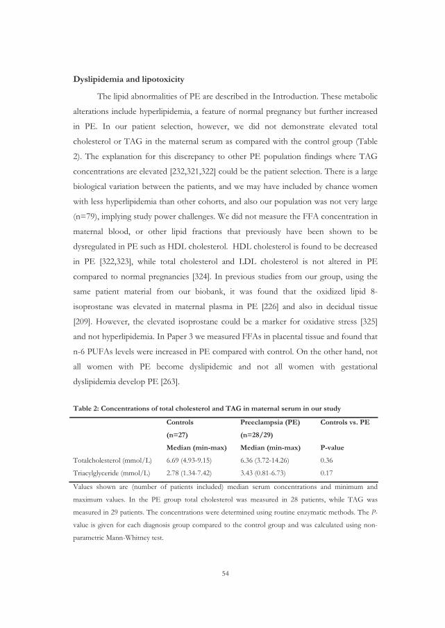
54
Dyslipidemia and lipotoxicity
The lipid abnormalities of PE are described in the Introduction. These metabolic
alterations include hyperlipidemia, a feature of normal pregnancy but further increased
in PE. In our patient selection, however, we did not demonstrate elevated total
cholesterol or TAG in the maternal serum as compared with the control group (Table
2). The explanation for this discrepancy to other PE population findings where TAG
concentrations are elevated [232,321,322] could be the patient selection. There is a large
biological variation between the patients, and we may have included by chance women
with less hyperlipidemia than other cohorts, and also our population was not very large
(n=79), implying study power challenges. We did not measure the FFA concentration in
maternal blood, or other lipid fractions that previously have been shown to be
dysregulated in PE such as HDL cholesterol. HDL cholesterol is found to be decreased
in PE [322,323], while total cholesterol and LDL cholesterol is not altered in PE
compared to normal pregnancies [324]. In previous studies from our group, using the
same patient material from our biobank, it was found that the oxidized lipid 8-
isoprostane was elevated in maternal plasma in PE [226] and also in decidual tissue
[209]. However, the elevated isoprostane could be a marker for oxidative stress [325]
and not hyperlipidemia. In Paper 3 we measured FFAs in placental tissue and found that
n-6 PUFAs levels were increased in PE compared with control. On the other hand, not
all women with PE become dyslipidemic and not all women with gestational
dyslipidemia develop PE [263].
Table 2: Concentrations of total cholesterol and TAG in maternal serum in our study
Controls
(n=27)
Preeclampsia (PE)
(n=28/29)
Controls vs. PE
Median (min-max) Median (min-max) P-value
Totalcholesterol (mmol/L) 6.69 (4.93-9.15) 6.36 (3.72-14.26) 0.36
Triacylglyceride (mmol/L) 2.78 (1.34-7.42) 3.43 (0.81-6.73) 0.17
Values shown are (number of patients included) median serum concentrations and minimum and
maximum values. In the PE group total cholesterol was measured in 28 patients, while TAG was
measured in 29 patients. The concentrations were determined using routine enzymatic methods. The P-
value is given for each diagnosis group compared to the control group and was calculated using non-
parametric Mann-Whitney test.
55
Accumulation of excess lipids in non-adipose tissues is termed lipotoxicity and
can lead to impaired cell signaling, cell dysfunction and cell death [241]. Lipid
accumulation in heart, skeletal muscle, pancreas, liver and kidney is associated with
diseases such as heart failure, obesity and diabetes [320]. Lipotoxicity has also been
suggested to play a role in insulin resistance, and hyperlipidemia [242], conditions
associated with both DM and PE.
In women who later develop PE, the concentrations of OA, LA and PA in the
maternal circulation is elevated already at week 16-20 of gestation [228,232]. We
incubated BeWo cells with these FAs and found both mRNA and protein expression of
ADRP increased with OA and LA, but only slightly with PA (Paper 4). In cell culture,
OA supplementation leads to TAG accumulation and is well tolerated, whereas PA is
poorly incorporated into triglyceride and causes apoptosis [326]. In Paper 1 we found
that OA induced the protein expression of ADRP, formation of lipid droplets and
accumulation of intracellular TAG and to a greater extent than DHA. Hence, the
increased ADRP expression in PE could be due to the higher concentrations of
circulating OA and LA found in the maternal circulation. It would be useful to measure
the concentration of these FAs in maternal blood in the samples from our biobank in
order to support this proposition. However, oxidative stress in the placenta or the
maternal circulation could also be responsible for the increased ADRP expression in
preeclamptic placenta.
Some studies suggest that lipid droplets may have protective effects in cells
against lipotoxicity [326,327]. In cardiomyocytes lipid droplets have a protective effect
against ischemia-reperfusion injury by sequestering FFAs [328]. In muscle cells lipid
droplets are reported to have a protective effect against the damaging effects of FAs on
insulin action and glucose tolerance [329,330]. Recently, Gubern et al studied stress
induced lipid droplet biogenesis without any external source of FAs and found that lipid
droplet biogenesis was not dependent on de novo FA synthesis, instead structural
membrane phospholipids were directed to the lipid droplets. The authors suggest that
this represents a survival strategy whereby the cell recycles membrane lipids to use as
energy generating substrates as a response to stress such as glucose depravation [331].
This is supported by the view that lipid droplets have a role in the management of
membrane lipids. The composition of lipids in the lipid droplets is complex and
56
includes a variety of different neutral lipids and phospholipids, and the large amount of
lysophospholids in the lipid droplets implies a role for them in the recycling of
phospholipids [332,333]. Possibly, recycling of lipids from cell membranes does not
occur in situations where the cell is in an environment with excess lipids.
The actual role of the alterations in the lipid metabolism in pregnancy and PE is
not fully understood. In normal pregnancy the hyperlipidemia might be important for
adequate fetal nutrition. In PE it might contribute to mobilize extra energy from the
mother that can be taken up and stored by the placenta as a support against the stressful
PE environment. Another way of looking at the maternal hyperlipidemia is that it is
harmful for the placenta, and therefore the placenta sequesters these extra lipids as a
protective mechanism to save the feto-placental unit from lipotoxicity.
Oxidative stress and inflammation
Oxidative stress is a feature of PE and is believed to be important in the
pathophysiology of the syndrome. We induced oxidative stress in BeWo cells using
different concentrations of H2O2, and found that the number and size of ADRP coated
lipid droplets increased with increasing doses and incubation times with H2O2 (Paper 4).
This finding is supported by several studies where different types of oxidative stress
induced accumulation of lipid droplets in different cell types and species [334,335,336].
Hence, we speculate that the increased ADRP gene expression in PE could be due to
increased oxidative stress in the placenta. Possibly, the combination of hyperlipidemia
and oxidative stress is even more potent in inducing accumulation of lipid droplets.
ROS could be involved in oxidation of the excess lipids in the maternal circulation or in
oxidative attack on lipids in cell membranes in the placental tissue.
Lipid droplets are normally sparse in normal cells (except adipocytes), but
increase in number and size in cells associated with inflammation [337]. The
accumulation of lipid droplets is induced by different inflammatory agents such as LPS
or cytokines in different cell types [338,339,340], and is also induced in inflammatory
and infectious diseases [339,341]. In placenta, the number of lipid droplets in amnion
epithelium increases during labor, indicating that lipid droplets are induced by stress
[342]. PE is associated with a more extreme maternal inflammatory response than
occurs in normal pregnancy. The systemic inflammatory response involves an increase

57
in circulating cytokines. We measured the cytokine concentrations of TNF-� and IL-6 in
maternal plasma in order to confirm the presence of inflammation in our PE group. We
found that both TNF-� and IL-6 concentrations were increased in plasma from our
patients with PE as compared to controls (Table 3). It is possible that these pro-
inflammatory cytokines also could be involved in inducing lipid droplet accumulation in
preeclamptic placenta; however, presently we have no evidence for this. Over-
expression of ADRP increase the expression of cytokines in macrophages and this is
suggested to be important in the development of atherosclerosis [343]. Hence, it would
be interesting to study the effect of TNF-� and IL-6 on ADRP expression and lipid
droplet accumulation in trophoblast cells and vice versa.
Table 3: Concentrations of cytokines in maternal plasma in our study
Controls (n=30) Preeclampsia (PE) (n=30) Controls vs. PE
Median (min-max) Median (min-max) P-value
IL-6 (pg/mL) 0.7 (0.1-3.5) 1.3 (0.8-13.1) 0.023*
sTNF R1 (pg/mL) 1184 (762-2383) 1535 (793-3438) 0.008*
Values shown are (number of patients included) median plasma concentrations and minimum and
maximum values. Enzyme linked immunosorbent assays (ELISA) for IL-6 (HS600B, Quantikine HS)
and sTNF R1 (DRT100, Quantikine) both from R&D Systems Europe (UK) was performed in
duplicates on EDTA-plasma samples according to the manufacturer’s instructions. The sTNF R1
ELISA measures the total amount of free soluble receptor plus the total amount of soluble receptor
bound to TNF present in the samples. The circulating levels of the receptor increase in response to
TNF-� production, and because the half-life of the receptor in plasma is much longer than the half-life
of TNF, the receptor concentration is considered to be a reflection of the TNF-� activity [344]. The P-
value is given for each diagnosis group compared to the control group (*P <0.05), and was calculated
using non-parametric Mann-Whitney test.
There is also increasing evidence that lipid droplets are important regulators of
inflammatory processes. Several eicosanoid-forming enzymes, involved in the
conversion of AA into eicosanoids, have been detected both in vitro and in vivo, and
accumulation of lipid droplets correlates with the capacity of cells to produce
eicosanoids [345].
In the preeclamptic situation, there is both a dysregulation of the lipid
metabolism with excess availability of lipids from the maternal circulation, and also
58
excess inflammation and oxidative stress. Possibly lipid droplets are accumulated in
placenta during pregnancy due to excess FFA in the circulation and oxidative stress in
the placenta. We speculate that lipid droplets could have a protective role in placenta,
whereby excess lipids are stored in the droplets in order to prevent them to exert
harmful effects intracellularly. Another hypothesis is that the lipid droplet could serve as
a survival mechanism for the placental cells, whereby it is storing lipids as an energy
reserve. Possibly, the increased maternal circulating FAs could serve as an adaptive
protective measure against the oxidative stress and inflammation in the placenta during
pregnancy.
Fibrinoid tissue
In Paper 4, ADRP immunostaining was mainly found to stain clusters of
droplets in the fibrinoid tissue of the placental extracellular matrix. This result is in
agreement with Meadows et al who also found that ADRP was localized to fibrinoid
tissue in the placenta, in a punctuate pattern corresponding to the size and location of
lipid droplets [346]. The fibrinoid tissue we observed with IHC is most probably of
fibrin-type fibrinoid, derived from the coagulation cascade. Fibrin-type fibrinoid is more
prevalent in PE placentas [347], and is associated with apoptosis in trophoblast cells in
PE [348]. In cultured primary term trophoblasts fibrin protects against hypoxic injury
[349].
Many functions for the fibrin-type-fibrinoid in the placenta have been suggested,
such as mechanical stability for anchoring the stem villi, as a regulator of intervillous
circulation, a barrier to limit the invasiveness of the trophoblasts and as a facilitator of
materno-fetal transport of macromolecules [3]. Alphafetoprotein has been shown to
pass through fibrin-type fibrinoid gaps in the syncytiotrophoblast layer [350]. Ackerman
et al identified numerous large lipid droplets in trophoblasts, which increased in size and
numbers the closer they were in proximity to the maternal interface, and they were also
associated to regions of necrotic cellular debris [351]. Possibly the lipid droplets could
be transported out of the placenta via areas of fibrinoid tissue and subsequently shed to
the maternal circulation in the same manner as STBMs. There is also the possibility that
the lipid droplets are transported from the placenta to the decidua where they are
engulfed by macrophages associated with acute atherosis. Katabuchi et al observed areas
59
of fibrin deposition and accumulation of foam cells in the spiral arteries of the decidua,
predominantly in patients with PE [352]. These foam cells were filled with lipid droplets
of various sizes.
5.4. Dysregulation of FA transport genes in preeclampsia
In Paper 3 and 4 we studied the expression of genes involved in lipid metabolism
in preeclamptic placenta. We found that several genes were dysregulated. Here we will
discuss the regulation of three of these that are involved in the uptake of FAs. FATP1,
CD36, and CAV1 were all significantly decreased in preeclamptic placenta (Paper 3 and
4).
FATP1 is a 63 kDa transport protein that mediates FA uptake and is expressed
in a wide variety of cell types and tissues. It is involved in transport of long chained FAs
with a preference for very long chained FAs and is linked with acyl-CoA synthetase
activity [353]. Characterization of purified mouse FATP1 protein conveyed that FATP1
has ACS activity [354]. The protein has a transmembrane domain and several membrane
associated domains [353]. FATP1 was first identified in placental membranes [47] and
the expression is induced by PPAR� and RXR agonists in primary trophoblasts [162].
We found the FATP1 gene expression to be decreased in PE, unfortunately we
were unable to quantify the protein expression and to observe the intracellular location
by immunohistochemistry in term placenta. Previously FATP1 has been found localized
to the plasma membrane and to small vesicles in the cytoplasm [62]. Over-expression of
FATP1 and CD36 in muscle cells resulted in increased transport of OA and PA [355].
FATP1 was localized throughout the cytosol in a reticular pattern, partly co-localized
with the Golgi system, and CD36 was localized to the extracellular membrane, neither
of the proteins were co-localized with lipid droplets [355].
FATP1 is downregulated by TNF-� in the liver [314]. In line with this we
speculate that inflammation could be involved in down-regulating FATP1 in placenta in
PE, as we did not observe any regulation of FATPs with FAs in Paper 2. Also, FATP1
and CAV1 were not regulated by the FAs (PA, OA and LA) that are found up-regulated
in the maternal circulation in PE, and neither by LCPUFAs (AA and DHA) (Figure 9).

60
Figure 9: Gene expression of ADRP, FATP1 and CAV1 in BeWo cells after 24 h incubation with 100
μM of different fatty acids (FAs). Gene expression was analyzed using quantitative real-time RT-PCR.
TBP was used as endogenous control. The P-value is given for each FA compared to the control (*P
<0.05) and was calculated using Students T-test.
CD36 is reported to be localized to lipid rafts (detergent resistant membranes)
[73] and in caveolae [86]. Pohl et al showed that caveolin-1 targets CD36 to the plasma
membrane in mice embryonic fibroblasts, and that in caveolin-1 knock-out mice CD36
was not localized to the plasma membrane [356]. Interestingly, we found that mRNA
expression of CD36 and caveolin-1, both associated with caveolae, were decreased in
PE. In Paper 4 we found caveolin-1 localized mainly to endothelial cells in placental
sections, but we also observed some occasional staining of cytotrophoblasts. However,
we found no difference between the sections from preeclamptic pregnancies compared
to controls (Paper 4). Vandré et al (2007) found caveolin-1 protein expression associated
with endothelial cells and stromal cells in placenta [357], and Byrne et al found by
immuno-gold labeling that caveolin-1 in placenta was mostly expressed in endothelial
cells, and only infrequently in trophoblast cells [358]. The gold particles were localized
to the basal and apical membrane of the endothelium, mostly on the basal side,
suggesting a role in active lipid transport from the maternal side to the fetal side. We
speculate that the reason for the decreased expression of these genes is connected
somehow to the FA transport system of the placenta, and could be a response to excess
FFA in the maternal circulation in PE. However, regulation of gene expression is

61
complex and may involve many other factors. The down-regulation could be a
protective mechanism against further lipid accumulation in the placenta, or it could be a
reduction of lipid transport from the placenta to the fetus either because the excess
lipids can be harmful for the fetus or because the placenta needs to accumulate lipids to
protect itself under the stressful PE environment. Our observation that caveolin-1 is
mainly expressed in fetal endothelial cells of the placenta (Paper 4) supports this notion.

62
6. Conclusions
The main conclusions of this study are as follows:
1. LCPUFAs (AA and DHA) are more efficiently transported across BeWo cells
compared to non-essential FAs (PA and OA).
2. The non-essential FA (OA) is incorporated into TAG fractions and induces
accumulation of lipid droplets in BeWo cells to a greater extent than LCPUFAs
(DHA).
3. LCPUFAs modulate the uptake of both LCPUFAs and OA in BeWo cells. The
uptake of all FAs increase after preincubation with LCPUFAs and the FAs are
preferentially incorporated into phospholipids.
4. Incubation of BeWo cells with LCPUFAs increased the gene expression of long-
chain acyl-CoA synthetases, ACSL1 and ACSL5.
5. There is a dysregulation of LXR�, LXR� and PPAR� in preeclamptic placenta,
the gene expression is significantly decreased for all three transcription factors.
6. ADRP gene expression is increased, while FATP1 and CAV1 expression is
decreased in preeclamptic placenta, and ADRP is also increased on the protein
level.
7. The protein expression of ADRP in placenta tissue is localized to clusters of
vacuoles in the fibrinoid tissue and also to small vesicles inside trophoblast cells.
8. ADRP expression is regulated by FAs and oxidative stress (H2O2) in BeWo cells.
In conclusion, this study supports the previous observations that LCPUFAs are
selectively enriched in the fetal circulation. Our results suggest that there are different
transport mechanisms for LCPUFAS (DHA) and non-essential FAs (OA) in the
trophoblasts of the placenta. The transport mechanism for OA may involve
accumulation of TAG in lipid droplets, while DHA possibly to some extent circumvents
this step. Our results support the concept of a special system for active transport of
LCPUFA across the placenta. We also found that LCPUFAs influence the uptake of
FAs in trophoblast cells and this could have important implications for fetal nutrition
and for dietary advice given to pregnant women. Our results suggest that ACSL1 and
63
ACSL5 could be involved in FA uptake and that conversion of FFAs to acyl-CoA is an
important step in the LCPUFA influenced uptake of FAs in the placenta.
We also aimed at exploring the dysregulation of the lipid metabolism in
pregnancies complicated by PE and/or DM and we show that both PPARs and LXRs
are dysregulated in placenta in PE. We suggest that these transcription factors could be
involved in the PE situation, but further research is required for definite conclusions.
We also show that the lipid droplet associated protein ADRP is dysregulated in PE. The
increased expression of ADRP is possibly induced by oxidative stress in the placenta,
even though dyslipidemia may also play a role in this regard.

64
7. Future perspectives
At the end of the road to a Ph.D. project, there are many questions surfacing that would
be interesting to study further. Here are some thoughts:
� What happens at earlier time points in the pregnancy? It would be interesting to
perform a longitudinal study of placenta tissue samples, both gene expression
patterns of our proteins of interest and immunohistochemistry analysis could
provide valuable insights into the metabolic development and adaptations of the
placenta during the pregnancy. Possibly, excess chorionic villi sampling tissue
used for prenatal diagnosis could be used for such longitudinal sampling,
although this material is very difficult to obtain in reasonable amounts.
� Do LCPUFAs modulate increase in the uptake of all FAs? In this study we
analyzed the effects on LCPUFA uptake and OA, but what about PA or other
short chained FAs that are not considered to beneficial in the diet? Is it correct
to recommend dietary supplementation of n-3 LCPUFA for pregnant women if
these FAs enhance the uptake of other FAs in the placenta?
� How is the FA uptake and transport in primary trophoblast cells and
differentiated BeWo cells? Could there be other cells in the placenta, in addition
to trophoblasts, involved in the preferential LCPUFA transport? It would be
interesting to isolate primary endothelial cells from placenta to compare the FA
transport in these with other endothelial cells, for example from the umbilical
cord and aorta.
� Are lipid droplet associated proteins involved in the development of acute
atherosis? Decidual tissue with areas of acute atherosis can be used as a model
for studying the process of atherosclerosis in general, because of similarities in
the development of lipid deposition in decidua and other tissues. It has been
shown that ADRP is involved in the formation of plaque in atherosclerosis [359].
Hence, it would be interesting to investigate the possible involvement of ADRP
and other LDAPs in decidua in areas of acute atherosis.

65
Placenta remains the key factor to a successful pregnancy and is also an
important predictor of future maternal and offspring health. Further placental
research will be essential for solving the riddle of PE and other pregnancy
complications, important for the wellbeing of both the mother and the fetus. The
similarities in the pathophysiology of PE with other diseases, such as diabetes and
atherosclerosis, makes further research on PE important also when the focus is on
development of other diseases.
66
8. References
[1] Desoye G and Shafrir E. Placental metabolism and its regulation in health and diabetes. Mol Aspects Med 1994; 15:505-682.
[2] Strickland S and Richards WG. Invasion of the trophoblasts. Cell 1992; 71:355-357.
[3] Benirschke K and Kaufmann P. Pathology of the Human Placenta. 4th ed. New York, USA. Springer-Verlag Inc. 2000.
[4] Redman CW and Sargent IL. Pre-eclampsia, the placenta and the maternal systemic inflammatory response--a review. Placenta 2003; 24 Suppl A:S21-S27.
[5] Moffett-King A. Natural killer cells and pregnancy. Nat Rev Immunol 2002; 2:656-663.
[6] Coppens M, Loquet P, Kollen M, De NF, and Buytaert P. Longitudinal evaluation of uteroplacental and umbilical blood flow changes in normal early pregnancy. Ultrasound Obstet Gynecol 1996; 7:114-121.
[7] Burton GJ, Jauniaux E, and Watson AL. Maternal arterial connections to the placental intervillous space during the first trimester of human pregnancy: the Boyd collection revisited. Am J Obstet Gynecol 1999; 181:718-724.
[8] Jauniaux E, Watson AL, Hempstock J, Bao YP, Skepper JN, and Burton GJ. Onset of maternal arterial blood flow and placental oxidative stress. A possible factor in human early pregnancy failure. Am J Pathol 2000; 157:2111-2122.
[9] Genbacev O, Joslin R, Damsky CH, Polliotti BM, and Fisher SJ. Hypoxia alters early gestation human cytotrophoblast differentiation/invasion in vitro and models the placental defects that occur in preeclampsia. J Clin Invest 1996; 97:540-550.
[10] Burton GJ. Oxygen, the Janus gas; its effects on human placental development and function. J Anat 2009; 215:27-35.
[11] Herrera E. Lipid metabolism in pregnancy and its consequences in the fetus and newborn. Endocrine 2002; 19:43-55.
[12] Wassall SR and Stillwell W. Polyunsaturated fatty acid-cholesterol interactions: domain formation in membranes. Biochim Biophys Acta 2009; 1788:24-32.
[13] Funk CD. Prostaglandins and leukotrienes: advances in eicosanoid biology. Science 2001; 294:1871-1875.
[14] Spector AA. Essentiality of fatty acids. Lipids 1999; 34 Suppl:S1-S3.
[15] Bagga D, Wang L, Farias-Eisner R, Glaspy JA, and Reddy ST. Differential effects of prostaglandin derived from omega-6 and omega-3 polyunsaturated fatty acids on COX-2 expression and IL-6 secretion. Proc Natl Acad Sci U S A 2003; 100:1751-1756.
[16] Kinsella JE, Broughton KS, and Whelan JW. Dietary unsaturated fatty acids: interactions and possible needs in relation to eicosanoid synthesis. J Nutr Biochem 1990; 1:123-141.
[17] Schmitz G and Ecker J. The opposing effects of n-3 and n-6 fatty acids. Prog Lipid Res 2008; 47:147-155.

67
[18] Shand JH and Noble RC. The characterization of the linoleic acid desaturation and elongation system in ovine placental tissue. Int J Biochem 1983; 15:1367-1371.
[19] Chambaz J, Ravel D, Manier MC, Pepin D, Mulliez N, and Bereziat G. Essential fatty acids interconversion in the human fetal liver. Biol Neonate 1985; 47:136-140.
[20] Duttaroy AK. Transport of fatty acids across the human placenta: a review. Prog Lipid Res 2009; 48:52-61.
[21] de Groot RH, Hornstra G, van Houwelingen AC, and Roumen F. Effect of alpha-linolenic acid supplementation during pregnancy on maternal and neonatal polyunsaturated fatty acid status and pregnancy outcome. Am J Clin Nutr 2004; 79:251-260.
[22] Carlson SE. Docosahexaenoic acid supplementation in pregnancy and lactation. Am J Clin Nutr 2009; 89:678S-684S.
[23] Greenberg JA, Bell SJ, and Ausdal WV. Omega-3 Fatty Acid supplementation during pregnancy. Rev Obstet Gynecol 2008; 1:162-169.
[24] Bourre JM. Dietary omega-3 fatty acids for women. Biomed Pharmacother 2007; 61:105-112.
[25] Ikonen E. Cellular cholesterol trafficking and compartmentalization. Nat Rev Mol Cell Biol 2008; 9:125-138.
[26] Tabas I. Consequences of cellular cholesterol accumulation: basic concepts and physiological implications. J Clin Invest 2002; 110:905-911.
[27] Mesmin B and Maxfield FR. Intracellular sterol dynamics. Biochim Biophys Acta 2009; 1791:636-645.
[28] Brown MS and Goldstein JL. The SREBP pathway: regulation of cholesterol metabolism by proteolysis of a membrane-bound transcription factor. Cell 1997; 89:331-340.
[29] Feng B, Yao PM, Li Y, Devlin CM, Zhang D, Harding HP, Sweeney M, Rong JX, Kuriakose G, Fisher EA, Marks AR, Ron D, and Tabas I. The endoplasmic reticulum is the site of cholesterol-induced cytotoxicity in macrophages. Nat Cell Biol 2003; 5:781-792.
[30] Napoli C, D'Armiento FP, Mancini FP, Postiglione A, Witztum JL, Palumbo G, and Palinski W. Fatty streak formation occurs in human fetal aortas and is greatly enhanced by maternal hypercholesterolemia. Intimal accumulation of low density lipoprotein and its oxidation precede monocyte recruitment into early atherosclerotic lesions. J Clin Invest 1997; 100:2680-2690.
[31] Haggarty P. Placental regulation of fatty acid delivery and its effect on fetal growth--a review. Placenta 2002; 23 Suppl A:S28-S38.
[32] Hubel CA. Oxidative stress in the pathogenesis of preeclampsia. Proc Soc Exp Biol Med 1999; 222:222-235.
[33] Biale Y. Lipolytic activity in the placentas of chronically deprived fetuses. Acta Obstet Gynecol Scand 1985; 64:111-114.
[34] Wittmaack FM, Gafvels ME, Bronner M, Matsuo H, McCrae KR, Tomaszewski JE, Robinson SL, Strickland DK, and Strauss JF, III. Localization and regulation of the human very low density lipoprotein/apolipoprotein-E receptor: trophoblast expression predicts a role for the receptor in placental lipid transport. Endocrinology 1995; 136:340-348.
68
[35] Bonet B, Brunzell JD, Gown AM, and Knopp RH. Metabolism of very-low-density lipoprotein triglyceride by human placental cells: the role of lipoprotein lipase. Metabolism 1992; 41:596-603.
[36] Alsat E, Bouali Y, Goldstein S, Malassine A, Laudat MH, and Cedard L. Characterization of specific low-density lipoprotein binding sites in human term placental microvillous membranes. Mol Cell Endocrinol 1982; 28:439-453.
[37] Furuhashi M, Seo H, Mizutani S, Narita O, Tomoda Y, and Matsui N. Expression of low density lipoprotein receptor gene in human placenta during pregnancy. Mol Endocrinol 1989; 3:1252-1256.
[38] Malassine A, Besse C, Roche A, Alsat E, Rebourcet R, Mondon F, and Cedard L. Ultrastructural visualization of the internalization of low density lipoprotein by human placental cells. Histochemistry 1987; 87:457-464.
[39] Farrugia W, Aitken MA, van DF, Wong MH, Brennecke SP, Scott KF, and Rice GE. Type II phospholipase A2 in human gestational tissues: subcellular distribution of placental immuno- and catalytic activity. Biochim Biophys Acta 1993; 1166:77-83.
[40] Rice GE, Wong MH, Farrugia W, and Scott KF. Contribution of type II phospholipase A2 to in vitro phospholipase A2 enzymatic activity in human term placenta. J Endocrinol 1998; 157:25-31.
[41] Magnusson AL, Waterman IJ, Wennergren M, Jansson T, and Powell TL. Triglyceride hydrolase activities and expression of fatty acid binding proteins in the human placenta in pregnancies complicated by intrauterine growth restriction and diabetes. J Clin Endocrinol Metab 2004; 89:4607-4614.
[42] Tabano S, Alvino G, Antonazzo P, Grati FR, Miozzo M, and Cetin I. Placental LPL gene expression is increased in severe intrauterine growth-restricted pregnancies. Pediatr Res 2006; 59:250-253.
[43] Wadsack C, Tabano S, Maier A, Hiden U, Alvino G, Cozzi V, Huttinger M, Schneider WJ, Lang U, Cetin I, and Desoye G. Intrauterine growth restriction is associated with alterations in placental lipoprotein receptors and maternal lipoprotein composition. Am J Physiol Endocrinol Metab 2007; 292:E476-E484.
[44] Gauster M, Hiden U, Blaschitz A, Frank S, Lang U, Alvino G, Cetin I, Desoye G, and Wadsack C. Dysregulation of placental endothelial lipase and lipoprotein lipase in intrauterine growth-restricted pregnancies. J Clin Endocrinol Metab 2007; 92:2256-2263.
[45] Dutta-Roy AK. Cellular uptake of long-chain fatty acids: role of membrane-associated fatty-acid-binding/transport proteins. Cell Mol Life Sci 2000; 57:1360-1372.
[46] Dutta-Roy AK. Transport mechanisms for long-chain polyunsaturated fatty acids in the human placenta. Am J Clin Nutr 2000; 71:315S-322S.
[47] Campbell FM, Bush PG, Veerkamp JH, and Dutta-Roy AK. Detection and cellular localization of plasma membrane-associated and cytoplasmic fatty acid-binding proteins in human placenta. Placenta 1998; 19:409-415.
[48] Larque E, Demmelmair H, Berger B, Hasbargen U, and Koletzko B. In vivo investigation of the placental transfer of (13)C-labeled fatty acids in humans. J Lipid Res 2003; 44:49-55.

69
[49] Berghaus TM, Demmelmair H, and Koletzko B. Essential fatty acids and their long-chain polyunsaturated metabolites in maternal and cord plasma triglycerides during late gestation. Biol Neonate 2000; 77:96-100.
[50] Friedman Z, Danon A, Lamberth EL, Jr., and Mann WJ. Cord blood fatty acid composition in infants and in their mothers during the third trimester. J Pediatr 1978; 92:461-466.
[51] Benassayag C, Mignot TM, Haourigui M, Civel C, Hassid J, Carbonne B, Nunez EA, and Ferre F. High polyunsaturated fatty acid, thromboxane A2, and alpha-fetoprotein concentrations at the human feto-maternal interface. J Lipid Res 1997; 38:276-286.
[52] Haggarty P, Ashton J, Joynson M, Abramovich DR, and Page K. Effect of maternal polyunsaturated fatty acid concentration on transport by the human placenta. Biol Neonate 1999; 75:350-359.
[53] Haggarty P, Page K, Abramovich DR, Ashton J, and Brown D. Long-chain polyunsaturated fatty acid transport across the perfused human placenta. Placenta 1997; 18:635-642.
[54] Veerkamp JH, van Moerkerk HT, Prinsen CF, and van Kuppevelt TH. Structural and functional studies on different human FABP types. Mol Cell Biochem 1999; 192:137-142.
[55] Biron-Shental T, Schaiff WT, Ratajczak CK, Bildirici I, Nelson DM, and Sadovsky Y. Hypoxia regulates the expression of fatty acid-binding proteins in primary term human trophoblasts. Am J Obstet Gynecol 2007; 197:516-516.
[56] Campbell FM, Gordon MJ, and Dutta-Roy AK. Placental membrane fatty acid-binding protein preferentially binds arachidonic and docosahexaenoic acids. Life Sci 1998; 63:235-240.
[57] Campbell FM, Taffesse S, Gordon MJ, and Dutta-Roy AK. Plasma membrane fatty-acid-binding protein in human placenta: identification and characterization. Biochem Biophys Res Commun 1995; 209:1011-1017.
[58] Campbell FM and Dutta-Roy AK. Plasma membrane fatty acid-binding protein (FABPpm) is exclusively located in the maternal facing membranes of the human placenta. FEBS Lett 1995; 375:227-230.
[59] Uchiyama A, Aoyama T, Kamijo K, Uchida Y, Kondo N, Orii T, and Hashimoto T. Molecular cloning of cDNA encoding rat very long-chain acyl-CoA synthetase. J Biol Chem 1996; 271:30360-30365.
[60] Watkins PA, Pevsner J, and Steinberg SJ. Human very long-chain acyl-CoA synthetase and two human homologs: initial characterization and relationship to fatty acid transport protein. Prostaglandins Leukot Essent Fatty Acids 1999; 60:323-328.
[61] Pei Z, Fraisl P, Berger J, Jia Z, Forss-Petter S, and Watkins PA. Mouse very long-chain Acyl-CoA synthetase 3/fatty acid transport protein 3 catalyzes fatty acid activation but not fatty acid transport in MA-10 cells. J Biol Chem 2004; 279:54454-54462.
[62] Schaffer JE and Lodish HF. Expression cloning and characterization of a novel adipocyte long chain fatty acid transport protein. Cell 1994; 79:427-436.
[63] Stahl A, Hirsch DJ, Gimeno RE, Punreddy S, Ge P, Watson N, Patel S, Kotler M, Raimondi A, Tartaglia LA, and Lodish HF. Identification of the major intestinal fatty acid transport protein. Mol Cell 1999; 4:299-308.
70
[64] Steinberg SJ, Wang SJ, McGuinness MC, and Watkins PA. Human liver-specific very-long-chain acyl-coenzyme A synthetase: cDNA cloning and characterization of a second enzymatically active protein. Mol Genet Metab 1999; 68:32-42.
[65] Larque E, Krauss-Etschmann S, Campoy C, Hartl D, Linde J, Klingler M, Demmelmair H, Cano A, Gil A, Bondy B, and Koletzko B. Docosahexaenoic acid supply in pregnancy affects placental expression of fatty acid transport proteins. Am J Clin Nutr 2006; 84:853-861.
[66] Larque E, Demmelmair H, Klingler M, De JS, Bondy B, and Koletzko B. Expression pattern of fatty acid transport protein-1 (FATP-1), FATP-4 and heart-fatty acid binding protein (H-FABP) genes in human term placenta. Early Hum Dev 2006; 82:697-701.
[67] Jia Z, Pei Z, Maiguel D, Toomer CJ, and Watkins PA. The fatty acid transport protein (FATP) family: very long chain acyl-CoA synthetases or solute carriers? J Mol Neurosci 2007; 33:25-31.
[68] Schaiff WT, Bildirici I, Cheong M, Chern PL, Nelson DM, and Sadovsky Y. Peroxisome proliferator-activated receptor-gamma and retinoid X receptor signaling regulate fatty acid uptake by primary human placental trophoblasts. J Clin Endocrinol Metab 2005; 90:4267-4275.
[69] Abumrad NA, el-Maghrabi MR, Amri EZ, Lopez E, and Grimaldi PA. Cloning of a rat adipocyte membrane protein implicated in binding or transport of long-chain fatty acids that is induced during preadipocyte differentiation. Homology with human CD36. J Biol Chem 1993; 268:17665-17668.
[70] Endemann G, Stanton LW, Madden KS, Bryant CM, White RT, and Protter AA. CD36 is a receptor for oxidized low density lipoprotein. J Biol Chem 1993; 268:11811-11816.
[71] Habets DD, Coumans WA, El HM, Zarrinpashneh E, Bertrand L, Viollet B, Kiens B, Jensen TE, Richter EA, Bonen A, Glatz JF, and Luiken JJ. Crucial role for LKB1 to AMPKalpha2 axis in the regulation of CD36-mediated long-chain fatty acid uptake into cardiomyocytes. Biochim Biophys Acta 2009; 1791:212-219.
[72] Silverstein RL and Febbraio M. CD36, a scavenger receptor involved in immunity, metabolism, angiogenesis, and behavior. Sci Signal 2009; 2:re3-
[73] Pohl J, Ring A, Korkmaz U, Ehehalt R, and Stremmel W. FAT/CD36-mediated long-chain fatty acid uptake in adipocytes requires plasma membrane rafts. Mol Biol Cell 2005; 16:24-31.
[74] Niyogi K and Hildreth JE. Characterization of new syncytium-inhibiting monoclonal antibodies implicates lipid rafts in human T-cell leukemia virus type 1 syncytium formation. J Virol 2001; 75:7351-7361.
[75] Linton EA, Rodriguez-Linares B, Rashid-Doubell F, Ferguson DJ, and Redman CW. Caveolae and caveolin-1 in human term villous trophoblast. Placenta 2003; 24:745-757.
[76] Rashid-Doubell F, Tannetta D, Redman CW, Sargent IL, Boyd CA, and Linton EA. Caveolin-1 and Lipid Rafts in Confluent BeWo Trophoblasts: Evidence for Rock-1 Association with Caveolin-1. Placenta 2006;
[77] Sargiacomo M, Scherer PE, Tang Z, Kubler E, Song KS, Sanders MC, and Lisanti MP. Oligomeric structure of caveolin: implications for caveolae membrane organization. Proc Natl Acad Sci U S A 1995; 92:9407-9411.
[78] Anderson RG. Caveolae: where incoming and outgoing messengers meet. Proc Natl Acad Sci U S A 1993; 90:10909-10913.

71
[79] Schnitzer JE, Oh P, Pinney E, and Allard J. Filipin-sensitive caveolae-mediated transport in endothelium: reduced transcytosis, scavenger endocytosis, and capillary permeability of select macromolecules. J Cell Biol 1994; 127:1217-1232.
[80] Massimino ML, Griffoni C, Spisni E, Toni M, and Tomasi V. Involvement of caveolae and caveolae-like domains in signalling, cell survival and angiogenesis. Cell Signal 2002; 14:93-98.
[81] Razani B, Schlegel A, Liu J, and Lisanti MP. Caveolin-1, a putative tumour suppressor gene. Biochem Soc Trans 2001; 29:494-499.
[82] Pol A, Martin S, Fernandez MA, Ferguson C, Carozzi A, Luetterforst R, Enrich C, and Parton RG. Dynamic and regulated association of caveolin with lipid bodies: modulation of lipid body motility and function by a dominant negative mutant. Mol Biol Cell 2004; 15:99-110.
[83] Pol A, Martin S, Fernandez MA, Ingelmo-Torres M, Ferguson C, Enrich C, and Parton RG. Cholesterol and fatty acids regulate dynamic caveolin trafficking through the Golgi complex and between the cell surface and lipid bodies. Mol Biol Cell 2005; 16:2091-2105.
[84] Trigatti BL, Anderson RG, and Gerber GE. Identification of caveolin-1 as a fatty acid binding protein. Biochem Biophys Res Commun 1999; 255:34-39.
[85] Ehehalt R, Fullekrug J, Pohl J, Ring A, Herrmann T, and Stremmel W. Translocation of long chain fatty acids across the plasma membrane - lipid rafts and fatty acid transport proteins. Mol Cell Biochem 2006; 284:135-140.
[86] Kolleck I, Guthmann F, Ladhoff AM, Tandon NN, Schlame M, and Rustow B. Cellular cholesterol stimulates acute uptake of palmitate by redistribution of fatty acid translocase in type II pneumocytes. Biochemistry (Mosc) 2002; 41:6369-6375.
[87] Pohl J, Ring A, and Stremmel W. Uptake of long-chain fatty acids in HepG2 cells involves caveolae: analysis of a novel pathway. J Lipid Res 2002; 43:1390-1399.
[88] Razani B, Combs TP, Wang XB, Frank PG, Park DS, Russell RG, Li M, Tang B, Jelicks LA, Scherer PE, and Lisanti MP. Caveolin-1-deficient mice are lean, resistant to diet-induced obesity, and show hypertriglyceridemia with adipocyte abnormalities. J Biol Chem 2002; 277:8635-8647.
[89] Ring A, Pohl J, Volkl A, and Stremmel W. Evidence for vesicles that mediate long-chain fatty acid uptake by human microvascular endothelial cells. J Lipid Res 2002; 43:2095-2104.
[90] Soupene E and Kuypers FA. Mammalian long-chain acyl-CoA synthetases. Exp Biol Med (Maywood ) 2008; 233:507-521.
[91] Watkins PA. Fatty acid activation. Prog Lipid Res 1997; 36:55-83.
[92] Van Horn CG, Caviglia JM, Li LO, Wang S, Granger DA, and Coleman RA. Characterization of recombinant long-chain rat acyl-CoA synthetase isoforms 3 and 6: identification of a novel variant of isoform 6. Biochemistry (Mosc) 2005; 44:1635-1642.
[93] Kang MJ, Fujino T, Sasano H, Minekura H, Yabuki N, Nagura H, Iijima H, and Yamamoto TT. A novel arachidonate-preferring acyl-CoA synthetase is present in steroidogenic cells of the rat adrenal, ovary, and testis. Proc Natl Acad Sci U S A 1997; 94:2880-2884.
[94] Tong F, Black PN, Coleman RA, and DiRusso CC. Fatty acid transport by vectorial acylation in mammals: roles played by different isoforms of rat long-chain acyl-CoA synthetases. Arch Biochem Biophys 2006; 447:46-52.

72
[95] Coleman RA, Lewin TM, Van Horn CG, and Gonzalez-Baro MR. Do long-chain acyl-CoA synthetases regulate fatty acid entry into synthetic versus degradative pathways? J Nutr 2002; 132:2123-2126.
[96] Larigauderie G, Furman C, Jaye M, Lasselin C, Copin C, Fruchart JC, Castro G, and Rouis M. Adipophilin enhances lipid accumulation and prevents lipid efflux from THP-1 macrophages: potential role in atherogenesis. Arterioscler Thromb Vasc Biol 2004; 24:504-510.
[97] Liu P, Ying Y, Zhao Y, Mundy DI, Zhu M, and Anderson RG. Chinese hamster ovary K2 cell lipid droplets appear to be metabolic organelles involved in membrane traffic. J Biol Chem 2004; 279:3787-3792.
[98] Londos C, Brasaemle DL, Schultz CJ, Segrest JP, and Kimmel AR. Perilipins, ADRP, and other proteins that associate with intracellular neutral lipid droplets in animal cells. Semin Cell Dev Biol 1999; 10:51-58.
[99] Mishra R, Emancipator SN, Miller C, Kern T, and Simonson MS. Adipose differentiation-related protein and regulators of lipid homeostasis identified by gene expression profiling in the murine db/db diabetic kidney. Am J Physiol Renal Physiol 2004; 286:F913-F921.
[100] Umlauf E, Csaszar E, Moertelmaier M, Schuetz GJ, Parton RG, and Prohaska R. Association of stomatin with lipid bodies. J Biol Chem 2004; 279:23699-23709.
[101] Kimmel AR, Brasaemle DL, McAndrews-Hill M, Sztalryd C, and Londos C. Adoption of PERILIPIN as a unifying nomenclature for the mammalian PAT-family of intracellular, lipid storage droplet proteins. J Lipid Res 2009;
[102] Miura S, Gan JW, Brzostowski J, Parisi MJ, Schultz CJ, Londos C, Oliver B, and Kimmel AR. Functional conservation for lipid storage droplet association among Perilipin, ADRP, and TIP47 (PAT)-related proteins in mammals, Drosophila, and Dictyostelium. J Biol Chem 2002; 277:32253-32257.
[103] Brasaemle DL, Barber T, Wolins NE, Serrero G, Blanchette-Mackie EJ, and Londos C. Adipose differentiation-related protein is an ubiquitously expressed lipid storage droplet-associated protein. J Lipid Res 1997; 38:2249-2263.
[104] Gao J and Serrero G. Adipose differentiation related protein (ADRP) expressed in transfected COS-7 cells selectively stimulates long chain fatty acid uptake. J Biol Chem 1999; 274:16825-16830.
[105] Eisinger DP and Serrero G. Structure of the gene encoding mouse adipose differentiation-related protein (ADRP). Genomics 1993; 16:638-644.
[106] Heid HW, Schnolzer M, and Keenan TW. Adipocyte differentiation-related protein is secreted into milk as a constituent of milk lipid globule membrane. Biochem J 1996; 320 ( Pt 3):1025-1030.
[107] Gao J, Ye H, and Serrero G. Stimulation of adipose differentiation related protein (ADRP) expression in adipocyte precursors by long-chain fatty acids. J Cell Physiol 2000; 182:297-302.
[108] Serrero G, Frolov A, Schroeder F, Tanaka K, and Gelhaar L. Adipose differentiation related protein: expression, purification of recombinant protein in Escherichia coli and characterization of its fatty acid binding properties. Biochim Biophys Acta 2000; 1488:245-254.
[109] Atshaves BP, Starodub O, McIntosh A, Petrescu A, Roths JB, Kier AB, and Schroeder F. Sterol carrier protein-2 alters high density lipoprotein-mediated cholesterol efflux. J Biol Chem 2000; 275:36852-36861.
73
[110] Dalen KT, Schoonjans K, Ulven SM, Weedon-Fekjaer MS, Bentzen TG, Koutnikova H, Auwerx J, and Nebb HI. Adipose tissue expression of the lipid droplet-associating proteins S3-12 and perilipin is controlled by peroxisome proliferator-activated receptor-gamma. Diabetes 2004; 53:1243-1252.
[111] Bildirici I, Roh CR, Schaiff WT, Lewkowski BM, Nelson DM, and Sadovsky Y. The lipid droplet-associated protein adipophilin is expressed in human trophoblasts and is regulated by peroxisomal proliferator-activated receptor-gamma/retinoid X receptor. J Clin Endocrinol Metab 2003; 88:6056-6062.
[112] Tarrade A, Schoonjans K, Guibourdenche J, Bidart JM, Vidaud M, Auwerx J, Rochette-Egly C, and Evain-Brion D. PPAR gamma/RXR alpha heterodimers are involved in human CG beta synthesis and human trophoblast differentiation. Endocrinology 2001; 142:4504-4514.
[113] Schild RL, Schaiff WT, Carlson MG, Cronbach EJ, Nelson DM, and Sadovsky Y. The activity of PPAR gamma in primary human trophoblasts is enhanced by oxidized lipids. J Clin Endocrinol Metab 2002; 87:1105-1110.
[114] Kotokorpi P, Venteclef N, Ellis E, Gustafsson JA, and Mode A. The human ADFP gene is a direct LXR target gene and differentially regulated by synthetic LXR ligands. Mol Pharmacol 2009;
[115] Babu MM, Luscombe NM, Aravind L, Gerstein M, and Teichmann SA. Structure and evolution of transcriptional regulatory networks. Curr Opin Struct Biol 2004; 14:283-291.
[116] Germain P, Staels B, Dacquet C, Spedding M, and Laudet V. Overview of nomenclature of nuclear receptors. Pharmacol Rev 2006; 58:685-704.
[117] Olefsky JM. Nuclear receptor minireview series. J Biol Chem 2001; 276:36863-36864.
[118] Mangelsdorf DJ, Thummel C, Beato M, Herrlich P, Schutz G, Umesono K, Blumberg B, Kastner P, Mark M, Chambon P, and Evans RM. The nuclear receptor superfamily: the second decade. Cell 1995; 83:835-839.
[119] Evans RM. The steroid and thyroid hormone receptor superfamily. Science 1988; 240:889-895.
[120] Mangelsdorf DJ and Evans RM. The RXR heterodimers and orphan receptors. Cell 1995; 83:841-850.
[121] Ingraham HA and Redinbo MR. Orphan nuclear receptors adopted by crystallography. Curr Opin Struct Biol 2005; 15:708-715.
[122] Wolf IM, Heitzer MD, Grubisha M, and DeFranco DB. Coactivators and nuclear receptor transactivation. J Cell Biochem 2008; 104:1580-1586.
[123] Gurevich I, Flores AM, and Aneskievich BJ. Corepressors of agonist-bound nuclear receptors. Toxicol Appl Pharmacol 2007; 223:288-298.
[124] Chawla A, Repa JJ, Evans RM, and Mangelsdorf DJ. Nuclear receptors and lipid physiology: opening the X-files. Science 2001; 294:1866-1870.
[125] Bishop-Bailey D and Wray J. Peroxisome proliferator-activated receptors: a critical review on endogenous pathways for ligand generation. Prostaglandins Other Lipid Mediat 2003; 71:1-22.
[126] Chen YE, Fu M, Zhang J, Zhu X, Lin Y, Akinbami MA, and Song Q. Peroxisome proliferator-activated receptors and the cardiovascular system. Vitam Horm 2003; 66:157-188.

74
[127] Gurnell M, Savage DB, Chatterjee VK, and O'Rahilly S. The metabolic syndrome: peroxisome proliferator-activated receptor gamma and its therapeutic modulation. J Clin Endocrinol Metab 2003; 88:2412-2421.
[128] Sorensen HN, Treuter E, and Gustafsson JA. Regulation of peroxisome proliferator-activated receptors. Vitam Horm 1998; 54:121-166.
[129] Issemann I and Green S. Activation of a member of the steroid hormone receptor superfamily by peroxisome proliferators. Nature 1990; 347:645-650.
[130] Dreyer C, Krey G, Keller H, Givel F, Helftenbein G, and Wahli W. Control of the peroxisomal beta-oxidation pathway by a novel family of nuclear hormone receptors. Cell 1992; 68:879-887.
[131] Beaven SW and Tontonoz P. Nuclear receptors in lipid metabolism: targeting the heart of dyslipidemia. Annu Rev Med 2006; 57:313-329.
[132] Tontonoz P, Hu E, and Spiegelman BM. Stimulation of adipogenesis in fibroblasts by PPAR gamma 2, a lipid-activated transcription factor. Cell 1994; 79:1147-1156.
[133] Rosen ED, Walkey CJ, Puigserver P, and Spiegelman BM. Transcriptional regulation of adipogenesis. Genes Dev 2000; 14:1293-1307.
[134] Kliewer SA, Sundseth SS, Jones SA, Brown PJ, Wisely GB, Koble CS, Devchand P, Wahli W, Willson TM, Lenhard JM, and Lehmann JM. Fatty acids and eicosanoids regulate gene expression through direct interactions with peroxisome proliferator-activated receptors alpha and gamma. Proc Natl Acad Sci U S A 1997; 94:4318-4323.
[135] Collins JL, Fivush AM, Watson MA, Galardi CM, Lewis MC, Moore LB, Parks DJ, Wilson JG, Tippin TK, Binz JG, Plunket KD, Morgan DG, Beaudet EJ, Whitney KD, Kliewer SA, and Willson TM. Identification of a nonsteroidal liver X receptor agonist through parallel array synthesis of tertiary amines. J Med Chem 2002; 45:1963-1966.
[136] Nagy L, Tontonoz P, Alvarez JG, Chen H, and Evans RM. Oxidized LDL regulates macrophage gene expression through ligand activation of PPARgamma. Cell 1998; 93:229-240.
[137] Kliewer SA, Sundseth SS, Jones SA, Brown PJ, Wisely GB, Koble CS, Devchand P, Wahli W, Willson TM, Lenhard JM, and Lehmann JM. Fatty acids and eicosanoids regulate gene expression through direct interactions with peroxisome proliferator-activated receptors alpha and gamma. Proc Natl Acad Sci U S A 1997; 94:4318-4323.
[138] Forman BM, Tontonoz P, Chen J, Brun RP, Spiegelman BM, and Evans RM. 15-Deoxy-delta 12, 14-prostaglandin J2 is a ligand for the adipocyte determination factor PPAR gamma. Cell 1995; 83:803-812.
[139] Devchand PR, Keller H, Peters JM, Vazquez M, Gonzalez FJ, and Wahli W. The PPARalpha-leukotriene B4 pathway to inflammation control. Nature 1996; 384:39-43.
[140] Lehmann JM, Lenhard JM, Oliver BB, Ringold GM, and Kliewer SA. Peroxisome proliferator-activated receptors alpha and gamma are activated by indomethacin and other non-steroidal anti-inflammatory drugs. J Biol Chem 1997; 272:3406-3410.
[141] Lenhard JM. PPAR gamma/RXR as a molecular target for diabetes. Receptors Channels 2001; 7:249-258.
[142] Lehmann JM, Moore LB, Smith-Oliver TA, Wilkison WO, Willson TM, and Kliewer SA. An antidiabetic thiazolidinedione is a high affinity ligand for peroxisome proliferator-activated receptor gamma (PPAR gamma). J Biol Chem 1995; 270:12953-12956.

75
[143] Castrillo A and Tontonoz P. Nuclear receptors in macrophage biology: at the crossroads of lipid metabolism and inflammation. Annu Rev Cell Dev Biol 2004; 20:455-480.
[144] Tontonoz P, Nagy L, Alvarez JG, Thomazy VA, and Evans RM. PPARgamma promotes monocyte/macrophage differentiation and uptake of oxidized LDL. Cell 1998; 93:241-252.
[145] Barish GD. Peroxisome proliferator-activated receptors and liver X receptors in atherosclerosis and immunity. J Nutr 2006; 136:690-694.
[146] Tontonoz P and Mangelsdorf DJ. Liver X receptor signaling pathways in cardiovascular disease. Mol Endocrinol 2003; 17:985-993.
[147] Ulven SM, Dalen KT, Gustafsson JA, and Nebb HI. LXR is crucial in lipid metabolism. Prostaglandins Leukot Essent Fatty Acids 2005; 73:59-63.
[148] Li AC and Glass CK. J Lipid Res 2004; 45:2161-2173.
[149] Jamroz-Wisniewska A, Wojcicka G, Horoszewicz K, and Beltowski J. Liver X receptors (LXRs). Part II: non-lipid effects, role in pathology, and therapeutic implications. Postepy Hig Med Dosw (Online ) 2007; 61:760-785.
[150] Lehmann JM, Kliewer SA, Moore LB, Smith-Oliver TA, Oliver BB, Su JL, Sundseth SS, Winegar DA, Blanchard DE, Spencer TA, and Willson TM. Activation of the nuclear receptor LXR by oxysterols defines a new hormone response pathway. J Biol Chem 1997; 272:3137-3140.
[151] Auboeuf D, Rieusset J, Fajas L, Vallier P, Frering V, Riou JP, Staels B, Auwerx J, Laville M, and Vidal H. Tissue distribution and quantification of the expression of mRNAs of peroxisome proliferator-activated receptors and liver X receptor-alpha in humans: no alteration in adipose tissue of obese and NIDDM patients. Diabetes 1997; 46:1319-1327.
[152] Janowski BA, Willy PJ, Devi TR, Falck JR, and Mangelsdorf DJ. An oxysterol signalling pathway mediated by the nuclear receptor LXR alpha. Nature 1996; 383:728-731.
[153] Janowski BA, Grogan MJ, Jones SA, Wisely GB, Kliewer SA, Corey EJ, and Mangelsdorf DJ. Structural requirements of ligands for the oxysterol liver X receptors LXRalpha and LXRbeta. Proc Natl Acad Sci U S A 1999; 96:266-271.
[154] Olkkonen VM and Lehto M. Oxysterols and oxysterol binding proteins: role in lipid metabolism and atherosclerosis. Ann Med 2004; 36:562-572.
[155] Qiu C, Phung TT, Vadachkoria S, Muy-Rivera M, Sanchez SE, and Williams MA. Oxidized low-density lipoprotein (Oxidized LDL) and the risk of preeclampsia. Physiol Res 2006; 55:491-500.
[156] Schultz JR, Tu H, Luk A, Repa JJ, Medina JC, Li L, Schwendner S, Wang S, Thoolen M, Mangelsdorf DJ, Lustig KD, and Shan B. Role of LXRs in control of lipogenesis. Genes Dev 2000; 14:2831-2838.
[157] Joseph SB, McKilligin E, Pei L, Watson MA, Collins AR, Laffitte BA, Chen M, Noh G, Goodman J, Hagger GN, Tran J, Tippin TK, Wang X, Lusis AJ, Hsueh WA, Law RE, Collins JL, Willson TM, and Tontonoz P. Synthetic LXR ligand inhibits the development of atherosclerosis in mice. Proc Natl Acad Sci U S A 2002; 99:7604-7609.
[158] Yoshikawa T, Shimano H, Yahagi N, Ide T, memiya-Kudo M, Matsuzaka T, Nakakuki M, Tomita S, Okazaki H, Tamura Y, Iizuka Y, Ohashi K, Takahashi A, Sone H, Osuga JJ, Gotoda T, Ishibashi S, and Yamada N. Polyunsaturated fatty acids suppress sterol regulatory element-
76
binding protein 1c promoter activity by inhibition of liver X receptor (LXR) binding to LXR response elements. J Biol Chem 2002; 277:1705-1711.
[159] Wang Q, Fujii H, and Knipp GT. Expression of PPAR and RXR isoforms in the developing rat and human term placentas. Placenta 2002; 23:661-671.
[160] Barak Y, Sadovsky Y, and Shalom-Barak T. PPAR Signaling in Placental Development and Function. PPAR Res 2008; 2008:142082-
[161] Barak Y, Nelson MC, Ong ES, Jones YZ, Ruiz-Lozano P, Chien KR, Koder A, and Evans RM. PPAR gamma is required for placental, cardiac, and adipose tissue development. Mol Cell 1999; 4:585-595.
[162] Schaiff WT, Carlson MG, Smith SD, Levy R, Nelson DM, and Sadovsky Y. Peroxisome proliferator-activated receptor-gamma modulates differentiation of human trophoblast in a ligand-specific manner. J Clin Endocrinol Metab 2000; 85:3874-3881.
[163] Tarrade A, Schoonjans K, Pavan L, Auwerx J, Rochette-Egly C, Evain-Brion D, and Fournier T. PPARgamma/RXRalpha heterodimers control human trophoblast invasion. J Clin Endocrinol Metab 2001; 86:5017-5024.
[164] Pavan L, Hermouet A, Tsatsaris V, Therond P, Sawamura T, Evain-Brion D, and Fournier T. Lipids from oxidized low-density lipoprotein modulate human trophoblast invasion: involvement of nuclear liver X receptors. Endocrinology 2004; 145:4583-4591.
[165] Levine RJ, Lam C, Qian C, Yu KF, Maynard SE, Sachs BP, Sibai BM, Epstein FH, Romero R, Thadhani R, and Karumanchi SA. Soluble endoglin and other circulating antiangiogenic factors in preeclampsia. N Engl J Med 2006; 355:992-1005.
[166] Staff AC, Braekke K, Johnsen GM, Karumanchi SA, and Harsem NK. Circulating concentrations of soluble endoglin (CD105) in fetal and maternal serum and in amniotic fluid in preeclampsia. Am J Obstet Gynecol 2007; 197:176-176.
[167] Henry-Berger J, Mouzat K, Baron S, Bernabeu C, Marceau G, Saru JP, Sapin V, Lobaccaro JM, and Caira F. Endoglin (CD105) expression is regulated by the liver X receptor alpha (NR1H3) in human trophoblast cell line JAR. Biol Reprod 2008; 78:968-975.
[168] Weedon-Fekjaer MS, Duttaroy AK, and Nebb HI. Liver X receptors mediate inhibition of hCG secretion in a human placental trophoblast cell line. Placenta 2005; 26:721-728.
[169] Stefulj J, Panzenboeck U, Becker T, Hirschmugl B, Schweinzer C, Lang I, Marsche G, Sadjak A, Lang U, Desoye G, and Wadsack C. Human endothelial cells of the placental barrier efficiently deliver cholesterol to the fetal circulation via ABCA1 and ABCG1. Circ Res 2009; 104:600-608.
[170] ACOG practice bulletin. Diagnosis and management of preeclampsia and eclampsia. Number 33, January 2002. American College of Obstetricians and Gynecologists. Int J Gynaecol Obstet 2002; 77:67-75.
[171] Roberts JM and Redman CW. Pre-eclampsia: more than pregnancy-induced hypertension. Lancet 1993; 341:1447-1451.
[172] Duley L. Pre-eclampsia and the hypertensive disorders of pregnancy. Br Med Bull 2003; 67:161-176.
[173] Irgens HU, Reisaeter L, Irgens LM, and Lie RT. Long term mortality of mothers and fathers after pre-eclampsia: population based cohort study. BMJ 2001; 323:1213-1217.

77
[174] Goswami D, Tannetta DS, Magee LA, Fuchisawa A, Redman CW, Sargent IL, and von DP. Excess syncytiotrophoblast microparticle shedding is a feature of early-onset pre-eclampsia, but not normotensive intrauterine growth restriction. Placenta 2006; 27:56-61.
[175] Sitras V, Paulssen RH, Gronaas H, Leirvik J, Hanssen TA, Vartun A, and Acharya G. Differential placental gene expression in severe preeclampsia. Placenta 2009; 30:424-433.
[176] Weinstein L. Syndrome of hemolysis, elevated liver enzymes, and low platelet count: a severe consequence of hypertension in pregnancy. Am J Obstet Gynecol 1982; 142:159-167.
[177] Cetin I, Foidart JM, Miozzo M, Raun T, Jansson T, Tsatsaris V, Reik W, Cross J, Hauguel-de-Mouzon S, Illsley N, Kingdom J, and Huppertz B. Fetal growth restriction: a workshop report. Placenta 2004; 25:753-757.
[178] Skjaerven R, Gjessing HK, and Bakketeig LS. Birthweight by gestational age in Norway. Acta Obstet Gynecol Scand 2000; 79:440-449.
[179] Johnsen SL, Rasmussen S, Wilsgaard T, Sollien R, and Kiserud T. Longitudinal reference ranges for estimated fetal weight. Acta Obstet Gynecol Scand 2006; 85:286-297.
[180] Alberti KG and Zimmet PZ. Definition, diagnosis and classification of diabetes mellitus and its complications. Part 1: diagnosis and classification of diabetes mellitus provisional report of a WHO consultation. Diabet Med 1998; 15:539-553.
[181] Redman CW. Current topic: pre-eclampsia and the placenta. Placenta 1991; 12:301-308.
[182] Vince GS, Starkey PM, Austgulen R, Kwiatkowski D, and Redman CW. Interleukin-6, tumour necrosis factor and soluble tumour necrosis factor receptors in women with pre-eclampsia. Br J Obstet Gynaecol 1995; 102:20-25.
[183] Conrad KP, Miles TM, and Benyo DF. Circulating levels of immunoreactive cytokines in women with preeclampsia. Am J Reprod Immunol 1998; 40:102-111.
[184] Venkatesha S, Toporsian M, Lam C, Hanai J, Mammoto T, Kim YM, Bdolah Y, Lim KH, Yuan HT, Libermann TA, Stillman IE, Roberts D, D'Amore PA, Epstein FH, Sellke FW, Romero R, Sukhatme VP, Letarte M, and Karumanchi SA. Soluble endoglin contributes to the pathogenesis of preeclampsia. Nat Med 2006; 12:642-649.
[185] Levine RJ, Maynard SE, Qian C, Lim KH, England LJ, Yu KF, Schisterman EF, Thadhani R, Sachs BP, Epstein FH, Sibai BM, Sukhatme VP, and Karumanchi SA. Circulating angiogenic factors and the risk of preeclampsia. N Engl J Med 2004; 350:672-683.
[186] Maynard SE, Min JY, Merchan J, Lim KH, Li J, Mondal S, Libermann TA, Morgan JP, Sellke FW, Stillman IE, Epstein FH, Sukhatme VP, and Karumanchi SA. Excess placental soluble fms-like tyrosine kinase 1 (sFlt1) may contribute to endothelial dysfunction, hypertension, and proteinuria in preeclampsia. J Clin Invest 2003; 111:649-658.
[187] Redman CW and Sargent IL. Placental debris, oxidative stress and pre-eclampsia. Placenta 2000; 21:597-602.
[188] Redman CW, Sacks GP, and Sargent IL. Preeclampsia: an excessive maternal inflammatory response to pregnancy. Am J Obstet Gynecol 1999; 180:499-506.
[189] Sacks GP, Studena K, Sargent K, and Redman CW. Normal pregnancy and preeclampsia both produce inflammatory changes in peripheral blood leukocytes akin to those of sepsis. Am J Obstet Gynecol 1998; 179:80-86.

78
[190] Roberts JM, Taylor RN, Musci TJ, Rodgers GM, Hubel CA, and McLaughlin MK. Preeclampsia: an endothelial cell disorder. Am J Obstet Gynecol 1989; 161:1200-1204.
[191] Redman CW and Sargent IL. Latest advances in understanding preeclampsia. Science 2005; 308:1592-1594.
[192] Huppertz B. Placental origins of preeclampsia: challenging the current hypothesis. Hypertension 2008; 51:970-975.
[193] Roberts JM and Lain KY. Recent Insights into the pathogenesis of pre-eclampsia. Placenta 2002; 23:359-372.
[194] Burton GJ, Woods AW, Jauniaux E, and Kingdom JC. Rheological and physiological consequences of conversion of the maternal spiral arteries for uteroplacental blood flow during human pregnancy. Placenta 2009; 30:473-482.
[195] Sargent IL, Borzychowski AM, and Redman CW. Immunoregulation in normal pregnancy and pre-eclampsia: an overview. Reprod Biomed Online 2006; 13:680-686.
[196] HUNTER CA, Jr., HOWARD WF, and McCORMICK CO, Jr. Amelioration of the hypertension of toxemia by postpartum curettage. Am J Obstet Gynecol 1961; 81:884-889.
[197] Magann EF, Martin JN, Jr., Isaacs JD, Perry KG, Jr., Martin RW, and Meydrech EF. Immediate postpartum curettage: accelerated recovery from severe preeclampsia. Obstet Gynecol 1993; 81:502-506.
[198] Sibai BM. Treatment of hypertension in pregnant women. N Engl J Med 1996; 335:257-265.
[199] Poston L, Briley AL, Seed PT, Kelly FJ, and Shennan AH. Vitamin C and vitamin E in pregnant women at risk for pre-eclampsia (VIP trial): randomised placebo-controlled trial. Lancet 2006; 367:1145-1154.
[200] Chappell LC, Seed PT, Briley AL, Kelly FJ, Lee R, Hunt BJ, Parmar K, Bewley SJ, Shennan AH, Steer PJ, and Poston L. Effect of antioxidants on the occurrence of pre-eclampsia in women at increased risk: a randomised trial. Lancet 1999; 354:810-816.
[201] Villar J, Abdel-Aleem H, Merialdi M, Mathai M, Ali MM, Zavaleta N, Purwar M, Hofmeyr J, Nguyen TN, Campodonico L, Landoulsi S, Carroli G, and Lindheimer M. World Health Organization randomized trial of calcium supplementation among low calcium intake pregnant women. Am J Obstet Gynecol 2006; 194:639-649.
[202] Jensen CL. Effects of n-3 fatty acids during pregnancy and lactation. Am J Clin Nutr 2006; 83:1452S-1457S.
[203] Olsen SF, Secher NJ, Tabor A, Weber T, Walker JJ, and Gluud C. Randomised clinical trials of fish oil supplementation in high risk pregnancies. Fish Oil Trials In Pregnancy (FOTIP) Team. BJOG 2000; 107:382-395.
[204] Askie LM, Duley L, Henderson-Smart DJ, and Stewart LA. Antiplatelet agents for prevention of pre-eclampsia: a meta-analysis of individual patient data. Lancet 2007; 369:1791-1798.
[205] Redman CW and Sargent IL. Preeclampsia and the systemic inflammatory response. Semin Nephrol 2004; 24:565-570.
[206] Rusterholz C, Hahn S, and Holzgreve W. Role of placentally produced inflammatory and regulatory cytokines in pregnancy and the etiology of preeclampsia. Semin Immunopathol 2007; 29:151-162.

79
[207] Myatt L and Cui X. Oxidative stress in the placenta. Histochem Cell Biol 2004; 122:369-382.
[208] Gupta S, Agarwal A, and Sharma RK. The role of placental oxidative stress and lipid peroxidation in preeclampsia. Obstet Gynecol Surv 2005; 60:807-816.
[209] Staff AC, Halvorsen B, Ranheim T, and Henriksen T. Elevated level of free 8-iso-prostaglandin F2alpha in the decidua basalis of women with preeclampsia. Am J Obstet Gynecol 1999; 181:1211-1215.
[210] Walsh SW, Vaughan JE, Wang Y, and Roberts LJ. Placental isoprostane is significantly increased in preeclampsia. FASEB J 2000; 14:1289-1296.
[211] Gupta S, Aziz N, Sekhon L, Agarwal R, Mansour G, Li J, and Agarwal A. Lipid peroxidation and antioxidant status in preeclampsia: a systematic review. Obstet Gynecol Surv 2009; 64:750-759.
[212] Many A and Roberts JM. Increased xanthine oxidase during labour--implications for oxidative stress. Placenta 1997; 18:725-726.
[213] Myatt L, Rosenfield RB, Eis AL, Brockman DE, Greer I, and Lyall F. Nitrotyrosine residues in placenta. Evidence of peroxynitrite formation and action. Hypertension 1996; 28:488-493.
[214] Tsukimori K, Maeda H, Ishida K, Nagata H, Koyanagi T, and Nakano H. The superoxide generation of neutrophils in normal and preeclamptic pregnancies. Obstet Gynecol 1993; 81:536-540.
[215] Zimmerman RJ, Chan A, and Leadon SA. Oxidative damage in murine tumor cells treated in vitro by recombinant human tumor necrosis factor. Cancer Res 1989; 49:1644-1648.
[216] Maziere C, Auclair M, and Maziere JC. Tumor necrosis factor enhances low density lipoprotein oxidative modification by monocytes and endothelial cells. FEBS Lett 1994; 338:43-46.
[217] Tan S, Yokoyama Y, Dickens E, Cash TG, Freeman BA, and Parks DA. Xanthine oxidase activity in the circulation of rats following hemorrhagic shock. Free Radic Biol Med 1993; 15:407-414.
[218] Roberts JM and Hubel CA. Is oxidative stress the link in the two-stage model of pre-eclampsia? Lancet 1999; 354:788-789.
[219] Cindrova-Davies T, Yung HW, Johns J, Spasic-Boskovic O, Korolchuk S, Jauniaux E, Burton GJ, and Charnock-Jones DS. Oxidative stress, gene expression, and protein changes induced in the human placenta during labor. Am J Pathol 2007; 171:1168-1179.
[220] Simpson ER and MacDonald PC. Endocrine physiology of the placenta. Annu Rev Physiol 1981; 43:163-188.
[221] Zygmunt M, Herr F, Keller-Schoenwetter S, Kunzi-Rapp K, Munstedt K, Rao CV, Lang U, and Preissner KT. Characterization of human chorionic gonadotropin as a novel angiogenic factor. J Clin Endocrinol Metab 2002; 87:5290-5296.
[222] Williams C and Coltart TM. Adipose tissue metabolism in pregnancy: the lipolytic effect of human placental lactogen. Br J Obstet Gynaecol 1978; 85:43-46.
[223] Potter JM and Nestel PJ. The hyperlipidemia of pregnancy in normal and complicated pregnancies. Am J Obstet Gynecol 1979; 133:165-170.
80
[224] Alvarez JJ, Montelongo A, Iglesias A, Lasuncion MA, and Herrera E. Longitudinal study on lipoprotein profile, high density lipoprotein subclass, and postheparin lipases during gestation in women. J Lipid Res 1996; 37:299-308.
[225] Skryten A, Johnson P, Samsioe G, and Gustafson A. Studies in diabetic pregnancy. I. Serum lipids. Acta Obstet Gynecol Scand 1976; 55:211-215.
[226] Harsem NK, Braekke K, and Staff AC. Augmented oxidative stress as well as antioxidant capacity in maternal circulation in preeclampsia. Eur J Obstet Gynecol Reprod Biol 2006; 128:209-215.
[227] Barden A, Ritchie J, Walters B, Michael C, Rivera J, Mori T, Croft K, and Beilin L. Study of plasma factors associated with neutrophil activation and lipid peroxidation in preeclampsia. Hypertension 2001; 38:803-808.
[228] Lorentzen B, Drevon CA, Endresen MJ, and Henriksen T. Fatty acid pattern of esterified and free fatty acids in sera of women with normal and pre-eclamptic pregnancy. Br J Obstet Gynaecol 1995; 102:530-537.
[229] Lorentzen B, Endresen MJ, Henriksen T, and Clausen T. Fasting serum free fatty acids and triglycerides are increased before 20 weeks of gestation in women who later develop preeclampsia. 1994; 13:103-109.
[230] Gratacos E, Casals E, Sanllehy C, Cararach V, Alonso PL, and Fortuny A. Variation in lipid levels during pregnancy in women with different types of hypertension. Acta Obstet Gynecol Scand 1996; 75:896-901.
[231] Chappell LC, Seed PT, Briley A, Kelly FJ, Hunt BJ, Charnock-Jones DS, Mallet AI, and Poston L. A longitudinal study of biochemical variables in women at risk of preeclampsia. Am J Obstet Gynecol 2002; 187:127-136.
[232] Lorentzen B, Endresen MJ, Henriksen T, and Clausen T. Fasting serum free fatty acids and triglycerides are increased before 20 weeks of gestation in women who later develop preeclampsia. Hypertens Pregnancy 1994; 103-109.
[233] Gratacos E, Casals E, Sanllehy C, Cararach V, Alonso PL, and Fortuny A. Variation in lipid levels during pregnancy in women with different types of hypertension. Acta Obstet Gynecol Scand 1996; 75:896-901.
[234] Ray JG, Diamond P, Singh G, and Bell CM. Brief overview of maternal triglycerides as a risk factor for pre-eclampsia. BJOG 2006; 113:379-386.
[235] Soria A, Bocos C, and Herrera E. Opposite metabolic response to fenofibrate treatment in pregnant and virgin rats. J Lipid Res 2002; 43:74-81.
[236] Pipe NG, Smith T, Halliday D, Edmonds CJ, Williams C, and Coltart TM. Changes in fat, fat-free mass and body water in human normal pregnancy. Br J Obstet Gynaecol 1979; 86:929-940.
[237] Coleman RA and Haynes EB. Synthesis and release of fatty acids by human trophoblast cells in culture. J Lipid Res 1987; 28:1335-1341.
[238] Lockwood CJ, Krikun G, and Schatz F. The decidua regulates hemostasis in human endometrium. Semin Reprod Endocrinol 1999; 17:45-51.
[239] Harsem NK, Roald B, Braekke K, and Staff AC. Acute atherosis in decidual tissue: not associated with systemic oxidative stress in preeclampsia. Placenta 2007; 28:958-964.

81
[240] Lain KY and Catalano PM. Metabolic changes in pregnancy. Clin Obstet Gynecol 2007; 50:938-948.
[241] Gregor MF and Hotamisligil GS. Thematic review series: Adipocyte Biology. Adipocyte stress: the endoplasmic reticulum and metabolic disease. J Lipid Res 2007; 48:1905-1914.
[242] Unger RH. Minireview: weapons of lean body mass destruction: the role of ectopic lipids in the metabolic syndrome. Endocrinology 2003; 144:5159-5165.
[243] Walsh SW. Lipid peroxidation in pregnancy. Hypertens pregnancy 1994; 13 (suppl 1):1-32.
[244] Bonet B, Hauge-Gillenwater H, Zhu XD, and Knopp RH. LDL oxidation and human placental trophoblast and macrophage cytotoxicity. Proc Soc Exp Biol Med 1998; 217:203-211.
[245] Hubel CA, Roberts JM, Taylor RN, Musci TJ, Rogers GM, and McLaughlin MK. Lipid peroxidation in pregnancy: new perspectives on preeclampsia. Am J Obstet Gynecol 1989; 161:1025-1034.
[246] Branch DW, Mitchell MD, Miller E, Palinski W, and Witztum JL. Pre-eclampsia and serum antibodies to oxidised low-density lipoprotein. Lancet 1994; 343:645-646.
[247] Johnson RD, Polakoski KL, Huang X, Sadovsky Y, and Nelson DM. The release of 15-hydroxyeicosatetraenoic acid by human placental trophoblast is increased in preeclampsia. Am J Obstet Gynecol 1998; 178:54-58.
[248] Mitchell MD and Koenig JM. Increased production of 15-hydroxyeicosatetraenoic acid by placentae from pregnancies complicated by pregnancy-induced hypertension. Prostaglandins Leukot Essent Fatty Acids 1991; 43:61-62.
[249] Halvorsen B, Staff AC, Henriksen T, Sawamura T, and Ranheim T. 8-iso-prostaglandin F(2alpha) increases expression of LOX-1 in JAR cells. Hypertension 2001; 37:1184-1190.
[250] Labarrere CA. Acute atherosis. A histopathological hallmark of immune aggression? Placenta 1988; 9:95-108.
[251] Hanssens M, Pijnenborg R, Keirse MJ, Vercruysse L, Verbist L, and Van Assche FA. Renin-like immunoreactivity in uterus and placenta from normotensive and hypertensive pregnancies. Eur J Obstet Gynecol Reprod Biol 1998; 81:177-184.
[252] Harsem NK, Braekke K, Roald B, and Staff AC. Acute atherosis, not only associated with preeclampsia. Placenta 2007;
[253] Khong TY, Pearce JM, and Robertson WB. Acute atherosis in preeclampsia: maternal determinants and fetal outcome in the presence of the lesion. Am J Obstet Gynecol 1987; 157:360-363.
[254] Levy RA, Avvad E, Oliveira J, and Porto LC. Placental pathology in antiphospholipid syndrome. Lupus 1998; 7 Suppl 2:S81-S85.
[255] Staff AC, Ranheim T, and Halvorsen B. Augmented PLA2 activity in pre-eclamptic decidual tissue--a key player in the pathophysiology of 'acute atherosis' in pre-eclampsia? Placenta 2003; 24:965-973.
[256] Kaaja R. Insulin resistance syndrome in preeclampsia. Semin Reprod Endocrinol 1998; 16:41-46.

82
[257] Patrono C and FitzGerald GA. Isoprostanes: potential markers of oxidant stress in atherothrombotic disease. Arterioscler Thromb Vasc Biol 1997; 17:2309-2315.
[258] Schram MT, Chaturvedi N, Schalkwijk C, Giorgino F, Ebeling P, Fuller JH, and Stehouwer CD. Vascular risk factors and markers of endothelial function as determinants of inflammatory markers in type 1 diabetes: the EURODIAB Prospective Complications Study. Diabetes Care 2003; 26:2165-2173.
[259] Garner PR, D'Alton ME, Dudley DK, Huard P, and Hardie M. Preeclampsia in diabetic pregnancies. Am J Obstet Gynecol 1990; 163:505-508.
[260] Barden A, Singh R, Walters BN, Ritchie J, Roberman B, and Beilin LJ. Factors predisposing to pre-eclampsia in women with gestational diabetes. J Hypertens 2004; 22:2371-2378.
[261] Roach VJ, Hin LY, Tam WH, Ng KB, and Rogers MS. The incidence of pregnancy-induced hypertension among patients with carbohydrate intolerance. Hypertens Pregnancy 2000; 19:183-189.
[262] King JC. Maternal obesity, metabolism, and pregnancy outcomes. Annu Rev Nutr 2006; 26:271-291.
[263] Ness RB and Roberts JM. Heterogeneous causes constituting the single syndrome of preeclampsia: a hypothesis and its implications. Am J Obstet Gynecol 1996; 175:1365-1370.
[264] Kirwan JP, Hauguel-De MS, Lepercq J, Challier JC, Huston-Presley L, Friedman JE, Kalhan SC, and Catalano PM. TNF-alpha is a predictor of insulin resistance in human pregnancy. Diabetes 2002; 51:2207-2213.
[265] Ehrenberg HM, Huston-Presley L, and Catalano PM. The influence of obesity and gestational diabetes mellitus on accretion and the distribution of adipose tissue in pregnancy. Am J Obstet Gynecol 2003; 189:944-948.
[266] Seely EW and Solomon CG. Insulin resistance and its potential role in pregnancy-induced hypertension. J Clin Endocrinol Metab 2003; 88:2393-2398.
[267] Solomon CG, Carroll JS, Okamura K, Graves SW, and Seely EW. Higher cholesterol and insulin levels in pregnancy are associated with increased risk for pregnancy-induced hypertension. Am J Hypertens 1999; 12:276-282.
[268] Wolf M, Sandler L, Munoz K, Hsu K, Ecker JL, and Thadhani R. First trimester insulin resistance and subsequent preeclampsia: a prospective study. J Clin Endocrinol Metab 2002; 87:1563-1568.
[269] Nien JK, Mazaki-Tovi S, Romero R, Kusanovic JP, Erez O, Gotsch F, Pineles BL, Friel LA, Espinoza J, Goncalves L, Santolaya J, Gomez R, Hong JS, Edwin S, Soto E, Richani K, Mazor M, and Hassan SS. Resistin: a hormone which induces insulin resistance is increased in normal pregnancy. J Perinat Med 2007; 35:513-521.
[270] Trayhurn P and Beattie JH. Physiological role of adipose tissue: white adipose tissue as an endocrine and secretory organ. Proc Nutr Soc 2001; 60:329-339.
[271] Hoggard N, Haggarty P, Thomas L, and Lea RG. Leptin expression in placental and fetal tissues: does leptin have a functional role? Biochem Soc Trans 2001; 29:57-63.
[272] Yura S, Sagawa N, Itoh H, Kakui K, Nuamah MA, Korita D, Takemura M, and Fujii S. Resistin is expressed in the human placenta. J Clin Endocrinol Metab 2003; 88:1394-1397.

83
[273] Pattillo RA and Gey GO. The establishment of a cell line of human hormone-synthesizing trophoblastic cells in vitro. Cancer Res 1968; 28:1231-1236.
[274] Fuchs R and Ellinger I. Endocytic and transcytotic processes in villous syncytiotrophoblast: role in nutrient transport to the human fetus. Traffic 2004; 5:725-738.
[275] Pattillo RA, Hussa RO, Ruckert AC, Kurtz JW, Cade JM, and Rinke ML. Human chorionic gonadotropin in BeWo trophoblastic cells after 12 years in continuous culture: retention of intact human chorionic gonadotropin secretion in mechanically versus enzyme-dispersed cells. Endocrinology 1979; 105:967-974.
[276] Tobin KA, Harsem NK, Dalen KT, Staff AC, Nebb HI, and Duttaroy AK. Regulation of ADRP expression by long-chain polyunsaturated fatty acids in BeWo cells, a human placental choriocarcinoma cell line. J Lipid Res 2006; 47:815-823.
[277] Moe AJ, Furesz TC, and Smith CH. Functional characterization of L-alanine transport in a placental choriocarcinoma cell line (BeWo). Placenta 1994; 15:797-802.
[278] Jones HN, Ashworth CJ, Page KR, and McArdle HJ. Expression and adaptive regulation of amino acid transport system A in a placental cell line under amino acid restriction. Reproduction 2006; 131:951-960.
[279] Eaton BM and Sooranna SR. In vitro modulation of L-arginine transport in trophoblast cells by glucose. Eur J Clin Invest 1998; 28:1006-1010.
[280] Eaton BM and Sooranna SR. Transport of large neutral amino acids into BeWo cells. Placenta 2000; 21:558-564.
[281] Vardhana PA and Illsley NP. Transepithelial glucose transport and metabolism in BeWo choriocarcinoma cells. Placenta 2002; 23:653-660.
[282] Schmid KE, Davidson WS, Myatt L, and Woollett LA. Transport of cholesterol across a BeWo cell monolayer: implications for net transport of sterol from maternal to fetal circulation. J Lipid Res 2003; 44:1909-1918.
[283] Liu F, Soares MJ, and Audus KL. Permeability properties of monolayers of the human trophoblast cell line BeWo. Am J Physiol 1997; 273:C1596-C1604.
[284] Campbell FM, Clohessy AM, Gordon MJ, Page KR, and Dutta-Roy AK. Uptake of long chain fatty acids by human placental choriocarcinoma (BeWo) cells: role of plasma membrane fatty acid-binding protein. J Lipid Res 1997; 38:2558-2568.
[285] Brown MA, Lindheimer MD, de SM, Van AA, and Moutquin JM. The classification and diagnosis of the hypertensive disorders of pregnancy: statement from the International Society for the Study of Hypertension in Pregnancy (ISSHP). Hypertens Pregnancy 2001; 20:IX-XIV.
[286] Raijmakers MT, Roes EM, Steegers EA, van der Wildt B, and Peters WH. Umbilical glutathione levels are higher after vaginal birth than after cesarean section. J Perinat Med 2003; 31:520-522.
[287] Vakilian K, Ranjbar A, Zarganjfard A, Mortazavi M, Vosough-Ghanbari S, Mashaiee S, and Abdollahi M. On the relation of oxidative stress in delivery mode in pregnant women; a toxicological concern. Toxicol Mech Methods 2009; 19:94-99.
[288] Rogers MS, Mongelli JM, Tsang KH, Wang CC, and Law KP. Lipid peroxidation in cord blood at birth: the effect of labour. Br J Obstet Gynaecol 1998; 105:739-744.

84
[289] Winn VD, Gormley M, Paquet AC, Kjaer-Sorensen K, Kramer A, Rumer KK, Haimov-Kochman R, Yeh RF, Overgaard MT, Varki A, Oxvig C, and Fisher SJ. Severe preeclampsia-related changes in gene expression at the maternal-fetal interface include sialic acid-binding immunoglobulin-like lectin-6 and pappalysin-2. Endocrinology 2009; 150:452-462.
[290] Sitras V, Paulssen RH, Gronaas H, Vartun A, and Acharya G. Gene expression profile in labouring and non-labouring human placenta near term. Mol Hum Reprod 2008; 14:61-65.
[291] Staff AC, Ranheim T, Khoury J, and Henriksen T. Increased contents of phospholipids, cholesterol, and lipid peroxides in decidua basalis in women with preeclampsia. Am J Obstet Gynecol 1999; 180:587-592.
[292] Harsem NK, Staff AC, He L, and Roald B. The decidual suction method: a new way of collecting decidual tissue for functional and morphological studies. Acta Obstet Gynecol Scand 2004; 83:724-730.
[293] Winn VD, Haimov-Kochman R, Paquet AC, Yang YJ, Madhusudhan MS, Gormley M, Feng KT, Bernlohr DA, McDonagh S, Pereira L, Sali A, and Fisher SJ. Gene expression profiling of the human maternal-fetal interface reveals dramatic changes between midgestation and term. Endocrinology 2007; 148:1059-1079.
[294] Xu G, Sztalryd C, Lu X, Tansey JT, Gan J, Dorward H, Kimmel AR, and Londos C. Post-translational regulation of adipose differentiation-related protein by the ubiquitin/proteasome pathway. J Biol Chem 2005; 280:42841-42847.
[295] Casagrandi D, Bearfield C, Geary J, Redman CW, and Muttukrishna S. Inhibin, activin, follistatin, activin receptors and beta-glycan gene expression in the placental tissue of patients with pre-eclampsia. Mol Hum Reprod 2003; 9:199-203.
[296] Meller M, Qiu C, Vadachkoria S, Abetew DF, Luthy DA, and Williams MA. Changes in placental adipocytokine gene expression associated with gestational diabetes mellitus. Physiol Res 2006; 55:501-512.
[297] Cleal JK, Day P, Hanson MA, and Lewis RM. Measurement of housekeeping genes in human placenta. Placenta 2009; 30:1002-1003.
[298] Livak KJ and Schmittgen TD. Analysis of relative gene expression data using real-time quantitative PCR and the 2(-Delta Delta C(T)) Method. Methods 2001; 25:402-408.
[299] Applied Biosystems. Using TaqMan Endogenous Control Assays to select an Endogenous Control for Experimental Studies. Applied Biosystems Application Note. Publication 127AP08-01. Foster City, USA. 2006.
[300] Applied Biosystems. User Bulletin #2: ABI Prism 7700 Sequence Detection System. User Bulletin 2. Foster City, USA. 2001.
[301] Oikawa E, Iijima H, Suzuki T, Sasano H, Sato H, Kamataki A, Nagura H, Kang MJ, Fujino T, Suzuki H, and Yamamoto TT. A novel acyl-CoA synthetase, ACS5, expressed in intestinal epithelial cells and proliferating preadipocytes. J Biochem 1998; 124:679-685.
[302] Marszalek JR, Kitidis C, Dararutana A, and Lodish HF. Acyl-CoA synthetase 2 overexpression enhances fatty acid internalization and neurite outgrowth. J Biol Chem 2004; 279:23882-23891.
[303] Marszalek JR, Kitidis C, DiRusso CC, and Lodish HF. Long-chain acyl-CoA synthetase 6 preferentially promotes DHA metabolism. J Biol Chem 2005; 280:10817-10826.
85
[304] Milger K, Herrmann T, Becker C, Gotthardt D, Zickwolf J, Ehehalt R, Watkins PA, Stremmel W, and Fullekrug J. Cellular uptake of fatty acids driven by the ER-localized acyl-CoA synthetase FATP4. J Cell Sci 2006; 119:4678-4688.
[305] Li LO, Mashek DG, An J, Doughman SD, Newgard CB, and Coleman RA. Overexpression of rat long chain acyl-coa synthetase 1 alters fatty acid metabolism in rat primary hepatocytes. J Biol Chem 2006; 281:37246-37255.
[306] Mashek DG, McKenzie MA, Van Horn CG, and Coleman RA. Rat long chain acyl-CoA synthetase 5 increases fatty acid uptake and partitioning to cellular triacylglycerol in McArdle-RH7777 cells. J Biol Chem 2006; 281:945-950.
[307] Richards MR, Harp JD, Ory DS, and Schaffer JE. Fatty acid transport protein 1 and long-chain acyl coenzyme A synthetase 1 interact in adipocytes. J Lipid Res 2006; 47:665-672.
[308] Brasaemle DL, Dolios G, Shapiro L, and Wang R. Proteomic analysis of proteins associated with lipid droplets of basal and lipolytically stimulated 3T3-L1 adipocytes. J Biol Chem 2004; 279:46835-46842.
[309] Lewin TM, Kim JH, Granger DA, Vance JE, and Coleman RA. Acyl-CoA synthetase isoforms 1, 4, and 5 are present in different subcellular membranes in rat liver and can be inhibited independently. J Biol Chem 2001; 276:24674-24679.
[310] Malhotra KT, Malhotra K, Lubin BH, and Kuypers FA. Identification and molecular characterization of acyl-CoA synthetase in human erythrocytes and erythroid precursors. Biochem J 1999; 344 Pt 1:135-143.
[311] Pasini EM, Kirkegaard M, Mortensen P, Lutz HU, Thomas AW, and Mann M. In-depth analysis of the membrane and cytosolic proteome of red blood cells. Blood 2006; 108:791-801.
[312] Gargiulo CE, Stuhlsatz-Krouper SM, and Schaffer JE. Localization of adipocyte long-chain fatty acyl-CoA synthetase at the plasma membrane. J Lipid Res 1999; 40:881-892.
[313] Bae TJ, Kim MS, Kim JW, Kim BW, Choo HJ, Lee JW, Kim KB, Lee CS, Kim JH, Chang SY, Kang CY, Lee SW, and Ko YG. Lipid raft proteome reveals ATP synthase complex in the cell surface. Proteomics 2004; 4:3536-3548.
[314] Stahl A, Evans JG, Pattel S, Hirsch D, and Lodish HF. Insulin causes fatty acid transport protein translocation and enhanced fatty acid uptake in adipocytes. Dev Cell 2002; 2:477-488.
[315] Elchalal U, Schaiff WT, Smith SD, Rimon E, Bildirici I, Nelson DM, and Sadovsky Y. Insulin and fatty acids regulate the expression of the fat droplet-associated protein adipophilin in primary human trophoblasts. Am J Obstet Gynecol 2005; 193:1716-1723.
[316] Meadows JW, Eis AL, Brockman DE, and Myatt L. Expression and localization of prostaglandin E synthase isoforms in human fetal membranes in term and preterm labor. J Clin Endocrinol Metab 2003; 88:433-439.
[317] Zehmer JK, Huang Y, Peng G, Pu J, Anderson RG, and Liu P. A role for lipid droplets in inter-membrane lipid traffic. Proteomics 2009; 9:914-921.
[318] Brasaemle DL. Thematic review series: adipocyte biology. The perilipin family of structural lipid droplet proteins: stabilization of lipid droplets and control of lipolysis. J Lipid Res 2007; 48:2547-2559.
[319] Lygren B, Tasken K, and Carlson CR. A fast and sensitive method for isolation of detergent-resistant membranes from T cells. J Immunol Methods 2005; 305:199-205.

86
[320] Schaffer JE. Lipotoxicity: when tissues overeat. Curr Opin Lipidol 2003; 14:281-287.
[321] Hubel CA, McLaughlin MK, Evans RW, Hauth BA, Sims CJ, and Roberts JM. Fasting serum triglycerides, free fatty acids, and malondialdehyde are increased in preeclampsia, are positively correlated, and decrease within 48 hours post partum. Am J Obstet Gynecol 1996; 174:975-982.
[322] Sattar N, Bendomir A, Berry C, Shepherd J, Greer IA, and Packard CJ. Lipoprotein subfraction concentrations in preeclampsia: pathogenic parallels to atherosclerosis. Obstet Gynecol 1997; 89:403-408.
[323] Kaaja R, Tikkanen MJ, Viinikka L, and Ylikorkala O. Serum lipoproteins, insulin, and urinary prostanoid metabolites in normal and hypertensive pregnant women. Obstet Gynecol 1995; 85:353-356.
[324] Hubel CA, Lyall F, Weissfeld L, Gandley RE, and Roberts JM. Small low-density lipoproteins and vascular cell adhesion molecule-1 are increased in association with hyperlipidemia in preeclampsia. Metabolism 1998; 47:1281-1288.
[325] Roberts LJ and Morrow JD. Measurement of F(2)-isoprostanes as an index of oxidative stress in vivo. Free Radic Biol Med 2000; 28:505-513.
[326] Listenberger LL, Han X, Lewis SE, Cases S, Farese RV, Jr., Ory DS, and Schaffer JE. Triglyceride accumulation protects against fatty acid-induced lipotoxicity. Proc Natl Acad Sci U S A 2003; 100:3077-3082.
[327] Urahama Y, Ohsaki Y, Fujita Y, Maruyama S, Yuzawa Y, Matsuo S, and Fujimoto T. Lipid droplet-associated proteins protect renal tubular cells from fatty acid-induced apoptosis. Am J Pathol 2008; 173:1286-1294.
[328] Barba I, Chavarria L, Ruiz-Meana M, Mirabet M, Agullo E, and Garcia-Dorado D. Effect of intracellular lipid droplets on cytosolic Ca2+ and cell death during ischaemia-reperfusion injury in cardiomyocytes. J Physiol 2009; 587:1331-1341.
[329] de WJ, Smit E, Snepvangers FJ, de Wit NW, Mohren R, Hulshof MF, and Mariman EC. Adipophilin protein expression in muscle - a possible protective role against insulin resistance. FEBS J 2009;
[330] Phillips SA, Choe CC, Ciaraldi TP, Greenberg AS, Kong AP, Baxi SC, Christiansen L, Mudaliar SR, and Henry RR. Adipocyte differentiation-related protein in human skeletal muscle: relationship to insulin sensitivity. Obes Res 2005; 13:1321-1329.
[331] Gubern A, Barcelo-Torns M, Casas J, Barneda D, Masgrau R, Picatoste F, Balsinde J, Balboa MA, and Claro E. Lipid droplet biogenesis induced by stress involves triacylglycerol synthesis that depends on group VIA phospholipase A2. J Biol Chem 2009; 284:5697-5708.
[332] Tauchi-Sato K, Ozeki S, Houjou T, Taguchi R, and Fujimoto T. The surface of lipid droplets is a phospholipid monolayer with a unique Fatty Acid composition. J Biol Chem 2002; 277:44507-44512.
[333] Bartz R, Li WH, Venables B, Zehmer JK, Roth MR, Welti R, Anderson RG, Liu P, and Chapman KD. Lipidomics reveals that adiposomes store ether lipids and mediate phospholipid traffic. J Lipid Res 2007; 48:837-847.
[334] Witt SN and Flower TR. alpha-Synuclein, oxidative stress and apoptosis from the perspective of a yeast model of Parkinson's disease. FEMS Yeast Res 2006; 6:1107-1116.

87
[335] Zoula S, Rijken PF, Peters JP, Farion R, Van der Sanden BP, Van der Kogel AJ, Decorps M, and Remy C. Pimonidazole binding in C6 rat brain glioma: relation with lipid droplet detection. Br J Cancer 2003; 88:1439-1444.
[336] Khatchadourian A and Maysinger D. Lipid droplets: their role in nanoparticle-induced oxidative stress. Mol Pharm 2009; 6:1125-1137.
[337] Fujimoto T, Ohsaki Y, Cheng J, Suzuki M, and Shinohara Y. Lipid droplets: a classic organelle with new outfits. Histochem Cell Biol 2008; 130:263-279.
[338] May GL, Sztelma K, and Sorrell TC. The presence of cytoplasmic lipid droplets is not sufficient to account for neutral lipid signals in the 1H MR spectra of neutrophils. Magn Reson Med 1994; 31:212-217.
[339] Pacheco P, Bozza FA, Gomes RN, Bozza M, Weller PF, Castro-Faria-Neto HC, and Bozza PT. Lipopolysaccharide-induced leukocyte lipid body formation in vivo: innate immunity elicited intracellular Loci involved in eicosanoid metabolism. J Immunol 2002; 169:6498-6506.
[340] Maya-Monteiro CM, Almeida PE, D'Avila H, Martins AS, Rezende AP, Castro-Faria-Neto H, and Bozza PT. Leptin induces macrophage lipid body formation by a phosphatidylinositol 3-kinase- and mammalian target of rapamycin-dependent mechanism. J Biol Chem 2008; 283:2203-2210.
[341] Mattos KA, D'Avila H, Rodrigues LS, Oliveira VG, Sarno EN, Atella GC, Pereira GM, Bozza PT, and Pessolani MC. Lipid droplet formation in leprosy: Toll-like receptor-regulated organelles involved in eicosanoid formation and Mycobacterium leprae pathogenesis. J Leukoc Biol 2009;
[342] Sonek J, Gabbe SG, Iams JD, and Kniss DA. Morphologic changes in the human amnion epithelium that accompany labor as seen with scanning and transmission electron microscopy. Am J Obstet Gynecol 1991; 164:1174-1180.
[343] Chen FL, Yang ZH, Wang XC, Liu Y, Yang YH, Li LX, Liang WC, Zhou WB, and Hu RM. Adipophilin affects the expression of TNF-alpha, MCP-1, and IL-6 in THP-1 macrophages. Mol Cell Biochem 2009;
[344] Schipper EJ, Bolte AC, Schalkwijk CG, Van Geijn HP, and Dekker GA. TNF-receptor levels in preeclampsia--results of a longitudinal study in high-risk women. J Matern Fetal Neonatal Med 2005; 18:283-287.
[345] Bozza PT, Magalhaes KG, and Weller PF. Leukocyte lipid bodies - Biogenesis and functions in inflammation. Biochim Biophys Acta 2009; 1791:540-551.
[346] Meadows JW, Pitzer B, Brockman DE, and Myatt L. Expression and localization of adipophilin and perilipin in human fetal membranes: association with lipid bodies and enzymes involved in prostaglandin synthesis. J Clin Endocrinol Metab 2005; 90:2344-2350.
[347] Kanfer A, Bruch JF, Nguyen G, He CJ, Delarue F, Flahault A, Nessmann C, and Uzan S. Increased placental antifibrinolytic potential and fibrin deposits in pregnancy-induced hypertension and preeclampsia. Lab Invest 1996; 74:253-258.
[348] Allaire AD, Ballenger KA, Wells SR, McMahon MJ, and Lessey BA. Placental apoptosis in preeclampsia. Obstet Gynecol 2000; 96:271-276.
[349] Humphrey RG, Smith SD, Pang L, Sadovsky Y, and Nelson DM. Fibrin enhances differentiation, but not apoptosis, and limits hypoxic injury of cultured term human trophoblasts. Placenta 2005; 26:491-497.

88
[350] Brownbill P, Edwards D, Jones C, Mahendran D, Owen D, Sibley C, Johnson R, Swanson P, and Nelson DM. Mechanisms of alphafetoprotein transfer in the perfused human placental cotyledon from uncomplicated pregnancy. J Clin Invest 1995; 96:2220-2226.
[351] Ackerman WE, Robinson JM, and Kniss DA. Association of PAT proteins with lipid storage droplets in term fetal membranes. Placenta 2007; 28:465-476.
[352] Katabuchi H, Yih S, Ohba T, Matsui K, Takahashi K, Takeya M, and Okamura H. Characterization of macrophages in the decidual atherotic spiral artery with special reference to the cytology of foam cells. Med Electron Microsc 2003; 36:253-262.
[353] Pohl J, Ring A, Ehehalt R, Herrmann T, and Stremmel W. New concepts of cellular fatty acid uptake: role of fatty acid transport proteins and of caveolae. Proc Nutr Soc 2004; 63:259-262.
[354] Hall AM, Smith AJ, and Bernlohr DA. Characterization of the Acyl-CoA synthetase activity of purified murine fatty acid transport protein 1. J Biol Chem 2003; 278:43008-43013.
[355] Garcia-Martinez C, Marotta M, Moore-Carrasco R, Guitart M, Camps M, Busquets S, Montell E, and Gomez-Foix AM. Impact on fatty acid metabolism and differential localization of FATP1 and FAT/CD36 proteins delivered in cultured human muscle cells. Am J Physiol Cell Physiol 2005; 288:C1264-C1272.
[356] Ring A, Le LS, Pohl J, Verkade P, and Stremmel W. Caveolin-1 is required for fatty acid translocase (FAT/CD36) localization and function at the plasma membrane of mouse embryonic fibroblasts. Biochim Biophys Acta 2006; 1761:416-423.
[357] Vandre DD, Ackerman WE, Kniss DA, Tewari AK, Mori M, Takizawa T, and Robinson JM. Dysferlin is expressed in human placenta but does not associate with caveolin. Biol Reprod 2007; 77:533-542.
[358] Byrne S, Ahenkorah J, Hottor B, Lockwood C, and Ockleford CD. Immuno-electron microscopic localisation of caveolin 1 in human placenta. Immunobiology 2007; 212:39-46.
[359] Nuotio K, Isoviita PM, Saksi J, Ijas P, Pitkaniemi J, Sonninen R, Soinne L, Saimanen E, Salonen O, Kovanen PT, Kaste M, and Lindsberg PJ. Adipophilin expression is increased in symptomatic carotid atherosclerosis: correlation with red blood cells and cholesterol crystals. Stroke 2007; 38:1791-1798.

Paper 1

This article is removed.


Paper 2

This article is removed.


Paper 3

1
Expression of liver X receptors in pregnancies complicated by preeclampsia
M.S. Weedon-Fekjæra*, G.M. Johnsena, E.H. Anthonisena, M. Sugulleb, H.I. Nebba, A.K. Duttaroya, A.C. Staffb,c
aDepartment of Nutrition, University of Oslo, Norway, bDepartment of Obstetrics and Gynaecology, Oslo
University Hospital, Ulleval and cFaculty of Medicine, University of Oslo, Norway.
*Corresponding Author: Mina Susanne Weedon-Fekjær, Department of Nutrition, University of Oslo, P. O. Box. 1946 Blindern, 0316 Oslo, Norway.
Fax. number: +47 22851341, Cell phone: +47 97684482; E-Mail-address: [email protected]
Running title: Expression of LXRs in preeclamptic pregnancies
Abstract Preeclampsia is a pregnancy specific disorder associated with hyperlipidemia. Liver
X receptor (LXR) � and LXR� are key regulators of lipid homeostasis. In the current study, we investigated expression of LXR�, LXR� and their target genes in human term placenta, decidua and subcutaneous adipose tissue from pregnancies complicated by preeclampsia. Furthermore, we analyzed the protein levels of LXR� and LXR� in placenta. We also analyzed lipid concentrations in term placental tissue. Gene expression of LXR�, LXR� and fatty acid transporter CD36 was significantly decreased in placental tissues while increased expression was observed for LXR� in adipose tissue from pregnancies complicated by preeclampsia. The placental protein level of LXR� was reduced, and there was a positive correlation between placental LXR� mRNA expression and placental free fatty acids in preeclampsia. Our results suggest a possible role for LXR� as a transcriptional regulator in the preeclamptic situation.
1. Introduction
Preeclampsia is a pregnancy-specific disorder affecting 3-10% of all pregnancies and a significant cause of maternal and neonatal morbidity and mortality. It is clinically defined by hypertension and proteinuria developing after week 20 of gestation. The pathogenesis of preeclampsia is still not fully understood and is most likely multifactorial. However, a key role for the placenta in the etiology of the disease is widely acknowledged (reviewed in [1]). Hyperlipidemia of pregnancy develops in every pregnant woman, but is significantly increased in women with preeclampsia relative to healthy
pregnancies, also prior to clinical onset of the disease [2]. The lipid abnormalities of preeclampsia include hypertriglyceridemia, increased circulating free fatty acids (FFA), increased concentration of small low density lipoproteins and the presence of oxidized low density lipoproteins in maternal circulation [3-5], lipids that could add to the endothelial dysfunction observed in preeclampsia. However, the regulation of lipid metabolism in preeclamptic placentas has not been studied extensively. Liver X receptors (LXR) are ligand-activated transcription factors belonging to the nuclear receptor superfamily. Two isoforms are known; LXR� and LXR�, and both are activated by oxidized cholesterol
2
derivatives, oxysterols [6]. They form obligate heterodimers with the nuclear receptors retinoid X receptors (RXRs). The LXRs have been identified as key regulators of lipid metabolism through the transcriptional regulation of genes involved in de novo fatty acid metabolism, triacylglycerol (TAG) synthesis and cholesterol homeostasis (reviewed in [7,8]). Other transcription factors that are important regulators of lipid metabolism include nuclear receptors of the peroxisome proliferator-activated receptor (PPAR) family and the sterol regulatory element binding protein (SREBP) family [9,10]. We previously found that LXR increased the synthesis of fatty acids and inhibited secretion of human chorionic gonadotropin in human placental BeWo cells [11]. A role for the LXRs in placentation and trophoblast invasion has also recently been described [12], as well as in regulation of placental cholesterol transport [13,14]. These findings suggest that the LXRs may be important in human placentation and feto-placental lipid transport and metabolism. The LXRs have been extensively studied in rodents in vivo, while clinical data in humans are limited. In order to explore the role of LXR in preeclampsia, we investigated the mRNA and protein expression in placenta, decidua and subcutaneous adipose tissue of LXR� and LXR�, their target genes, and other transcription factors involved in regulation of lipid metabolism. We found a statistically significant lower placental mRNA expression of LXR� and LXR�, as well as lower LXR� protein levels and lower concentrations of placental FFAs in preeclampsia compared to controls, and a correlation between placental mRNA LXR� expression and placental FFAs in the preeclamptic group. Based on these findings we speculate that placental LXR� may have a role in regulating FFA levels in the preeclamptic placenta.
2. Materials and Methods
2.1. Patient selection
Samples were obtained from an ongoing biobank collection of patient samples from complicated and uncomplicated pregnancies at Oslo University Hospital, Ulleval. Women with singleton pregnancy undergoing caesarean section (n=61) were included in this study; including 33 women with uncomplicated pregnancy (controls) and 28 with preeclamptic pregnancy. No women with chronic hypertension or renal disease were included. All patients were fasted for a minimum of 6 hours; none were in active labor, had ruptured membranes or clinical signs of infection. Preeclampsia was defined as blood pressure augmentation after 20 weeks’ gestation to >140/90 on � two occasions six hours apart in a previously normotensive woman, combined with proteinuria. Proteinuria was defined as protein dip stick � 1+ on � two midstream urine samples six hours apart or a 24-hour urine excretion of � 0.3 g protein, in the absence of urinary infection. Severe preeclampsia was defined by the American College of Obstetricians and Gynecologists criteria (ACOG) [15], including women with blood pressure of 160 mmHg systolic or higher. The newborn birth weight percentiles were calculated according to national birth registry data [16] or a ultrasound based weight percentile [17]. The study protocol was approved by the Regional Committee of Medical Research Ethics in Eastern Norway, and informed written consent was obtained from each patient. 2.3. Tissue samples All tissue samples were obtained during cesarean section. Subcutaneous adipose tissue biopsies were sampled adjacent to the lower abdominal incision. Placenta biopsies from a macroscopically normal looking,

3
centrally located cotyledon were collected, omitting the decidual layer. Decidual tissue was collected through vacuum suctioning of the uterine wall underlying the placenta (corresponding to the superficial layer of the placental bed, including minimal myometrial tissue), as described previously [18]. All tissues were snap-frozen in liquid nitrogen and stored at –80°C. 2.4. RNA isolation from tissue and cDNA synthesis
The tissues were pulverized in liquid nitrogen and ~15 mg of tissue was homogenized in 800 μl of RNA lysis buffer using an Ultra-Thurrax homogenizer for 30 sec. Total RNA was extracted from placental and decidual tissues using ABI6100 (Applied Biosystems, Foster City, CA) and adipose tissue using RNeasy Lipid Tissue Mini kit (Qiagen, Netherland) according to the manufacturers' instructions. The quality and quantity of the RNA was determined using spectrophotometer (NanoDrop 1000, NanoDrop Technologies, Boston, MA) and capillary electrophoresis (Agilent 2100 Bioanalyzer, Agilent Technologies, Palo Alto, CA) according to manufacturer’s protocol, and was found to be sufficient for the gene expression analysis with 260/280 and 260/230 ratios above 2 and RNA integrity numbers above 7. cDNA was synthesized (20 �l) from extracted total RNA (400 ng) using the High-Capacity cDNA Reverse Transcription Kit (Applied Biosystems) according to the manufacturer’s instructions.
2.6. mRNA gene analyses by quantitative reverse transcriptase polymerase chain reaction
Quantitative real-time PCR (qRT-PCR) was performed using custom-made 384-well microfluid cards (TaqMan Low Density Array) and Gene Expression Master Mix (both from Applied Biosystems). The RT-PCR was performed using the ABI Prism 7900HT Sequence Detection System
(Applied Biosystems), and data acquisition and analysis were done according to the manufacturer’s instructions, using the ��Ct method except for Figure 1, where a slightly different �Ct method version was used. Briefly, the arbitrary values were calculated using the Ct value for each qPCR reaction according to the following equation ((2(-Ct)) x 109. The multiplication with 109 was done to get a number close to 1 instead of a very small number that would be difficult to read. The following TaqMan gene expression assays were employed:
LXR� (Hs00172885_m1), LXR� (Hs00173195_m1), RXR� (Hs01067640_m1), PPAR� (Hs00602622_m1), PPAR� (Hs01115513_m1), SREBP-1 (Hs01088691), fatty acid synthase (FAS) (Hs00188012_m1), fatty acid elongase 5 (Elovl5) (Hs01094711_m1), CD36 (Hs00169627_m1), lipoprotein lipase (LPL) (Hs00173425_m1), low density lipoprotein receptor (LDLR) (Hs01092525_m1), apolipoprotein E (ApoE) (Hs00171168_m1), ATP-binding cassette (ABC), sub-family A, member 1 (ABCA1) (Hs00194045_m1), ABC, sub-family G, member 1 (ABCG1) (Hs01555189_m1), leptin (Hs00174877_m1). Four genes, all commonly used as endogenous controls; 18S (Hs99999901-s1), glyceraldehyde-3-phosphate dehydrogenase (Hs99999905_m1), TATA box binding protein (TBP) (Hs99999910_m1), tyrosine 3-monooxygenase/tryptophan 5-monooxygenase activation protein, zeta polypeptide (YWHAZ) (Hs00237047_m1) were included in the expression analysis and the expression values of each investigated gene in all tissue samples were normalized against the most stabile endogenous control in each tissue, which was YWHAZ for placenta and decidual tissue and TBP for adipose tissue. For the comparison of LXR� and LXR� between the tissues, all four endogenous controls were found to be differentially expressed between the tissues. If the chosen endogenous controls are regulated, in worst case the choice of controls could produce results merely reflecting the regulation of the endogenous control, not that of the target gene. Our thorough testing of the material assured us that we included equal amount of RNA in each sample and that there was no

4
inhibition of the qRT-PCR reaction, hence, that the amount of total RNA initially measured would directly represent the level of expression for each gene. We therefore believe that the use of Ct values, not normalized to the regulated endogenous control, when calculating the difference in expression between the tissues, is the most correct way of measuring the gene expression between the tissues in our experiment (Figure 1). 2.7. Western blots: Protein analysis of LXR� and LXR�
A ~5 mg piece of placental tissue was added to a tube containing 1 spoon of glass beads and 300 �l of lysis buffer on ice (1% Nonidet-P40, 0.5% sodium deoxycholate, 0.1% SDS and 15 �l/ml of proteinase inhibitor cocktail). The tissue was homogenized using a Precellys24 homogenizer (Bertin Technologies, France) at 5000 rpm for 2 x 20 seconds. Protein concentration was quantified using the Bio-Rad colorimetric assay system with BSA as protein standard (Bio-Rad Laboratories, Hercules, USA). Fifty μg of total proteins were separated on SDS polyacrylamide gels (Bio-Rad Laboratories, Inc.), and LXR� and LXR� levels were determined by blotting with mouse monoclonal anti-human LXR antibodies (R&D Systems, Perseus Proteomics, Inc., Japan: PP-K8607-00 (LXR�) or PP-K8917-00 (LXR�), 1: 500). Levels of �-actin were determined using mouse monoclonal anti-�-actin antibody (Sigma-Aldrich Inc.: A5441, 1: 10000). Secondary anti-mouse IgG antibody (Abcam plc, UK: ab6728, 1: 10000).
2.8. Measurement of lipids and the fatty acid profile in placental tissues Lipid class analysis
Homogenized placenta tissue (100 -200 mg), that was initially crushed under liquid nitrogen, was removed from -80�C storage, transferred to a frozen vial with a frozen spatel and immediately added 1.5 ml
methanol and vortex-mixed to avoid metabolism of free fatty acids. Lipids were then extracted by Folch, evaporated to dryness by vacuum centrifugation and the lipids dissolved in 200 μl hexane. The hexane containing the lipids was aliquoted into two vials, one for polar lipid analysis and one for apolar lipids analysis. Lipid classes were separated by normal phase HPLC and detected with Evaporative Light Scattering Detector. Apolar lipids were separated with 0.5 % acetic acid in heptane delivered at 2.0 ml /min on a Merck Purospher Si, 100 mm x 4.6 mm, while polar lipids were analyzed in separate method using the same column but with a mobile phase consisting of 0.5 % acetic acid and 5 % methyl tert butyl ether in heptane. Fatty acid analysis
Approximately 40 mg placenta tissue powder, that was initially crushed under liquid nitrogen, was removed from -80�C storage, transferred to a frozen vial with a frozen spatel and immediately added 0.9 ml HCl Methanol and vortex-mixed. Transmethylation was performed in an ultrasound bath held at 70�C for 30 min, then for additional 120 min at 80�C without ultrasound. After cooling and neutralization by KOH, fatty acid methyl esters were extracted with 500 μl hexane. Gas chromatographic analysis
Analyses were performed using a 6890N GC with a split/splitless injector, a 7683B automatic liquid sampler, and flame ionization detection (Agilent Technologies, Palo Alto, CA). Separation was performed with a Supelco SP2380 (30 m × 0.25 mm i.d. × 0.25 μm film thickness) GC column. Temperature program, initial: 90ºC with 0.5 min hold, ramp 50ºC/min to 150ºC, 10ºC/min to 225ºC, 120 ºC/min to 245ºC with hold 3 minutes. Carrier gas was H2 with a flow of 2.2 ml/min. Fatty acid analysis was performed by auto injection of 0.5 μl of each sample at a split ratio of 0.1:1, constant flow mode, injector temperature 250�C. The flame ionization detector
5
temperature was 270ºC. The sampling frequency was 10 Hz. The run time for a single sample was 12.37 min. Theoretical response factors were used. 2.9. Statistical analysis
Statistical analyses were performed
with the Statistical Package for the Social Sciences (SPSS-PC), version 16.0. For gene expression data and lipid concentration data, the results were normally distributed and the significance was calculated using Student's t-test and correlations using linear regression. For the patient characteristics, differences in continuous variables between the control and preeclamptic group were tested by non-parametric Mann-Whitney tests. A probability level of <0.05 was considered statistically significant. 3. Results 3.1. Clinical characteristics
Clinical characteristics of the 28 preeclamptic and 33 control pregnant women included in the study on placenta are shown in Table 1. Not all samples were available or used for all analyses (decidua: 30 controls and 27 preeclampsia; adipose tissue: 29 controls and 28 with preeclampsia; placenta tissue lipid analysis: 12 controls and 12 with preeclampsia, western blot analysis: 6 controls and 6 with preeclampsia). In the patient groups used for decidual and placental lipid analyses, there were no significant differences in diastolic blood pressure <week 20 between the preeclamptic group and the control group. In the placental lipid analysis group, there was also no difference in body mass index (BMI) prior to pregnancy or at delivery between preeclamptic and controls. The remaining clinical variations between the patient groups were not significantly different from the results presented in Table 1. All preeclamptic patients had severe preeclampsia according to ACOG criteria [15]. In the preeclampsia group, 20 patients
delivered prematurely, before week 37, and 17 of these delivered prior to week 34. In the uncomplicated pregnancy group, there were no premature deliveries. Further, three of the preeclamptic patients had evidence of HELLP (hemolysis, elevated liver enzymes and low platelets) syndrome [15]. There was a significantly higher median BMI (weight (kg)/height (meter)2) in the preeclamptic group compared to controls, while birth weight, neonatal weight percentile and gestational age at delivery were significantly lower in the preeclamptic group as compared to controls.
3.2. mRNA expression of transcription factors in placenta, decidua and adipose tissue in healthy subjects
mRNA expression of LXR�, LXR�, PPAR�, PPAR�, RXR� and SREBP-1 in placenta, decidua and adipose tissue from healthy controls are shown in Figure 1. Judged by Ct values, LXR� and LXR� were similarly expressed in the gestational tissues, with an approximately equal expression in placenta and decidua, and an equal or lower expression than for the rest of the transcription factors investigated. In adipose tissue, LXR� was similarly expressed, whereas higher expression of LXR� was observed compared to placenta and decidua. 3.3. Expression of genes in placenta, decidua and adipose tissue in pregnancies complicated by preeclampsia
The results from the placental transcription factor expression analysis according to patient groups are shown in Figure 2a. We found a statistically significant lower mean level of expression of LXR�, LXR�, RXR� and PPAR� in the placentas of the preeclamptic group compared to those of the control group. However, a significantly higher expression of SREBP-1 was observed. There was no difference in the expression of PPAR�. The results from the placental expression

6
analysis of genes involved in lipid metabolism are shown in Figure 2b. There was a significantly lower expression of both CD36/FAT and ApoE, and a significantly higher expression of LDLR in preeclamptic placentas as compared with the controls (Figure 2b). We included leptin as a positive control because it is upregulated in preeclampsia in both serum and placental tissue [19] and found a 12-times higher expression in preeclamptic placentas compared to control placentas (P=.0001) (data not shown). The results from the decidua expression analysis according to patient groups are shown in Figure 2c and 2d. We found a statistically significant lower mean level of expression of ABCG1 in the decidua of the preeclamptic group compared to the control group. There were no differences in the expression of any of the other genes that were analyzed. The results from the adipose tissue expression analysis according to patient groups are shown in Figure 2e and 2f. We found a statistically significant higher mean level of expression of LXR�, RXR�, PPAR� and ABCA1 in the preeclamptic group compared with those of control group. There were no differences in the expression of any of the other genes that were analyzed. 3.4. Placental protein levels of LXR� and LXR� in pregnancies complicated by preeclampsia
To dissect the biological implications of LXR in placenta, we next investigated the protein levels of LXR� and LXR� in both healthy controls and pregnancies complicated by preeclampsia. Total proteins (50 μg) from 6 patients in each group were analysed for LXR� and LXR� expression using specific LXR antibodies (Figure 3). Both LXR� and LXR� were detected in the control group. The protein level of LXR� was clearly down-regulated in the preeclamptic group compared to the control group. In the case of LXR�, no difference in the expression level was seen between the controls and the preeclamptic group.
Comparing mean LXR� and LXR� mRNA levels for the whole patient groups to the mean LXR� and LXR� mRNA levels for the selected patient groups used for protein analyses (data not shown) did not reveal any selection bias for the group used for protein analysis, indicating that this was a representative selection of the total study group. 3.5. Placenta concentrations of lipids and fatty acid profiles in pregnancies complicated by preeclampsia
The results from the analysis of placental lipid classes are shown in Table 2a. A statistically significant lower mean concentration of FFAs was observed in placenta tissues from the preeclampsia group compared to the control group. No statistically significant difference in concentration was found for any of the other classes of placental lipids between the patient groups. We further analysed the total fatty acid profile (esterified and non-esterified fatty acids together) in placenta, which included all saturated fatty acids, monounsaturated fatty acids and polyunsaturated fatty acids (PUFAs). We found a small but significantly higher concentration of total n-6 PUFAs (the total of linoleic acid, 18:2n-6 (LA), dihomo gamma linolenic acid, 20:3n-6 (DGLA) and arachidonic acid, 20:4n-6) (ARA) in placenta tissues from pregnancies complicated by preeclampsia compared to controls (Table 2b). No statistically significant difference in concentration was observed for any of the other fatty acids or groups of fatty acids in preeclamptic placentas compared to controls (data not shown). 3.6. Correlation between LXR expression and concentrations of lipids
There was a significant positive correlation between LXR� expression in preeclamptic placentas and FFA
7
concentration in preeclamptic placentas (Figure 3). The same, but statistically non-significant trend, was observed between placental concentrations of FFA and placental expression of LXR� and CD36 in preeclampsia (data not shown). There was no correlation between placental expression of LXR�, LXR� or CD36 and placental FFA concentration in the control group (data not shown).
4. Discussion
There is an increasing interest regarding the role of LXR� and LXR� in placental biology [11,14,20-23], however, their regulation in placenta in pathological pregnancy has to the best of our knowledge not previously been addressed. In this paper, we show a statistically significant lower mRNA expression of LXR�, LXR� and their heterodimeric partner RXR� in preeclamptic placentas as compared to controls. Furthermore, we detected both LXR� and LXR� proteins expression in healthy controls, and we found the protein level of LXR� to be down-regulated in the preeclamptic placentas compared to controls. However, we did not find any regulation of LXR� protein expression in preeclamptic placentas as compared to controls. The observed discrepancies between the transcript and protein level could suggest mRNA processing or post-translational modifications of LXR� in placenta dissimilar for that of LXR�. LXR� has previously been reported to be differentially expressed between mRNA and protein levels in macrophages [24], and recently the amount of LXR� protein was shown to be highly regulated by LXR� agonists, preventing the protein from degradation by ubiquitination [25]. The clear difference between expression of LXR� in controls and in preeclamptic placentas, point towards a role of LXR� in normal placenta function. Rodie et al have previously reported no consistent difference in placental mRNA and protein levels of RXR�, PPAR� or
PPAR� compared to controls [26], while we detected a statistically significant lower mRNA expression of PPAR� in preeclamptic placentas compared to controls. The discrepancy could be due to differences in the two study populations. We did not investigate the expression of PPAR� in our study as it has lower level of expression in placenta compared to PPAR� [27]. We also analyzed the expression of SREBP-1 (a master regulator of genes in de novo fatty acid biosynthesis). There was a higher expression of SREBP-1 in preeclamptic placentas as compared to controls. Although SREBP-1 is a direct LXR target gene in many tissues, the two transcription factors have also previously been shown to independently regulate de novo lipidogenesis [28]. Taken together, our data suggest that the LXR�, PPAR�, their heterodimeric partner RXR�, and SREBP-1c might play regulatory roles in lipid metabolism during preeclampsia as these genes have a dysregulated or altered mRNA expression, and protein level (LXR�), in preeclamptic placentas compared to controls, in contrast to the other lipid regulating transcription factors investigated in this study. Small changes in gene expression, especially of transcription factors, can have major differences in vivo as they can alter the expression of many different genes [29]. We observed a decreased expression of CD36 in preeclamptic placentas compared to controls. CD36 is a direct LXR target gene in liver and a fatty acid transport protein [30]. However, Laivuori et al did not find any difference in expression of CD36 in preeclamptic placentas compared with those of controls [31]. This contrasting data may be due to study differences in mode of delivery, differences in gestational age or other differences between the study populations. CD36 is important for the LXR mediated increase in liver FFA concentrations in mice [30]. We demonstrated a statistically significant decreased concentration of FFA in preeclamptic placentas compared to
8
controls. LXR activation is previously reported to increase FFA levels in pancreatic � cells and liver tissue [30,32], in our study we observed a positive correlation in placenta between expression of LXR� and levels of FFA in preeclamptic subjects. Taken together, these data suggest that LXR� could play a role in the control of preeclamptic placental concentrations of FFA. We found significantly higher concentrations of total n-6 PUFAs (esterified and non-esterified fatty acids together) in preeclamptic placentas as compared to controls. This could indicate a selective uptake or reduced oxidation of n-6 PUFAs by the placenta in order to meet the fetal demands of long-chain PUFAs. The elevated PUFAs in preeclamptic placentas could also reflect differences in food intake between the preeclamptic and control mothers. Circulating free fatty acid levels of linoleic acid has been reported to be increased in preeclampsia in one study [33]. Wang et al found lower concentrations of total non-esterified n-6 and n-3 PUFAs mainly due to lower concentrations of ARA and docosahexaenoic acid (DHA) in preeclamptic placentas compared to controls [34]. This discrepancy between our findings is probably due to the difference in measuring non-esterified fatty acids alone or esterified and non-esterified fatty acids together. The expression of LXR� and LXR� is influenced both by the fasting-refeeding situation [35,36] and by oxidative stress in different tissues [37]. A strength of our study is that our results were not confounded by the influence of variations in fasting-feeding as all women were fasted for a minimum of 6 hours. In addition, elective cesarean section secured no variation in labor duration and thereby possible variations in oxidative stress and stretching of fetal membranes. There is a significant difference in gestational age between our two study groups with the preeclamptic group delivering earlier than the control group.
Premature deliveries of uncomplicated pregnancies are ethically unacceptable and not available as a control group. Similarly, premature deliveries are normally due to pathological conditions, such as inflammation/infection and therefore not suitable as controls. Correcting for gestational age is mathematically possible but it is not necessarily biologically correct, as premature delivered women with preeclampsia will generally have a more severe form of the disease than women delivered at term [38]. Still we cannot exclude that differences in gestational length between the study groups could potentially affect the conclusions. Placenta is composed of a number of different cell types. LXR� and LXR� are reported to be expressed in both trophoblasts, macrophages and endothelial cells, which are also present in the placenta [11,39,40]. To explore which placental cells are important for the differences in gene expression and protein levels observed between the preeclamptic and control group, it would be necessary to investigate the protein expression of LXR� and LXR� in the different placental cell types, e.g. using immunohistochemistry (IHC). However, due to the relative high background detecting LXRs by western blotting, and the less controlled antibody-specificity in IHC (due to lack of size controls), better LXR antibodies need to be developed. These experiments are ongoing in our laboratory. In summary, the present study shows a down-regulation of LXR� and LXR� mRNA levels and LXR� protein levels in preeclamptic placentas compared to controls. Further, it suggests a role for placental LXR� in regulating placental FFA concentrations in third trimester preeclamptic pregnancy. Acknowledgements The work is supported by research grants from the Medical faculty, University of Oslo, the Regional Health Authority of South-Eastern Norway, Oslo University

9
Hospital, Ulleval and the Norwegian Research Council. We are grateful for the contribution of Drs Nina K. Harsem and Kristin Brække in recruiting some of the patients included in the study, as well as to the patient inclusion and biobank organization of Ms Lise Levy, all from Oslo University Hospital, Ulleval.
References [1] Myatt L. Role of placenta in preeclampsia.
Endocrine 2002; 1:103-111.
[2] Lorentzen B, Endresen MJ, Henriksen T and Clausen T. Fasting serum free fatty acids and triglycerides are increased before 20 weeks of gestation in women who later develop preeclampsia. Hypertens Pregnancy 1994; 13:103-109.
[3] Hubel CA, McLaughlin MK, Evans RW, Hauth BA, Sims CJ and Roberts JM. Fasting serum triglycerides, free fatty acids, and malondialdehyde are increased in preeclampsia, are positively correlated, and decrease within 48 hours post partum. Am J Obstet Gynecol 1996; 3:975-982.
[4] Ogura K, Miyatake T, Fukui O, Nakamura T, Kameda T and Yoshino G. Low-density lipoprotein particle diameter in normal pregnancy and preeclampsia. J Atheroscler Thromb 2002; 1:42-47.
[5] Qiu C, Phung TT, Vadachkoria S, Muy-Rivera M, Sanchez SE and Williams MA. Oxidized low-density lipoprotein (Oxidized LDL) and the risk of preeclampsia. Physiol Res 2006; 5:491-500.
[6] Janowski BA, Willy PJ, Devi TR, Falck JR and Mangelsdorf DJ. An oxysterol signalling pathway mediated by the nuclear receptor LXR alpha. Nature 1996; 6602:728-731.
[7] Tontonoz P and Mangelsdorf DJ. Liver X receptor signaling pathways in cardiovascular disease. Mol Endocrinol 2003; 6:985-993.
[8] Ulven SM, Dalen KT, Gustafsson JA and Nebb HI. LXR is crucial in lipid metabolism. Prostaglandins Leukot Essent Fatty Acids 2005; 1:59-63.
[9] Li AC and Glass CK. J Lipid Res 2004; 12:2161-2173.
[10] Shimano H. Sterol regulatory element-binding proteins (SREBPs): transcriptional regulators of lipid synthetic genes. Prog Lipid Res 2001; 6:439-452.
[11] Weedon-Fekjaer MS, Duttaroy AK and Nebb HI. Liver X receptors mediate inhibition of hCG secretion in a human placental trophoblast cell line. Placenta 2005; 10:721-728.
[12] Pavan L, Hermouet A, Tsatsaris V, Therond P, Sawamura T, Evain-Brion D and Fournier T. Lipids from oxidized low-density lipoprotein modulate human trophoblast invasion: involvement of nuclear liver X receptors. Endocrinology 2004; 10:4583-4591.
[13] Burke KT, Colvin PL, Myatt L, Graf GA, Schroeder F and Woollett LA. Transport of maternal cholesterol to the fetus is affected by maternal plasma cholesterol concentrations in the golden syrian hamster. J Lipid Res 2009.
[14] Lindegaard ML, Wassif CA, Vaisman B, Amar M, Wasmuth EV, Shamburek R, Nielsen LB, Remaley AT and Porter FD. Characterization of placental cholesterol transport: ABCA1 is a potential target for in utero therapy of Smith-Lemli-Opitz syndrome. Hum Mol Genet 2008; 23:3806-3813.
[15] . ACOG practice bulletin. Diagnosis and management of preeclampsia and eclampsia. Number 33, January 2002. American College of Obstetricians and Gynecologists. Int J Gynaecol Obstet 2002; 1:67-75.
[16] Skjaerven R, Gjessing HK and Bakketeig LS. Birthweight by gestational age in Norway. Acta Obstet Gynecol Scand 2000; 6:440-449.
[17] Johnsen SL, Rasmussen S, Wilsgaard T, Sollien R and Kiserud T. Longitudinal reference ranges for estimated fetal weight. Acta Obstet Gynecol Scand 2006; 3:286-297.
[18] Harsem NK, Staff AC, He L and Roald B. The decidual suction method: a new way of collecting decidual tissue for functional and morphological studies. Acta Obstet Gynecol Scand 2004; 8:724-730.
10
[19] Poston L. Leptin and preeclampsia. Semin Reprod Med 2002; 2:131-138.
[20] Fournier T, Handschuh K, Tsatsaris V, Guibourdenche J and Evain-Brion D. Role of nuclear receptors and their ligands in human trophoblast invasion. J Reprod Immunol 2008; 2:161-170.
[21] Henry-Berger J, Mouzat K, Baron S, Bernabeu C, Marceau G, Saru JP, Sapin V, Lobaccaro JM and Caira F. Endoglin (CD105) expression is regulated by the liver X receptor alpha (NR1H3) in human trophoblast cell line JAR. Biol Reprod 2008; 6:968-975.
[22] Plosch T, van Straten EM and Kuipers F. Cholesterol transport by the placenta: placental liver X receptor activity as a modulator of fetal cholesterol metabolism? Placenta 2007; 7:604-610.
[23] Stefulj J, Panzenboeck U, Becker T, Hirschmugl B, Schweinzer C, Lang I, Marsche G, Sadjak A, Lang U, Desoye G and Wadsack C. Human endothelial cells of the placental barrier efficiently deliver cholesterol to the fetal circulation via ABCA1 and ABCG1. Circ Res 2009; 5:600-608.
[24] Watanabe Y, Jiang S, Takabe W, Ohashi R, Tanaka T, Uchiyama Y, Katsumi K, Iwanari H, Noguchi N, Naito M, Hamakubo T and Kodama T. Expression of the LXRalpha protein in human atherosclerotic lesions. Arterioscler Thromb Vasc Biol 2005; 3:622-627.
[25] Kim KH, Yoon JM, Choi AH, Kim WS, Lee GY and Kim JB. Liver X receptor ligands suppress ubiquitination and degradation of LXRalpha by displacing BARD1/BRCA1. Mol Endocrinol 2009; 4:466-474.
[26] Rodie VA, Young A, Jordan F, Sattar N, Greer IA and Freeman DJ. Human placental peroxisome proliferator-activated receptor delta and gamma expression in healthy pregnancy and in preeclampsia and intrauterine growth restriction. J Soc Gynecol Investig 2005; 5:320-329.
[27] Berry EB, Eykholt R, Helliwell RJ, Gilmour RS, Mitchell MD and Marvin KW. Peroxisome proliferator-activated receptor isoform expression changes in human gestational tissues with labor at term. Mol Pharmacol 2003; 6:1586-1590.
[28] Liang G, Yang J, Horton JD, Hammer RE, Goldstein JL and Brown MS. Diminished hepatic response to fasting/refeeding and liver X receptor agonists in mice with selective deficiency of sterol regulatory element-binding protein-1c. J Biol Chem 2002; 11:9520-9528.
[29] Baranowski M. Biological role of liver X receptors. J Physiol Pharmacol 2008; 31-55.
[30] Zhou J, Febbraio M, Wada T, Zhai Y, Kuruba R, He J, Lee JH, Khadem S, Ren S, Li S, Silverstein RL and Xie W. Hepatic fatty acid transporter Cd36 is a common target of LXR, PXR, and PPARgamma in promoting steatosis. Gastroenterology 2008; 2:556-567.
[31] Laivuori H, Gallaher MJ, Collura L, Crombleholme WR, Markovic N, Rajakumar A, Hubel CA, Roberts JM and Powers RW. Relationships between maternal plasma leptin, placental leptin mRNA and protein in normal pregnancy, pre-eclampsia and intrauterine growth restriction without pre-eclampsia. Mol Hum Reprod 2006; 9:551-556.
[32] Choe SS, Choi AH, Lee JW, Kim KH, Chung JJ, Park J, Lee KM, Park KG, Lee IK and Kim JB. Chronic activation of liver X receptor induces beta-cell apoptosis through hyperactivation of lipogenesis: liver X receptor-mediated lipotoxicity in pancreatic beta-cells. Diabetes 2007; 6:1534-1543.
[33] Lorentzen B, Drevon CA, Endresen MJ and Henriksen T. Fatty acid pattern of esterified and free fatty acids in sera of women with normal and pre-eclamptic pregnancy. Br J Obstet Gynaecol 1995; 7:530-537.
[34] Wang JL, Yang Z, Wang R and Zhu JM. [Interaction among abnormal fatty acid oxidation, endothelial function disorder, and oxidative stress in the onset of severe preeclampsia]. Zhonghua Yi Xue Za Zhi 2008; 21:1471-1475.
[35] Kamei Y, Miura S, Suganami T, Akaike F, Kanai S, Sugita S, Katsumata A, Aburatani H, Unterman TG, Ezaki O and Ogawa Y. Regulation of SREBP1c gene expression in skeletal muscle: role of retinoid X receptor/liver X receptor and forkhead-O1 transcription factor. Endocrinology 2008; 5:2293-2305.
11
[36] Oosterveer MH, van Dijk TH, Grefhorst A, Bloks VW, Havinga R, Kuipers F and Reijngoud DJ. Lxralpha deficiency hampers the hepatic adaptive response to fasting in mice. J Biol Chem 2008; 37:25437-25445.
[37] Marcil V, Delvin E, Sane AT, Tremblay A and Levy E. Oxidative stress influences cholesterol efflux in THP-1 macrophages: role of ATP-binding cassette A1 and nuclear factors. Cardiovasc Res 2006; 3:473-482.
[38] Sitras V, Paulssen RH, Gronaas H, Leirvik J, Hanssen TA, Vartun A and Acharya G. Differential Placental Gene Expression in Severe Preeclampsia. Placenta 2009.
[39] Costet P, Luo Y, Wang N and Tall AR. Sterol-dependent transactivation of the ABC1 promoter by the liver X receptor/retinoid X receptor. J Biol Chem 2000; 36:28240-28245.
[40] Hoekstra M, Kruijt JK, Van EM and Van Berkel TJ. Specific gene expression of ATP-binding cassette transporters and nuclear hormone receptors in rat liver parenchymal, endothelial, and Kupffer cells. J Biol Chem 2003; 28:25448-25453.

12
Figure and Table legends
Figure 1. Gene expression of transcription factors in placenta, decidua and adipose tissue in healthy subjects. Gene expression was analyzed on total RNA extracted from placenta, decidua and adipose tissue (controls only) using qRT-PCR. The arbitrary values on the y-axis were calculated as follows: (2(-Ct)) 109. The results are presented as mean fold change ± standard error of the mean (SEM). Figure 2. Gene expression of transcription factors and LXR target genes in preeclampsia in placenta, decidua and fat compared to controls. Gene expression was analyzed in total RNA extracted from preeclamptic (PE) and control tissues, using qRT-PCR normalized to YWHAZ for placenta and decidua and TBP for fat tissue. A) Transcription factors in placenta B) LXR target genes in placenta C) Transcription factors in decidua D) LXR target genes in decidua E) Transcription factors in fat tissue F) LXR target genes in fat tissue. The results are presented as mean fold change ±SEM relative to controls. P-values * < 0.05 and ** < 0.01. Figure 3. Placental protein levels of LXR� and LXR� in preeclampsia compared to controls. Fifty μg total proteins isolated from six patients in the control group (C1-C6) and six patients in the preeclampsia group (PE1-PE6) were subjected to SDS-PAGE and blotted using anti-LXR�, anti-LXR� or anti-�-actin antibodies as indicated. The control lane (C) contains exogenously expressed human LXR� / LXR� in a human hepatoma cell line and is used for positive control. Arrow points at LXR�, LXR� or �-actin proteins. * = unspecific band. Figure 4. Correlation between expression of LXR� and concentrations of FFA. Linear regression between placental LXR� expression and placental concentrations of FFA. Preeclamptic group alone, r2 = 0.237, p-value <0.05 Table 1. Clinical characteristics of the control and preeclamptic (PE) patient groups included in the placental gene expression analysis (n=61). Values shown are median (and minimum-maximum). The p-values are given for PE compared to control. P-values * < 0.05 and ** < 0.01. n.s. = not statistically significant. Table 2. Concentration of different classes of lipids and PUFAs in placenta and maternal serum lipids from preeclamptic (PE) compared to control pregnancies. The results are shown as mean and 95% confidence interval concentrations. A) Placental lipids (mg/g placental wet weight) B) Placental PUFAs (esterified and non-esterified together, g/100 g fatty acid methyl ester (FAME)). The p-values are given for PE compared to controls. P-values * < 0.05. n.s. = not statistically significant.

13
Figures and Tables Figure 1.
Figure 2a. Placenta
0
0,2
0,4
0,6
0,8
1
1,2
1,4
LXRa LXRb PPARg PPARd RXRa SREBP-1
Rel
ativ
e m
RN
A e
xpre
ssio
n
ControlsPE
**
**
*
*
LXR� RXR�LXR� PPAR�PPAR� SREBP-1
*
Rel
ativ
e m
RN
Aex
pres
sion
0
0,2
0,4
0,6
0,8
1
1,2
1,4
LXRa LXRb PPARg PPARd RXRa SREBP-1
Rel
ativ
e m
RN
A e
xpre
ssio
n
ControlsPE
**
**
*
*
LXR� RXR�LXR� PPAR�PPAR� SREBP-1
*
Rel
ativ
e m
RN
Aex
pres
sion
0
5
10
15
20
25
30
35
40
LXRa LXRb PPARg PPARd RXRa SREBP-1
Rel
ativ
e m
RN
A ex
pres
sion
(arb
itrar
y un
its)
PlacentaDeciduaFat
LXR� RXR�LXR� PPAR� SREBP-1PPAR�Rel
ativ
e m
RN
Aex
pres
sion
(arb
itrar
yun
its)
0
5
10
15
20
25
30
35
40
LXRa LXRb PPARg PPARd RXRa SREBP-1
Rel
ativ
e m
RN
A ex
pres
sion
(arb
itrar
y un
its)
PlacentaDeciduaFat
LXR� RXR�LXR� PPAR� SREBP-1PPAR�Rel
ativ
e m
RN
Aex
pres
sion
(arb
itrar
yun
its)

14
Figure 2b. Placenta
Figure 2c. Decidua
0
0,5
1
1,5
2
2,5
FAS ELOVL5 CD36 LPL LDLR APOE ABCA1 ABCG1
Rel
ativ
e m
RN
A ex
pres
sion
ControlsPE
**
*
*
Rel
ativ
e m
RN
Aex
pres
sion
FAS Elo5 CD36 LPL LDLR ApoE ABCA1 ABCG10
0,5
1
1,5
2
2,5
FAS ELOVL5 CD36 LPL LDLR APOE ABCA1 ABCG1
Rel
ativ
e m
RN
A ex
pres
sion
ControlsPE
**
*
*
Rel
ativ
e m
RN
Aex
pres
sion
FAS Elo5 CD36 LPL LDLR ApoE ABCA1 ABCG1
0
0,2
0,4
0,6
0,8
1
1,2
1,4
LXRa LXRb PPARg PPARd RXRa SREBP-1
Rel
ativ
e m
RN
A ex
pres
sion
ControlsPE
LXR� RXR�LXR� PPAR�PPAR� SREBP-1
Rel
ativ
e m
RN
Aex
pres
sion
0
0,2
0,4
0,6
0,8
1
1,2
1,4
LXRa LXRb PPARg PPARd RXRa SREBP-1
Rel
ativ
e m
RN
A ex
pres
sion
ControlsPE
LXR� RXR�LXR� PPAR�PPAR� SREBP-1
Rel
ativ
e m
RN
Aex
pres
sion
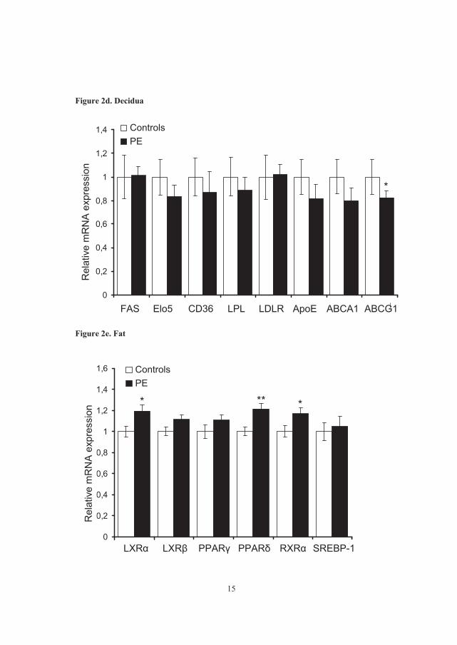
15
Figure 2d. Decidua
Figure 2e. Fat
0
0,2
0,4
0,6
0,8
1
1,2
1,4
FAS ELOVL5 CD36 LPL LDLR APOE ABCA1 ABCG1
Rel
ativ
e m
RN
A ex
pres
sion
ControlsPE
FAS Elo5 CD36 LPL LDLR ApoE ABCA1 ABCG1
Rel
ativ
e m
RN
Aex
pres
sion
*
0
0,2
0,4
0,6
0,8
1
1,2
1,4
FAS ELOVL5 CD36 LPL LDLR APOE ABCA1 ABCG1
Rel
ativ
e m
RN
A ex
pres
sion
ControlsPE
FAS Elo5 CD36 LPL LDLR ApoE ABCA1 ABCG1
Rel
ativ
e m
RN
Aex
pres
sion
*
0
0,2
0,4
0,6
0,8
1
1,2
1,4
1,6
LXRa LXRb PPARg PPARd RXRa SREBP-1
Rel
ativ
e m
RN
A ex
pres
sion
ControlsPE
** **
LXR� RXR�LXR� PPAR�PPAR� SREBP-1
Rel
ativ
e m
RN
Aex
pres
sion
0
0,2
0,4
0,6
0,8
1
1,2
1,4
1,6
LXRa LXRb PPARg PPARd RXRa SREBP-1
Rel
ativ
e m
RN
A ex
pres
sion
ControlsPE
** **
LXR� RXR�LXR� PPAR�PPAR� SREBP-1
Rel
ativ
e m
RN
Aex
pres
sion

16
Figure 2f. Fat
Figure 3.
0
0,2
0,4
0,6
0,8
1
1,2
1,4
1,6
FAS ELOVL5 CD36 LPL LDLR APOE ABCA1 ABCG1
Rel
ativ
e m
RN
A ex
pres
sion
ControlsPE *
FAS Elo5 CD36 LPL LDLR ApoE ABCA1 ABCG1
Rel
ativ
e m
RN
Aex
pres
sion
0
0,2
0,4
0,6
0,8
1
1,2
1,4
1,6
FAS ELOVL5 CD36 LPL LDLR APOE ABCA1 ABCG1
Rel
ativ
e m
RN
A ex
pres
sion
ControlsPE *
FAS Elo5 CD36 LPL LDLR ApoE ABCA1 ABCG1
Rel
ativ
e m
RN
Aex
pres
sion
C1
C2
C3
C4
C5
C6
PE1
PE2
PE3
PE4 PE
5
PE6
CControl PE
�-LXR�
�-LXR�
�-�-actin
*
C1
C2
C3
C4
C5
C6
PE1
PE2
PE3
PE4 PE
5
PE6
CControl PE
�-LXR�
�-LXR�
�-�-actin
*

17
Figure 4. Correlation between placental expression of LXR� and placental concentrations of FFA.
0,05
0,1
0,15
0,2
0,25
0,3
0,6 0,8 1 1,2Placental relative LXR� expression
Pla
cent
al F
FA (m
g/g
plac
enta
)
Table 1. Clinical characteristics of subjects
Control (n=33) PE (n=28) PE vs. control Median
(min.-max.) Median
(min.-max.) p-value
Patient age at delivery (years)
33 (25-40)
31 (18-42)
n.s.
BMI before pregnancy (kg/m2)
21.7 (18-30.7)
24.9 (19.4-41.1)
<0.05*
BMI at delivery (kg/m2) 27.9 (22.8-37.5)
31.3 (24.0-49.6)
<0.05*
Parity 0 (0-3)
0 (0-3)
n.s.
Gestational age at delivery (weeks)
38.7 (37-41.7)
32.6 (24.9-39.3)
<0.001**
Systolic BP <week 20 (mmHg)
110 (90-135)
117 (95-135)
<0.05*
Diastolic BP <week 20 (mmHg)
65 (55-93)
73 (50-89)
<0.05*
Systolic BP at delivery (mmHg)
120 (100-153)
161 (140-220)
<0.001*
Diastolic BP at delivery (mmHg)
70 (55-92)
100 (90-119)
<0.001*
Neonatal weight (g) 3600 (2800-4376)
1710 (540-5036)
<0.001*
Neonatal weight percentile1
62.0 (13.6-99.5)
34.8 (1.0-99.5)
<0.001*
Neonatal weight percentile2
77.7 (9.0-99.5)
7.2 (0.5-99.5)
<0.001*
1 Neonatal weight percentile according to national birth registry data [16] 2 Neonatal weight percentile according to ultrasound based weight percentile [17]
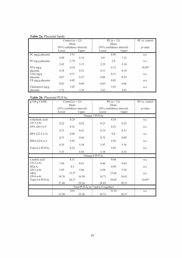
18
Table 2a. Placental lipids
Control (n = 12) PE (n = 12) Mean Mean
PE vs. control
(95% confidence interval) (95% confidence interval) p-value
Lower Upper Lower Upper 5.51 6.06 n.s. PC mg/g placenta
4.89 6.14 4.9 7.22 2.79 2.8 n.s. PE mg/g placenta
2.42 3.15 2.25 3.34 0.24 0.15 <0.05* FFA mg/g
placenta 0.18 0.31 0.11 0.19 0.17 0.31 n.s. TAG mg/g
placenta 0.07 0.27 0.06 0.55 0.05 0.05 n.s. CE mg/g placenta
0.03 0.06 0.03 0.06 3.05 3.03 n.s. Cholesterol mg/g
placenta 2.72 3.38 2.42 3.63 Table 2b. Placental PUFAs
Control (n = 12) PE (n = 12) Mean Mean
PE vs. control
(95% confidence interval) (95% confidence interval) p-value
g/100 g FAME
Lower Upper Lower Upper Omega-3 PUFAs
0.24 0.24 n.s. �-linolenic acid (18:3 n-6) 0.22 0.25 0.23 0.25
0.32 0.25 n.s. EPA (20:5 n-3 0.23 0.41 0.19 0.31
0.84 0.8 n.s. DPA (22:5 n-3) 0.73 0.94 0.72 0.89
4.83 4.56 n.s. DHA (22:6 n-3 4.29 5.38 3.97 5.16
6.23 5.85 n.s. Total n-3 PUFAs 5.53 6.92 5.18 6.52
Omega-6 PUFAs 8.31 9.04 n.s. Linoleic acid
(18:2 n-6) 7.80 8.81 8.44 9.64 4.5 4.99 n.s. DGLA
(20:3 n-6) 3.95 5.04 4.58 5.39 15.57 15.66 n.s. ARA
(20:4 n-6) 14.76 16.38 14.71 16.61 28.37 29.69 <0.05* Total n-6 PUFAs
27.48 29.26 28.85 30.53 Total PUFAs (n-3 and n-6 together)
34.6 35.54 n.s. 33.94 35.26 34.71 36.37

Paper 4

This article is removed.
