fcγ-receptor iia-mediated src signaling pathway is
TRANSCRIPT
RESEARCH ARTICLE
Fcγ-receptor IIa-mediated Src Signaling
Pathway Is Essential for the Antibody-
Dependent Enhancement of Ebola Virus
Infection
Wakako Furuyama1, Andrea Marzi2, Aaron B. Carmody3, Junki Maruyama1,
Makoto Kuroda1¤, Hiroko Miyamoto1, Asuka Nanbo4, Rashid Manzoor1, Reiko Yoshida1,
Manabu Igarashi1,5, Heinz Feldmann2, Ayato Takada1,5,6*
1 Division of Global Epidemiology, Research Center for Zoonosis Control, Hokkaido University, Sapporo,
Japan, 2 Laboratory of Virology, Division of Intramural Research, National Institute of Allergy and Infectious
Diseases, National Institutes of Health, Rocky Mountain Laboratories, Hamilton, MT, United States of
America, 3 Research Technologies Branch, Division of Intramural Research, National Institute of Allergy and
Infectious Diseases, National Institutes of Health, Hamilton, MT, United States of America, 4 Department of
Cell Physiology, Hokkaido University Graduate School of Medicine, Sapporo, Japan, 5 Global Station for
Zoonosis Control, Global Institution for Collaborative Research and Education, Hokkaido University, Sapporo,
Japan, 6 School of Veterinary Medicine, the University of Zambia, Lusaka, Zambia
¤ Current address: Department of Pathobiological Sciences, School of Veterinary Medicine, University of
Wisconsin-Madison, Madison, WI, United States of America
Abstract
Antibody-dependent enhancement (ADE) of Ebola virus (EBOV) infection has been demon-
strated in vitro, raising concerns about the detrimental potential of some anti-EBOV antibod-
ies. ADE has been described for many viruses and mostly depends on the cross-linking of
virus-antibody complexes to cell surface Fc receptors, leading to enhanced infection. How-
ever, little is known about the molecular mechanisms underlying this phenomenon. Here we
show that Fcγ-receptor IIa (FcγRIIa)-mediated intracellular signaling through Src family pro-
tein tyrosine kinases (PTKs) is required for ADE of EBOV infection. We found that deletion
of the FcγRIIa cytoplasmic tail abolished EBOV ADE due to decreased virus uptake into cel-
lular endosomes. Furthermore, EBOV ADE, but not non-ADE infection, was significantly
reduced by inhibition of the Src family protein PTK pathway, which was also found to be
important to promote phagocytosis/macropinocytosis for viral uptake into endosomes. We
further confirmed a significant increase of the Src phosphorylation mediated by ADE. These
data suggest that antibody-EBOV complexes bound to the cell surface FcγRIIa activate the
Src signaling pathway that leads to enhanced viral entry into cells, providing a novel per-
spective for the general understanding of ADE of virus infection.
Author Summary
Antibody-dependent enhancement (ADE), a phenomenon in which viral infectivity is
increased by virus-specific antibodies, is observed in vitro for a large number of viruses.
PLOS Pathogens | DOI:10.1371/journal.ppat.1006139 December 30, 2016 1 / 22
a1111111111
a1111111111
a1111111111
a1111111111
a1111111111
OPENACCESS
Citation: Furuyama W, Marzi A, Carmody AB,
Maruyama J, Kuroda M, Miyamoto H, et al. (2016)
Fcγ-receptor IIa-mediated Src Signaling Pathway Is
Essential for the Antibody-Dependent
Enhancement of Ebola Virus Infection. PLoS
Pathog 12(12): e1006139. doi:10.1371/journal.
ppat.1006139
Editor: Thomas Geisbert, University of Texas
Medical Branch, UNITED STATES
Received: September 13, 2016
Accepted: December 19, 2016
Published: December 30, 2016
Copyright: This is an open access article, free of all
copyright, and may be freely reproduced,
distributed, transmitted, modified, built upon, or
otherwise used by anyone for any lawful purpose.
The work is made available under the Creative
Commons CC0 public domain dedication.
Data Availability Statement: All relevant data are
within the paper and its Supporting Information
files.
Funding: This work was supported by KAKENHI
(14J06186 and 15H01249), Grants-in-Aid for
Scientific Research from the Ministry of Education,
Culture, Sports, Science and Technology (MEXT)
and the Japan Society for the Promotion of Science
(JSPS), Japan, and partly by the Japan Initiative for
Global Research Network on Infectious Diseases
For some of these viruses, ADE often become an issue for disease control by vaccination.
It has also been shown that some human sera convalescent from Ebola virus disease con-
tain ADE antibodies. ADE has been shown mostly to depend on the cross-linking of
virus-antibody complexes to cell surface Fc receptor, which activate various signaling
pathways involved in the reorganization of the actin cytoskeleton and membrane remod-
eling. In this study, we demonstrate that Fc receptor-mediated intracellular signaling is a
key factor for ADE of Ebola virus infection. We found that the antibody-virus complexes
bound to the cell surface Fc receptors triggered the phosphorylation of particular protein-
tyrosine kinases that activated signaling pathways leading to enhanced viral uptake into
cells through phagocytosis and/or macropinocytosis. Our study provides new insights
into mechanisms of ADE and also offer a potential new cellular target to develop treat-
ments for ADE-associated diseases such as dengue hemorrhagic fever and possibly Zika
virus infection.
Introduction
Ebola virus (EBOV), a member of the family Filoviridae, causes severe hemorrhagic fever in
humans and nonhuman primates, with human case fatality rates of up to 90% [1]. EBOV
expresses a glycoprotein (GP) that is the only viral surface protein and important for both
receptor binding and membrane fusion [2,3]. EBOV entry is initiated by viral attachment to
cell surface molecules such as T-cell immunoglobulin and mucin domain 1 (TIM-1) and C-
type lectins [4,5], followed by internalization of the virus particle into cells via macropinocyto-
sis [6–8]. In the late endosome, EBOV GP is cleaved by host proteases such as cathepsins L
and B [9], exposing the GP receptor binding site that then binds to the receptor, Niemann-
Pick C1 (NPC1), followed by membrane fusion [10,11].
In addition to the direct interaction between GP and host cell receptors, it has been demon-
strated that EBOV exploits some GP-specific antibodies for its entry into cells, leading to
increased infectivity in vitro [12,13]. This phenomenon has been described for a number of
viruses and is known as antibody-dependent enhancement (ADE) [14–17]. For some of these
viruses, ADE has become a great concern to disease control by vaccination. Particularly, con-
valescent human sera have been shown to contain ADE antibodies [12,13], raising concerns
about potential detrimental effects of passive immunization with convalescent human sera,
which is currently under consideration for treatment of Ebola virus disease. Importantly, it
was recently demonstrated that therapeutic treatment with convalescent sera having in vitro
neutralizing activities was not sufficient for protection against EBOV infection in nonhuman
primates [18]. Although ADE was not evaluated in vitro and any enhanced pathogenicity in
the treated animals was not observed, it might be possible that ADE antibodies counterbal-
anced the neutralizing activity as suggested previously [17]. Two distinct pathways of EBOV
ADE, one mediated by Fc receptors and the other by complement component C1q and its
ligands, are known [13,17]. In particular, the Fcγ receptor (FcγR) is commonly involved in
ADE of virus infections [19,20]. However, the molecular mechanisms underlying ADE-medi-
ated virus entry through FcγR are not fully understood.
Three classes of FcγR, FcγRI (CD64), FcγRII (CD32), and FcγRIII (CD16), are expressed in
various human immune cells such as dendritic cells, monocytes, and B lymphocytes [21].
Among these FcγRs, FcγRII is a key molecule for EBOV ADE of infection in human leukemia
K562 cells [17]. Human FcγRII exists in two isoforms, FcγRIIa and FcγRIIb, which differ in
their signal peptides and cytoplasmic tails. FcγRIIa is the active form of FcγRII and contains
Fc Receptor-Mediated Signaling Pathway Essential for Ebola Virus ADE
PLOS Pathogens | DOI:10.1371/journal.ppat.1006139 December 30, 2016 2 / 22
(J-GRID) from Ministry of Education, Culture,
Sport, Science & Technology in Japan, and Japan
Agency for Medical Research and Development
(AMED), and the Division of Intramural Research,
National Institute of Allergy and Infectious
Diseases, National Institutes of Health, USA. The
funders had no role in study design, data collection
and analysis, decision to publish, or preparation of
the manuscript.
Competing Interests: The authors have declared
that no competing interests exist.
an immunoreceptor tyrosine-based activation motif (ITAM) in its cytoplasmic tail [21]. The
cytoplasmic tail of FcγRIIa is known to contribute to the activitation of two structurally and
functionally distinct protein-tyrosine kinase (PTK) classes, the sarcoma (Src) family PTKs
[22,23] and spleen tyrosine kinase (Syk) [24]. In addition, Syk is reported to participate in acti-
vation of enzymes such as rat sarcoma (Ras), phosphatidylinositol 3-kinase (PI3K), and Bru-
ton’s tyrosine kinase (Btk) [21,25]. These signaling pathways are known to be important for
the induction of phagocytic and endocytic processes to internalize immune complexes
[21,25,26].
In this study, we focused on the role of FcγRIIa and investigated the contribution of
FcγRIIa-mediated signaling to the ADE of EBOV infection. We show that Src family PTKs are
essential for EBOV ADE-mediated entry. Our data indicate that binding of antibody-virus
complexes to the cell surface FcγRIIa triggers phosphorylation of Src family PTKs and activates
subsequent signaling pathways, leading to enhanced viral uptake through phagocytosis and/or
macropinocytosis.
Results
Cytoplasmic tail of FcγRIIa is required for ADE of EBOV infection
To investigate the role of FcγRIIa and, in particular, the importance of its cytoplasmic tail in
EBOV ADE, we compared the functions of wild-type FcγRIIa and a deletion mutant of FcγRIIa
lacking its cytoplasmic tail (FcγRIIaΔCT). Both molecules were expressed on Jurkat T cells,
which are known to be poorly permissive for EBOV infection [27] and lack this Fc receptor
[28]. Jurkat T cells were transduced with full-length FcγRIIa or FcγRIIaΔCT genes using a retro-
virus vector (Fig 1A and 1B) and subsequently infected with vesicular stomatitis virus (VSV)
pseudotyped with EBOV GP (VSV-EBOV GP) and infectious EBOV in the presence or absence
of the GP-specific monoclonal antibody (MAb) ZGP12/1.1, which is known to induce EBOV
ADE [12] (Fig 1C and 1D). We found that viral infectivity was almost undetectable in naive and
control vector-transduced Jurkat T cells but the expression of wild-type FcγRIIa significantly
enhanced the infectivity of VSV-EBOV GP and EBOV in the presence of ZGP12/1.1, though
not control IgG (CTR IgG). Interestingly, the infection rate of FcγRIIaΔCT-expressing cells was
significantly lower than that of cells expressing wild-type FcγRIIa. These results indicated that
the FcγRIIa-MAb complex functioned as a receptor-like molecule on this poorly permissive cell
line and efficiently promoted infection through the ADE of EBOV entry. More importantly, the
results suggested that signaling pathways via the FcγRIIa cytoplasmic tail were likely involved in
the ADE of EBOV entry into cells.
Cytoplasmic tail of FcγRIIa is important for enhanced viral uptake
FcγRIIa is known to modulate phagocytosis/macropinocytosis through signaling pathways via
its cytoplasmic tail [29,30]. Therefore, to analyze viral binding and intracellular uptake in
more detail, we produced lipophilic tracer (DiI)-labeled virus-like particles (VLPs) consisting
of the major EBOV structural proteins, GP, matrix protein (VP40), and nucleoprotein (NP),
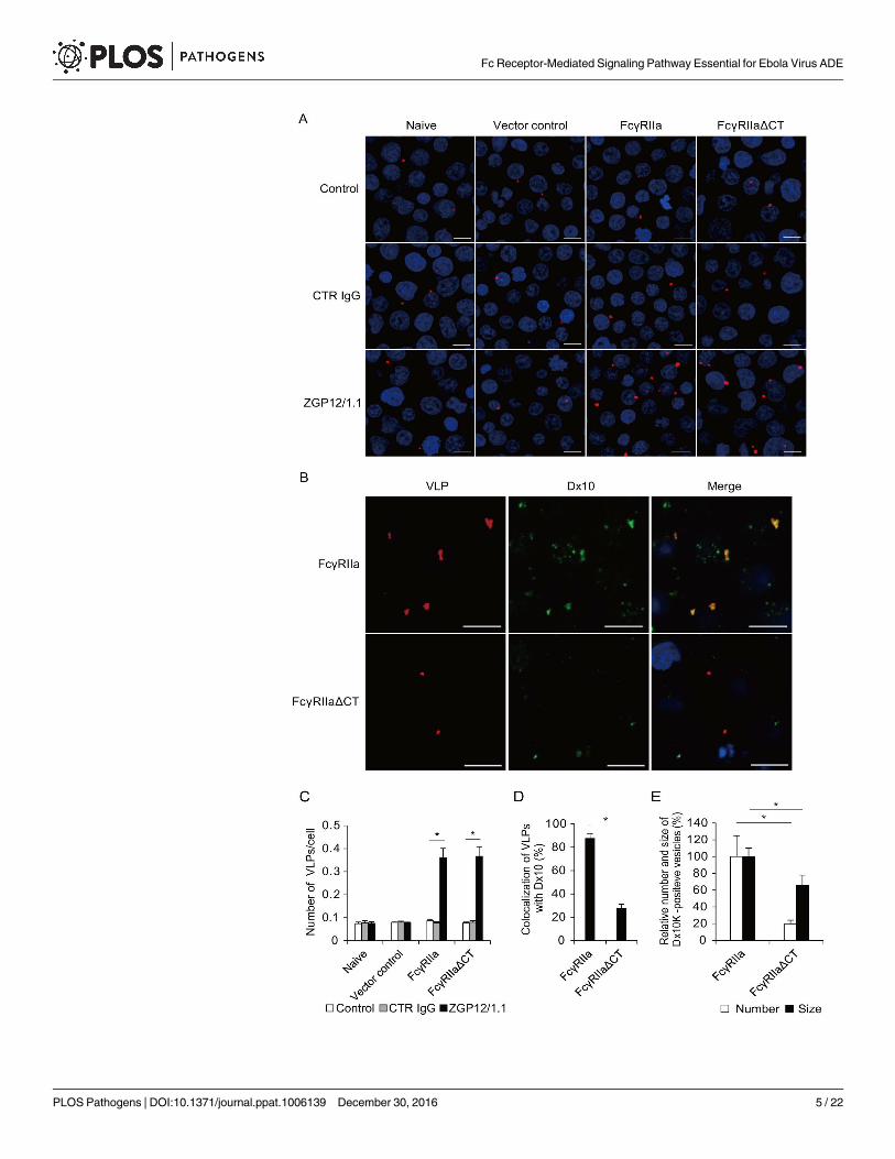
and monitored the localization of VLPs in each transduced Jurkat T cell line (Fig 2). The num-
ber of VLPs attached to the surface of naive and empty vector-transduced Jurkat T cells was
not significantly different irrespective of the presence of CTR IgG and ZGP12/1.1 (Fig 2A and
2C). In contrast, the attachment of VLPs was significantly enhanced to similar extents in Jurkat
T cells expressing FcγRIIa and FcγRIIaΔCT in the presence of ZGP12/1.1 but not CTR IgG
(Fig 2A and 2C), suggesting that the FcγRIIa ectodomain expressed on Jurkat T cells had the
ability to increase the VLP attachment mediated by ZGP12/1.1. Next, we assessed the number
of VLPs incorporated into intracellular vesicles along with Alexa Fluor 647 (Alexa647)-labeled
Fc Receptor-Mediated Signaling Pathway Essential for Ebola Virus ADE
PLOS Pathogens | DOI:10.1371/journal.ppat.1006139 December 30, 2016 3 / 22
Fig 1. Importance of FcγRIIa and its cytoplasmic tail in EBOV ADE. (A) Schematic representation of FcγRIIa and
FcγRIIaΔCT. TM, transmembrane region; CT, cytoplasmic tail. (B) Cell surface expression of the FcγRIIa ectodomain on
Jurkat T cell lines. Jurkat T cells stably expressing FcγRIIa and FcγRIIaΔCT were stained with an anti-CD32 antibody and
analyzed by flow cytometry. Black lines represent vector-transduced, FcγRIIa-, and FcγRIIaΔCT-expressing cells. Gray
shading represents naive Jurkat T cells. (C, D) Infectivity of VSV-EBOV GP and EBOV in Jurkat T cell lines. VSV-EBOV
GP (C) and EBOV (D) were incubated with medium alone (Control), CTR IgG, or ZGP12/1.1, followed by inoculation into
each Jurkat T cell line. At 24 h (C) and 72 h (D) after inoculation, GFP-positive cells were counted. The relative infectivity in
each cell line was calculated by setting the IU value of untreated (Control), CTR IgG-, and ZGP12/1.1-treated viruses in
naive Jurkat T cells to 100%, respectively. The mean and standard deviation of three independent experiments are shown.
Statistical analysis was performed using Student’s t-test (*p<0.05).
doi:10.1371/journal.ppat.1006139.g001
Fc Receptor-Mediated Signaling Pathway Essential for Ebola Virus ADE
PLOS Pathogens | DOI:10.1371/journal.ppat.1006139 December 30, 2016 4 / 22
Fc Receptor-Mediated Signaling Pathway Essential for Ebola Virus ADE
PLOS Pathogens | DOI:10.1371/journal.ppat.1006139 December 30, 2016 5 / 22
dextran Mw 10,000 (Dx10), a specific probe for visualizing phagocytotic and macropinocytotic
vesicles [6,31]. After incubation for 2 h, ZGP12/1.1-treated VLPs efficiently colocalized with
Dx10 in Jurkat T cells expressing wild-type FcγRIIa, but not in cells expressing FcγRIIaΔCT
(Fig 2B and 2D). Viral uptake was not observed drastically in the absence of FcγRIIa and
ZGP12/1.1. Furthermore, the number and size of Dx10-positive vesicles incorporated into Jur-
kat T cells expressing FcγRIIaΔCT were significantly smaller than those in cells expressing
FcγRIIa, indicating the importance of the FcγRIIa cytoplasmic tail in activating the phagocyto-
sis/macropinocytosis (Fig 2B and 2E). These data suggested that the ADE infection of FcγRIIa-
expressing Jurkat T cells was associated with enhanced viral uptake into cellular vesicles, most
likely due to the activation of FcγRIIa-mediated signaling via its cytoplasmic tail.
Intracellular signaling via Src family PTKs contributes to ADE of EBOV
infection
To identify the intracellular signaling pathway involved in the ADE of EBOV entry, we ana-
lyzed the effects of different inhibitors of signaling pathways in K562 cells, which naturally
express FcγRIIa. Consistent with previous studies [17,32], K562 cells were permissive to
VSV-EBOV GP and EBOV infections, and viral infection rates were significantly enhanced in
the presence of ZGP12/1.1 (S1A and S1B Fig). We then tested inhibitors of Syk and Src family
PTKs (R788 and PP2, respectively) as these PTKs are known to be principally involved in sig-
naling pathways downstream of FcγRIIa, and in particular to play important roles in inducing
FcγR-mediated phagocytosis/macropinocytosis [26,33]. We found that the ADE of VSV-E-
BOV GP infection was significantly reduced in K562 cells treated with these inhibitors in a
dose-dependent manner. In contrast, only a limited reduction was seen in non-ADE infection
at the highest concentrations of the inhibitors (Fig 3). The ADE-specific inhibitory effect was
more prominent in cells treated with PP2 than in those treated with R788.
Since Syk is reported to participate in the activation of signaling through PI3K, Btk, and
Ras, we further examined which pathways downstream of Syk contributed to the ADE of
VSV-EBOV GP infection using specific inhibitors of PI3K (LY294002), Btk (LFM-A13), and
Ras (Manumycin A) (Fig 3). However, both ADE and non-ADE infections by VSV-EBOV
GP were dose-dependently reduced by LY294002 and Manumycin A, respectively. LFM-A13
showed little effect on the infectivity of VSV-EBOV GP. Subsequently, we confirmed the
effects of these inhibitors using infectious EBOV. Consistent with the data for VSV-EBOV
GP, PP2 selectively reduced the ADE, but not the non-ADE infection. Interestingly, R788
showed no effects on the ADE of EBOV infection and rather enhanced the non-ADE infection.
LFM-A13 slightly reduced both the ADE and the non-ADE infections, whereas LY294002 and
Manumycin A did not inhibit the ADE infection, though Manumycin A slightly inhibited the
non-ADE infection.
Fig 2. Importance of FcγRIIa and its cytoplasmic tail in ADE-mediated VLP uptake into cells. (A, B)
Fluorescent images of attachment and internalization of VLPs. (C-E) Quantified fluorescent signals of VLP
and Dx10. Untreated (Control), CTR IgG-, and ZGP12/1.1-treated DiI-labeled VLPs were inoculated into each
Jurkat T cell line and incubated for 30 min on ice. After adsorption, the cells were incubated for 0 h (A, C) or
incubated with Alexa647-labeled Dx10 for 2 h at 37˚C (B, D, E). VLPs (red) on the cell surface (A, C) and
VLPs (red) and Dx10 (green) in the cytoplasm (B, D, E) were monitored by confocal laser scanning
microscopy. Scale bars represent 10 μm. Nuclei of cells are visualized with DAPI (blue). The number of VLPs
attached to the cell surface (C), the colocalization of VLPs (DiI) and Dx10 (Alexa647) signals (D), and the
number and size of Dx10-positive vesicles (E) were quantified. The number and size were calculated by
setting the value of FcγRIIa-expressing Jurkat T cells in the presence of ZGP12/1.1-treated VLPs to 100%.
The mean and standard deviation of three independent experiments are shown. Statistical analysis was
performed using Student’s t-test (*p<0.05).
doi:10.1371/journal.ppat.1006139.g002
Fc Receptor-Mediated Signaling Pathway Essential for Ebola Virus ADE
PLOS Pathogens | DOI:10.1371/journal.ppat.1006139 December 30, 2016 6 / 22
Fig 3. Identification of FcγRIIa-mediated signaling pathway for EBOV ADE. K562 cells were treated with the indicated concentrations of
R788, PP2, LFM-A13, LY294002, or Manumycin A for 1 h at 37˚C and inoculated with VSV-EBOV GP or EBOV preincubated with CTR IgG,
or ZGP12/1.1. The relative percentage of infectivity was calculated by setting the IU value of the viruses in DMSO or ethanol-treated cells to
Fc Receptor-Mediated Signaling Pathway Essential for Ebola Virus ADE
PLOS Pathogens | DOI:10.1371/journal.ppat.1006139 December 30, 2016 7 / 22
To further investigate the role of Src- and Syk-mediated signaling in EBOV ADE, we gener-
ated Src and Syk knockdown K562 cells. K562 cells were transduced with retroviral vectors
expressing small hairpin RNAs (shRNAs) for silencing Src or Syk genes (Fig 4A and 4B) and
infected with VSV-EBOV GP in the presence or absence of ZGP12/1.1 (Fig 4C). We found
that transduced cells stably expressing Src shRNAs (shSrc3 and shSrc4) showed approximately
50% reduction in protein levels (Fig 4A) and ZGP12/1.1-mediated ADE was significantly
decreased in these cell lines (Fig 4C). In contrast, no significant difference was seen between
ADE (ZGP12/1.1) and non-ADE (CTR IgG) infections in Syk knockdown cells although 2 of
the Syk shRNAs (shSyk3 and shSyk4) significantly reduced the expression of the Syk protein
(Fig 4B and 4C).
These results demonstrated that FcγRIIa-mediated signaling through the activation of Src
family PTKs contributed to the ADE of EBOV infection, but Syk-related signaling including
PI3K, Btk, and Ras did not seem to be specifically involved. We further tested the effect of PP2
in K562 cell lines stably expressing dendritic cell-specific ICAM-3-grabbing non-integrin
(DC-SIGN) or human macrophage galactose-type C-type lectin (hMGL), both of which have
been shown to act as attachment receptors for EBOV [32, 34], and found that PP2 had limited
effects on the infectivity of VSV-EBOV GP in these cell lines (S2 Fig).
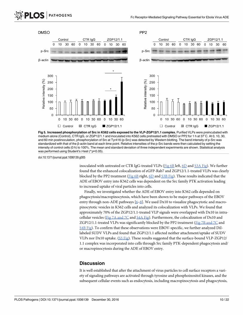
VLP-MAb complexes activate phosphorylation of Src in K562 cells
To directly detect the activation of Src family PTKs, we quantified the phosphorylation levels
of Src in K562 cells (Fig 5). We found no significant difference in the cells inoculated with
intact VLPs alone at each time point. Likewise, inoculation of CTR IgG-treated VLPs did not
enhance Src phosphorylation levels. However, a significant increase of the Src phosphorylation
was detected at 30 and 60 min after K562 cells were exposed to ZGP12/1.1-treated VLPs (Fig 5
left). Furthermore, the enhanced phosphorylation was completely blocked by the Src family
PTK inhibitor, PP2 (Fig 5 right). These findings suggested that Src were activated by the inter-
action of the VLP-ZGP12/1.1 complex with FcγRIIa.
The ADE of EBOV entry depends on phagocytosis and/or
macropinocytosis mediated by Src family PTKs
To further characterize the role of the Src family PTK-dependent signaling in the ADE of
EBOV infection, we analyzed the effect of PP2 on the attachment and uptake of DiI-labeled
VLPs using K562 cells. We first compared the number of VLPs attached to the cell surface
among untreated, CTR IgG-, and ZGP12/1.1-treated VLPs. Since the overexpression of
FcγRIIa in Jurkat T cells increased the attachment of VLPs to the cell surface in the presence of
ZGP12/1.1 (Fig 2), we hypothesized that ZGP12/1.1 would enhance the VLP attachment to
K562 cells. However, the number of VLPs attached to the cell surface was not significantly dif-
ferent in the presence or absence of ZGP12/1.1 (Fig 6A left and 6C), and was not affected by
the PP2 treatment (Fig 6A right and 6C), indicating that EBOV ADE in K562 cells did not
result from increased viral attachment to the cell surface. For the visualization of the VLP
uptake into endosomes, K562 cells expressing enhanced green fluorescent protein fused to
Rab7 (eGFP-Rab7), a late endosome marker, were used to analyze colocalization of eGF-
P-Rab7 and internalized VLPs. We found that ZGP12/1.1-treated VLPs were efficiently coloca-
lized with eGFP-Rab7 in K562 cells, whereas only 10–20% colocalization was seen in the cells
100%. The mean and standard deviation of three independent experiments are shown. Statistical analysis was performed using Student’s t-
test (*p<0.05).
doi:10.1371/journal.ppat.1006139.g003
Fc Receptor-Mediated Signaling Pathway Essential for Ebola Virus ADE
PLOS Pathogens | DOI:10.1371/journal.ppat.1006139 December 30, 2016 8 / 22
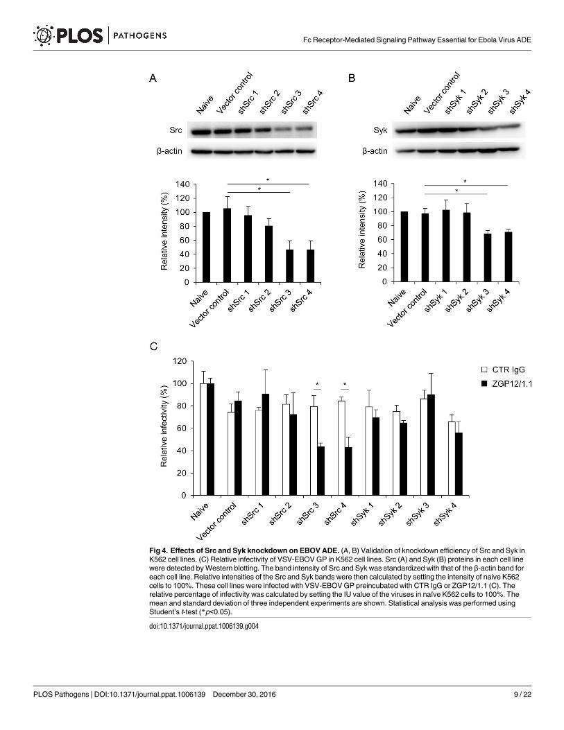
Fig 4. Effects of Src and Syk knockdown on EBOV ADE. (A, B) Validation of knockdown efficiency of Src and Syk in
K562 cell lines. (C) Relative infectivity of VSV-EBOV GP in K562 cell lines. Src (A) and Syk (B) proteins in each cell line
were detected by Western blotting. The band intensity of Src and Syk was standardized with that of the β-actin band for
each cell line. Relative intensities of the Src and Syk bands were then calculated by setting the intensity of naive K562
cells to 100%. These cell lines were infected with VSV-EBOV GP preincubated with CTR IgG or ZGP12/1.1 (C). The
relative percentage of infectivity was calculated by setting the IU value of the viruses in naïve K562 cells to 100%. The
mean and standard deviation of three independent experiments are shown. Statistical analysis was performed using
Student’s t-test (*p<0.05).
doi:10.1371/journal.ppat.1006139.g004
Fc Receptor-Mediated Signaling Pathway Essential for Ebola Virus ADE
PLOS Pathogens | DOI:10.1371/journal.ppat.1006139 December 30, 2016 9 / 22
inoculated with untreated or CTR IgG-treated VLPs (Fig 6B left, 6D and S3A Fig). We further
found that the enhanced colocalization of eGFP-Rab7 and ZGP12/1.1-treated VLPs was clearly
blocked by the PP2 treatment (Fig 6B right, 6D and S3B Fig). These results indicated that the
ADE of EBOV entry into K562 cells was dependent on the Src family PTK activation leading
to increased uptake of viral particles into cells.
Finally, we investigated whether the ADE of EBOV entry into K562 cells depended on
phagocytosis/macropinocytosis, which have been shown to be major pathways of the EBOV
entry through non-ADE pathways [6–8]. We used Dx10 to visualize phagocytotic and macro-
pinocytotic vesicles in K562 cells and analyzed its colocalization with VLPs. We found that
approximately 70% of the ZGP12/1.1-treated VLP signals were overlapped with Dx10 in intra-
cellular vesicles (Fig 7A and 7C and S4A Fig). Furthermore, the colocalization of Dx10 and
ZGP12/1.1-treated VLPs was significantly blocked by the PP2 treatment (Fig 7B and 7C and
S4B Fig). To confirm that these observations were EBOV-specific, we further analyzed DiI-
labeled SUDV VLPs and found that ZGP12/1.1 affected neither attachment/uptake of SUDV
VLPs nor Dx10 uptake. (S5 Fig). These results suggested that the surface-bound VLP-ZGP12/
1.1 complex was incorporated into cells through Src family PTK-dependent phagocytosis and/
or macropinocytosis during the ADE of EBOV entry.
Discussion
It is well established that after the attachment of virus particles to cell surface receptors a vari-
ety of signaling pathways are activated through tyrosine and phosphoinositol kinases, and the
subsequent cellular events such as endocytosis, including macropinocytosis and phagocytosis,
Fig 5. Increased phosphorylation of Src in K562 cells exposed to the VLP-ZGP12/1.1 complex. Purified VLPs were preincubated with
medium alone (Control), CTR IgG, or ZGP12/1.1 and inoculated into K562 cells pretreated with DMSO or PP2 for 1 h at 37˚C. At 0, 10, 30,
and 60 min postinoculation, phosphorylation of Src at Tyr416 (p-Src) was detected by Western blotting. The band intensity of p-Src was
standardized with that of the β-actin band at each time point. Relative intensities of the p-Src bands were then calculated by setting the
intensity of control cells (0 h) to 100%. The mean and standard deviation of three independent experiments are shown. Statistical analysis
was performed using Student’s t-test (*p<0.05).
doi:10.1371/journal.ppat.1006139.g005
Fc Receptor-Mediated Signaling Pathway Essential for Ebola Virus ADE
PLOS Pathogens | DOI:10.1371/journal.ppat.1006139 December 30, 2016 10 / 22
Fig 6. Enhanced VLP uptake into endosomes in K562 cells during ADE. (A, B) Fluorescent images of attachment and internalization
of VLPs. (C, D) Quantified fluorescent signals of VLP and Rab-7. K562 cells expressing eGFP-Rab7 were incubated with DMSO or PP2
Fc Receptor-Mediated Signaling Pathway Essential for Ebola Virus ADE
PLOS Pathogens | DOI:10.1371/journal.ppat.1006139 December 30, 2016 11 / 22
are important for the entry of viruses [35,36]. Likewise, it has been suggested that signaling path-
ways via FcγR are involved in ADE of virus infections [20]. However, there is limited informa-
tion on the detailed molecular mechanisms of intracellular signaling pathways required for ADE
of virus infection. It has been shown that the non-ADE entry of EBOV requires host factors such
as PI3K, the Rho family, and protein kinase C [6,7,37], although the virus-specific receptor mole-
cules involved in these signaling pathways are not yet identified. In the present study, we focused
on the ADE of EBOV entry and found that the FcγRIIa-mediated signaling pathway was essen-
tial for this process, which is distinct from those required for the non-ADE entry.
The FcγRIIa cytoplasmic tail has been shown to be essential for ADE of dengue virus entry
and the involvement of Syk cascade in ADE entry has been reported [38–40]. Our data indicate
that the cytoplasmic tail of FcγRIIa is crucial for the ADE of EBOV infection and that Src family
PTK-dependent signaling is important to enhance viral uptake into cellular vesicles during ADE-
mediated entry of EBOV. Src family PTKs are non-receptor tyrosine kinases involved in the regu-
lation of diverse cellular functions like proliferation, differentiation, adhesion, and phagocytosis
[41,42]. Importantly, this signaling pathway is known to regulate endocytic machinery by trigger-
ing the reorganization of the actin cytoskeleton and membrane remodeling [21,26]. Indeed, previ-
ous studies have demonstrated that Src family PTK-dependent signaling is required for the non-
ADE entry of some viruses into host cells [43,44]. Our data indicate that this signaling is also used
to promote viral particle uptake during the FcγRIIa-mediated ADE of EBOV entry.
It was noted that there was a significant increase (approximately 400%) in infectivity of
VSV-EBOV GP when FcγRIIaΔCT is expressed on Jurkat T cells, although it was not as high
as wildtype FcγRIIa. This observation suggest that the binding provided by the external por-
tion of FcγRIIa may also have some importance and that the signaling function provided by
the cytoplasmic portion of FcγRIIa further enhances the ADE effect. It might also be possible
that the FcγRIIa associated with lipid rafts activates some FcγRIIa-mediated signals through its
transmembrane domain as described previously [45].
Interestingly, we found that the Syk inhibitor R788 reduced the ADE efficiency of VSV-E-
BOV GP but not EBOV. While the VSV pseudotype system is widely used to study EBOV GP
functions, it has been suggested that pseudotyped VSV and authentic EBOV can utilize differ-
ent entry pathways since the particle size and structure of pseudotyped VSV do not accurately
recapitulate those of EBOV [6,46]. Thus, it may be possible that the EBOV entry primarily
relies on macropinocytosis as shown previously [6–8], whereas VSV-EBOV GP can also be
incorporated into smaller vesicles. We assume that this difference could influence the effect of
the inhibitor since Syk-dependent signaling might be associated with caveolin-mediated endo-
cytosis [47,48]. Another difference found between VSV-EBOV GP and authentic EBOV was
that while the Syk inhibitor did not affect EBOV ADE, non-ADE infection was significantly
enhanced in the presence of this inhibitor, an effect not observed for VSV-EBOV GP. This
might be due to the effect on post-entry mechanisms such as antiviral cellular responses, as
proposed by a recent study demonstrating that dengue virus-antibody complexes decreased
type-I interferon-stimulated gene expression triggered by the FcγR-mediated signaling path-
way through Syk, leading to enhanced replication of the virus [39,40]. Since such an antiviral
response could be different between VSV- and EBOV-infected cells, it is possible that the Syk
inhibitor specifically affected the replication of EBOV, but not VSV, RNA genomes.
for 1 h at 37˚C. Untreated (Control), CTR IgG-, and ZGP12/1.1-treated DiI-labeled VLPs were inoculated into cells and VLPs (red) on the
cell surface at 0 h (A, C) and VLPs (red) and eGFP-Rab7 (green) in the cytoplasm at 2 h (B, D) after adsorption were monitored by
confocal laser scanning microscopy. Scale bars represent 10μm. Nuclei of cells are visualized with DAPI (blue). The number of VLPs on
the cell surface (C) and the colocalization of VLPs (DiI) and eGFP-Rab7 signals (D) were quantified. The mean and standard deviation of
three independent experiments are shown. Statistical analysis was performed using Student’s t-test (*p<0.05).
doi:10.1371/journal.ppat.1006139.g006
Fc Receptor-Mediated Signaling Pathway Essential for Ebola Virus ADE
PLOS Pathogens | DOI:10.1371/journal.ppat.1006139 December 30, 2016 12 / 22
Fc Receptor-Mediated Signaling Pathway Essential for Ebola Virus ADE
PLOS Pathogens | DOI:10.1371/journal.ppat.1006139 December 30, 2016 13 / 22
Previous studies have demonstrated that cellular C-type lectins such as DC-SIGN and
hMGL serve as attachment receptors and promote the entry of EBOV into cells [5,32,34].
These C-type lectins, as well as Fc receptors, are thought to initiate phagocytic pathways for
uptake of microorganisms, cell debris, and apoptotic cells [49,50]. Interestingly, both C-type
lectins and ADE-antibodies, including ZGP12/1.1, mainly bind to the mucin-like region of
EBOV GP, which has a number of N- and O-linked glycosylation sites in the middle portion
of the protein [17,32,34], suggesting a similarity in the mechanism of the virus entry mediated
by C-type lectins and ADE. However, the Src family PTK inhibitor PP2 showed limited effects
on the infectivity of VSV-EBOV GP in the cell lines expressing DC-SIGN or hMGL. Taken
together, our data suggest that the FcγRIIa-mediated EBOV ADE principally depends on sig-
naling pathways distinct from those for C-type lectin-mediated entry, while both C-type lectins
and ADE-antibody-FcγRIIa complexes are assumed to serve as attachment receptors and sub-
sequent processes for membrane fusion (i.e., cathepsin cleavage and NPC1 binding) appear
also to be similar.
In conclusion, our data indicate that EBOV ADE is not simply dependent on increased
viral attachment through interaction between FcγRIIa and virus-antibody complexes, and that
the induction of FcγRIIa-mediated signaling associated with the activation of Src family PTKs
is essential for EBOV ADE. This signaling pathway most likely promotes macropinocytosis,
the major entry pathway of EBOV, and leads to enhanced viral uptake into cells. The contribu-
tion of Src family PTKs to FcγRIIa-mediated ADE of virus entry has not been demonstrated
previously. Discovery of this ADE mechanism provides a novel perspective for the general
understanding of ADE of virus infection. Although the impact of ADE on disease progression
remains unclear for many viruses, our findings may offer a potential new target to develop
treatments for ADE-associated diseases such as dengue hemorrhagic fever and possibly Zika
virus infection [16,51–53] since signaling pathways are known to be essential for virus entry
into cells and some signaling inhibitors have been considered to be potential treatment options
for virus infections [54–56]. However, since non-ADE entry mechanisms of these viruses are
different from EBOV, it is required to investigate whether the Src family PTK-mediated ADE
mechanism can be generally applied for other viruses known to utilize ADE entry into cells.
Materials and Methods
Cells and viruses
African green monkey kidney Vero E6 cells and human embryonic kidney (HEK) 293T cells
were grown in Dulbecco’s modified Eagle’s medium (DMEM) (Sigma), and human chronic
myelogenous leukemia K562, K562/DC-SIGN, and K562/hMGL cell lines [32,57] and human
leukemic Jurkat T cells were grown in Roswell Park Memorial Institute (RPMI) 1640 medium
(Sigma). These media were supplemented with 10% fetal calf serum (FCS) (Cell Culture Bio-
science), 100 U/ml penicillin, and 0.1 mg/ml streptomycin (Gibco). These cells were obtained
from an already-existing collection in the Research Center for Zoonosis Control, Hokkaido
Fig 7. Colocalization of Dx10 and ZGP12/1.1-treated VLPs. (A, B) Fluorescent images of internalization of
VLPs and Dx10. c, Quantified fluorescent signals of VLPs and Dx10. K562 cells were incubated with DMSO
(A) or PP2 (B) for 1 h at 37˚C. Untreated (Control), CTR IgG-, and ZGP12/1.1-treated DiI-labeled VLPs were
inoculated into cells and incubated for 30 min on ice. After adsorption, cells were incubated with
Alexa647-labeld Dx10 for 1 h at 37˚C in the presence of DMSO (A) or PP2 (B). VLPs (red) and Dx10 (green)
in the cytoplasm were monitored by confocal laser scanning microscopy. The colocalization of VLPs (DiI) and
Dx10 (Alexa647) signals was quantified (C). Scale bars represent 10 μm. Nuclei of cells are visualized with
DAPI (blue). The mean and standard deviation of three independent experiments are shown. Statistical
analysis was performed using Student’s t-test (*p<0.05).
doi:10.1371/journal.ppat.1006139.g007
Fc Receptor-Mediated Signaling Pathway Essential for Ebola Virus ADE
PLOS Pathogens | DOI:10.1371/journal.ppat.1006139 December 30, 2016 14 / 22
University. EBOV expressing GFP [58] was propagated in Vero E6 cells and stored at -80˚C.
Replication-incompetent VSV pseudotyped with EBOV GP containing GFP instead of the
VSV G gene (VSV-EBOV GP) was generated as described previously [3,59]. Virus titers in
EBOV ADE cell line were determined as infectious units (IUs) by counting GFP-positive cells.
All infectious work with EBOV was performed in the biosafety level 4 laboratory at the Inte-
grated Research Facility of the Rocky Mountain Laboratories, Division of Intramural Research,
National Institute of Allergy and Infectious Diseases, National Institutes of Health, Hamilton,
Montana, USA.
Generation of FcγRIIa- or FcγRIIaΔCT-expressing Jurkat T cells
The FcγRIIa gene was PCR-amplified from a full-length cDNA library prepared from K562
cells using the primers, EcoRI-FcγRIIa (5’-GGGAATTCGGATGACTATGGAGACCCAA-3’)
and FcγRIIa-XhoI (5’-ATTTCTCGAGTTTGTCATCCACTCAGCAAG-3’). Mutant FcγRIIa
lacking its cytoplasmic tail (amino acid positions 241–317) was generated using a PrimeSTAR
Mutagenesis Basal Kit (Takara). After sequence confirmation, these PCR products were cloned
into a murine leukemia virus-based retroviral vector, pMXs-Puro Retroviral Vector (Cell Bio-
labs). To generate the retrovirus, 293T-derived Platinum-GP (Plat-GP) cells (Cell Biolabs) were
cotransfected with pMXs-puro encoding FcγRIIa or FcγRIIaΔCT and the expression plasmid
pCAGGS encoding the VSV G protein using Lipofectamine 2000 (Invitrogen). Forty-eight h
later, the culture supernatants containing retroviruses were collected, clarified through 0.45-μm
filters, and then used to infect Jurkat T cells. Jurkat T cell lines stably expressing FcγRIIa or
FcγRIIaΔCT were selected with RPMI medium containing 10% FCS, 100 U/ml penicillin, 0.1
mg/ml streptomycin, and 10 μg/ml puromycin (Sigma-Aldrich). For some experiments, each
Jurkat T cell line was cloned by limiting dilution to enrich the population of FcγRIIa-expressing
cells. To check the expression levels of FcγRIIa and FcγRIIaΔCT, these cells were incubated
with a mouse anti-CD32 monoclonal antibody (GeneTex) for 1 h at room temperature. After
washing 3 times with phosphate-buffered saline (PBS), binding of the primary antibody was
detected with Alexa647-conjugated F(ab’)2-goat anti-mouse IgG (H+L) (Jackson ImmunoRe-
search). After further washing 3 times with PBS, the fluorescent intensity of the cells was ana-
lyzed using a FACS Canto flow cytometer (BD Biosciences) and FlowJo software (Tree Star).
ADE assays
EBOV was appropriately diluted to provide 50–100 IUs/50 μl in K562 cells and then incubated
for 30 min-1 h at 37˚C with or without 10 μg/ml MAbs. The anti-EBOV GP MAb ZGP12/1.1
(IgG2a), which is known to enhance EBOV infection in vitro, was used as the ADE MAb [12].
S139/1 (IgG2a), a MAb specific to influenza A virus hemagglutinin, was used as the CTR IgG
[60]. K562 and Jurkat T cells were inoculated with EBOV alone or EBOV/MAb mixtures and
incubated for 72 h. Virus infectivity was measured by counting the number of GFP-positive
cells in FACS and analyzed using FlowJo software. VSV-EBOV GP appropriately diluted to
yield 50–100 IUs/50 μl in K562 cells was incubated for 1 h at room temperature with or with-
out 1 μg/ml MAbs, and then inoculated into K562 and Jurkat T cells. Twenty-four h later,
GFP-positive cells were counted using an IN Cell Analyzer 2000 (GE Healthcare). To reduce
the background (i.e., residual) infectivity of the parent VSV, VSV-EBOV GP was treated with
a neutralizing MAb to VSV G protein (VSV-G[N]1–9) before use.
Purification and DiI-labeling of VLPs
For purification of VLPs, HEK293T cells were transfected with equal amounts of the expres-
sion plasmids encoding EBOV or SUDV GP, VP40, and NP using TransIT LT-1 (Mirus)
Fc Receptor-Mediated Signaling Pathway Essential for Ebola Virus ADE
PLOS Pathogens | DOI:10.1371/journal.ppat.1006139 December 30, 2016 15 / 22
according to the manufacturer’s instructions. Forty-eight h after transfection, the culture
supernatant was harvested and centrifuged at 3,500 rpm for 15 min at 4˚C to remove cell
debris. VLPs were precipitated through a 25% sucrose cushion by centrifugation at 11,000 rpm
for 1 h at 4˚C with an SW32Ti rotor (Beckman). Pelleted VLPs were suspended in PBS, and
fractionated through a 20–50% sucrose gradient in PBS at 28,000 rpm with an SW41 rotor
(Beckman) for 2 h at 4˚C. One ml of 1 μg/ml fractionated VLPs was incubated with 0.6 μl of
100 μM 1,1’-dioctadecyl-3,3,3’,3’-tetramethylindocarbocyanine perchlorate (DiI) (Molecular
Probes) in the dark for 1 h at room temperature with gentle agitation [6,61].
Imaging of attachment and internalization of DiI-labeled VLPs
The eGFP-Rab7 fusion protein gene was cloned into a Moloney murine leukemia virus-based
retrovirus plasmid [6,62], and recombinant retroviruses for the expression of eGFP-Rab7 were
produced and used to infect K562 cells as described above. K562 and Jurkat T cell lines were
cultured in 35 mm glass-bottom dishes (MatTek Corporation) precoated with borate buffer
containing 0.1 mg/ml poly-L-lysine (Sigma). The cells were washed with 200 μl phenol red-
free RPMI (Gibco) and inoculated with 100 μl of 1 μg/ml DiI-labeled VLPs treated with 20 μg/
ml ZGP12/1.1 or CTR IgG for 1 h at room temperature, followed by incubation for 30 min on
ice. They were then washed twice with the same medium to remove unbound DiI-labeled
VLPs and incubated with 200 μl phenol red-free RPMI containing 2% FCS and 4% bovine
serum albumin (BSA) for 0 and 2 h at 37˚C to analyze DiI-labeled VLP attachment and inter-
nalization, respectively. To count the number of DiI-labeled VLPs, the cells were fixed with 4%
paraformaldehyde for 15 min at room temperature. Then the nuclei were stained with 1 μg/ml
4’,6-diamidino-2-phenylindole, dihydrochloride (DAPI) (Molecular Probes) for 10 min at room
temperature. Microscopic images were acquired with a 63× oil objective lens on a Zeiss LSM780
inverted microscope and ZEN 2010 software (Carl Zeiss). For measurement of the number of
DiI-labeled VLPs, images of 4–20 optical sections were acquired in 1 micron steps. The number
of DiI-labeled VLPs was determined in approximately 100 individual cells using MetaMorph
software (Molecular Devices) and the average number per cell was calculated for each condition.
For colocalization analysis, the percentage of DiI-labeled VLPs that colocalized with eGFP-Rab7
(Both DiI- and eGFP-positive pixels/DiI-positive pixels × 100) was measured in approximately
100 individual cells using the Coloc module in ZEN 2010 software (Carl Zeiss).
Dextran uptake assays
One μg/ml DiI-labeled VLPs were treated with 20 μg/ml CTR IgG or ZGP12/1.1 for 1 h at
room temperature. K562 and Jurkat T cell lines were cultured in poly-L-lysine-coated glass-
bottom culture dishes and incubated with 100 μl untreated, CTR IgG-, or ZGP12/1.1-treated
DiI-labeled VLPs for 30 min on ice. The cells were washed twice with 200 μl phenol red-free
RPMI and then incubated with 200 μl phenol red-free RPMI containing 2% FCS, 4% BSA, and
0.5 mg/ml Dextran, Alexa Fluor 647, 10,000 MW (Alexa647-labeled Dx10) (Molecular Probes)
for 1–2 h at 37˚C. After washing twice with 200 μl phenol red-free RPMI to remove surface-
unbound DiI-labeled VLPs and Alexa647-labeled Dx10, and the cells were fixed with 4% para-
formaldehyde for 15 min at room temperature. Then, the nuclei were stained with 1 μg/ml
DAPI for 10 min at room temperature. Internalized DiI-labeled VLPs and Alexa647-labeled
Dx10 were analyzed by confocal laser scanning microscopy as described above. The percentage
of DiI-labeled VLPs that colocalized with Alexa647-labeled Dx10 (Both DiI- and Alexa647-po-
sitive pixels/DiI-positive pixels × 100) was measured in approximately 100 individual cells
using the Coloc module in ZEN 2010 software. The number and size of Dx10-positive vesicles
were analyzed with MetaMorph software.
Fc Receptor-Mediated Signaling Pathway Essential for Ebola Virus ADE
PLOS Pathogens | DOI:10.1371/journal.ppat.1006139 December 30, 2016 16 / 22
Inhibitor treatments
For infection assays, the Syk inhibitor R788 (Santa Cruz), Src family PTK inhibitor PP2
(Tocris), BTK inhibitor LFM-A13 (Focus Biomolecules), PI3K inhibitor LY294002 (Wako),
and Ras inhibitor Manumycin A (Santa Cruz) were used for treatments of K562 cells. R788,
LFM-A13, and LY294002 were used at 0.15–40 μM. PP2 and Manumycin A were used at 0.15–
10 μM. For imaging analysis, K562 cell lines were cultured in 35 mm glass-bottom dishes pre-
coated with poly-L-lysine, and then treated with 20 μM PP2 for 1 h at 37˚C. PP2-treated cells
were washed with phenol red-free RPMI and inoculated with untreated, CTR IgG-, or ZGP12/
1.1-treated DiI-labeled VLPs for 30 min on ice in the presence of 20 μM PP2 in the same
medium. The cells were then washed twice with the same medium and incubated with phenol
red-free RPMI containing 2% FCS, 4% BSA, and 20 μM PP2 for 0 and 2 h at 37˚C. Then they
were fixed and analyzed by confocal laser scanning microscopy as described above. Dimethyl
sulfoxide (DMSO, Sigma-Aldrich) or ethanol (Kanto Chemical) was used as a solvent control.
Generation of Src or Syk knockdown K562 cells
Plat-GP cells (Cell Biolabs) were cotransfected with pRS (retroviral plasmids) encoding human
Src or Syk shRNA (ORIGENE) and the expression plasmid pCAGGS encoding the VSV G
protein using Lipofectamine 2000 (Invitrogen). ShSrc target sequences were: shSrc1:5’-GGAG
GCTTCAACTCCTCGGACACCGTCAC-3’, shSrc2: 5’-AAGAAAGGCGAGCGGCTCCAGA
TTGTCAA-3’, shSrc3: 5’-GCAGTTGTATGCTGTGGTTTCAGAGGAGC-3’, shSrc4: 5’-CTG
GAGGCAATCAAGCAGACATAGAAGAG-3’. ShSyk target sequences were: shSyk1:5’- GAA
TATGTGAAGCAGACATGGAACCTGCA-3’, shSyk2: 5’- GGAGGAGGCAGAAGATTAC
CTGGTCCAGG-3’, shSyk3: 5’- TGTCATTCAATCCGTATGAGCCAGAACTT-3’, shSyk4:
5’- CTCTGGCAGCTAGTCGAGCATTATTCTTA-3’. After incubation for 48 h, culture
supernatants containing the retroviruses expressing human Src or Syk shRNAs were collected,
clarified through 0.45-μm filters, and then used to infect K562 cells. Transduced K562 cell
lines were selected with RPMI medium containing 10% FCS, 100 U/ml penicillin, 0.1 mg/ml
streptomycin, and 5 μg/ml puromycin (Sigma-Aldrich). To check the knockdown efficiency
for Src and Syk, cells were collected and washed once with PBS and treated with lysis buffer
(0.1% Nonidet P-40, 150 mM NaCl, 1 mM EDTA, 10 mM Tris HCl, pH 7.8) in the presence
of a protease inhibitor cocktail, Complete mini (Roche). Then the lysates were mixed with
SDS-PAGE sample buffer (Bio-Rad) with 5% 2-mercaptoethanol (Wako) and boiled for 5 min.
The samples were electrophoresed by SDS-PAGE on 5 to 20% gradient polyacrylamide gel,
SuperSep Ace (Wako), and separated proteins were blotted on a polyvinylidene difluoride
membrane (Millipore). The membrane was blocked for at least 1 h at room temperature with
Tris-buffered saline containing 0.1% Tween 20 (TBST) and 1% BSA. Then the membrane was
incubated with a rabbit anti-Src antibody (36D10: Cell Signaling) or mouse anti-Syk antibody
(4D10.1: Abcam) in TBST containing 1% BSA, followed by incubation with peroxidase-conju-
gated donkey anti-rabbit IgG (H+L) or peroxidase-conjugated goat anti-mouse IgG (H+L)
(Jackson ImmunoResearch), respectively, and visualization by Immobilon Western (Milli-
pore). Band intensities were analyzed with a VersaDoc Imaging System (Bio-Rad) and quanti-
fied with Image Lab version 3.0 software (Bio-Rad).
Phosphorylation assay
One μg/ml purified VLPs were treated with 20 μg/ml ZGP12/1.1 or CTR IgG for 1 h at room
temperature. K562 cells were incubated with DMSO or 20 μM PP2 for 1 h at 37˚C. Untreated
or PP2-treated K562 cells were inoculated with untreated, CTR IgG-, or ZGP12/1.1-treated
VLPs and incubated for 0, 10, 30, or 60 min at 37˚C. At each time point, cells were collected
Fc Receptor-Mediated Signaling Pathway Essential for Ebola Virus ADE
PLOS Pathogens | DOI:10.1371/journal.ppat.1006139 December 30, 2016 17 / 22
and washed once in PBS and treated with lysis buffer (0.1% Nonidet P-40, 150 mM NaCl, 1
mM EDTA, 10 mM Tris HCl, pH 7.8) in the presence of a protease inhibitor cocktail, Com-
plete mini (Roche), and a phosphatase inhibitor cocktail, PhosSTOP (Roche). Then the lysates
were mixed with SDS-PAGE sample buffer (Bio-Rad) with 5% 2-mercaptoethanol (Wako) and
boiled for 5 min. The samples were electrophoresed by SDS-PAGE on 5 to 20% gradient poly-
acrylamide gel, SuperSep Ace (Wako), and separated proteins were blotted on a polyvinylidene
difluoride membrane (Millipore). The membrane was blocked for at least 1 h at room temper-
ature with Tris-buffered saline containing 0.1% Tween 20 (TBST) and 1% BSA. Then the
membrane was incubated with a phospho-Src family (Tyr416) antibody (Cell Signaling) in
TBST containing 1% BSA, followed by visualization using peroxidase-conjugated donkey anti-
rabbit IgG (H+L) (Jackson ImmunoResearch) and Immobilon Western (Millipore). Band
intensities were analyzed with a VersaDoc Imaging System (Bio-Rad) and quantified with
Image Lab version 3.0 software (Bio-Rad).
Statistical analysis
All data were analyzed using Excel software. In all experiments, Student’s t-test was used to
evaluate statistical differences. P values of less than 0.05 were considered to be significant.
Supporting Information
S1 Fig. EBOV ADE in K562 cells. K562 cells were infected with VSV-EBOV GP or EBOV fol-
lowing incubation with CTR IgG or ZGP12/1.1 for 30 min-1 h at 37˚C. After incubation for 24
(VSV-EBOV GP) or 72 (EBOV) h, GFP-positive cells were counted and IUs of viruses were
determined (A). The relative percentage of infectivity was calculated by setting the IU value of
the viruses in CTR IgG-treated cells to 100% (B). The mean and standard deviation of three
independent experiments are shown.
(PDF)
S2 Fig. Effects of PP2 on the VSV-EBOV GP infectivity in K562 cell lines stably expressing
C-type lectin. K562/DC-SIGN, K562/hMGL, and mock control K562 cells were treated with
DMSO or PP2 for 1 h at 37˚C and infected with VSV-EBOV GP in the presence of the inhibi-
tor. After incubation for 24 h, GFP-positive cells were counted. The relative percentage of
infectivity was calculated by setting the IU value of the virus in DMSO-treated cells to 100%.
The mean and standard deviation of three independent experiments are shown.
(PDF)
S3 Fig. Magnified images of DiI-labeled VLPs and eGFP-Rab7 shown in Fig 6. K562 cells
expressing eGFP-Rab7 were incubated with DMSO or PP2 for 1 h at 37˚C. Untreated (Con-
trol), CTR IgG-, and ZGP12/1.1-treated DiI-labeled VLPs were inoculated into the cells and
incubated for 30 min on ice. After adsorption, the cells were incubated for 2 h at 37˚C in the
presence of DMSO (A) or PP2 (B). VLPs (red) and eGFP-Rab7 (green) in the cytoplasm were
monitored by confocal laser scanning microscopy. Scale bars represent 10 μm. Nuclei of cells
are visualized with DAPI (blue).
(PDF)
S4 Fig. Magnified images of DiI-labeled VLPs and Alexa647-labeled Dx10 shown in Fig 7.
K562 cells were incubated with DMSO (A) or PP2 (B) for 1 h at 37˚C. Untreated (Control),
CTR IgG-, and ZGP12/1.1-treated DiI-labeled VLPs were inoculated into cells and incubated
for 30 min on ice. After adsorption, cells were incubated with Alexa647-labeled Dx10 for 1 h at
37˚C in the presence of DMSO (A) or PP2 (B). VLPs (red) and Dx10 (green) in the cytoplasm
were monitored by confocal laser scanning microscopy. Scale bars represent 10 μm. Nuclei of
Fc Receptor-Mediated Signaling Pathway Essential for Ebola Virus ADE
PLOS Pathogens | DOI:10.1371/journal.ppat.1006139 December 30, 2016 18 / 22
cells are visualized with DAPI (blue).
(PDF)
S5 Fig. Attachment, uptake, and localization of DiI-labeled SUDV VLPs. Untreated (Con-
trol), CTR IgG-, and ZGP12/1.1-treated DiI-labeled SUDV VLPs were inoculated into K562
cell lines and SUDV VLPs (red) on the cell surface at 0 h (A, D) and VLPs (red) and eGF-
P-Rab7 (B, E) (green) or Dx10 (C, F) (green) in the cytoplasm at 2 h after adsorption were
monitored by confocal laser scanning microscopy. Scale bars represent 10 μm. Nuclei of cells
are visualized with DAPI (blue). The number of SUDV VLPs on the cell surface (D) and the
colocalization of SUDV VLPs (DiI) and eGFP-Rab7 (E) or Dx10 (F) signals were quantified.
The mean and standard deviation of three independent experiments are shown. Statistical
analysis was performed using Student’s t-test (�p<0.05).
(PDF)
Acknowledgments
We thank M. Ishijima and SJ. Robertson for technical assistance and K. Barrymore for editing
the manuscript.
Author Contributions
Conceptualization: WF AT.
Formal analysis: WF AM AN AT.
Investigation: WF AM ABC MK HM.
Methodology: WF AT.
Resources: AM ABC HF.
Writing – original draft: WF AT.
Writing – review & editing: WF AM JM MK HM AN RM RY MI HF AT.
References1. Feldmann H, Geisbert TW. Ebola haemorrhagic fever. Lancet. 2011; 377(9768):849–62. doi: 10.1016/
S0140-6736(10)60667-8 PMID: 21084112
2. Wool-Lewis RJ, Bates P. Characterization of Ebola virus entry by using pseudotyped viruses: identifica-
tion of receptor-deficient cell lines. J Virol. 1998; 72(4):3155–60. PMID: 9525641
3. Takada A, Robison C, Goto H, Sanchez A, Murti KG, Whitt MA, et al. A system for functional analysis of
Ebola virus glycoprotein. Proc Natl Acad Sci U S A. 1997; 94(26):14764–9. PMID: 9405687
4. Kondratowicz AS, Lennemann NJ, Sinn PL, Davey RA, Hunt CL, Moller-Tank S, et al. T-cell immuno-
globulin and mucin domain 1 (TIM-1) is a receptor for Zaire Ebolavirus and Lake Victoria Marburgvirus.
Proc Natl Acad Sci U S A. 2011; 108(20):8426–31. doi: 10.1073/pnas.1019030108 PMID: 21536871
5. Alvarez CP, Lasala F, Carrillo J, Muñiz O, Corbı AL, Delgado R. C-type lectins DC-SIGN and L-SIGN
mediate cellular entry by Ebola virus in cis and in trans. J Virol. 2002; 76(13):6841–4. doi: 10.1128/JVI.
76.13.6841-6844.2002 PMID: 12050398
6. Nanbo A, Imai M, Watanabe S, Noda T, Takahashi K, Neumann G, et al. Ebolavirus is internalized into
host cells via macropinocytosis in a viral glycoprotein-dependent manner. PLoS Pathog. 2010; 6(9):
e1001121. doi: 10.1371/journal.ppat.1001121 PMID: 20886108
7. Saeed MF, Kolokoltsov AA, Albrecht T, Davey RA. Cellular entry of Ebola virus involves uptake by a
macropinocytosis-like mechanism and subsequent trafficking through early and late endosomes. PLoS
Pathog. 2010; 6(9):e1001110. doi: 10.1371/journal.ppat.1001110 PMID: 20862315
8. Aleksandrowicz P, Marzi A, Biedenkopf N, Beimforde N, Becker S, Hoenen T, et al. Ebola virus enters
host cells by macropinocytosis and clathrin-mediated endocytosis. J Infect Dis. 2011; 204(Suppl 3):
S957–67.
Fc Receptor-Mediated Signaling Pathway Essential for Ebola Virus ADE
PLOS Pathogens | DOI:10.1371/journal.ppat.1006139 December 30, 2016 19 / 22
9. Chandran K, Sullivan NJ, Felbor U, Whelan SP, Cunningham JM. Endosomal proteolysis of the Ebola
virus glycoprotein is necessary for infection. Science. 2005; 308(5728):1643–5. doi: 10.1126/science.
1110656 PMID: 15831716
10. Carette JE, Raaben M, Wong AC, Herbert AS, Obernosterer G, Mulherkar N, et al. Ebola virus entry
requires the cholesterol transporter Niemann-Pick C1. Nature. 2011; 477(7364):340–3. doi: 10.1038/
nature10348 PMID: 21866103
11. Cote M, Misasi J, Ren T, Bruchez A, Lee K, Filone CM, et al. Small molecule inhibitors reveal Niemann-
Pick C1 is essential for Ebola virus infection. Nature. 2011; 477(7364):344–8. doi: 10.1038/
nature10380 PMID: 21866101
12. Takada A, Watanabe S, Okazaki K, Kida H, Kawaoka Y. Infectivity-enhancing antibodies to Ebola virus
glycoprotein. J Virol. 2001; 75(5):2324–30. doi: 10.1128/JVI.75.5.2324-2330.2001 PMID: 11160735
13. Takada A, Feldmann H, Ksiazek TG, Kawaoka Y. Antibody-dependent enhancement of Ebola virus
infection. J Virol. 2003; 77(13):7539–44. doi: 10.1128/JVI.77.13.7539-7544.2003 PMID: 12805454
14. Takada A, Kawaoka Y. Antibody-dependent enhancement of viral infection: molecular mechanisms
and in vivo implications. Rev Med Virol. 2003; 13(6):387–98. doi: 10.1002/rmv.405 PMID: 14625886
15. Halstead SB. Dengue antibody-dependent enhancement: knowns and unknowns. Microbiol Spectr.
2014; 2(6): 249–71.
16. Halstead SB. Antibody, macrophages, dengue virus infection, shock, and hemorrhage: A pathogenetic
cascade. Rev Infect Dis. 1989; 11(Suppl 4):S830–9.
17. Takada A, Ebihara H, Feldmann H, Geisbert TW, Kawaoka Y. Epitopes required for antibody-depen-
dent enhancement of Ebola virus infection. J Infect Dis. 2007; 196(Suppl 2):S347–56.
18. Mire CE, Geisbert JB, Agans KN, Thi EP, Lee AC, et al. Passive immunotherapy: assessment of conva-
lescent serum against Ebola virus Makona infection in nonhuman primates. J Infect Dis. 2016; 214
(Suppl 3):S367–74. doi: 10.1093/infdis/jiw333 PMID: 27571900
19. Taylor A, Foo S-S, Bruzzone R, Vu Dinh L, King NJC, Mahalingam S. Fc receptors in antibody-depen-
dent enhancement of viral infections. Immunol Rev. 2015; 268(1):340–64. doi: 10.1111/imr.12367
PMID: 26497532
20. Halstead SB, Mahalingam S, Marovich MA, Ubol S, Mosser DM. Intrinsic antibody-dependent enhance-
ment of microbial infection in macrophages: disease regulation by immune complexes. Lancet Infect
Dis. 2010; 10(10):712–22. doi: 10.1016/S1473-3099(10)70166-3 PMID: 20883967
21. Nimmerjahn F, Ravetch JV. Fcγ receptors as regulators of immune responses. Nat Rev Immunol.
2008; 8(1):34–47. doi: 10.1038/nri2206 PMID: 18064051
22. Hamada F, Aoki M, Akiyama T, Toyoshima K. Association of immunoglobulin G Fc receptor II with Src-
like protein-tyrosine kinase Fgr in neutrophils. Proc Natl Acad Sci U S A. 1993; 90(13):6305–9. PMID:
8327512
23. Ghazizadeh S, Bolen JB, Fleit HB. Physical and functional association of Src-related protein tyrosine
kinases with Fc gamma RII in monocytic THP-1 cells. J Biol Chem. 1994; 269(12):8878–84. PMID:
8132624
24. Kiener PA, Rankin BM, Burkhardt AL, Schieven GL, Gilliland LK, Rowley RB, et al. Cross-linking of Fc
gamma receptor I (Fc gamma RI) and receptor II (Fc gamma RII) on monocytic cells activates a signal
transduction pathway common to both Fc receptors that involves the stimulation of p72 Syk protein tyro-
sine kinase. J Biol Chem. 1993; 268(32):24442–8. PMID: 8226994
25. Joshi T, Butchar JP, Tridandapani S. FcγReceptor Signaling in Phagocytes. Int J Hematol. 2006; 84
(3):210–6. doi: 10.1532/IJH97.06140 PMID: 17050193
26. May RC, Machesky LM. Phagocytosis and the actin cytoskeleton. J Cell Sci. 2001; 114(6):1061–77.
27. Chan SY, Speck RF, Ma MC, Goldsmith MA. Distinct Mechanisms of Entry by Envelope Glycoproteins
of Marburg and Ebola (Zaire) Viruses. J Virol. 2000; 74(10):4933–7. PMID: 10775638
28. Brooks DG, Qiu WQ, Luster AD, Ravetch JV. Structure and expression of human IgG FcRII(CD32).
Functional heterogeneity is encoded by the alternatively spliced products of multiple genes. J Exp Med.
1989; 170(4):1369–85. PMID: 2529342
29. Worth RG, Mayo-Bond L, Kim M-K, van de Winkel JGJ, Todd RF, Petty HR, et al. The cytoplasmic
domain of FcγRIIA (CD32) participates in phagolysosome formation. Blood. 2001; 98(12):3429–34.
PMID: 11719384
30. Araki N, Hatae T, Furukawa A, Swanson JA. Phosphoinositide-3-kinase-independent contractile activi-
ties associated with Fcγ-receptor-mediated phagocytosis and macropinocytosis in macrophages. J Cell
Sci. 2003; 116(2):247–57.
31. Worth RG, Kim M-K, Kindzelskii AL, Petty HR, Schreiber AD. Signal sequence within FcγRIIA controls
calcium wave propagation patterns: Apparent role in phagolysosome fusion. Proc Natl Acad Sci U S A.
2003; 100(8):4533–8. doi: 10.1073/pnas.0836650100 PMID: 12676989
Fc Receptor-Mediated Signaling Pathway Essential for Ebola Virus ADE
PLOS Pathogens | DOI:10.1371/journal.ppat.1006139 December 30, 2016 20 / 22
32. Takada A, Fujioka K, Tsuiji M, Morikawa A, Higashi N, Ebihara H, et al. Human macrophage C-type lec-
tin specific for galactose and N-acetylgalactosamine promotes filovirus entry. J Virol. 2004; 78(6):2943–
7. doi: 10.1128/JVI.78.6.2943-2947.2004 PMID: 14990712
33. Kasahara K, Nakayama Y, Sato I, Ikeda K, Hoshino M, Endo T, et al. Role of Src-family kinases in for-
mation and trafficking of macropinosomes. J Cell Physiol. 2007; 211(1):220–32. doi: 10.1002/jcp.20931
PMID: 17167779
34. Simmons G, Reeves JD, Grogan CC, Vandenberghe LH, Baribaud F, Whitbeck JC, et al. DC-SIGN and
DC-SIGNR Bind Ebola Glycoproteins and Enhance Infection of Macrophages and Endothelial Cells.
Virology. 2003; 305(1):115–23. PMID: 12504546
35. Greber FU. Signalling in viral entry. Cell Mol Life Sci. 2002; 59(4):608–26. PMID: 12022470
36. Mercer J, Helenius A. Virus entry by macropinocytosis. Nat Cell Biol. 2009; 11(5):510–20. doi: 10.1038/
ncb0509-510 PMID: 19404330
37. Saeed MF, Kolokoltsov AA, Freiberg AN, Holbrook MR, Davey RA. Phosphoinositide-3 Kinase-Akt
Pathway Controls Cellular Entry of Ebola Virus. PLoS Pathog. 2008; 4(8):e1000141. doi: 10.1371/
journal.ppat.1000141 PMID: 18769720
38. Moi ML, Lim C-K, Takasaki T, Kurane I. Involvement of the Fcγ receptor IIA cytoplasmic domain in anti-
body-dependent enhancement of dengue virus infection. J Gen Virol. 2010; 91(1):103–11.
39. Chan KR, Ong EZ, Tan HC, Zhang SL-X, Zhang Q, Tang KF, et al. Leukocyte immunoglobulin-like
receptor B1 is critical for antibody-dependent dengue. Proc Natl Acad Sci U S A. 2014; 111(7):2722–7.
doi: 10.1073/pnas.1317454111 PMID: 24550301
40. Callaway JB, Smith SA, McKinnon KP, de Silva AM, Crowe JE Jr., et al. Spleen tyrosine kinase (Syk)
mediates IL-1beta induction by primary human monocytes during antibody-enhanced dengue virus
infection. J Biol Chem. 2015; 290(28):17306–20. doi: 10.1074/jbc.M115.664136 PMID: 26032420
41. Berton G, Mocsai A, Lowell CA. Src and Syk kinases: key regulators of phagocytic cell activation.
Trends Immunol. 2005; 26(4):208–14. doi: 10.1016/j.it.2005.02.002 PMID: 15797511
42. Okutani D, Lodyga M, Han B, Liu M. Src protein tyrosine kinase family and acute inflammatory
responses. Am J Physiol Lung Cell Mol Physiol. 2006; 291(2):L129–41. doi: 10.1152/ajplung.00261.
2005 PMID: 16581827
43. Krishnan HH, Sharma-Walia N, Streblow DN, Naranatt PP, Chandran B. Focal adhesion kinase is criti-
cal for entry of Kaposi’s sarcoma-associated herpesvirus into target cells. J Virol. 2006; 80(3):1167–80.
doi: 10.1128/JVI.80.3.1167-1180.2006 PMID: 16414994
44. Li H, Wang F, Han Z, Gao Q, Li H, Shao Y, et al. Genome-wide gene expression analysis identifies the
proto-oncogene tyrosine-protein kinase Src as a crucial virulence determinant of infectious laryngotra-
cheitis virus in chicken cells. J Virol. 2016; 90(1):9–21.
45. Garcia-Garcia E, Brown EJ, Rosales C. Transmembrane mutations to FcgammaRIIA alter its association
with lipid rafts: implications for receptor signaling. J Immunol. 2007; 178(5):3048–58. PMID: 17312151
46. Cureton DK, Massol RH, Whelan SPJ, Kirchhausen T. The Length of Vesicular Stomatitis Virus Parti-
cles Dictates a Need for Actin Assembly during Clathrin-Dependent Endocytosis. PLoS Pathog. 2010; 6
(9):e1001127. doi: 10.1371/journal.ppat.1001127 PMID: 20941355
47. Samaj J, Baluska F, Voigt B, Schlicht M, Volkmann D, Menzel D. Endocytosis, actin cytoskeleton, and
signaling. Plant Physiol. 2004; 135(3):1150–61. doi: 10.1104/pp.104.040683 PMID: 15266049
48. Utskarpen A, Massol R, van Deurs B, Lauvrak SU, Kirchhausen T, Sandvig K. Shiga toxin increases for-
mation of clathrin-coated pits through Syk kinase. PLoS ONE. 2010; 5(7):e10944. doi: 10.1371/journal.
pone.0010944 PMID: 20668539
49. Stuart LM, Ezekowitz RAB. Phagocytosis: elegant complexity. Immunity. 2005; 22(5):539–50. doi: 10.
1016/j.immuni.2005.05.002 PMID: 15894272
50. Kerrigan AM, Brown GD. C-type lectins and phagocytosis. Immunobiology. 2009; 214(7):562–75. doi:
10.1016/j.imbio.2008.11.003 PMID: 19261355
51. Dejnirattisai W, Supasa P, Wongwiwat W, Rouvinski A, Barba-Spaeth G, Duangchinda T, et al. Dengue
virus sero-cross-reactivity drives antibody-dependent enhancement of infection with zika virus. Nat
Immunol. 2016;advance online publication.
52. Priyamvada L, Quicke KM, Hudson WH, Onlamoon N, Sewatanon J, Edupuganti S, et al. Human anti-
body responses after dengue virus infection are highly cross-reactive to Zika virus. Proc Natl Acad Sci
U S A. 2016; 113(28):7852–7. doi: 10.1073/pnas.1607931113 PMID: 27354515
53. Kawiecki AB, Christofferson RC. Zika-induced antibody response enhances dengue serotype 2 replica-
tion in vitro. J infect Dis. 2016;
54. Qatsha KA, Rudolph C, Marme D, Schachtele C, May WS. Go 6976, a selective inhibitor of protein
kinase C, is a potent antagonist of human immunodeficiency virus 1 induction from latent/low-level-pro-
ducing reservoir cells in vitro. Proc Natl Acad Sci U S A. 1993; 90(10):4674–8. PMID: 7685108
Fc Receptor-Mediated Signaling Pathway Essential for Ebola Virus ADE
PLOS Pathogens | DOI:10.1371/journal.ppat.1006139 December 30, 2016 21 / 22
55. Cooper L, Longnecker R. Inhibition of host kinase activity altered by the LMP2A signalosome-a thera-
peutic target for Epstein-Barr virus latency and associated disease. Antiviral Res. 2002; 56(3):219–31.
PMID: 12406506
56. Planz O. Development of cellular signaling pathway inhibitors as new antivirals against influenza. Antivi-
ral Res. 2013; 98(3):457–68. doi: 10.1016/j.antiviral.2013.04.008 PMID: 23603495
57. Matsuno K, Kishida N, Usami K, Igarashi M, Yoshida R, Nakayama E, et al. Different potential of C-type
lectin-mediated entry between Marburg virus strains. J Virol. 2010; 84(10):5140–7. doi: 10.1128/JVI.
02021-09 PMID: 20219911
58. Ebihara H, Theriault S, Neumann G, Alimonti JB, Geisbert JB, Hensley LE, et al. In vitro and in vivo
characterization of recombinant Ebola viruses expressing enhanced green fluorescent protein. J Infect
Dis. 2007; 196(Suppl 2):S313–22.
59. Takada A, Feldmann H, Stroeher U, Bray M, Watanabe S, Ito H, et al. Identification of protective epi-
topes on Ebola virus glycoprotein at the single amino acid level by using recombinant vesicular stomati-
tis viruses. J Virol. 2003; 77(2):1069–74. doi: 10.1128/JVI.77.2.1069-1074.2003 PMID: 12502822
60. Yoshida R, Igarashi M, Ozaki H, Kishida N, Tomabechi D, Kida H, et al. Cross-protective potential of a
novel monoclonal antibody directed against antigenic site B of the hemagglutinin of Influenza A viruses.
PLoS Pathog. 2009; 5(3):e1000350. doi: 10.1371/journal.ppat.1000350 PMID: 19300497
61. Furuyama W, Marzi A, Nanbo A, Haddock E, Maruyama J, Miyamoto H, et al. Discovery of an antibody
for pan-ebolavirus therapy. Sci Rep. 2016; 6:20514. doi: 10.1038/srep20514 PMID: 26861827
62. Kennedy G, Sugden B. EBNA-1, a bifunctional transcriptional activator. Mol Cell Biol. 2003; 23
(19):6901–8. doi: 10.1128/MCB.23.19.6901-6908.2003 PMID: 12972608
Fc Receptor-Mediated Signaling Pathway Essential for Ebola Virus ADE
PLOS Pathogens | DOI:10.1371/journal.ppat.1006139 December 30, 2016 22 / 22