fdm training program mod 7 * fdmt561c: physiology of the ...function of the hypothalamus, pituitary...
TRANSCRIPT
FDM Training Program Mod 7 * FDMT561C: Physiology of the Hypothalamus and Pituitary Gland
(Diseases of the Pituitary) (Ectopic Hormone Production/Tumor Markers)
Wayne L. Sodano, D.C., D.A.B.C.I., & Ron Grisanti, D.C., D.A.B.C.O., M.S.
©Sequoia Educations Systems, Inc.
http://www.FunctionalMedicineUniversity.com 1
Functional Diagnostic Medicine
Training Program
Mod 7 * FDMT 561C
Physiology of the Hypothalamus and Pituitary Gland
(Diseases of the Pituitary)
(Ectopic Hormone Production/Tumor Markers)
Wayne L. Sodano, D.C., D.A.B.C.I.
&
Ron Grisanti, D.C., D.A.B.C.O., M.S.
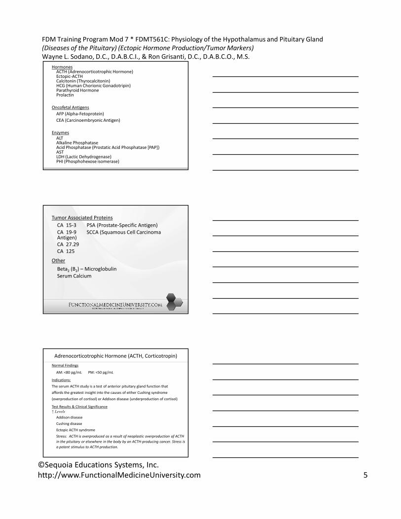
Sagittal view of the hypothalamic region of the brainLifeArt Collection of Images Baltimore MD reprinted with permission
The hypothalamus:
• coordinates drive-related activities that include
hormonal, emotional and autonomic
• is connected to the limbic system (emotional)
• has outputs to the anterior and posterior lobes of
the pituitary (hormonal)
• has connections to visceral and somatic nuclei of the
brainstem and spinal cord (autonomic)

FDM Training Program Mod 7 * FDMT561C: Physiology of the Hypothalamus and Pituitary Gland
(Diseases of the Pituitary) (Ectopic Hormone Production/Tumor Markers)
Wayne L. Sodano, D.C., D.A.B.C.I., & Ron Grisanti, D.C., D.A.B.C.O., M.S.
©Sequoia Educations Systems, Inc.
http://www.FunctionalMedicineUniversity.com 2
Light Therapy
Light therapy (aka phototherapy) has been used to
treat the following conditions: seasonal affective
disorder (SAD), obsessive-compulsive disorders, jet-
lag, sleep disorders, and attention-
deficit/hyperactivity disorder (ADHD)
Functional Anatomy of the Hypothalamus and
the Pituitary Gland
The pituitary gland, also known as the hypophysis,
has two distinct parts called the adenohypophysis
(anterior pituitary) and the neurohypophysis
(posterior pituitary).
Hypothalamic Releasing and Inhibitory
Hormones (Control of the Anterior Pituitary)
• Thyrotropin-releasing hormone (TRH)
• Gonadotropin-releasing hormone (GnRH)
• Corticotrophin-releasing hormone (CRH)
• Growth hormone-releasing hormone( GHRH)
• Growth hormone-inhibitory hormone (somatostatin)
• Prolactin-inhibiting hormone (PIH)

FDM Training Program Mod 7 * FDMT561C: Physiology of the Hypothalamus and Pituitary Gland
(Diseases of the Pituitary) (Ectopic Hormone Production/Tumor Markers)
Wayne L. Sodano, D.C., D.A.B.C.I., & Ron Grisanti, D.C., D.A.B.C.O., M.S.
©Sequoia Educations Systems, Inc.
http://www.FunctionalMedicineUniversity.com 3
Hormones of the Anterior Pituitary
• Growth hormone (GH) (somatotropin)
• Adrenocorticotropic hormone (ACTH)
(corticotrophin)
• Thyroid-stimulating hormone (TSH) (thyrotropin)
• Follicle-stimulating hormone (FSH)
• Luteinizing hormone (LH)
• Prolactin (PRL)
Growth Hormone-Insulin-Liver Connection
Growth hormone’s metabolic effects, aside from
causing growth, include:
– Increases the rate of protein synthesis
– Increases mobilization of fatty acids from the
adipose tissue (fatty acids to acetyl-CoA)
– Increases fatty acid metabolism for energy
production
– Decreases the rate of carbohydrate utilization
Ghrelin
• stimulates growth hormone secretion
• regulates energy balance by affecting the
hypothalamus

FDM Training Program Mod 7 * FDMT561C: Physiology of the Hypothalamus and Pituitary Gland
(Diseases of the Pituitary) (Ectopic Hormone Production/Tumor Markers)
Wayne L. Sodano, D.C., D.A.B.C.I., & Ron Grisanti, D.C., D.A.B.C.O., M.S.
©Sequoia Educations Systems, Inc.
http://www.FunctionalMedicineUniversity.com 4
The Hypothalamus and the Posterior Pituitary
The posterior pituitary acts as a supporting structure
for nerve fibers that originate in the hypothalamus.
The nerve endings in the posterior pituitary secrete
two hormones; antidiuretic hormone (ADH), also
known as vasopressin, and oxytocin.
Disorders of the Hypothalamus
and Pituitary Gland
Diseases of the endocrine glands typically cause
hormone deficiencies or excesses.
Tumor Markers
• Tumor markers include a group of hormones,
oncofetal proteins, enzymes and tumor antigens
measured primarily in the blood, and sometimes in
urine.
• Tumor markers are not in themselves specific
enough to permit a diagnosis of malignancy to be
made, but once a malignancy has been diagnosed
and shown to be associated with elevated levels of
tumor marker, the marker can be used to assess
response to treatment.
FDM Training Program Mod 7 * FDMT561C: Physiology of the Hypothalamus and Pituitary Gland
(Diseases of the Pituitary) (Ectopic Hormone Production/Tumor Markers)
Wayne L. Sodano, D.C., D.A.B.C.I., & Ron Grisanti, D.C., D.A.B.C.O., M.S.
©Sequoia Educations Systems, Inc.
http://www.FunctionalMedicineUniversity.com 5
HormonesACTH (Adrenocorticotrophic Hormone)Ectopic-ACTHCalcitonin (Thyrocalcitonin)HCG (Human Chorionic Gonadotripin)Parathyroid Hormone Prolactin
Oncofetal Antigens
AFP (Alpha-Fetoprotein)
CEA (Carcinoembryonic Antigen)
Enzymes
ALTAlkaline Phosphatase Acid Phosphatase (Prostatic Acid Phosphatase [PAP])AST LDH (Lactic Dehydrogenase)PHI (Phosphohexose isomerase)
Tumor Associated Proteins
CA 15-3 PSA (Prostate-Specific Antigen)
CA 19-9 SCCA (Squamous Cell Carcinoma Antigen)
CA 27.29
CA 125
Other
Beta2 (B2) – Microglobulin
Serum Calcium
Adrenocorticotrophic Hormone (ACTH, Corticotropin)
Normal Findings
AM: <80 pg/mL PM: <50 pg/mL
Indications:
The serum ACTH study is a test of anterior pituitary gland function that
affords the greatest insight into the causes of either Cushing syndrome
(overproduction of cortisol) or Addison disease (underproduction of cortisol)
Test Results & Clinical Significance
↑ Levels
Addison disease
Cushing disease
Ectopic ACTH syndrome
Stress: ACTH is overproduced as a result of neoplastic overproduction of ACTH
in the pituitary or elsewhere in the body by an ACTH-producing cancer. Stress is
a potent stimulus to ACTH production.
FDM Training Program Mod 7 * FDMT561C: Physiology of the Hypothalamus and Pituitary Gland
(Diseases of the Pituitary) (Ectopic Hormone Production/Tumor Markers)
Wayne L. Sodano, D.C., D.A.B.C.I., & Ron Grisanti, D.C., D.A.B.C.O., M.S.
©Sequoia Educations Systems, Inc.
http://www.FunctionalMedicineUniversity.com 6
Test Explanation:
An elaborate feedback mechanism for cortisol coordinates the
function of the hypothalamus, pituitary gland, and adrenal glands.
In the patient with Cushing syndrome an elevated ACTH level can be
caused by a pituitary ACTH-producing tumor or a nonpituitary
(ectopic) ACTH-producing tumor, usually in the lung, pancreas,
thymus, or ovary. ACTH levels greater than 200 pg/mL usually
indicate ectopic ACTH production. If the ACTH level is below normal
in a patient with Cushing syndrome, an adrenal adenoma or
carcinoma is probably the cause of the hyperfunction.
In patients with Addison disease an elevated ACTH level indicates
primary adrenal gland failure, as in adrenal gland destruction cause
by infection, hemorrhage, or autoimmunity.
Ref: Mosby’s Manual of Diagnostic & Laboratory Tests, 3rd edition
Calcitonin (Human Calcitonin [HCT], Thyrocalcitonin)
Normal Findings
Males: ≤19 pg/mL Females: ≤14 pg/mL
Indications
This test is usually indicated to evaluate persons with suspected medullary
carcinoma of the thyroid. Calcitonin is useful in monitoring response to
therapy and predicting recurrences of medullary thyroid cancer. It is also
useful as a screening test for those with a family history of medullary
cancer.
Test Results & Clinical Significance↑ LevelsMedullary carcinoma of thyroid Primary hyperparathyroidismC-cell hyperplasia Secondary hyperparathyroidismOat cell carcinoma of lung Pernicious AnemiaBreast carcinoma Thyroiditis
Pancreatic cancer: these cancers can act as an autonomous ectopic site of calcitonin production.
Test explanation:
Calcitonin is a hormone secreted by the parafollicular or
C cells of the thyroid gland. Secretion is stimulated by
elevated serum calcium levels. Calcitonin contributes to
calcium homeostasis. It decreases serum calcium levels
by inhibiting bone resorption and increasing calcium
excretion by the kidneys.
Ref: Ibid

FDM Training Program Mod 7 * FDMT561C: Physiology of the Hypothalamus and Pituitary Gland
(Diseases of the Pituitary) (Ectopic Hormone Production/Tumor Markers)
Wayne L. Sodano, D.C., D.A.B.C.I., & Ron Grisanti, D.C., D.A.B.C.O., M.S.
©Sequoia Educations Systems, Inc.
http://www.FunctionalMedicineUniversity.com 7
Human Chorionic Gonadotropin {HCG}, Beta SubunitNormal Findings
Qualitative: negative: positive in pregnancy
Beta subunit: depends on the method & test used.
Indications
This test is used to diagnose pregnancy. It is also helpful in the monitoring of ‘high-
risk’ pregnancies. It can also be used as a tumor marker for certain cancers. (trophoblastic)
Test Results & Clinical Significance
↑ Levels
Pregnancy
Ectopic Pregnancy:
Highest beta HCG levels (.30,000 milli-international units/mL) are recorded in pregnancy. Lowest amounts are generally seen in ectopic pregnancy.
Hydatidiform mole of uterus
Choriocarcinoma of uterus
Germ cell (choriocarcinoma, teratomas, embryonal cell) tumors of testes or ovaries
Other tumors (poorly differentiated tumors, such as hematoma and lymphoma.
Test explanation
Ectopic pregnancy, hydatidiform mole of the uterus, and
choriocarcinoma of the uterus can all produce HCG. Germ cell tumors
(choriocarcinoma, embryonal cell cancers) of the testes or ovaries can
produce HCG in men and nonpregnant women, respectively. Primary
liver cell cancers (hepatoma) can also make HCG. In these tumors, HCG
is used as a valuable tumor marker to aid in tumor identification. For
example, HCG is serially measured in patients with cirrhosis, because
they have a high chance of developing a hepatoma. If detected early
enough, the tumor can be removed and the patient cured. HCG is also
used to monitor the therapy and disease progression and/or the
regression of these tumors.
Ref: Ibid
Parathyroid Hormone (PTH, Parathormone)
Indications
PTH is measured to assist in the evaluation of hypercalcemia or hypocalcemia. It is routinely monitored in patients with chronic renal failure (CRF)
Test Results & Clinical Significance
↑ Levels
Hyperparathyroidism secondary to adenoma or carcinoma of the parathyroid gland.
Non-PTH-producing tumors (paraneoplastic syndrome) commonly noted with lung, kidney, or breast carcinoma (These tumors produce a ‘PTH-related protein’ that acts like PTH.)
↓ Levels
Hypercalcemia
Metastatic bone tumor
Hypercalcemia of malignancy (most often with lung, breast, or lymphoma cancer)
Sarcoidosis: These patients can develop elevated serum calcium levels, thus
reducing PTH.
.
FDM Training Program Mod 7 * FDMT561C: Physiology of the Hypothalamus and Pituitary Gland
(Diseases of the Pituitary) (Ectopic Hormone Production/Tumor Markers)
Wayne L. Sodano, D.C., D.A.B.C.I., & Ron Grisanti, D.C., D.A.B.C.O., M.S.
©Sequoia Educations Systems, Inc.
http://www.FunctionalMedicineUniversity.com 8
Test Explanation:
PTH is the only hormone secreted by the parathyroid gland in response to hypocalcemia. PTH is one of the major factors affecting calcium metabolism.
Primary hyperparathyroidism is most often caused by a parathyroid adenoma and only rarely from parathyroid cancer. These patients have high PTH and calcium levels.
Ref: Ibid
Prolactin Level (PRL)
Normal Findings
Adult male: 0-20 ng/mL
Adult female: 0-25 ng/mL Pregnant female: 20-400 ng/mL
Indications
Prolactin levels are used to diagnose and monitor prolactin-secreting pituitary
adenomas.
Test Results & Clinical Significance
↑ Levels
Galactorrhea
Amenorrhea: Patients who have had normal menses and then stop having menses may be found to have elevated prolactin levels. Many are subsequently found to have prolactin-
secreting pituitary adenomas.
Prolactin-secreting pituitary tumor
Metastatic cancer of pituitary gland
Hypothyroidism
Paraneoplastic syndrome: Associated with ectopic production of prolactin
Stress
Polycystic ovary syndrome
Test explanation
Prolactin is a hormone secreted by the anterior pituitary
gland (adenohypophysis). In females, prolactin promotes
lactation.
Ref: Ibid
FDM Training Program Mod 7 * FDMT561C: Physiology of the Hypothalamus and Pituitary Gland
(Diseases of the Pituitary) (Ectopic Hormone Production/Tumor Markers)
Wayne L. Sodano, D.C., D.A.B.C.I., & Ron Grisanti, D.C., D.A.B.C.O., M.S.
©Sequoia Educations Systems, Inc.
http://www.FunctionalMedicineUniversity.com 9
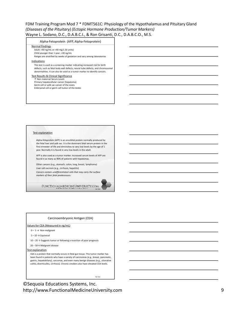
Alpha-Fetoprotein (AFP, Alpha-Fetoprotein)
Normal Findings
Adult: <40 ng/mL or <40 mg/L (SI units)
Child younger than 1 year: <30 ng/mL
Ranges are stratified by weeks of gestation and vary among laboratories
Indications
This test is used as a screening marker indicating increased risk for birth
defects, such as fetal body wall defects, neural tube defects, and chromosomal
abnormalities. It can also be used as a tumor marker to identify cancers.
Test Results & Clinical Significance↑ Non maternal Serum Levels
Primary hepatocellular cancer (hepatoma)
Germ cell or yolk sac cancer of the ovary
Embryonal cell or germ cell tumor of the testes
Test explanation
Alpha-fetoprotein (AFP) is an oncofetal protein normally produced by
the fetal liver and yolk sac. It is the dominant fetal serum protein in the
first trimester of life and diminishes to very low levels by the age of 1
year. Normally it is found in very low levels in the adult.
AFP is also used as a tumor marker. Increased serum levels of AFP are
found in as many as 90% of patients with hepatomas.
Other cancers (e.g., stomach, colon, lung, breast, lymphoma)
Liver cell necrosis (e.g., cirrhosis, hepatitis)
Cancers contain undifferentiated cells that may carry the surface
markers of their fetal predecessors.
Ref: Ibid
Carcinoembryonic Antigen (CEA)
Values for CEA (Measured in ng/mL)
0 – 5 → Non-malignant
5 – 10 → Equivocal
10 – 20 → Suggests tumor or following a resection of poor prognosis
20 – 50→ Malignant disease
Test explanation
CEA is a protein that normally occurs in fetal gut tissue. This tumor marker has
been found in patients who have a variety of carcinomas (e.g., breast, pancreatic,
gastric, hepatobiliary), sarcomas, and even many benign diseases (e.g., ulcerative
colitis, diverticulitis, cirrhosis). Chronic smokers also have elevated CEA levels.
Ref: Ibid

FDM Training Program Mod 7 * FDMT561C: Physiology of the Hypothalamus and Pituitary Gland
(Diseases of the Pituitary) (Ectopic Hormone Production/Tumor Markers)
Wayne L. Sodano, D.C., D.A.B.C.I., & Ron Grisanti, D.C., D.A.B.C.O., M.S.
©Sequoia Educations Systems, Inc.
http://www.FunctionalMedicineUniversity.com 10
Acid Phosphatase (Prostatic Acid Phosphatase [PAP])
Normal Findings
Adult/elderly: 0.13 – 0.63 units/L
Child: 8.6 – 12.6 units/mL
Newborn: 10.4 – 16.4 units/mL
IndicationsTotal acid phosphatase and specifically the PAP isoenzyme is primarily used to stage prostatic carcinoma and to monitor the efficacy of treatment.
Test Results & Clinical Significance↑ Levels
Prostatic carcinoma
Benign prostatic hypertrophy
Prostatitis
Multiple myeloma
Thrombocytosis
Lysosomal disorders
Renal diseases
Liver diseases, such as cirrhosis
Ref: Ibid
Test explanation
Usually (but not always) elevated levels are seen in
patients with prostatic cancer that has metastasized
beyond the capsule to other parts of the body,
especially bone. Since PAP is not elevated in early
stage prostate disease, this test is not recommended
for screening.
Ref: Ibid
Phosphohexose Isomerase (PHI): (enzyme found in muscle used forglucose metabolism)
Diseases of the heart, liver, skeletal muscles as well as certain malignancies, (especially carcinoma of the breast, prostate, and intestines) may cause an elevation of serum PHI. It is thought in active cancer the elevation may be due to liver or bone metastasis. Although PHI may be elevated from several different causes it is useful as a screening device to establish the presence or absence of certain types of cancer as well as progress of treatment.
Squamous Cell Carcinoma Antigen:
SCCA has been shown to be elevated with squamous cell carcinoma of the cervix, and in squamous cell carcinomas of the lung, pharynx, larynx, palate, tongue, and neck.
Alanine Aminotransferase (ALT or SGPT):
A specific liver enzyme found increased in hepatitis, liver metastasis, obstructive jaundice, hepatic congestion, and infectious mononucleosis. ALT is decreased in pyridoxine deficiency.
Aspartate Aminotranfserease (AST or SGOT):
Found elevated in hepatic disease, muscle damage, pancreatitis, and neoplasia. It is found decreased in pyridoxine deficiency and terminal stages of liver cancer.
( Ref: The Merck Manual, 16th edition)
FDM Training Program Mod 7 * FDMT561C: Physiology of the Hypothalamus and Pituitary Gland
(Diseases of the Pituitary) (Ectopic Hormone Production/Tumor Markers)
Wayne L. Sodano, D.C., D.A.B.C.I., & Ron Grisanti, D.C., D.A.B.C.O., M.S.
©Sequoia Educations Systems, Inc.
http://www.FunctionalMedicineUniversity.com 11
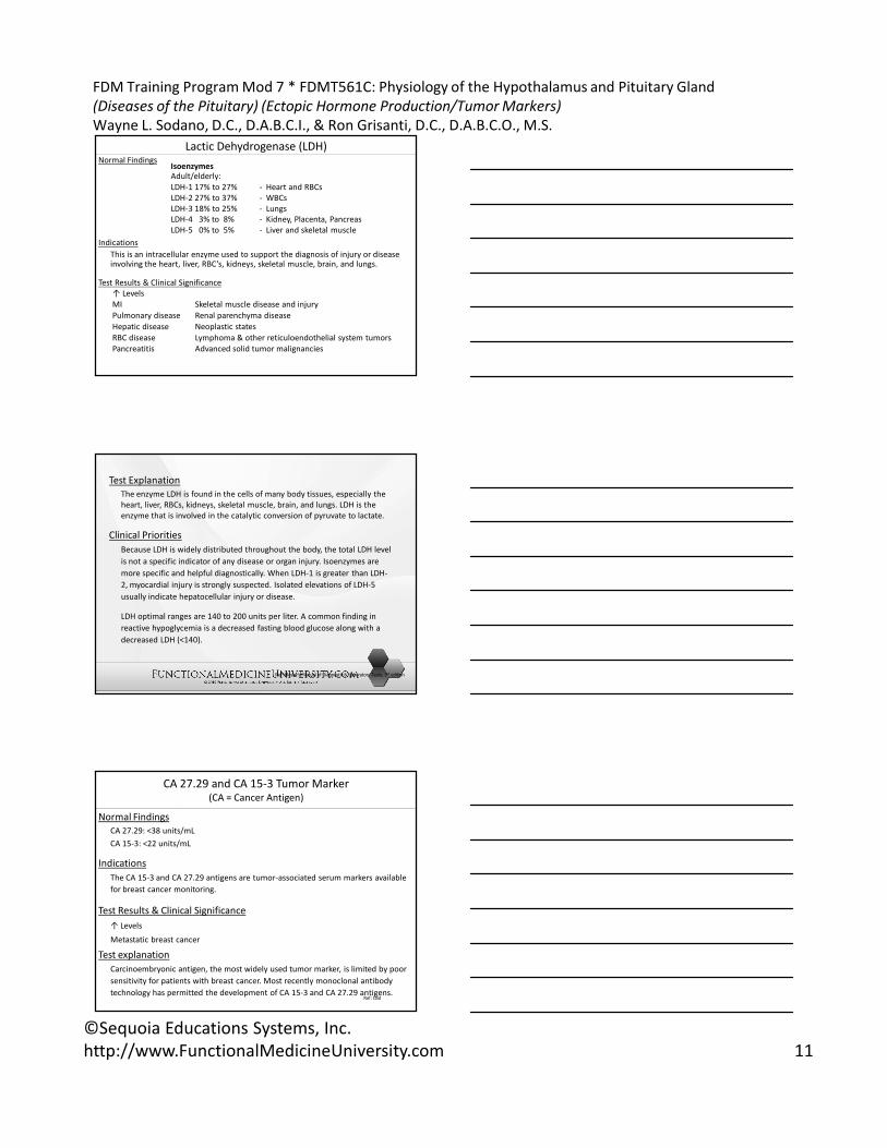
Lactic Dehydrogenase (LDH)Normal Findings
Isoenzymes
Adult/elderly:
LDH-1 17% to 27% - Heart and RBCs
LDH-2 27% to 37% - WBCs
LDH-3 18% to 25% - Lungs
LDH-4 3% to 8% - Kidney, Placenta, Pancreas
LDH-5 0% to 5% - Liver and skeletal muscle
Indications
This is an intracellular enzyme used to support the diagnosis of injury or disease involving the heart, liver, RBC’s, kidneys, skeletal muscle, brain, and lungs.
Test Results & Clinical Significance
↑ Levels
MI Skeletal muscle disease and injury
Pulmonary disease Renal parenchyma disease
Hepatic disease Neoplastic states
RBC disease Lymphoma & other reticuloendothelial system tumors
Pancreatitis Advanced solid tumor malignancies
Test Explanation
The enzyme LDH is found in the cells of many body tissues, especially the
heart, liver, RBCs, kidneys, skeletal muscle, brain, and lungs. LDH is the
enzyme that is involved in the catalytic conversion of pyruvate to lactate.
Clinical Priorities
Because LDH is widely distributed throughout the body, the total LDH level
is not a specific indicator of any disease or organ injury. Isoenzymes are
more specific and helpful diagnostically. When LDH-1 is greater than LDH-
2, myocardial injury is strongly suspected. Isolated elevations of LDH-5
usually indicate hepatocellular injury or disease.
LDH optimal ranges are 140 to 200 units per liter. A common finding in
reactive hypoglycemia is a decreased fasting blood glucose along with a
decreased LDH (<140).
Ref: Mosby’s Manual of Diagnostic & Laboratory Tests, 3rd edition
CA 27.29 and CA 15-3 Tumor Marker(CA = Cancer Antigen)
Normal Findings
CA 27.29: <38 units/mL
CA 15-3: <22 units/mL
Indications
The CA 15-3 and CA 27.29 antigens are tumor-associated serum markers available
for breast cancer monitoring.
Test Results & Clinical Significance
↑ Levels
Metastatic breast cancer
Test explanation
Carcinoembryonic antigen, the most widely used tumor marker, is limited by poor
sensitivity for patients with breast cancer. Most recently monoclonal antibody
technology has permitted the development of CA 15-3 and CA 27.29 antigens. Ref: Ibid
FDM Training Program Mod 7 * FDMT561C: Physiology of the Hypothalamus and Pituitary Gland
(Diseases of the Pituitary) (Ectopic Hormone Production/Tumor Markers)
Wayne L. Sodano, D.C., D.A.B.C.I., & Ron Grisanti, D.C., D.A.B.C.O., M.S.
©Sequoia Educations Systems, Inc.
http://www.FunctionalMedicineUniversity.com 12
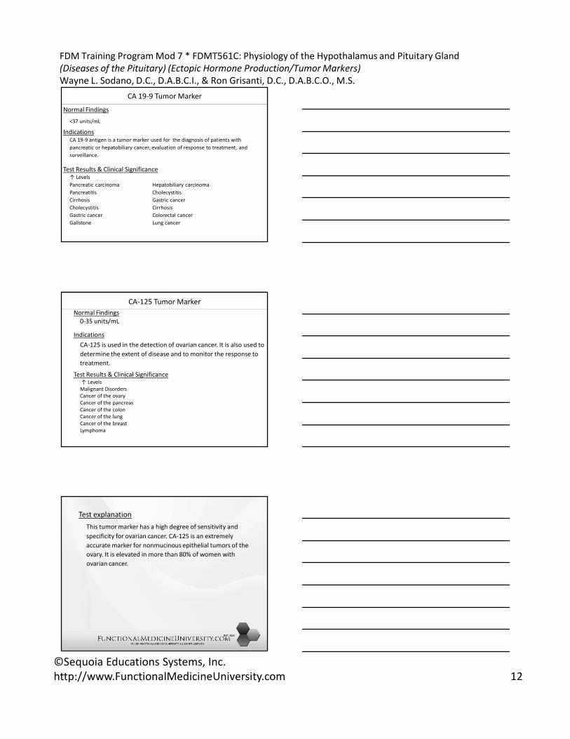
CA 19-9 Tumor Marker
Normal Findings
<37 units/mL
IndicationsCA 19-9 antigen is a tumor marker used for the diagnosis of patients with
pancreatic or hepatobiliary cancer, evaluation of response to treatment, and
surveillance.
Test Results & Clinical Significance↑ Levels
Pancreatic carcinoma Hepatobiliary carcinoma
Pancreatitis Cholecystitis
Cirrhosis Gastric cancer
Cholecystitis Cirrhosis
Gastric cancer Colorectal cancer
Gallstone Lung cancer
CA-125 Tumor Marker
Normal Findings
0-35 units/mL
Indications
CA-125 is used in the detection of ovarian cancer. It is also used to
determine the extent of disease and to monitor the response to
treatment.
Test Results & Clinical Significance↑ Levels
Malignant Disorders
Cancer of the ovary
Cancer of the pancreas
Cancer of the colon
Cancer of the lung
Cancer of the breast
Lymphoma
Test explanation
This tumor marker has a high degree of sensitivity and
specificity for ovarian cancer. CA-125 is an extremely
accurate marker for nonmucinous epithelial tumors of the
ovary. It is elevated in more than 80% of women with
ovarian cancer.
Ref: Ibid

FDM Training Program Mod 7 * FDMT561C: Physiology of the Hypothalamus and Pituitary Gland
(Diseases of the Pituitary) (Ectopic Hormone Production/Tumor Markers)
Wayne L. Sodano, D.C., D.A.B.C.I., & Ron Grisanti, D.C., D.A.B.C.O., M.S.
©Sequoia Educations Systems, Inc.
http://www.FunctionalMedicineUniversity.com 13
Prostate-Specific Antigen (PSA)Normal Findings
<4 ng/mL
Indications
This test is used as a screening method for early detection of prostatic cancer. When the PSA test is combined with a rectal examination, nearly 90% of clinically significant cancers can be detected. This test also used to monitor the disease after treatment.
Test Results & Clinical Significance
↑ Levels
Prostate Cancer
BPH
Prostatitis
Interfering FactorsRectal examinations are well known to falsely elevate PAP levels and they may also minimally elevate the PSA. To avoid this problem, the PSA should be drawn before rectal exam of the prostate or several hours afterward. Ejaculation within 24 hours of blood testing will be associated with elevated PSA levels
Test explanation
PSA is a glycoprotein found in high concentrations in the prostatic lumen. Levels greater than 4 ng/mL have been found in more than 80% of men with prostate cancer. The PSA assay is also a sensitive test for monitoring response to therapy.
The PSA is limited by a lack of specificity within the ‘diagnostic gray zone’ of 4 to 10 ng/mL. PSA levels also may be minimally elevated in patients with benign prostatic hypertrophy (BPH) and prostatitis. PSA levels greater than 10 ng/mL indicate a high probability of prostate cancer. Lower values may be compatible with BPH or early prostate cancer.
Percent free PSA (%FPSA) is also helpful in differentiating between cancer and benign prostate disorders in men with total PSA levels within this ‘diagnostic gray zone’. It has been
noted that when the %FPSA is <25%, there is a high likelihood of cancer.
Higher free PSA with BPH
Higher protein bound PSA with prostate cancer
Ref: Ibid
Calcium, Blood
(Total/Ionized Calcium, CA)
Test explanation
Serum calcium is necessary in many metabolic enzymatic pathways. It is vital for
muscle contractility, cardiac function, neural transmission, and blood clotting. The
most common cause of hypercalcemia is hyperparathyroidism.
Malignancy, the second most common cause of hypercalcemia, can cause elevated
calcium levels in two main ways. First, tumor metastasis (myeloma, lung, breast,
renal cell) to the bone can destroy the bone, causing resorption and pushing
calcium into the blood. Second, the cancer (lung, breast, renal cell) can produce a
PTH-like substance that drive the serum calcium up (ectopic PTH)
Beta2 (B2) – microglobulin
Levels may be raised in people with multiple myeloma or other cancers of blood
cells.
This test cannot be recommended for cancer screening.
Ref: Ibid
FDM Training Program Mod 7 * FDMT561C: Physiology of the Hypothalamus and Pituitary Gland
(Diseases of the Pituitary) (Ectopic Hormone Production/Tumor Markers)
Wayne L. Sodano, D.C., D.A.B.C.I., & Ron Grisanti, D.C., D.A.B.C.O., M.S.
©Sequoia Educations Systems, Inc.
http://www.FunctionalMedicineUniversity.com 14
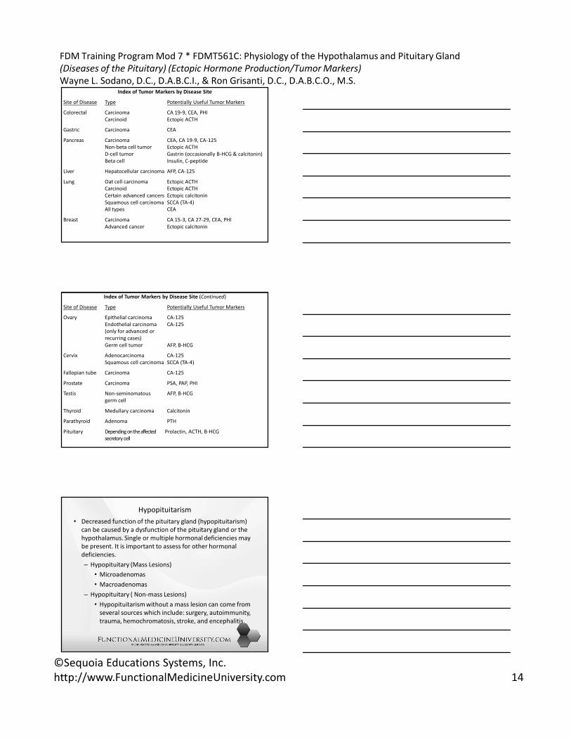
Index of Tumor Markers by Disease Site
Site of Disease Type Potentially Useful Tumor Markers
Colorectal Carcinoma CA 19-9, CEA, PHI
Carcinoid Ectopic ACTH
Gastric Carcinoma CEA
Pancreas Carcinoma CEA, CA 19-9, CA-125
Non-beta cell tumor Ectopic ACTH
D-cell tumor Gastrin (occasionally B-HCG & calcitonin)
Beta cell Insulin, C-peptide
Liver Hepatocellular carcinoma AFP, CA-125
Lung Oat cell carcinoma Ectopic ACTH
Carcinoid Ectopic ACTH
Certain advanced cancers Ectopic calcitonin
Squamous cell carcinoma SCCA (TA-4)
All types CEA
Breast Carcinoma CA 15-3, CA 27-29, CEA, PHI
Advanced cancer Ectopic calcitonin
Index of Tumor Markers by Disease Site (Continued)
Site of Disease Type Potentially Useful Tumor Markers
Ovary Epithelial carcinoma CA-125
Endothelial carcinoma CA-125
(only for advanced or
recurring cases)
Germ cell tumor AFP, B-HCG
Cervix Adenocarcinoma CA-125
Squamous cell carcinoma SCCA (TA-4)
Fallopian tube Carcinoma CA-125
Prostate Carcinoma PSA, PAP, PHI
Testis Non-seminomatous AFP, B-HCG
germ cell
Thyroid Medullary carcinoma Calcitonin
Parathyroid Adenoma PTH
Pituitary Depending on the affected Prolactin, ACTH, B-HCG
secretory cell
Hypopituitarism
• Decreased function of the pituitary gland (hypopituitarism)
can be caused by a dysfunction of the pituitary gland or the
hypothalamus. Single or multiple hormonal deficiencies may
be present. It is important to assess for other hormonal
deficiencies.
– Hypopituitary (Mass Lesions)
• Microadenomas
• Macroadenomas
– Hypopituitary ( Non-mass Lesions)
• Hypopituitarism without a mass lesion can come from
several sources which include: surgery, autoimmunity,
trauma, hemochromatosis, stroke, and encephalitis.

FDM Training Program Mod 7 * FDMT561C: Physiology of the Hypothalamus and Pituitary Gland
(Diseases of the Pituitary) (Ectopic Hormone Production/Tumor Markers)
Wayne L. Sodano, D.C., D.A.B.C.I., & Ron Grisanti, D.C., D.A.B.C.O., M.S.
©Sequoia Educations Systems, Inc.
http://www.FunctionalMedicineUniversity.com 15
Hyperprolactinemia
Excessive secretion of prolactin cause will cause
elevated serum prolactin or hyperprolactinemia.
About 70% of women with secondary amenorrhea
and galactorrhea (lactation in the absence of nursing)
have hyperprolactinemia. Symptoms in women
include: menstrual cycle disturbances, galactorrhea
and infertility. Men typically present with decrease
libido, erectile dysfunction, infertility and
hypogonadism (decreases testosterone production).
The following is a list of the potential causes
of hyperprolactinemia:
• Pharmacologic
• Physiologic
• Pathologic
Summary