fdss applications - hamamatsu photonics · amplex red (thermofisher)/luminol/ ros-glo (promega)...
TRANSCRIPT

Cell Health and Energy Metabolism
Protein-Protein Interaction
Safety/Toxicology
Cardiovascular Disease
GPCR
Ion Channel
Enzymatic Assay
New Screening Technology
Antibody
Optogenetic
High Speed Acquisition
New High Resolution
Camera
FDSS Applications

FDSS Applications2
Living-cell PPI assay is based on BRET (Bioluminescent Resonance Energy Transfer) screening technology
Protein-Protein Interaction (PPI)
Publication: Application Note Nr. 25 (CBCS, Karolinska Institutet, University of Gothenburg)
Identification of small molecule modulators of reactive oxygen release in TNF-α primed primarx human neutrophils. In the test system freshly isolated primary human neutrophils are first incubated with TNF-α. The cells are then triggered with cytochalasin B. The superoxide anions produced are measured by a chemiluminescence technique based on isoluminol using the single photon counting camera of the Hamamatsu FDSS 7000.
Oxidative Stress Assay (ROS) Assays for measuring real time production of ROS (Reactive oxygen species) as an indicator of cell heath or signalling events
Energy Metabolism Assay (NADPH) NAD(P)H molecules are important cofactors for different enzymes involved in cellular pathways. Measurement of this molecule can determine metabolic activity for cells affected by disease.
Cell Health and Energy Metabolism
Addition of TNF-α/compound treated cells to pre heated Cytochalasin B (130 µl)
Recording signal for 5 minutes
Therapeutic area
Drug discovery
FDSS Screening technology
BRET1 (Rluc8) (96/384), BRET2 (96 format only), NanoBret (Promega), NanoBit (Promega)
Therapeutic area
Research & Clinical application � Inflammatory disease � Monitoring inflammation of transplant before surgery
� Cancer (drug evaluation studies for Chemotherapy
FDSS Screening technology
Amplex red (Thermofisher)/Luminol/ ROS-Glo (Promega) NADPH-Glo/Pholasin®-based ABEL® (Knight Scientific)

3FDSS Applications
Membrane potential activity hiPS-derived cardiomyocytes in 96 wells
Presentation 3rd FDSS application Workshop, Barcelona, Stephane Bedut (ICM, Servier)
Any acute drug effect involved in primary or human iPSC-derived cardiomyocytes can be detected by monitoring [Ca2+] transients and electrical activity.
Safety/Toxicology and Cardiovascular Disease
Application Note Pluricyte® Cardiomyocytes
Acquired 10 s (black) and 3 min. (red) after ouabain treatmenz (96 format, FluoVolt dye)
0.1 % DMSO
0:00 1:00
1 µM Diltiazem
0:00 1:00
1 µM Bay K 8644
0:00 1:00
100 nM E 4031
0:00 1:00
1 µM Nifedipine
0:00 1:00
100 nM Isoproterenol
0:00 1:00
A B CBaseline Isoproterenol (1 µM) E-4031 (1 µM)
10,000
5,000
0
RFU
0 20 40 60Time (sec)
0 20 40 60Time (sec)
0 20 40 60Time (sec)
Application Protocol iCell® Cardiomyocytes2Application Note COR.4U®
Therapeutic area
Drug discovery, Safety pharmacology (Cardiac & Neuron), Assessment of cardiotoxocity and drug efficacy early in drug development process
FDSS Screening technology
[Ca2+] sensitive dye (Cal520, ATT Bioquest)/Voltage sensitive dye (FluoVolt, Molecular probes)
Vehicle
Ivabradine 1 µM
Ivabradine 2 µM
Lidocaine 50 µM
Ivabradine 1 µM
1 s 1 s
1000
F.u
.
1000
F.u
.
Dia
stol
ic s
lope
(F.u
.s-1)
Lidocaine 50 µM 1000
800
600
400
200
0
FDSS Applications4
GPCRs (G-protein-coupled receptors) are one of the most important classes of drug targets
Effect of GPCR pathway activation: intracellular Ca2+, cAMP, Beta Arrestin
GPCR
Therapeutic area
Drug discovery
FDSS Screening technology
[Ca2+] sensitive dye (Cal520,ATT Bioquest)/Voltage sensitive dye (FluoVolt, Molecular probes), BRET, cAMP (Glow sensor,Promega), Beta Arrestin (Discoverx)
Print screen FDSS software
GPCR assay: � 1536 assay – Ca2+ Aequorin � B2-Arrestin 2 Recruitment
� Full agonist vs. partial agonist: w/o full view –> misleading
� Efficacy vs. AUC vs. Slow binders –> kinetics
� Time course is key
Cpd1Cpd2
Cpd3
125
100
75
50
25
0
-25
125
100
75
50
25
0
-25
-50
100
75
50
25
0
-25
-50
BRET
(% v
s D
opam
ine
1 µM
)
BRET
(% v
s D
opam
ine
1 µM
)
BRET
indu
ced
by c
ompo
unds
(% v
s D
opam
ine
1 µM
)
Log [ligand] (M)-10 -9 -8 -7 -6 -5
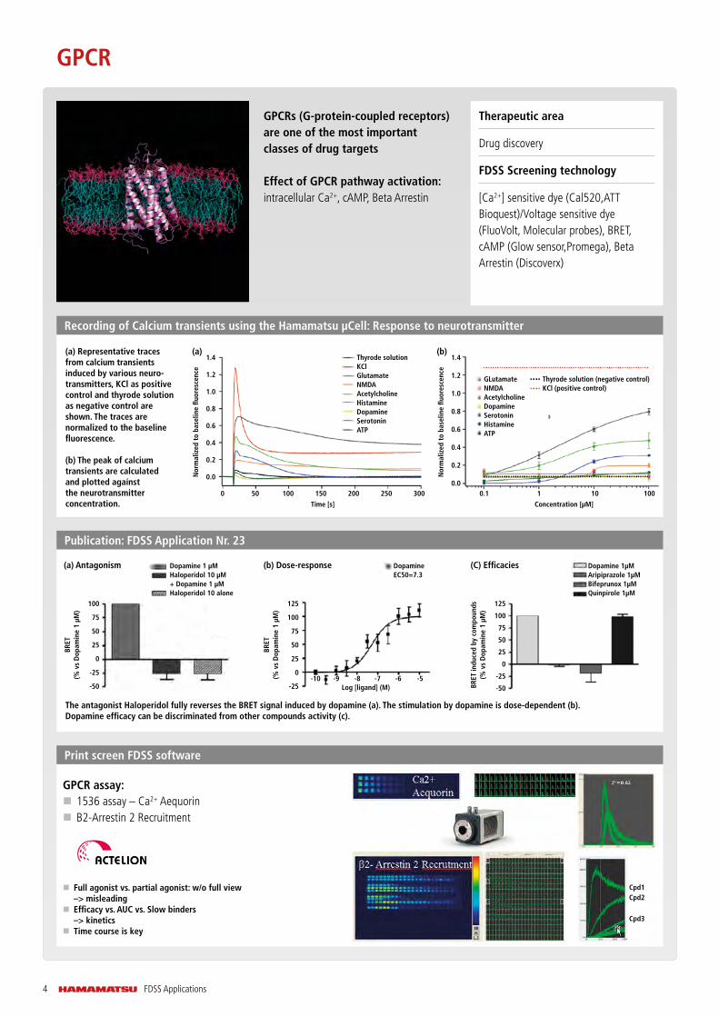
Publication: FDSS Application Nr. 23
The antagonist Haloperidol fully reverses the BRET signal induced by dopamine (a). The stimulation by dopamine is dose-dependent (b). Dopamine efficacy can be discriminated from other compounds activity (c).
(a) Antagonism (b) Dose-response (C) Efficacies DopamineEC50=7.3
Dopamine 1 µMHaloperidol 10 µM + Dopamine 1 µMHaloperidol 10 alone
Dopamine 1µMAripiprazole 1µMBifeprunox 1µMQuinpirole 1µM
Recording of Calcium transients using the Hamamatsu µCell: Response to neurotransmitter
(a)
Nor
mal
ized
to
base
line
fluor
esce
nce
1.4
1.2
1.0
0.8
0.6
0.4
0.2
0.0
0 50 200100 250150 300
Time [s]
Thyrode solutionKClGlutamateNMDAAcetylcholineHistamineDopamineSerotoninATP
(b)Dopamine 1µMAripiprazole 1µMBifeprunox 1µMQuinpirole 1µM
Nor
mal
ized
to
base
line
fluor
esce
nce
1.4
1.2
1.0
0.8
0.6
0.4
0.2
0.01010.1 100
Concentration [µM]
GLutamateNMDAAcetylcholineDopamineSerotoninHistamineATP
Thyrode solution (negative control)KCl (positive control)
(a) Representative traces from calcium transients induced by various neuro-transmitters, KCl as positive control and thyrode solution as negative control are shown. The traces are normalized to the baseline fluorescence.
(b) The peak of calcium transients are calculated and plotted agaínst the neurotransmitter concentration.

5FDSS Applications
Publication: YFP-halide assays for CFTR drug discovery using the FDSS/µcell
Thierry Christophe, PhD, Director - Biology, Galapagos NV Hamamatsu 11th FDSS User Meeting 11 June 2015
Detection of CFTR potentiatorsCells over-expressing F508d-CFTR and YFP-H148/I152L
Fsk + inactive cpd
Fsk + CFTR potentiator
Cl-
Cl-
Cl-
l-
l-
NaI
Vehicle/inactive
CFTR potentiator
Dose response CFTR potentiator0.8
0.6
0.4
0.2
0.0
1.0
0.9
0.8
log conc (M)-12 -10 -8 -6 -4
1-F/
F0
F/F0
time
Ion Channel
(Isomerase Inhibitor Screening)
Use of a Real-Time Fluorescence Monitoring System for High-Throughput Screening for Prolyl Isomerase InhibitorsTadashi Mori, Selma Itami, Tomotaka Yanagi, Yota Tatara, Mari Takamiya and Takafumi Uchida
Enzymatic Assay
The Km value of CypA with the peptide was 59 M. (C) Concentration dependence of CsA inhibition of CypA activity. Concentrations of CypA and CsA are expressed as follows: CypA none, CsA none (♦); CypA 7 nM, CsA none ( ); CypA 7 nM, CsA 3.13 nM (x); CypA 7 nM, CsA 12.5 nM ( ); CypA 7 nM, CsA 50 nM, (▲);and CypA 7 nM, CsA200 nM (•).
CypA none, CsA noneCypA 7 nM,CsA noneCypA 7 nM, CsA 3.2 nM
Conv
ersi
on R
atio
(%)
100
80
60
40
20
-3 -2 -1 40 51 65 7 93 8 10
CypA 7 nM, CsA 12.5 nMCypA 7 nM, CsA 50 nMCypA 7 nM, CsA 200 nM
Antibody (Functional Assay)
Therapeutic area
Drug discovery (ion channel/GPCR)
FDSS Screening technology
Ca2+ dyeCalcium Mobilization response: Inhibition of Ca2+ response induced by EC80 of Agonist in presence of different antibodies
AntibodyCa2+ buffer only
80 nM Agonist
Therapeutic area:
Drug discovery
FDSS Screening technology
� Na+/K+ channel: TEFLAB dye, Membrane potential dye (Molecular device)/FluxOR Potassium Ion Channel Assay (Thermo Fisher)/SBFI (Thermo Fisher)/ FRET VSP dye (Invitrogen)
� Cl- channel Fluorescence of Yellow Fluorescent Protein mutant (YFP-H148Q/I521L) quenched by halides
l-
l-

FDSS Applications6
New Screening Technology
Therapeutic area
Cardiac, CNS, Muscular diseases
FDSS Screening technology
Ca2+ dye/(FDSS/μcell + EFS + High speed options)
High-Throughput Fluorescence Measurements of Ca2+ Transients in primary rat and in human iPSC-derived cardiomyocytes
a) Primary rat cardiomyocytes (Cosmo Bio) were cultured in 96-well plate. The above figures show the intracellular Ca2+ concentration changes for 5 s in 96 wells in a microplate. In primary cultured cardiomyocytes, cells in each well beat at different rates and with different timing (left). When electrical field stimulation was applied (1.0 Hz, voltage 5 V, duration 5 ms), the result was uniform Ca2+ oscillations between all wells resulting in synchronized beating of cells (right).
b) Effects of ion channel blockers on Ca2+ transients in iPS cardio-mocytes: Beating rate is dependent on Electric Field Stimulation frequency.
Spontaneous beating(a) Rat primary cardiomyocytes
Electric Field Stimulation (EFS) of human iPSC-derived neuron using Hamamatsu FDSS/µCELL
The Ca2+ response is inhibited in the presence of a calcium channel blocker, Bepridil
With 1.0 Hz stimulation
(b) Ca2+ channel blocker: Verapamil
Electric fieldStimulation (EFS)
96-channel electrode array
� Stimulate all 96 wells simultaneously � Cylindrical electrodes � Stimulation voltage is changeable column by column
Bepridil, a calcium channel blocker, was added to the wells at the indicated concentration and incubated for 20 min, and then EFS pulses (5 ms of pulse width, 40 Hz) were given. The figures on the right show intracellular Ca2+ concentration changes. At more than 15 µM, the Ca2+ response was completely inhibited. The IC50 value was estimated to be 5.6 µM (graph on the left).
Caution Notice: The FDSS/µCELL EFS system should not be used for optically detecting/monitoring change in transmembrane potential of the cells. The FDSS/µCELL EFS system should not be used on any cell or cells in which the user or anyone else has expressed target ion channels.
0
10
20
30
40
50
60
70
withoutstimulation
1.0H z astimulation
P ra
te [/
min
]
P rate [/min]
0
20
40
60
80
100
w/o 0.5Hz 1.0Hz 1.5Hz 2.0Hz
P ra
te [/
min
]
P rate [/min]
0
500
1000
1500
2000
2500
3000
3500
w/o 0.5Hz 1.0Hz 1.5Hz 2.0Hz
P-P
time
[mse
c.]
P-P time [msec.]
0
20
40
60
80
100
120
140
w/o 0.5Hz 1.0Hz 1.5Hz 2.0Hz
P ra
te [/
min
]
P rate [/min]
0
500
1000
1500
2000
2500
3000
3500
w/o 0.5Hz 1.0Hz 1.5Hz 2.0Hz
P-P
time
[mse
c.]
P-P time [msec.]
0
5
10
15
20
w/o 0.5Hz 1.0Hz 1.5Hz 2.0Hz
Risin
g slo
pe [/
mse
c.]
Rising slope
0
50
100
150
200
w/o 0.5Hz 1.0Hz 2.0Hz 3.0Hz
P ra
te [/
min
]
P rate [/min]
0
500
1000
1500
2000
w/o 0.5Hz 1.0Hz 2.0Hz 3.0Hz
P-P
time
[mse
c.]
P-P time [msec.]
0
50
100
150
200
250
w/o 0.5Hz 1.0Hz 2.0Hz 3.0Hz
IPW
D50
[mse
c.]
PWD50
0500
10001500200025003000350040004500
w/o 0.5Hz 1.0Hz 1.5Hz 2.0Hz
Ampl
itude
[RFU
]
Ampulitude
N=96
1.0 Hzstimulation
70
60
50
40
30
20
10
0
0.08
0.06
0.04
0.02
0.00
-0.02
10-6 10-5 10-4
withoutstimulation
P rate [/min]
P ra
te [/
min
]
Ampl
itude
(F/
F 0)
Bepridil [M]Time (s)
IC50=5.6 μM
3020100 3020100
0.1
0.05
0
3020100 3020100
Fluo
resc
ence
inte
nsity
(
F/F 0)
Presentation CDI 12th FDSS Users Meeting
iCell SkM + EarlyTox calcium dye
iCell Neurons
iCell Skeletal Myoblasts
Compound Treatment Electric Field Stimulation (EFS)
2V 3V 4V 5V 6V
7FDSS Applications
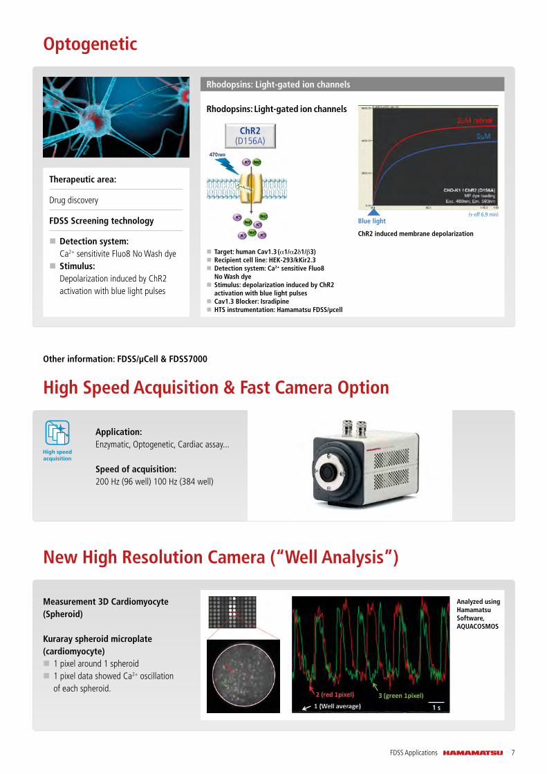
Rhodopsins: Light-gated ion channels
Optogenetic
Measurement 3D Cardiomyocyte (Spheroid)
Kuraray spheroid microplate (cardiomyocyte)
� 1 pixel around 1 spheroid � 1 pixel data showed Ca2+ oscillation of each spheroid.
Analyzed using Hamamatsu Software, AQUACOSMOS
New High Resolution Camera (“Well Analysis”)
Therapeutic area:
Drug discovery
FDSS Screening technology
� Detection system: Ca2+ sensitivite Fluo8 No Wash dye
� Stimulus: Depolarization induced by ChR2 activation with blue light pulses
Rhodopsins: Light-gated ion channels
ChR2(D156A)
� Target: human Cav1.3 (α1/α2δ1/β3) � Recipient cell line: HEK-293/kKir2.3 � Detection system: Ca2+ sensitive Fluo8 No Wash dye
� Stimulus: depolarization induced by ChR2 activation with blue light pulses
� Cav1.3 Blocker: Isradipine � HTS instrumentation: Hamamatsu FDSS/µcell
ChR2 induced membrane depolarization
Blue light(τ-off 6.9 min)
Other information: FDSS/µCell & FDSS7000
High Speed Acquisition & Fast Camera Option
Application: Enzymatic, Optogenetic, Cardiac assay...
Speed of acquisition: 200 Hz (96 well) 100 Hz (384 well)
High speedacquisition
1 s

www.hamamatsu.comE-mail: [email protected]