feasibility and validation of estimating global lv functional indices from limited projections using...
TRANSCRIPT

BioMed Central
Journal of Cardiovascular Magnetic Resonance
ss
Open AcceOral presentationFeasibility and validation of estimating Global LV functional indices from limited projections using a Modified Simpson's AlgorithmRamkumar Krishnamurthy*1, Amol Pednekar2, Benjamin Cheong3 and Raja Muthupillai3Address: 1Rice University, Houston, TX, USA, 2Philips Health Care, Houston, TX, USA and 3St. Luke's Episcopal Hospital, Houston, TX, USA
* Corresponding author
IntroductionA stack of 10-12 cine SSFP slices covering the LV are typi-cally acquired to estimate global LV function. But, ininstances such as dobutamine stress MR, it is difficult toacquire 10-12 contiguous short axis slices, and acquisitionis limited to cine imaging at three short-axis (located atbasal, mid and apical portions of the LV), and three longaxis orientations (2-, 3- and 4-chamber views) [1]. It isunclear if it is feasible obtain an estimate of global LVfunction, e.g., EDV, ESV, etc. from these limited views.
PurposeThe purpose of this work is to test the feasibility of devel-oping a modified Simpson's algorithm that can calculateLV volumes from a limited sub-set of cardiac cine MRimages (three short-axis views, and one long axis view),and validate the algorithm in human subjects.
MethodsData acquisition: In 20 subjects (14 M, age: 38+9 years) aset of contiguous cardiac cine SSFP images in the short-axis and in the three standard long-axis orientations wereacquired at 1.5 T. The acquisition parameters were:TR(ms)/TE(ms)/flip: 3.2/1.6/60°; acquired voxel-size: 2.5× 2.5 × 8 mm3; temporal resolution: 40-60 ms; breath-hold time: 5-8 s/slice.
from 13th Annual SCMR Scientific SessionsPhoenix, AZ, USA. 21-24 January 2010
Published: 21 January 2010
Journal of Cardiovascular Magnetic Resonance 2010, 12(Suppl 1):O46 doi:10.1186/1532-429X-12-S1-O46
<supplement> <title> <p>Abstracts of the 13<sup>th </sup>Annual SCMR Scientific Sessions - 2010</p> </title> <note>Meeting abstracts - A single PDF containing all abstracts in this Supplement is available <a href="http://www.biomedcentral.com/content/files/pdf/1532-429X-11-S1-full.pdf">here</a>.</note> <url>http://www.biomedcentral.com/content/files/pdf/1532-429X-11-S1-info</url> </supplement>
This abstract is available from: http://jcmr-online.com/content/12/S1/O46
© 2010 Krishnamurthy et al; licensee BioMed Central Ltd.
LV Cavity Geometric Model: The LV cavity between the mitral-valve annulus to basal slice was modeled as a cylinder of length L1, the two regions between the basal and mid, and mid and apical slices were moeled as tow cut-cones of lengths L2, and L3, and the apical region between the apical slice and the LV apex was modeled as a cone of length L4Figure 1LV Cavity Geometric Model: The LV cavity between the mitral-valve annulus to basal slice was modeled as a cylinder of length L1, the two regions between the basal and mid, and mid and apical slices were moeled as tow cut-cones of lengths L2, and L3, and the apical region between the apical slice and the LV apex was modeled as a cone of length L4. L1 and L4 were calculated directly from one of the long axis views, and the L2, and L3 were calcualted from the inter-slice gap pres-ribed during acquisition.
Page 1 of 2(page number not for citation purposes)
Journal of Cardiovascular Magnetic Resonance 2010, 12(Suppl 1):O46 http://jcmr-online.com/content/12/S1/O46
Publish with BioMed Central and every scientist can read your work free of charge
"BioMed Central will be the most significant development for disseminating the results of biomedical research in our lifetime."
Sir Paul Nurse, Cancer Research UK
Your research papers will be:
available free of charge to the entire biomedical community
peer reviewed and published immediately upon acceptance
cited in PubMed and archived on PubMed Central
yours — you keep the copyright
Submit your manuscript here:http://www.biomedcentral.com/info/publishing_adv.asp
BioMedcentral
Modified Simpson's algorithmThe LV cavity geometric model is described in Figure 1.The total volume (V) of the left ventricle was calculatedfrom the area (volume) of the LV cavity in the basal (Ab),mid (Am), and apical (Aa) slices by using the followingformula in the figure 1a.
Data AnalysisThe total LV volume was calculated using the modifiedSimpson's algorithm, and compared against the LV vol-ume extracted from the expert drawn manual contours onthe stack of contiguous short axis slices.
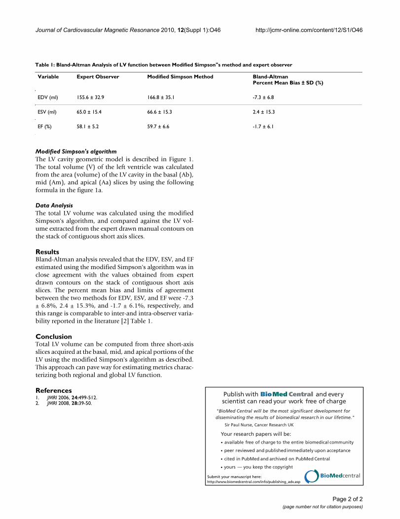
ResultsBland-Altman analysis revealed that the EDV, ESV, and EFestimated using the modified Simpson's algorithm was inclose agreement with the values obtained from expertdrawn contours on the stack of contiguous short axisslices. The percent mean bias and limits of agreementbetween the two methods for EDV, ESV, and EF were -7.3± 6.8%, 2.4 ± 15.3%, and -1.7 ± 6.1%, respectively, andthis range is comparable to inter-and intra-observer varia-bility reported in the literature [2] Table 1.
ConclusionTotal LV volume can be computed from three short-axisslices acquired at the basal, mid, and apical portions of theLV using the modified Simpson's algorithm as described.This approach can pave way for estimating metrics charac-terizing both regional and global LV function.
References1. JMRI 2006, 24:499-512.2. JMRI 2008, 28:39-50.
Table 1: Bland-Altman Analysis of LV function between Modified Simpson"s method and expert observer
Variable Expert Observer Modified Simpson Method Bland-AltmanPercent Mean Bias ± SD (%)
EDV (ml) 155.6 ± 32.9 166.8 ± 35.1 -7.3 ± 6.8
ESV (ml) 65.0 ± 15.4 66.6 ± 15.3 2.4 ± 15.3
EF (%) 58.1 ± 5.2 59.7 ± 6.6 -1.7 ± 6.1
Page 2 of 2(page number not for citation purposes)