female sterile mutations on the second chromosome of drosophila
TRANSCRIPT
Copyright 0 1991 by the Genetics Society of America
Female Sterile Mutations on the Second Chromosome of Drosophila melanogaster. 11. Mutations Blocking Oogenesis or
Altering Egg Morphology
Trudi Schupbach and Eric Wieschaus Department of Molecular Biology, Princeton University, Princeton, New Jersey 08544
Manuscript received July 8, 199 1 Accepted for publication August 24, 199 1
ABSTRACT In mutagenesis screens for recessive female sterile mutations on the second chromosome of
Drosophila melanogaster 528 lines were isolated which allow the homozygous females to survive but cause sterility. In 62 of these lines early stages of oogenesis are affected, and these females usually do not lay any eggs. In 333 lines oogenesis proceeds apparently normally to stage 8 of oogenesis, but morphological defects become often apparent during later stages of oogenesis, and are visible in the defective eggs produced by these females whereas 133 lay eggs that appear morphologically normal, but do not support normal embryonic development. Of the lines 341 have been genetically charac- terized and define a total of 140 loci on the second chromosome. Not all the loci are specific for oogenesis. From the numbers obtained we estimate that the second chromosome of Drosophila contains about 13 loci that are relatively specific for early oogenesis, 70 loci that are specifically required in mid to late oogenesis, and around 30 maternal-effect lethals.
0 OGENESIS is a highly regulated developmental process. In insects, as in dany metazoans, it
involves the close cooperation between the germ cells and a number of somatic cell types. Genetic and morphological studies of mutations that affect ooge- nesis have revealed that in Drosophila melanogaster a large number of genes are active during oogenesis to ensure the production of a normal egg capable of supporting the development of a normal embryo (GARCIA-BELLIDO and ROBBINS 1983; PERRIMON, ENCSTROM and MAHOWALD 1984). These genes can be grouped in several classes. On the one hand, all cells involved in oogenesis require the normal comple- ment of household genes that allow those cells to grow and divide. They also require some more specialized gene functions which are also expressed in other tis- sues. Because of their pleiotropic effects outside the ovary, mutations in all of these genes would usually cause lethality in homozygous individuals. A smaller, third group of genes are required more specifically for processes that only occur during oogenesis. Mu- tations in these genes will allow the homozygous car- rier female to survive, but cause defects in oogenesis. These mutations can be used to analyze various as- pects of pattern formation and differentiation proc- esses unique to oogenesis.
Smaller, previous screens for such female-sterile mutations were focussed mostly on genes on the X chromosome. These screens identified classes of fe- male-sterile mutations that seem to affect particular processes in oogenesis (GANS, AUDIT and MASON
Genetics 129 1 1 19-1 136 (December, 1991)
1975; MOHLER 1977; KOMITOPOULOU et al. 1983; PERRIMON et al. 1986; ORR et al. 1989). This present work describes the results of a large-scale mutagenesis experiment on the second chromosome of Drosophila and deals specifically with female sterile mutations affecting oogenesis. The larger number of mutations obtained in this screen allows us to compare the rela- tive number of mutations and loci affecting various developmental processes that occur in oogenesis. These comparisons suggest that only few genes in the genome are exclusively required for the survival of germline or gonadal primordia in the stages before adulthood. Another small group of genes appear to be required specifically for the initial establishment and maintenance of the 15 + 1 nurse cell-oocyte clus- ter. On the other hand, a larger number of genes appear to be specifically involved in follicle cell func- tions such as secretion of the egg shell, or the migra- tion patterns of the follicle cells within the developing egg chamber.
MATERIALS AND METHODS
Drosophila stocks and genetic crosses: The female-ster- ile mutations described in this work were all isolated after EMS mutagenesis of males carrying isogenized second chro- mosomes, and testing the Fs generation for fertility, as previously described (SCHUPBACH and WIESCHAUS 1989). These screens led to the isolation of 528 mutant lines [the number of 529, as reported in SCHUPBACH and WIFSCHAUS (1989), was adjusted after one of the lines was subsequently found to be fertile]. All marker mutations, chromosomal rearrangements and balancer chromosomes used in the
1120 T. Schupbach and E. Wieschaus
experiments are described and referenced in LINDSLEY and GRELL (1968) or LINDSLEY and ZIMM (1985, 1986, 1987, 1990), unless otherwise indicated. Genetic mapping of the mutations was performed using an a1 d p b pr c p x sp chro- mosome (“all-chromosome”) or in some cases a chromosome carrying the dominant markers S Sp B1, and using, in addi- tion, the markers cn and bw which are present on all muta- genized chromosomes. Once a female sterile mutation had been placed between two marker mutations, a larger set of additional recombinants between the two markers was iso- lated and tested in a second recombination experiment. This yielded a genetic map position with an error interval of less than 5 map units (P = 5%). After a particular mutation had been mapped, all chromosomal deficiencies within 10 map units on either side were tested in t rans to the mutation (Table 1). Finally, all mutations mapping be- tween the same two markers were complemented against each other in order to find all members of a complementa- tion group, regardless of the female sterile phenotype. In addition, a set of 14 deficiencies (indicated with an asterisk in Table 1) were crossed to all 528 sterile lines, in order to find all the mutations in the collection that are uncovered by these deficiencies. This analysis revealed that the gene vasa was actually represented by six rather than two alleles as previously reported (SCHWPBACH and WIFSCHAUS 1989), and, given the phenotype of the additional alleles, the gene had to be placed into the group of female sterile mutations affecting mid to late oogenesis. In addition, the gene previ- ously designated as mat(2)cellHK35 was found to be allelic to the gene squash which also affects mid to late oogenesis. The mutation designated as splicedRL3 was found to be allelic to torso by KLINGLER et al. (1 988), and by STRECKER et al. (1989). As compared to the previous report, the class of maternal effect lethals has therefore been reduced to 133 lines (64 loci) from the previously reported 136 lines (67 loci).
Phenotypic descriptions: Eggs were collected, fixed, and mounted in Hoyer’s medium for inspection with a com- pound microscope, as described in WIFSCHAUS and NUS- SLEIN-VOLHARD (1986). For mutant lines that did not lay any eggs, ovaries of several females were dissected, fixed for 5-10 min in 2% glutaraldehyde, transferred to phos- phate-buffered saline (PBS) and after further dissection of ovarioles and egg chambers, the ovaries were inspected with phase contrast optics, or DIC optics. The ovaries of selected mutations were stained with Hoechst stain for 5 min (1 rg/ rnl in PBS, after fixation with glutaraldehyde) and inspected with a fluorescence microscope. Enhancer trap lines ( i e . , lines carrying insertions of engineered P elements with lac- 2 inserts (BIER et al. 1989) which show expression patterns in ovarian cell types were crossed to some of the mutant lines, and the resulting lines were stained for &galactosidase expression as described by FASANO and KERRIDGE (1 988)l. The enhancer trap lines were obtained in our laboratory (T. SCHUPBACH, L. J. MANSEAU and E. SHADDIX, unpub- lished results) after mobilization of a P element insert con- taining the lac-Z gene on the Cy0 chromosome, which had been provided to us by the laboratories of L. Y . JAN and N. Y. JAN.
RESULTS
In screens for female-sterile mutations on the sec- ond chromosome of D. melanogaster 528 sterile lines were isolated from a total of 18,782 individual mu- tated chromosomes (SCHUPBACH and WIFSCHAUS
1989). In all these sterile lines the homozygous fe- males survive but fail to produce viable offspring. The female-sterile lines were grouped into three broad phenotypic categories, similar to the phenotypic groups defined for female-sterile lines on the X chro- mosome (KING and MOHLER 1975; GANS, AUDIT and MASON 1975; PERRIMON et al. 1986). In 133 lines the defects appeared to be restricted to embryogene- sis. These maternal-effect loci have been described earlier (SCHUPBACH and WIESCHAUS 1989). T h e re- maining 395 lines caused defects already visible at the time the egg is laid. They could be grouped into two general categories. In the first group of 62 mutant lines, early stages of oogenesis are often abnormal, and these females usually do not lay any eggs. In the second group of 333 lines, oogenesis apparently pro- ceeds relatively normally through stage 8 [for stages of oogenesis see KING (1970) and MAHOWALD and KAMBYSELLIS (1 980)]. Morphological defects become detectable at later stages of oogenesis and may involve a lack of transport of the nurse cell content into the oocyte, abnormal egg shape, abnormal migration of follicle cells, or abnormal secretion of the egg shell. T h e females in these lines will usually lay morpholog- ically defective eggs.
Within each group phenotypic subgroups were de- fined, and several lines from each subgroup were selected as representatives. T h e mutations in these representative lines were genetically mapped and crossed to all the remaining lines within the pheno- typic subgroup. This allowed us to define several loci, often with multiple alleles, within each subgroup. Subsequently, complementation tests were performed between alleles of loci which mapped to similar posi- tions on the chromosome regardless of their pheno- type, and the genetically characterized mutants were also tested over chromosomal deficiencies (Table 1). These tests revealed that a number of loci in the early defective class have alleles that had originally been put into the late defective class. Similarly, late defec- tive loci sometimes had alleles in the maternal-effect class. These complementation tests allowed us to de- fine 140 loci on the second chromosome which are represented by a total of 341 lines. In the following descriptions, loci have been placed into the categories defined by the allele that produced the earliest visible defect, given that such alleles were found to be closer to the amorphic state of the gene in all cases where alleles could be tested over chromosomal deficiencies. On the other hand, the classification of loci with only one mutant allele may sometimes be inaccurate.
To carry out a complete complementation analysis on a subset of loci, without relying on the phenotypic groups, a set of 14 chromosomal deficiencies were chosen and all 528 mutant lines were crossed to each of these deficiencies. (These deficiencies are indicated
Female Sterile Mutations
TABLE 1
Deficiencies on the second chromosome
1121
Deficiency breakpoints Deficiency
Loci uncovered Reference
Df2L)al Df2L)S'
Dfl2L)ast-2 *Dfl2L)edSz Dfl2L)ed-dphl
Dfl2L)cl 1 Dfl2L)cl 7 Dfl2L)GpdhA Dfl2LJTE62X2 Dfl2L)3OA;C Dfl2L)J"derZ
Dfl2L)ast-1
Dfl2L)M-zB
*Dfl2L)J-der27 *Dfl2L)Prl Dfl2L)prd 1.7 Dfl2L)b75 *DflZL)64j Dfl2L)75c Dfl2L)A446 *Dfl2L)osp29 * Dfl2L)r 10 *Dfl2L)H20 Dfl2L)TW137 *Dfl2L)TW50
Dfl2L)TW130 Dfl2L)E55 Dfl2L)TW2 *Dfl2L)TW65 Df2L)TW161 Dfl2LjPR31 (=C') * Dfl2R)p k 78s Dfl2R)pk78k Dfl2R)P?2 Dfl2RJST1 Dfl2R)eve 1.2 7 Dfl2R)enA Dfl2R)enB Dfl2R)og1?5 Dfl2R)UgC Dfl2R)olY * D f l W J 8 Df12R)LR+" Dfl2R)trix Dfl2R)XTE 18 Dfl2R)WMG *Dfl2R)PC4 * Dfl2R)D 1 7 Dfl2R)Pl3 Dfl2R)b~-D23
Dfl2R)bwS Dfl2R)Px' DflZR)M-c??a
21B8-Cl; 21C8-Dl 21C6-DI; 22A6-BI 21C7-8; 23A1-2 21D1-2; 22B2-3 24A3-4; 24D3-4 24C1,2-3; 25A1-4 24E2-Fl; 24F6-7 25D7-El; 25E6-F3 25D7-El; 26A7-8 25D7-El; 26A8-9 27E5-Fl; 28D3-4 30A; 30C 31A; 32A1,2
31D; 31F3 3211-3; 33F1-2 33B3-7; 34A1-2 34D4-6; 34E5-6 34D1-2; 35B9-Cl 35A1,2; 35D4-7 35B1-3; 35E6-F2 35B2-3; 35E6 ?5D1,2; 36A6,7 36A8-9; 36E1,2 36C2-4; 37BQ-Cl 36E4-Fl; 38A6-7
37BQ-Cl; 37D1-2 37D2-El; 37F5-38A1 37D2-El; 38E6-9 37F5-38A1; 39E2-Fl 38A6-Bl; 40A4-Bl 2L heterochromatin
42E3; 43C3 43A3; 43F6
42C1-7; 43F5-8
43B3-5; 43E1-8 46C3-4; 4669-1 1 47D3; 48B2-5 47E3-6; 48A4 48D-E; 49D-E 49B2-3; 49E7-Fl 49C1-2; 49E2-6 49D3-4; 50A2-5 50F-51A1; 51B 51A1,2; 51B6 51E3; 52C9-Dl 52A; 52D 55A; 55F 57B4; 58B 57B13-14; 57D8-9 59D4-5; 60A1-2
59D8-11; 60A7
59DIO-EI; 59E4-Fl 6OC5-6; 60D9-10 6OE2-3: 60E11-12
capu, fs(2)ltoQE45 capu, fd(2)ltoQE45
mat(2)earlyRS?2 fs(2)ltoQ 5 4 2
rem, mat(2)cellQC13, mat(2)cellRH?6 trk, mat(2)earlyQM47, rnat(2)synPJ50, errfs(2)ltoDG25,fs(2)ltoPl23
mat(2)earlyQM47, mat(2)synPJ50, err fs(2)ltoDG25,ji(2)ltoPl2?fs(2)ltoRV26 aret, LUC aret, zuc
fs(2)1toRU26
fs(2)ltoQL53 vas, fs(2)ltoRJ36 vas, Bic-C, fs(2)ltoRJ?6 vas, Bic-C fs(2)ltoRJ?6 Bit-C, cact, chif, mat(2)synHB5,fs(2)ltoRN48 dl , kel, qua, BicDfs(2)ltoHC44, mdy dl, pre, kel fs(2)TWl, pre,fs(2)ltoPM4?,fs(2)ltoHD43fs(2)ltoRE57,fs(2)ltoRV64
fs(2)TWl, fs(2)ltoPM4? ji(2)ltoRE57,fs(2)1toRV64 Val, del, spir, fs(2)ltoRE57, fs(2)1toHD43 fs(2)ltoRV64, fs(2)PP4? Val, del, spir, fs(2)ltoHD43, fs(2)PP43 Val, del, spir cta tor, crib, scra sax, mat(2)cellRE4? sat, fs(2)ltoRN7?
tor, scra, sat, mat(2)cellRE43, fs(2)ltoRN73 tor, scra, sat, fs(2)ltoRN7?
fs(2)ltoPP4?
stil
sie mat(2)earlyPB28
rnat(Z)synPL6? mat(2)cellQL46 fs(2)ltoPA77
eay, sub, stau hal, fs(2)ltoDF6 grau, tud, mat(2)N, p a t , top tud egl, qui, shu, pep, mi, retn, fs(2)ltoAHK?5fs(2)ltoAPV6?,fs(2)ltoDC?7
egl, qui, shu, pep. mi, retn, fs(2)ltoAHK?5 fs(Z)ltoAPV6?, fs(2)ltoDC37 fs(2)ltoHMll, fs(2)ltoRM7
fs(2)ltoHMll, fs(2)ltoRM7
a a a a b b a a a a 1
C
d
d a a a a a a a j a a a
a a a a a a a a e a a a a f a a a g g g g a h h a
a
a a a

1122 T. Schiipbach and E. Wieschaus
2 1 2 2 2 3 2 4 2 5 2 6 2 7 2 8 2 9 3 0 3 1 3 2 3 3 3 4 3 5 3 6 3 7 3 8 3 9 4 0
U ast-1 0 ast-2
30AC 0 J 2 n Prl 0 PR31
65
161
I / rem / / \
trk err
aub I ltoPN48 cellHK35 earlyRL4 synSE 10
cellQE 1 e0p13
4 1 4 2 4 3 4 4 4 5 4 6 4 7 4 8 4 9 5 0 5 1 5 2 5 3 5 4 5 5 5 6 5 7 5 8 5 9 6 0
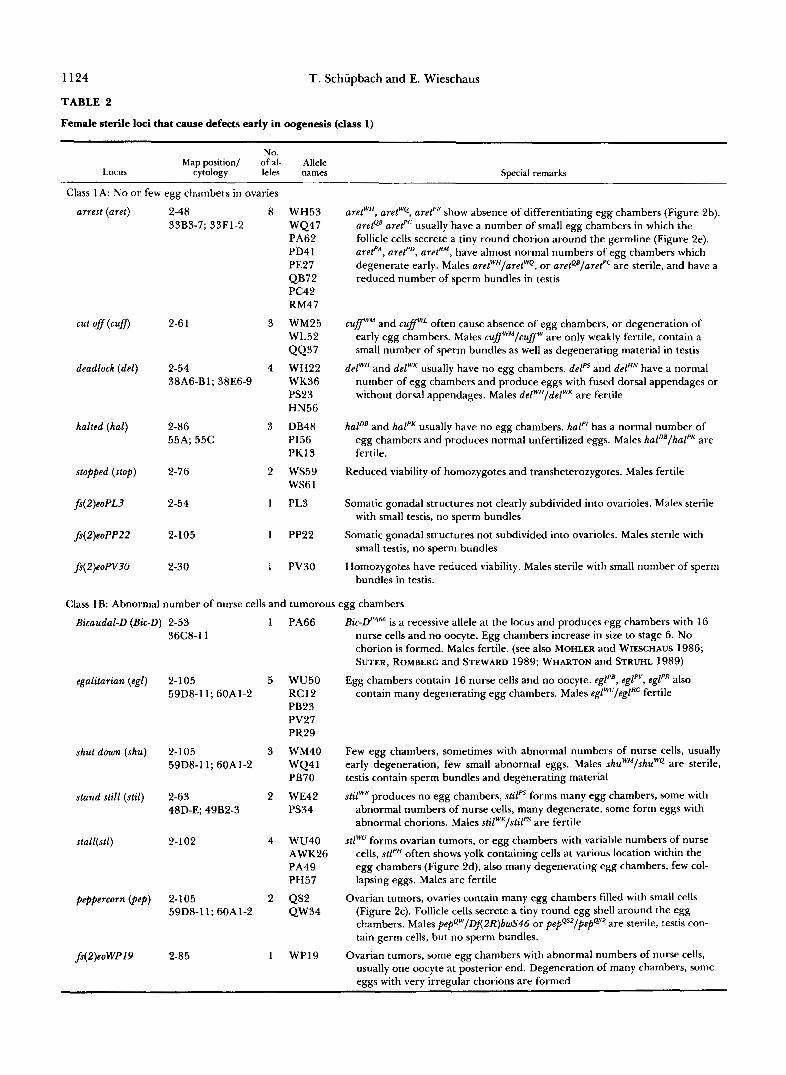
FIGURE 1.-A simplified map of the second chromosome indicating map positions and cytological localization of the 140 mapped loci. Deficiencies are represented by bars. The crosshatched bars indicate deficiencies that have been complementation tested against all 528 mutant lines. Cytologically defined loci are shown directly below the corresponding deficiencies, cytologically undefined loci are shown directly above or below the genetic map (modified from NWSSLEIN-VOLHARD, WIFSCHAUS and KLUDINC 1984).
with an asterix on Table 1, and a cross-hatched bar in Mutations affecting early oogenesis Figure 1 .) In total these deficiencies uncover approx- imately 532 bands on the second chromosome, cor- Class 1A. Production of few, defective germ cells:
regions on that chromosome^ of the mutant lines, loci contain small, underdeveloped ovaries which con- 163 (31 % of all lines, including the maternal-effect tain few, if any egg chambers 2)- The rare egg lines) failed to complement one of these deficiencies. chambers usually become abnormal at early stages of These 163 mutant lines define a total of 62 loci oogenesis and degenerate. Occasionally, single egg yielding an average allele frequency of 2.6 per locus. chambers can develop further, and an abnormal cho-
responding to approximately 27.4% of the banded homozygous for strong mutations at eight
Female Sterile Mutations 1123
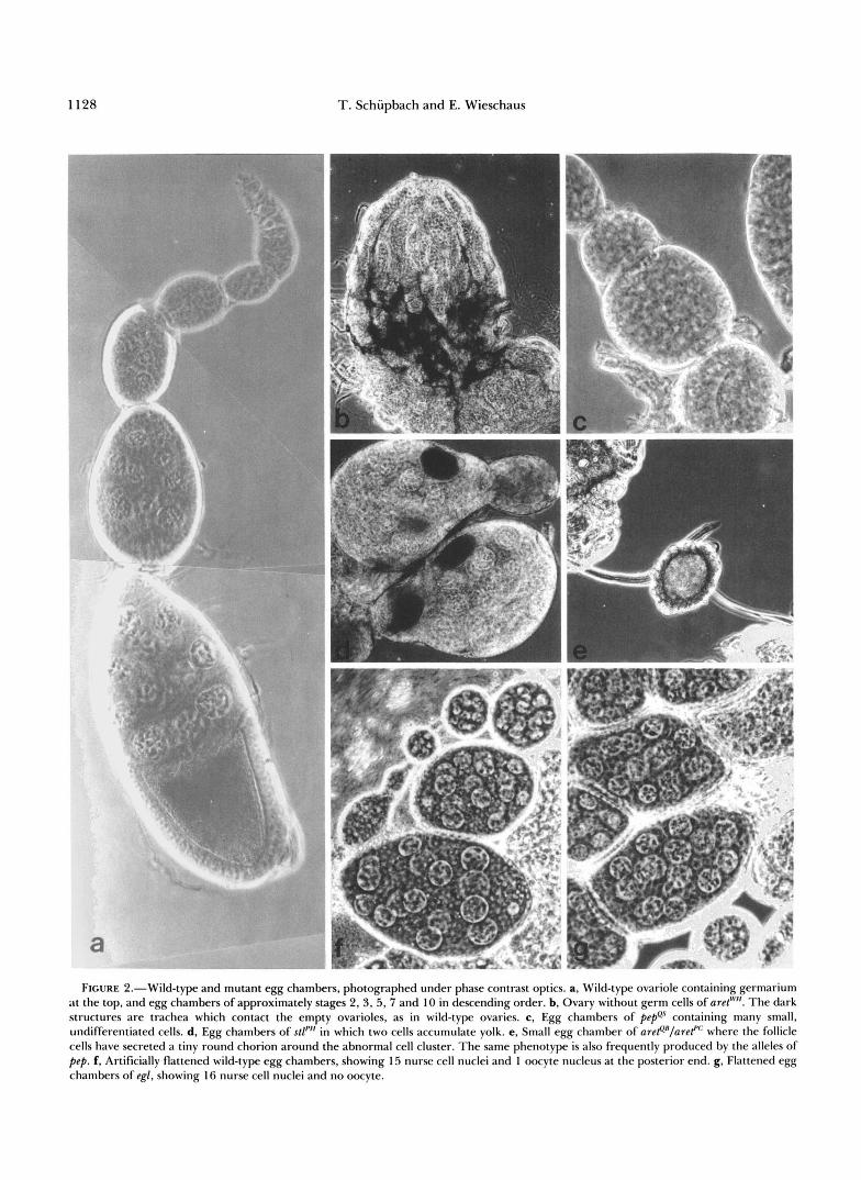
rion with reduced or fused dorsal appendages may be formed. The only locus that consistently caused com- plete absence of germ cells from all homozygous fe- males was aret. This phenotype would be expected of a locus specifically required for germline survival dur- ing embryonic, larval or pupal stages. Weaker alleles of aret, however, cause a range of different pheno- types, not easily explained if aret were only required for survival of germ cells in preadult stages. Zygoti- cally acting mutations that cause complete absence of germ cells from the adult gonads, therefore, appear to be rare. This may imply that most germline survival functions are not exclusively used by germ cells alone. Alternatively, more loci might be specific for germline survival, but may encode partially redundant func- tions.
For most of the mutations in this class the small, empty ovaries were subdivided into ovarioles, as are normal ovaries (Figure 2b). But in fs(2)eoZ'L3 and
fs(2)eoPP22 the ovaries were misformed and no sub- division into ovarioles was observed. The absence of developing germ cells in these females may therefore be a secondary consequence of a defect in the differ- entiation of the somatic portions of the ovary.
In several loci in this group, males homozygous or trans-heterozygous for different alleles were also found to be sterile (Table 2). This is in contrast to groups of female-sterile loci which produce later phe- notypes and usually affect only female fertility (data not shown). The higher incidence of male/female sterility in the early class may reflect the similarities observed in the the early stages of development in male and female germ cells.
Class 1 B. Production of egg chambers with abnor- mal numbers of nurse cells and ovarian tumors: In mutations at seven loci, egg chambers are formed that do not contain the normal complement of 15 nurse cells and 1 oocyte (Table 2). During normal oogenesis a stem cell division at the tip of the ovariole will produce one differentiating daughter cell, which undergoes four more mitotic divisions giving rise to a cluster of 16 cells. Normally one of these cells becomes the oocyte, the other 15 assume a nurse cell fate [for a review of oogenesis, see e.g., KING (1970) and MA- HOWALD and KAMBYSELLIS (198O)l. Mutations that produce egg chambers with abnormal numbers of nurse cells may interfere with these initial events. With the exception of egalitarian and Bicaudal-D, the actual number of nurse cells (or pseudo-nurse cells, KING and RILEY 1982) in all these lines appears variable. Usually the abnormal egg chambers degenerate. In many of the lines some egg chambers are found that contain normal numbers of nurse cells and may de- velop further and form abnormal eggs. The high degree of variability in these lines would suggest that these loci function in a mechanism that is not abso-
lutely necessary for germline divisions themselves, but are involved in a process that confers regularity to these divisions.
In two of the loci (stall and pep) ovarian tumors are formed. These ovaries show ovarioles which contain many small, undifferentiated cells. A similar pheno- type has been described in detail for the locus otu, a female sterile locus on the X chromosome (KING and RILEY 1982; STORTO and KING 1988). Like otu, the alleles of the locus stall show a range of egg chamber phenotypes ranging from tumorous egg chambers to egg chambers with abnormal numbers of nurse cells and egg chambers that will develop into variably ab- normal eggs. The allele stallpH often forms egg cham- bers in which two cells can be observed to take up yolk (Figure 2d). The allele pepes of peppercorn forms egg chambers which contain many small cells with large nuclei, somewhat reminiscent of the phenotype produced by germ cells homozygous for mutations at the locus Sex-lethal (SCHUPBACH 1985; Figure 2c). These egg chambers are surrounded by a monolayer of follicle cells which will eventually secrete a tiny round chorion around the cluster of undifferentiated cells.
In females homozygous for mutations at egalitarian or homozygous for the recessive loss-of-function allele Bicaudal-DPA66 all egg chambers contain 16 nurse cells and no oocyte (Figure 2g). These two loci therefore do not seem to be involved in controlling the correct number of the four mitotic germline divisions, but instead seem to be required for the establishment or maintenance of the oocyte (see also MOHLER and WIESCHAUS 1986; SUTER, ROMBERG and STEWARD 1989; WHARTON and STRUHL 1989).
Class 1C. Early degeneration: In mutations at four loci, and in 16 genetically uncharacterized lines, egg chambers are formed, but the cells in these egg cham- bers appear morphologically abnormal, and usually degenerate before yolk uptake begins. Already at early stages the cells in the egg chambers are often abnormally shaped and appear very refractile in phase contrast optics, indicating that they are beginning to degenerate. Since usually both the follicle cells as well as the nurse cells and oocyte appear abnormal, it is not easy to determine whether the primary defect in these lines results from germline or follicle cell mal- function.
Mutations affecting mid- and late stages of oogenesis
Class 2A. Oogenesis is arrested at stage 8/9: In one locus and eight genetically uncharacterized lines, early stages of oogenesis appear to develop normally, but egg chambers arrest around stage 8 or 9 and begin to degenerate. At this stage the follicle cell epithelium that surrounds the elongated egg chamber is of uniform thickness, and the oocyte has taken up
1124 T. Schiipbach and E. Wieschaus
TABLE 2
Female sterile loci that cause defects early in oogenesis (class 1)
Map position/ of al- Allele No.
Locus cytology leles names Special remarks
Class 1 A: N o or few egg chambers in ovaries
arrest (aret)
cut off(cufl
deadlock (del)
halted (hal)
stopped (stop)
fs(2)eoPL3
fs(2)eoPP22
fs(2)eoPV?O
2-48 8 33B3-7; 33F1-2
2-6 1 3
2-54 4 38A6-Bl; 38E6-9
2-86 3 55A; 55C
2-76 2
2-54 1
2-105 I
2-30 I
WH53 WQ47 PA62 PD4 1 PE27 QB72 PC42 RM47
WM25 WL52 QQ37 WH22 WK36 PS23 HN56
DB48 PI56 PKI 3
ws59 WS6 1
PL3
PP22
PV30
aretWH, aretWQ, aret"' show absence of differentiating egg chambers (Figure 2b). arete' aref" usually have a number of small egg chambers in which the follicle cells secrete a tiny round chorion around the germline (Figure 2e). arefA, aref", aref'", have almost normal numbers of egg chambers which degenerate early. Males aretWH/aretWQ, or aretQ'/aref'" are sterile, and have a reduced number of sperm bundles in testis
cuff'" and cuffwL often cause absence of egg chambers, or degeneration of early egg chambers. Males cuffwM/cuffw are only weakly fertile, contain a small number of sperm bundles as well as degenerating material in testis
delWH and delWK usually have no egg chambers. delPS and delHN have a normal number of egg chambers and produce eggs with fused dorsal appendages or without dorsal appendages. Males delWH/delWK are fertile
hal"' and halPK usually have no egg chambers. hal" has a normal number of egg chambers and produces normal unfertilized eggs. Males hal"'/halPK are fertile.
Reduced viability of homozygotes and transheterozygotes. Males fertile
Somatic gonadal structures not clearly subdivided into ovarioles. Males sterile with small testis, no sperm bundles
Somatic gonadal structures not subdivided into ovarioles. Males sterile with small testis, no sperm bundles
Homozygotes have reduced viability. Males sterile with small number of sperm bundles in testis.
Class 1B: Abnormal number of nurse cells and tumorous egg chambers
Bicaudal-D (Bic-D) 2-53
egalitarian (egl)
shut down (shu)
stand still (stil)
stall(st1)
peppercorn (pep)
fs(2)eoWP19
36C8-11
2-105 59D8-11; 60A1-2
2-1 05 59D8-11; 60A1-2
2-63 48D-E; 49B2-3
2-1 02
2-105 59D8-11; 60A1-2
2-85
1 PA66
5 WU50 RC12 PB23 PV27 PR29
3 WM40 WQ4 1 PB70
2 WE42 PS34
4 WU40 AWK26 PA49 PH57
2 QS2 QW34
1 WP19
is a recessive allele at the locus and produces egg chambers with 16 nurse cells and no oocyte. Egg chambers increase in size to stage 6. No chorion is formed. Males fertile. (see also MOHLER and WIFSCHAUS 1986; SUTER, ROMBERG and STEWARD 1989; WHARTON and STRUHL 1989)
Egg chambers contain 16 nurse cells and no oocyte. eglpB, egl'", eglpR also contain many degenerating egg chambers. Males eglWu/eglRC fertile
Few egg chambers, sometimes with abnormal numbers of nurse cells, usually early degeneration, few small abnormal eggs. Males shuWM/shuWQ are sterile, testis contain sperm bundles and degenerating material
stilwE produces no egg chambers, d l p s forms many egg chambers, some with abnormal numbers of nurse cells, many degenerate, some form eggs with abnormal chorions. Males stilwE/stilps are fertile
stlWU forms ovarian tumors, or egg chambers with variable numbers of nurse cells, d l P H often shows yolk containing cells at various location within the egg chambers (Figure 2d), also many degenerating egg chambers, few col- lapsing eggs. Males are fertile
Ovarian tumors, ovaries contain many egg chambers filled with small cells (Figure 2c). Follicle cells secrete a tiny round egg shell around the egg chambers. Males pepQw/DA2R)bwS46 or pepQsz/pepQsz are sterile, testis con- tain germ cells, but no sperm bundles.
Ovarian tumors, some egg chambers with abnormal numbers of nurse cells, usually one oocyte at posterior end. Degeneration of many chambers, some eggs with very irregular chorions are formed
Female Sterile Mutations 1125
~~
No.
Locus Map position/ of al- Allele
cytology leles names Special remarks
Class 1C Early degeneration
blocked (b lc ) 2-20 1 WE1 3 Males are sterile
close down ( d o ) 2-77 3 WG17 Males clowc/clowK fertile WK50 w u 5
fs( 2)eof 86 2-64 1 PB6 Males are sterile
in addition: 16 genetically undefined lines (WN32, WSlO, PE32, PK33, PM41, PN50, PQ16, PT37, PW21, PY64, QG27, QW63, RD13, RG62, RL36, RS53)
some yolk. It is not obvious what developmental proc- ess is primarily affected by these mutations; possibly the posterior migration of the follicle cells cannot be initiated, or alternatively, yolk synthesis or yolk uptake may be abnormal. The arrested egg chambers even- tually degenerate.
Class 2B. Mutations that interfere with normal follicle cell migration patterns: During mid- and late stages of oogenesis the follicle cells undergo a series of cell shape changes and migrations which result in the complex pattern of the mature egg shell (for descriptions see e.g., KING 1970, MAHOWALD and KAMBYSELLIS 1980; MARGARITIS 1985). A large num- ber of female sterile lines can cause abnormalities in follicle cell migration patterns resulting in abnormally patterned egg shells. This indicates that many differ- ent genes are required, either directly or indirectly, to produce the correct pattern of follicle cell migra- tion.
Open-ended chorion phenotype: Mutations at eight loci on the second chromosome cause an open-ended cho- rion phenotype in homozygous mutant females (Table 3). In these egg chambers the centripetal migration of the follicle cells, which should lead to the closing over of the anterior end of the egg shell, does not occur. In a normal egg chamber the follicle cells initially surround the nurse cell-oocyte complex in a uniform monolayer. During stage 8 and 9 of oogenesis the follicle cells at the posterior end of the chamber begin to change their shape. They become columnar and more densely packed over the oocyte. As more follicle cells come to overlie the oocyte, only few follicle cells are left to cover the nurse cells. These follicle cells assume a very flattened morphology. At the end of this process there is a clear boundary in the follicle cell epithelium distinguishing the columnar follicle cells from the flattened cells. In normal egg chambers this boundary coincides precisely with the boundary between the oocyte and the nurse cells. In the loci which cause the open ended chorion pheno- type this boundary in the follicle cell epithelium is frequently shifted anteriorly and lies over the nurse
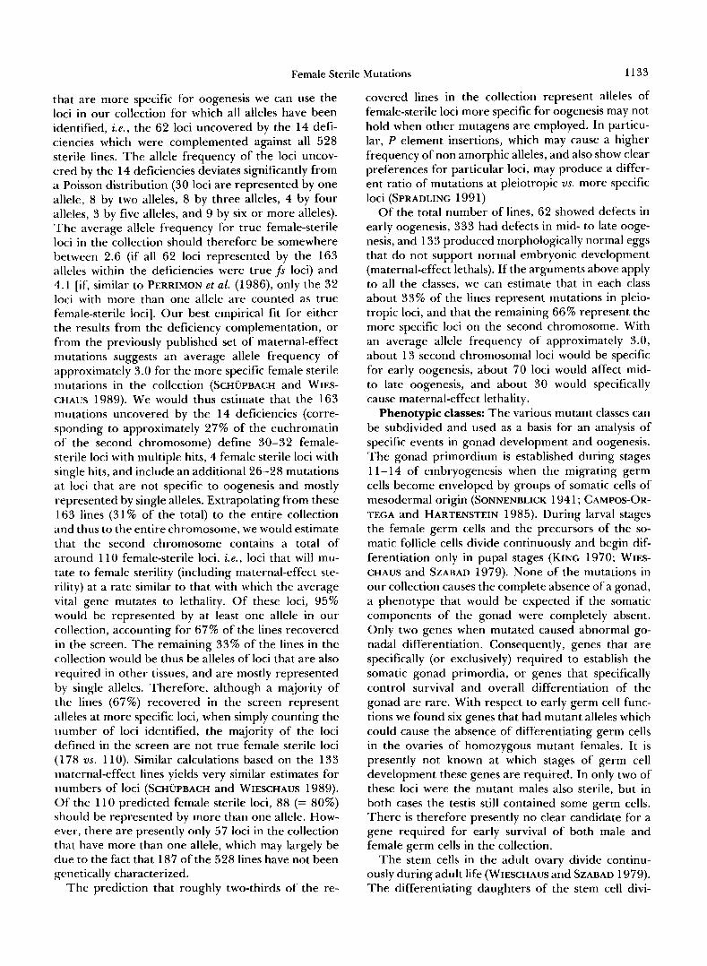
cells. In cup, chal and Bic-C the first tier of nurse cells is usually enclosed by columnar follicle cells (Figure 3). In kel and quit the follicle cell boundary is more variable and is often in the proper position. In strong mutant alleles at any of the five loci there is no “centripetal” migration of follicle cells in between the oocyte and nurse cells. The follicle cells remain on the outside of the nurse cell-oocyte complex. They continue to differentiate and secrete a structured, multilayered chorion, but in the absence of the cen- tripetal follicle cell migrations these egg shells remain open-ended (“cup-shaped”). In cup-shaped eggs dorsal appendages are not formed. Instead, a patch of dorsal appendage material is often found within the surface of the egg shell (Figure 3), indicating that a number of follicle cells were apparently determined to secrete dorsal appendage material, but did not migrate prop- erly out of the epithelium. In contrast, migration of border cells which occurs during stage 9 (KING 1970) is initiated normally in these mutations. However, the border cells will only migrate as far posterior as the plane defined by the boundary of flat us. columnar follicle cells, i.e., in these mutants, the border cells usually do not reach the anterior end of the oocyte. This indicates that the anterior-posterior pattern of the entire follicle cell population is affected in the mutants.
Mutations at the Bicaudal-C (Bic-C) locus also have a dominant haplo-insufficient phenotype and cause a low frequency of bicaudal embryos to form inside the morphologically normal looking eggs produced by heterozygous females (MOHLER and WIESCHAUS 1986). No bicaudal production was ever observed in heterozygous females of the other loci. Whereas strong mutations at the loci cup, chal, kel, Bic-C and quit consistently produce open ended chorions, weaker alleles at cup, chal and Bic-C lead to the pro- duction of more normal looking eggs which very frequently have abnormal anterior ends and fused and reduced dorsal appendages. Weaker alleles at the locus kelch, in contrast, will produce very small eggs with short, stubby appendages, a phenotype that is
1126
TABLE 3
T. Schupbach and E. Wieschaus
Female sterile loci that cause defects in midoogenesis
Locus of al- Allele
Map position/cytology leles names
No.
Special remarks
Class 2A: Female sterile loci that cause arrest and degeneration of egg chambers in midoogenesis
midway (mdy) 2-53 2 QX25 36A8-9; 36E1,2 RF48
in addition: 8 genetically undefined lines (WG18, WG44, PB7, PK9, PS66, QR7, RC24, RS37)
<:lass 28: Female sterile loci that cause abnormalities in follicle cell migration
i. Open ended chorion 2-37 chalice (chal)
3 WP46 chalWP shows strong phenotype. chalQc more variable, pro- QC23 duces some eggs with fused dorsal appendages. chal"' HC49 produces normal, unfertilized eggs
CUP ( C U P ) 2-23 37 -a Alleles vary from cupWQs2, cup""7 with strong phenotype to cuppB", cup"' which produce eggs with fused dorsal ap- pendages, to cup""' with mostly normal eggs and 10% normal embryos
a WQ52, PBl, PB53, PF21, PF63, PH69, P L l l , PN14, PS73, PV36, PW3, PY76, QD57, Q J9, Q 536, Q 565, QK4, QK12, QK24, QL54, QM66, QR55, QR56, QV7, QV50, QW45, RC11, RG6, RH13, RH24, RI77, RM12, RN61, RS2, RS50, RU45, RV4.
Bicaudal-C (Bic-C) 2-53 5 35D1,2; 35E6
kelch (ke l ) 2-53 36C2-4; 36E1,2
7
quit (qui) 2-105 3 59D8-11: 60A 1-2
fs(P)ltoHE#I 2-9 1 fs(Z)ltoQZ47 2-48 fs(Z)ltoRG3 2-4
ii. Abnormalities in dorso-ventral pattern of follicle cells
cappuccino (capu) 2-8 3 24C1,2-3; 24D3-4
spire (spir)
gurken (grk)
2-54 38A6-B 1; 38E6-9
2-30
4
7
torpedo (top) 2- 100 1
WC45 Bic-CW" all egg chambers are open ended, heterozygous PE37 females produce low frequency of bicaudal embryos.
QL53 dages RU35
Wi20 WB6 DE 1 HL39 PL25 QD64 RF4 1
QL24 PX6 1 APE36
PX 1 Other alleles may produce eggs with fused dorsal appen-
HE4 1 QZ47 RG3
RK12 HK3 HK38
Variable expansion (dorsalization) of dorsal appendages, at expense of main body egg shell. Embryos frequently dorsalized. In addition, in embryos absence of polar granules, pole cells and abdominal segments (MANSEAU and SCHUPBACH 1989)
Same phenotype as capu RP48 HPlO PJ56 QF70
WG41 Reduction or absence of dorsal appendages (ventraliza- DC29 tion) with increase in main body egg shell. Frequently HF48 micropyles at both ends of eggs. Embryos ventralized HG2 1 (SCHUPBACH 1987) HK36 HL23 Q166
QY 1 top@"' produces phenotype similar to gurken, but less ex- treme. Amorphic alleles at the locus are lethal (SCHUP- BACH 1987; CLIFFORD and SCHUPBACH 1989)
Female Sterile Mutations 1127
~ ~~~
No. of al- Allele
Locus Map position/cytology leles names Special remarks
i i i . Variable abnormalities in dorsal appendage formation
aubergine (aub)
gourd (gou)
okra ( o h )
zucchini (zuc)
squash (s9u)
vasa (vas)
ji(2)ltoRE57
fs(2)ltoWi42
ji(2,1toRJ36
fs(2)1toRN48
fs( 2)ltoRV64
2-39
2-5
2- 1
2-45 33B307; 33F1-2
2-53
2-5 1 35B2-3; 35D4-7
2-54 37D2-El; 37F5-38A1
2-100
2-50 35B2-3; 35D4-7
2-5 1 35E6; 36A6,7
3-54 37D2-El; 37F5-38A1
5
2
2
3
3
6
1
2
1
1
1
QC42 HM23 HN2 AHN56 AWE13
R133 QD67
RU47 WSl
RS49 HM27 SG63
PP32 HE47 HK35
PD23 HE1 PW72 QS17 RG53 AQB3
RE57
Wi42 Wi46
RJ36
RN48
RV64
Most extreme eggs are spindle shaped without dorsal ap- pendages. Other eggs have fused dorsal appendages, or are of normal morphology and remain unfertilized
Similar to aub
Similar to aub. Eggs of okrWs often have a second micro- pyle at posterior end
Similar to aub
Similar to aub
va~Q"''~, vasAQB3, produce eggs with fused dorsal appendages. Eggs sometimes small. and vas""' produce normal eggs. In all alleles, embryos lack polar granules, pole cells and abdominal segments (see also SCHUPBACH and WIESCHAUS 1986; LASKO and ASH- BURNER 1988; HAY, JAN andJAN 1988)
Similar to aub
Eggs of variable size, sometimes fused dorsal appendages
1
in addition: 33 genetically uncharacterized lines (Wig, WN49, AWD18, HE59, Hi36, DD23, PW41, QG56, Q16, 4134, QM57, QM63, QSlO, RH33, RI44, RI67, RJ27, SD22, Wi36,HF27, HL51, DB22, DC41, DD15, PR56, PU13, QG75, QQ64, RC7, RE42, RF14, RS12, RU44)
usually observed in mutations where the nurse cell content is not transported into the oocyte (see below). In spite of the phenotypic similarities seen in the egg shells produced by strong alleles at the four loci, the differences observed in the weaker, or heterozygous phenotypes therefore suggests that the five loci may affect different processes in oogenesis.
Mutations that cause alterations in the dorso-ventral pattern of the egg shell: Mutations at the loci gurken (grk ) and torpedo (top) cause a ventralization of the follicle cell epithelium and cause reduction or absence of the dorsal follicle cell populations that would nor- mally form operculum and dorsal appendages. Em- bryos which develop inside these eggs are also ven- tralized (SCHUPBACH 1987). All alleles of gurken con- sistently produce this ventralized egg shell phenotype, whereas amorphic alleles at the locus torpedo are lethal (CLIFFORD and SCHUPBACH 1989). Mutations at the
loci spire (spir) and cappuccino (capu) produce a vari- able egg shell phenotype with a fraction of the eggs showing a dorsalized egg shell where follicle cells from the entire circumference have secreted dorsal appen- dage material. Other eggs have one broadened dorsal appendage on the dorsal midline, some eggs show a reduced, fused dorsal appendage, and a fraction of the eggs are always of normal morphology (MANSEAU and SCHUPBACH 1989). Embryos developing inside spir or capu derived eggs lack polar granules and pole cells, show deletions in their abdominal segmentation pattern and are frequently dorsalized. Mutations at spir and capu therefore consistently block the forma- tion of the posterior pole plasm which should harbor the determinants for the germline as well as the de- terminants for abdominal segmentation, whereas the effect of the mutations on dorso-ventral pattern for-
1128 T. Schupbach and E. Wieschaus
FIGURE 2.-Wild-type and mutant egg chambers, photographed under phase contrast optics. a, Wild-type ovariole containing germarium at the top, and egg chambers of approximately stages 2,3,5,7 and 10 in descending order. b, Ovary without germ cells of aret'"". The dark structures are trachea which contact the empty ovarioles, as in wild-type ovaries. c, Egg chambers of pepQs containing many small, undifferentiated cells. d, Egg chambers of stl'" in which two cells accumulate yolk. e, Small egg chamber of are@'/areP where the follicle cells have secreted a tiny round chorion around the abnormal cell cluster. The same phenotype is also frequently produced by the alleles of pep. f, Artificially flattened wild-type egg chambers, showing 15 nurse cell nuclei and 1 oocyte nucleus at the posterior end. g, Flattened egg chambers of egl, showing 16 nurse cell nuclei and no oocyte.

Female Sterile Mutations 1129
R
E
FIGURE 3.-Egg chambers and chorions from wild type and from mutations that produce an open-ended chorion phenotype. a and b, egg chambers from females carrying the transposant BB127 which contains the lac-Z gene, stained with X-gal. The transposant UB127 expresses 8-galactosidase in nurse cells, and beginning at stage 10 of oogenesis, in a ring o f follicle cells at the boundary between columnar and flattened follicle cells. a, In wild-type egg chambers this ring of follicle cells is exactly aligned with the bound- ary between oocyte and nurse cells. b, In Eic-Cwc homozygous egg chambers this ring o f staining follicle cells encircles the nurse cells. c, Wild-type chorion with two dorsal appendages, d, open ended chorion from hel'vi, only a knob of dorsal appendage material is formed.
mation is more variable (MANSEAU and SCHUPBACH 1989).
Mutations which cause variable abnormalities in dorsal appendage formation: Mutations at eleven loci and 33 genetically uncharacterized lines produce eggs with variable dorsal appendage abnormalities. Eggs pro-
duced by females homozygous for these mutations often have only one fused dorsal appendage which may be reduced in size. A fraction of the eggs may lack dorsal appendages altogether. Such eggs are usu- ally spindle shaped, a phenotype which is similar to the eggs produced by the ventralizing mutations tor- pedo or gurken. In addition, eggs derived from okra (okr) homozygous females often show an anterior and a posterior micropyle, similar to eggs from strong gurken alleles (SCHUPBACH 1987). Despite the similar- ities to gurken and torpedo it is however not clear whether this variable fusion of dorsal appendages or incompletely penetrant spindle-shaped egg phenotype represents a ventralization of the follicle cell epithe- lium. The ventralized eggs of gurken and torpedo are larger than wild-type eggs, because more follicle cells contribute to the main body of the egg shell at the expense of the two populations of migrating appen- dage producing follicle cells. In contrast, the eggs produced by many of the mutations in this variable egg shell class are frequently smaller than wild-type eggs. Fusion or absence of dorsal appendages in eggs from these lines may therefore not necessarily reflect a ventralization of the egg shell, but may be caused by a variety of defects in germline and follicle cell development. In addition, this variable fused dorsal appendage phenotype is also frequently produced by weak alleles of loci that block oogenesis at early stages (Table 2). It is possible that the correct establishment and differentiation of the migrating dorsal appendage producing cell populations is a very sensitive process. Slight disturbances in various processes of oogenesis may lead to reductions in the number of cells com- mitted to this fate, or alternatively, may interfere with survival, migration, or differentiation of these partic- ular cell populations.
Based on our previous analysis of the two alleles vaf and vas"" the locus vasa was originally placed into the category of maternal-effect mutations that have no visible effect on oogenesis or egg shell patterning. Using the deficiency Dfl2L)osp29 to screen all mutant lines we found, however, four more alleles of vasa which cause variable defects in oogenesis and lead to the production of eggs with fused and reduced dorsal appendages. The eggs that are laid are also usually somewhat smaller than wild-type eggs. Similar defects in oogenesis were also observed for strong vasa alleles by LASKO and ASHBURNER (1990) and LEHMANN and NUSSLEIN-VOLHARD (1 99 1).
Class 2C. Mutations which cause the egg shell to be structurally abnormal: Thin, fragile chorion phe- notype: The eggs produced by females at 9 loci and in 14 genetically uncharacterized lines usually have a chorion that appears unstructured and fragile (Table 4). Often the chorions rupture at the time of egg deposition by the female and such eggs appear to be
1130 T. Schiipbach and E. Wieschaus
TABLE 4
Female sterile loci that affect the structure and integrity of the egg shell (class 2C)
Locus
~~
No. ofal- Allele leks names Special remarks
~~
Map position/cytology
i. Thin, fragile chorion
chqfon (chzfi
gauze (gau)
rayon (ray)
satin (sat)
f s ( 2 ) l t o A H E l fs(2)ltoHLZZ fs(Z)EtoPA77
2-53 35EB; 36A6,7
2-74
2-46
2-57 43C3; 43E1-8
2-7 1 2-55 2-72 5 1 E3; 52.4
2-5 1 2-54 37F5-38Al; 38A6-7
6
7
1 1
WD18 QW16
WF24 p555
DB23
PT6 1 HF19 PB55 PY16 QN68 RJ3 I RP65
Q N 54
5c46
QY42
AHEl h122 PA77 lethal over Deficiency
(see also UNDER- WOOD et al. 1990)
PN48 PP43 lethal over Deficiency
in addition 14 genetically uncharacterixed lines: HN12 PU34, PZ61, QE55, QL57, QM25, QU35, RB37, RE74, RM33, R031,RP65, SP28, ASB62
ii. Collapsed eggs
crepe (crpe) 2-59
muslin (mln) 2-28
fs(Z)ltoHD43 2-54 37F5-38A1; 38A6-7
fs(2)ltoHMI 1 2-105 59D8-11; 60A1-2
fs (2) l toQJ42 2-1 7 25E6-F3; 26.47-8
fs(Z)ltoRN73 2-57 43C3; 43E1-8
f(Z)EtoRU26 2-4 1 31D; 31F3
in addition 37 genetically uncharacterized lines: HB6. HE2. H H
6
2
1
1
1
1
1
15. HI
PZ3 HM38 DB3 p14
QY25 r135
PP4 1 h13
HD43
H M l l
QJ42
RN73
RU26
SAVANT and WARING (1989)
d32, HK29, HL48, HM22, HN43, DE17, DG18, PE70, PG42, PC69, PK75, PR67, PT44, PU48, QE31, QF3, Q F 5 7 , Q H l , Q15, QI56, QK35, QK36, QK52, QL73, QT48, QT68, RA37, RD30, RD77, RQ63, RS54, RU3, SGlO, SG71
surrounded only by a vitelline membrane. Upon in- absent (e.g., in eggs from chgoon and gauze). The spection of the chorion, the follicle cell imprints seem various layers of the normal egg shell are secreted by less distinct than in wild type, the dorsal appendages the follicle cells during middle and late stages of are thin and reduced in size, or they may be altogether oogenesis, a process which requires a well controlled
Female Sterile Mutations
TABLE 5
Female sterile loci that produce abnormally shaped eggs, or fail to lay eggs
1131
of al- No.
Allele Locus Map position/cytology leles names Special remarks
Class 2D: Abnormal egg size
i. Tiny eggs (failure in transport)
chickadee (chic) 2-23
quail (qua)
warbler (war)
minus (mi)
fs(2)ltoAHK35
fs(2)ltoDC37
fs(2)ltoDG25
fs(2)ltoQB3
fs(2)ltoQT50
fs(2)11oRM7
2-53 36C8-11
2-79
2-105 59D8-11; 60A1-2
2-105 592)s-11; 60A1-2
2-105 59D8-11; 60A1-2
2-4 1 31D; 31F3
2-62 2-54 37D2-El; 37F5-38Al
2-105 59D8-11; 60A1-2
w c 5 7 WF57 WK26
WP20 HK25 HM 14 PX42 QE24 Q T 2 1
RUlO
PT43 QX54
AHK35
DC37
DG25
QB3 QT50
RM7
Molecularly characterized by L. COOLEY (personal com- munication)
(see also STEWARD and NUS- SLEIN-VOLHARD 1986)
(Locus originally discovered by BIDDLE, see LINDSLEY and ZIMM 1990)
lethal over Deficiency, allelic to 1(2)7W37Bc. (T. WRIGHT, personal com- munication)
in addition 11 genetically uncharacterized lines: HL58, HP11, PE29, PI42, PV1, QH9, RCl, RC25, RD54, RF76, R03
ii. Short and variable egg size
2-105 1 APV63 59D8-11; 60A1-2
2-4 1 1 p123 31D; 31F3
2-8 1 QE45 24C1,2-3; 24D3-4
in addition 41 genetically uncharacterized lines: HA21, HD45, HF43, HP16, HP26, DB37, PA15, PE69, PF29, PJ47, PK16, PP52, PU5, PU6, PU36, QC43, QDI, QD21, QE12, QE44, QF9, QG48, Q176, QS61, QU33, QX48, QX63, RH53, RJ33, RK35, RM29, RP64, RR14, RS74, RU12, RV15, RV31, RV53, RV62, SD36, SG09
Class 2E: Egg retention
retained (retn) 2-105 2 R 0 4 4 59D8-11; 60A1-2 RU50
fs(2)ltoHC44 2-53 1 HC44 36A8-9; 36E1,2
fs(Z)ltoPM43 2-54 1 PM43 37B9-Cl; 37D1,2
in addition 27 genetically uncharacterized lines: WQ40, HB3, HC22, HD51, Hi6, HL56, PC46, PG49, PK50, PL21, PL22, PW34, QB4, QB62, QD44, QD76,Q07, QU70, RB30, RC32, RF15, RH61, R054, RQ69, RS35, RS44, RV57

1132 T. Schiipbach and E. Wieschaus
and coordinated pattern of gene expression in these cells (MARCARITIS, KAFATOS and PETRI 1980; MAR- GARITIS 1985; PARKS and SPRADLINC 1987; ROMANO et al. 1988). Most likely the mutations with thin and fragile egg shells cause abnormalities in the secretion of one or more layers of the egg shell. In fact, the egg shell of fs(2)ltoPA77 has been analyzed by UNDER- WOOD et al. (1990) and was found to show abnormal- ities in chorion gene expression. chqwas found to be defective in chorion gene amplification (J. TOWER and A . SPRADLING, personal communication and cited in SPRADLINC 199 1).
Collapsed, flaccid egg shells: In mutations at seven loci and in 37 genetically uncharacterized lines the mutant females lay eggs that appear collapsed (Table 4). In normal eggs the vitelline membrane and the waxy layer seal the egg from its surrounding and prevent the eggs from dehydration. Abnormalities in the deposition of the vitelline membrane or the waxy layer, or alternatively, failure to completely cover the anterior end of the oocyte via the late migrations of the anterior follicle cells might cause eggs to lose water and show the collapsed phenotype. fs(2)ltoQ142 has been analyzed by SAVANT and WARING (1989) and was found to cause absence of a major vitelline mem- brane protein. On the other hand, mutations in the yolk protein YP2 locus can also cause a collapsed egg phenotype (WILLIAMS et al. 1987). Alterations of var- ious steps in yolk production or yolk uptake might therefore also account for this phenotype.
Class 2D. Abnormalities in egg size and propor- tion: Tiny eggs: failure to transport nurse cell content into the oocyte: In normal eggs the nurse cells attain their largest size at stage 10B (KING 1970). Subse- quently the nurse cell content is transported into the oocyte via the system of interconnected ring canals. I n the egg chambers from mutant females at 10 loci and an additional 11 uncharacterized lines, the nurse cells remain large (Table 5). The oocyte arrests growth at around stage 10. The follicle cells proceed with their regular migration movements to enclose the anterior end of the oocyte and form dorsal appen- dages. Since the nurse cell content largely remains excluded from the oocyte, tiny eggs are produced. The follicle cell imprints on these eggs appear thick- ened, presumably because the follicle cells do not stretch to cover the growing oocyte, but instead, re- tained their columnar shape. The dorsal appendages are usually short and stubby, possibly due to a topo- logical barrier for the migrating follicle cells posed by the large nurse cells.
Short eggs: The mutant females in 44 lines lay eggs which are shorter and usually fatter than wild-type eggs. In contrast to the tiny eggs described above, these short eggs are frequently fertilized and often give rise to normal embryos, or to embryos that show
irregular reductions and fusions in their anterior- posterior segmentation pattern. Upon dissection of ovaries from such lines usually no consistent abnor- malities are visible and the nurse cells seem to regress normally at later stages. This short egg phenotype is very similar to the phenotype of the X chromosomal locus short egg (seg) which is caused by an abnormal follicle cell function (WIESCHAUS, AUDIT and MASSON 1981). The second chromosomal lines which cause this short egg phenotype were not further analyzed.
Class 2E. Mutations that cause retention of mor- phologically normal eggs in the ovary: The mutant females in three loci and 27 genetically uncharacter- ized lines very rarely lay any eggs (Table 5). Their ovaries contain, however, a large number of appar- ently mature eggs, similar to ovaries of females that, for lack of suitable egg laying medium, retain their eggs. It is possible that the oviducts of these females are not functional, or alternatively, their egg laying behavior may be abnormal.
DISCUSSION
Number of genes: In the mutagenesis screens de- scribed in this paper we recovered 528 female sterile lines from a total of 7351 lines tested. These 7,351 lines derived from 18,782 lines, 60% of which were lethal. A traditional way to estimate the number of female sterile loci is based on comparing the frequency of female sterile mutations to the frequency of lethals in a given screen (GANS, AUDIT and MASSON 1975; MOHLER 1977). Using this method we can calculate that the target size for lethality on the second chro- mosome is 12.5 times larger than the target size for female sterility (see also SCHUPBACH and WIESCHAUS 1989). Using an estimate of 1700 lethals on the second chromosome (NUSSLEIN-VOLHARD, WIESCHAUS and KLUDING 1984), this result would suggest that 140 loci on the second chromosome can mutate to female- sterility in a standard EMS mutagenesis. This uncor- rected number compares well with similar estimates derived from screens for female sterile mutations on the X chromosome (KING and MOHLER 1975; GANS, AUDIT and MASSON 1975). These calculations are, however, subject to major reservations. Similar to PERRIMON et al. (1986) we have previously argued that the distribution of alleles in such female-sterile screens represents the combination of alleles at two sets of loci: the more specific female-sterile loci with an allele distribution approximately following a Pois- son distribution, and a set of nonamorphic alleles at loci with functions in various tissues. Indeed, in our collection, there is a large excess of loci represented by only one allele, and a smaller group of loci with multiple alleles which are approximately Poisson dis- tributed (see also SCHUPBACH and WIESCHAUS 1989). In order to obtain an estimate of the number of loci
Female Sterile Mutations 1133
that are more specific for oogenesis we can use the loci in our collection for which all alleles have been identified, i e . , the 62 loci uncovered by the 14 defi- ciencies which were complemented against all 528 sterile lines. The allele frequency of the loci uncov- ered by the 14 deficiencies deviates significantly from a Poisson distribution (30 loci are represented by one allele, 8 by two alleles, 8 by three alleles, 4 by four alleles, 3 by five alleles, and 9 by six or more alleles). The average allele frequency for true female-sterile loci in the collection should therefore be somewhere between 2.6 (if all 62 loci represented by the 163 alleles within the deficiencies were true fs loci) and 4.1 [if, similar to PERRIMON et al. (1 986), only the 32 loci with more than one allele are counted as true female-sterile loci]. Our best empirical fit for either the results from the deficiency complementation, or from the previously published set of maternal-effect mutations suggests an average allele frequency of approximately 3.0 for the more specific female sterile mutations in the collection (SCHUPBACH and WIES- CHAUS 1989). We would thus estimate that the 163 mutations uncovered by the 14 deficiencies (corre- sponding to approximately 27% of the euchromatin of the second chromosome) define 30-32 female- sterile loci with multiple hits, 4 female sterile loci with single hits, and include an additional 26-28 mutations at loci that are not specific to oogenesis and mostly represented by single alleles. Extrapolating from these 163 lines (31% of the total) to the entire collection and thus to the entire chromosome, we would estimate that the second chromosome contains a total of around 1 10 female-sterile loci, i . e . , loci that will mu- tate to female sterility (including maternal-effect ste- rility) at a rate similar to that with which the average vital gene mutates to lethality. Of these loci, 95% would be represented by at least one allele in our collection, accounting for 67% of the lines recovered in the screen. The remaining 33% of the lines in the collection would be thus be alleles of loci that are also required in other tissues, and are mostly represented by single alleles. Therefore, although a majority of the lines (67%) recovered in the screen represent alleles at more specific loci, when simply counting the number of loci identified, the majority of the loci defined in the screen are not true female sterile loci (178 vs. 110). Similar calculations based on the 133 maternal-effect lines yields very similar estimates for numbers of loci (SCHUPBACH and WIESCHAUS 1989). Of the 1 10 predicted female sterile loci, 88 (= 80%) should be represented by more than one allele. How- ever, there are presently only 57 loci in the collection that have more than one allele, which may largely be due to the fact that 187 of the 528 lines have not been genetically characterized.
The prediction that roughly two-thirds of the re-
covered lines in the collection represent alleles of female-sterile loci more specific for oogenesis may not hold when other mutagens are employed. In particu- lar, P element insertions, which may cause a higher frequency of non amorphic alleles, and also show clear preferences for particular loci, may produce a differ- ent ratio of mutations at pleiotropic us. more specific loci (SPRADLING 199 1)
Of the total number of lines, 62 showed defects in early oogenesis, 333 had defects in mid- to late ooge- nesis, and 133 produced morphologically normal eggs that do not support normal embryonic development (maternal-effect lethals). If the arguments above apply to all the classes, we can estimate that in each class about 33% of the lines represent mutations in pleio- tropic loci, and that the remaining 66% represent the more specific loci on the second chromosome. With an average allele frequency of approximately 3.0, about 13 second chromosomal loci would be specific for early oogenesis, about 70 loci would affect mid- to late oogenesis, and about 30 would specifically cause maternal-effect lethality.
Phenotypic classes: The various mutant classes can be subdivided and used as a basis for an analysis of specific events in gonad development and oogenesis. The gonad primordium is established during stages 1 1-14 of embryogenesis when the migrating germ cells become enveloped by groups of somatic cells of mesodermal origin (SONNENBLICK 194 1 ; CAMPOS-OR- TEGA and HARTENSTEIN 1985). During larval stages the female germ cells and the precursors of the so- matic follicle cells divide continuously and begin dif- ferentiation only in pupal stages (KING 1970; WIES- CHAUS and SZABAD 1979). None of the mutations in our collection causes the complete absence of a gonad, a phenotype that would be expected if the somatic components of the gonad were completely absent. Only two genes when mutated caused abnormal go- nadal differentiation. Consequently, genes that are specifically (or exclusively) required to establish the somatic gonad primordia, or genes that specifically control survival and overall differentiation of the gonad are rare. With respect to early germ cell func- tions we found six genes that had mutant alleles which could cause the absence of differentiating germ cells in the ovaries of homozygous mutant females. It is presently not known at which stages of germ cell development these genes are required. In only two of these loci were the mutant males also sterile, but in both cases the testis still contained some germ cells. There is therefore presently no clear candidate for a gene required for early survival of both male and female germ cells in the collection.
The stem cells in the adult ovary divide continu- ously during adult life (WIESCHAUS and SZABAD 1979). The differentiating daughters of the stem cell divi-
1134 T. Schiipbach and E. Wieschaus
sions will undergo four incomplete mitotic divisions (“cystocyte divisions”) giving rise to a cluster of sixteen cells connected to each other by ring canals, the remnants of the mitotic division. One of the two cells in the cluster that have four ring canals will become the oocyte and will move from its initial central posi- tion within the cluster to the posterior end. The remaining fifteen cells will differentiate into nurse cells. Seven of the genes in our collection interfere at some level with the normal establishment of the 15 + 1 nurse cell-oocyte cluster. Three of these loci can be counted into the “ovarian tumor” class giving rise to egg chambers that contain many small, undif- ferentiated cells, similar to the phenotypes produced by mutations at the X chromosomal locus otu, or the third chromosomal locus 6ag-ofmarbles (KING and RI- LEY 1982; MCKEARIN and SPRADLING 1990). Clearly the control of the cystocyte divisions is affected in these mutants at some level. In one allele of st1 egg chambers with variable numbers of nurse cells are formed which will often contain more than one cell that accumulates yolk, strongly suggesting that more than one of the cells in the cluster has acquired the characteristics of an oocyte. Given that normally the cell lineage of the cystocyte divisions is fixed, and only one of two particular daughters within the cluster can become the future oocyte (see e.g., KING 1970), it is perhaps not unexpected that a mutation that disrupts the normal control of cell division could lead to egg chambers with more than one oocyte.
Mutations at the two loci egalitarian and Bic-D can give rise to egg chambers that contain 16 nurse cells and no oocyte. In these mutants the distinction be- tween oocyte and nurse cells is either never established or fails to be maintained. Bic-D has been molecularly cloned and its protein has been shown to show se- quence similarity to the tail portion of the myosin heavy chain. The RNA and the protein have also been shown to accumulate in the oocyte of wild-type egg chambers (SUTER, ROMBERG and STEWARD 1989; WHARTON and STRUHL 1989), but the gene products fail to accumulate in egl egg chambers (SUTTER and STEWARD 199 1). Given the additional observation that egl mutations will suppress the dominant gain-of- function phenotype of Bic-D alleles (MOHLER and WIESCHAUS 1986), it is possible that egl function is necessary for Bic-D localization, and that localization of Bic-D in turn is necessary to maintain the oocyte in its determined state.
After the initial cystoblast divisions the somatically derived follicle cells envelop the 16 cell cluster as it enters the lower portion (region 2B) of the germar- ium. Further development of the egg chamber will depend on the close cooperation between the three cell types-oocyte, nurse cells and follicle cells. The follicle cells contribute yolk to the developing oocyte,
and in mid-oogenesis they undergo a series of complex migrations and cell shape changes before beginning with the secretion of the various layers of the egg shell (see e.g., MARGARITIS 1985). A large fraction of the mutations recovered in this screen affects follicle cell behavior. Mutations at 23 loci and in 33 uncharacter- ized lines will cause abnormalities in follicle cell mi- gration patterns, as reflected in the mature secreted egg shell, and mutations in 16 loci and 5 1 uncharac- terized lines lead to the formation of a structurally abnormal egg shell. This demonstrates that a rela- tively large number of genes are involved in the development and the differentiation processes that take place in the follicle cell epithelium. This finding corresponds well with the results obtained from recent screens involving random insertion of lac-Z containing P elements into the genome (“enhancer-trap” screens) performed in various laboratories. The lines isolated in these screens have revealed a high complexity of transcriptional activity in the follicle cells during oog- enesis (FASANO and KERRIDGE 1988; GROSSNIKLAUS et al. 1989; SPRADLING 1992; T. SCHUPBACH, L. J. MAN- SEAU and E. SHADDIX, unpublished results). The ma- jority of the mutant lines affecting follicle cell behav- ior that were obtained in the present screen have very variable phenotypes. This variability may in part arise because many of the mutations may not be null alleles at the corresponding loci. In fact, genetic mapping and complementation tests revealed that several of the lines that showed variable effects on follicle cell migration leading to fusion and reduction of dorsal appendages, represented alleles at loci where strong alleles blocked oogenesis at earlier stages. However, there also exist several loci represented by multiple alleles in the collection, for which even the strong alleles when tested over a deficiency still produce variable egg shell phenotypes. Possibly there exists a certain amount of redundancy among the genes in- volved in late follicle cell functions such that single mutations will not completely block these later differ- entiation processes. Studies of genetically mosaic egg chambers have shown that the dorso-ventral devel- opment of the follicle cell epithelium depends on signals from the underlying germ line (WIESCHAUS, MARSH and GEHRING 1978; SCHUPBACH 1987). Since the movements of the follicle cells occur in relation to the underlying oocyte and nurse cells, it is conceivable that some of the mutations affecting follicle cell mi- gration would act in the germline whereas others might be expected to block follicle cell intrinsic proc- esses.
Mutations in seven loci cause a block of the centrip- etal migrations of the follicle cells that normally occur after stage 10 of oogenesis and lead to the formation of the anterior specialized regions of the eggshell (operculum, micropyle, ventral collar). The resulting
Female Sterile Mutations 1135
egg shell consequently remains open-ended (“cup- shaped”). Morphological examination of the pheno- type during oogenesis and use of “enhancer-trap” lines that express lac-Z in particular subsets of follicle cells have shown that as an initial defect in these lines the posterior migration of the follicle cells at stages 8 and 9 of oogenesis is arrested before the population of columnar follicle cells is properly aligned with the oocyte surface. Border cell migration in these lines is similarly arrested before the border cells reach the anterior end of the oocyte. It is therefore possible that in these lines the populations of columnar us. flat follicle cells mis-position themselves in the anterior- posterior axis with respect to the underlying germline. Alternatively, it is possible that oocyte growth is ar- rested or slowed down in these mutants such that although the follicle cells position themselves correctly within the egg chamber, the oocyte remains too small and thus causes the relative misalignment. In either case, the mutations reveal that follicle cells do not necessarily have to be in contact with the oocyte itself to make a clear distinction between the population attaining the columnar vs. flattened cell shape, and likewise, that the border cells can arrest their posterior migration without having made contact with the an- terior end of the oocyte itself. This observation argues against a strict chemotactic model for border cell migration with the oocyte providing the source of the signal. In the future, the morphological and mosaic study of such mutations in combination with available molecular techniques should allow a more precise understanding of the various signals and patterning processes that govern the development and differen- tiation of the somatic and germline components of the ovary of Drosophila.
We thank MARIA WEBER and NGOC LY for their help with the mutagenesis screen. We are very grateful to GORDON GRAY and CUBIT CASE for reliably providing fly food, and to NGOC LY, CHITRA VASHI and YAXIN YU for help with maintaing the stocks and performing complementation crosses. We thank EILEEN SHADDIX for her assistance with the “enhancer trap” @-galactosidase staining of ovaries. We thank CHRISTIANE NUSSLEIN-VOLHARD and the past and present members of the Princeton fly groups for many discus- sions and encouragement. For sending us deficiency stocks we would like to thank MICHAEL ASHBURNER, BRUCE BAKER, ELIOT GOLDSTEIN, TODD LAVERTY, Ross MACINTYRE, JANIS O’DONNELL, GUNTER REUTER, DAVID ROBERTS, SIEGFRIED ROTH, PAT SIMPSON, DONALD SINCLAIR, THEODORE F. WRIGHTand GEROLD WUSTMANN. This work was supported by research grants from the National Institute of Health (GM 40558 and HD 15587) and the National Science Foundation (DCB-8508917).
LITERATURE CITED
ASHBURNER, M., P. THOMSON, J. ROOTE, P. F. LASKO, Y. GRAU, M. EL MESSAL, S. ROTH and P. SIMPSON, 1990 The genetics of a small autosomal region of Drosophila melanogaster containing the structural gene for alcohol dehydrogenase. VI1 Character- ization of the region around the snail and cactus loci. Genetics 1 2 6 679-694.
BIER, E., H. VASSIN, S. SHEPHERD, K. LEE, K. MCCALL, S. BARBEL, L. ACKERMAN, R. CARRETTO, T. UEMURA, E. GRELL, L. Y. JAN
and Y. N. JAN, 1998 Searching for pattern and mutation in the Drosophila genome with a P-lacZ vector. Genes Dev. 3:
CAMPOS-ORTEGA, J. A,, and V. HARTENSTEIN, 1985 The Embryonic Development of Drosophila melanogaster. Springer Verlag, Berlin.
CLIFFORD, R. J., and T. SCHUPBACH, 1989 Coordinately and dif- ferentially mutable activities of torpedo, the Drosophila melano- gaster homolog of the vertebrate EGF receptor gene. Genetics
FASANO, L., and S. KERRIDGE, 1988 Monitoring positional infor- mation during oogenesis in adult Drosophila. Development 104:
CANS, M., C. AUDIT and M. MASSON, 1975 Isolation and charac- terization of sex-linked female sterile mutants in Drosophila melanogaster. Genetics 81: 683-704.
GARCIA-BELLIDO, A., and L. G. ROBBINS, 1983 Viability of female germ-line cells homozygous for zygotic lethals in Drosophila melanogaster. Genetics 103: 235-247.
GROSSNIKLAUS, U., H. J. BELLEN, C. W r r s o ~ and W. J. GEHRING, 1989 P-element-mediated enhancer detection applied to the study of oogenesis in Drosophila. Development 107: 189-200.
HAY, B., L. Y. JAN and Y. N. JAN, 1988 A protein component of Drosophila polar granules is encoded by vasa and has extensive sequence similarity to ATP-dependent helicases. Cell 5 5 577- 587.
KING, R. C., 1970 Ovarian Development in Drosophila melanogaster. Academic Press, New York.
KING, R. C., and J. D. MOHLER, 1975 The genetic analysis of oogenesis in Drosophila melanogaster, pp. 757-791 in Handbook of Genetics, Vol. 3, edited by R. C. KING. Plenum, New York.
KING, R. C., and S. F. RILEY, 1982 Ovarian pathologies generated by various alleles of the otu locus in Drosophila melanogaster. Dev. Genet. 3: 69-86.
KLINGLER M., M. ERDELYI, J. SZABAD and C. NUSSLEIN-VOLHARD, 1988 Function of torso in determining the terminal anlagen of the Drosophila embryo. Nature 335: 275-277.
KOMITOPOULOU K., M. CANS, L. H. MARGARITIS, F. C. KAFATOS and M. MASSON, 1983 Isolation and characterization of sex- linked female-sterile mutants in Drosophila melanogaster with special attention to eggshell mutants. Genetics 105 897-920.
LASKO, P. F., and M. ASHBURNER, 1988 The product of the Drosophila gene vasa is very similar to eukaryotic initiation factor-4A. Nature 335: 61 1-617.
LASKO, P. F., and M. ASHBURNER, 1990 Posterior localization of vasa protein correlates with, but is not sufficient for, pole cell developments. Genes Dev. 4 905-92 1.
LEHMANN, R., and C. NUSSLEIN-VOLHARD, 1991 The maternal gene nanos has a central role in posterior pattern formation of the Drosophila embryo. Development 112: 679-691 (1991).
LINDSLEY, D. L., and E. H. GRELL, 1968 Genetic Variations of Drosophila melanogaster. Carnegie Inst. Wash. Publ. 627.
LINDSLEY, D. L., and G. ZIMM, 1985 The genome of Drosophila melanogaster, Part 1: Genes A-K. Drosophila Inform. Serv. 62:
LINDSLEY, D. L., and G. ZIMM, 1986 The genome of Drosophila melanogaster, Part 2: Lethals, maps. Drosophila Inform. Serv.
LINDSLEY, D. L., and G. ZIMM, 1987 The genome of Drosophila melanogaster, Part 3: Rearrangements. Drosophila Inform. Serv. 65: 1-203.
LINDSLEY, D. L., and G. ZIMM, 1990 The genome of Drosophila melanogaster, Part 4: Genes L-Z, balancers, transposable ele- ments. Drosophila Inform. Serv. 68: 1-382.
MAHOWALD, A. P., and M. P. KAMBYSELLIS, 1980 Oogenesis, pp. 141-224 in The Genetics and Biology of Drosophila, Vol. 2d,
1273-1287.
123: 771-787.
245-253.
1-227.
6 4 1-158.

1136 T. Schiipbach and E. Wieschaus
edited by M. ASHBURNER and T . R. F. WRIGHT. Academic Press, London.
MANSEAU, L. J., and T. SCHUPBACH, 1989 cappuccino and spire: two unique maternal-effect loci required for both the antero- posterior and dorsoventral patterns of the Drosophila embryo. Genes Dev. 3: 1437-1452.
MARGARITIS, L. H., 1985 Structure and physiology of the egg shell, pp. 113-1 15 in Comprehensive Insect Physiology, Biochem- ist? and Pharmacology, Vol. 1, edited by G. A. KERKUT and L. I. GILBERT. Pergamon Press, Elmsford, N.Y.
MARGARITIS, L. H., F. KAFATOS and W. H. PETRI, 1980 The eggshell of Drosophila melanogaster. I. Fine structure of the layers and regions of the wild-type eggshell. J. Cell Sci. 43: 1- 35.
MCKEARIN, D. M., and SPRADLING, A. P., 1990 bag-of-marbles: a Drosophila gene required to initiate both male and female gametogenesis. Genes Dev. 4: 2242-2251.
MOHLER, J. D., 1977 Developmental genetics of the Drosophila egg. I . Identification of 50 sex-linked cistrons with maternal effects on embryonic development. Genetics 8 5 259-272.
MOHLER, J., and E. WIESCHAUS, 1986 Dominant maternal-effect mutations of Drosophila melanogaster causing the production of double-abdomen embryos. Genetics 112: 803-822.
NUSSLEIN-VOLHARD C., E. WIESCHAUS and H. KLUDING, 1984 Mutations affecting the pattern of the larval cuticle in Drosophila melanogaster. I. Zygotic loci on the second chromo- some. Roux’s Arch. Dev. Biol. 193: 267-282.
ORR, W. C., V. K. GALANOPOULOS, C. P. ROMANO and F. C. KAFATOS, 1989 A female sterile screen of the Drosophila melanogaster X chromosome using hybrid dysgenesis: Identifi- cation and characterization of egg morphology mutants. Ge- netics 122: 847-858.
PARKS, S., and A. SPRADLING, 1987 Spatially regulated expression of chorion genes during Drosophila oogenesis. Genes Dev. 1:
PERRIMON, N., L. ENGSTROM and A. P. MAHOWALD, 1984 The effects of zygotic lethal mutations on germline functions in Drosophila. Dev. Biol. 105 404-414.
PERRIMON, N., D. MOHLER, L. ENGSTROM and A. P. MAHOWALD, 1986 X-linked female-sterile loci in Drosophila melanogaster. Genetics 113: 695-7 12.
REUTER, G., and J. SZIDONIA, 1983 Cytogenetic analysis of varie- gation suppressors and a dominant temperature-sensitive lethal in region 23-26 of chromosome 2L in Drosophila melanogaster. Chromosoma 88: 277-285.
ROMANO, C. P., B. BIENZ-TADMOR, B. D. MARIANI and F. C. KAFATOS, 1988 Both early and late Drosophila chorion gene promoters confer correct temporal, tissue and sex specificity on a reporter Adh gene. EMBO J. 7: 783-790.
SAVANT, S. S., and G. L. WARING, 1989 Molecular analysis and rescue of a vitelline membrane mutant in Drosophila. Dev. Biol.
SCHUPBACH, T., 1985 Normal female germ cell differentiation requires the female X chromosome to autosome ratio and expression of Sex-lethal in Drosophila melanogaster. Genetics 109: 529-548.
SCHUPBACH, T., 1987 Germ line and soma cooperate during
497-509.
135 43-52.
oogenesis to establish the dorsoventral pattern of egg shell and embryo in Drosophila melanogaster. Cell 49: 699-707.
SCHUPBACH, T., and E. WIESCHAUS, 1989 Female sterile muta- tions on the second chromosome of Drosophila melanogaster. I . Maternal effect mutations. Genetics 121: 101-1 17.
SONNENBLICK, B. P., 1941 Germ cell movements and sex differ- entiation of the gonads in the Drosophila embryo. Proc. Natl. Acad. Sci. USA 27: 484-489.
SPRADLING, A., 1992 Developmental genetics of oogenesis, in The Development of Drosophila, edited by M. BATE and A. MARTINEZ- ARIAS. Cold Spring Harbor Laboratory, Cold Spring Harbor, N.Y. (in press).
STEWARD, R., and C. NUSSLEIN-VOLHARD, 1986 The genetics of the dorsal-Bicaudal-D region of Drosophila melanogaster. Ge- netics 113: 665-678.
STORTO, P. D., and R. C. KING, 1988 Multiplicity of functionsfor the otu gene products during Drosophila oogenesis. Dev. Genet.
STRECKER, T., S. R. HAISELL, W. FISHER and H. D. LIPSHITZ, 1989 Reciprocal effects of hyper- and hypoactivity mutations in the Drosophila pattern gene torso. Science 243: 1062-1066.
SUTER, B., L. M. ROMBERG and R. STEWARD, 1989 Bicaudal-D, a Drosophila gene involved in developmental asymmetry: local- ized transcript accumulation in ovaries and sequence similarity to myosin heavy chain tail domains. Genes Dev. 3: 1957-1968.
SUTER, B., and R. STEWARD 199 1 Requirement for phosphoryla- tion and localization of the bicaudal-D protein in Drosophila oocyte differentiation. Cell 276 (in press).
UNDERWOOD, E. M., A. S. BRIOT, K. 2. DOLL, R. L. LUDWICZAK, D. C. OTTESON, J. TOWER, K. B. VESSEY and K. Yu, 1990 Genetics of 5 1 D-52A, a region containing transcripts in Drosophila. Genetics 1 2 6 639-650.
WHARTON, R. P., and G. STRUHL, 1989 Structure of the Dro- sophila Bicaudal-D protein and its role in localizing the poste- rior determinant nanos. Cell 5 9 881-892.
WIESCHAUS, E., C. AUDIT and M. MASON, 1981 A clonal analysis of the roles of somatic cells and germ line during oogenesis in Drosophila. Dev. Biol. 8 8 92-103.
WIESCHAUS, E., J. L. MARSH and W. GEHRING, 1978 fs(1)KlO: a germlinedependent female sterile mutation causing abnormal chorion morphology in Drosophila melanogaster. Wilhelm Roux’ Arch. Dev. Biol. 184: 75-82.
WIESCHAUS, E., and C. NUSSLEIN-VOLHARD, 1986 Looking at embryos, pp. 199-227 in Drosophila, A Practical Approach, edited by D. ROBERTS. IRL Press, Washington, D.C.
WIESCHAUS, E., and J. SZABAD, 1979 The development and func- tion of the female germ line in Drosophila: a cell lineage study. Dev. Biol. 68: 29-46.
WILLIAMS, J. L., R. D. C. SAUNDERS, M. BOWNES and A. SCOTT, 1987 Identification of a female-sterile mutation affecting yolk protein 2 in Drosophila melanogaster. Mol. Gen. Genet. 20%
WUSTMANN, G., J. SZIDONIA, H. TAUBERT and G. REUTER, 1989 The genetics of position-effect variegation modifying loci in Drosophila melanogaster. Mol. Gen. Genet. 217: 520- 527.
9 91-120.
360-365.
Communicating editor: A. CHOVNICK