femoro acetabularimpingementsyndrome-130924081558-phpapp02
TRANSCRIPT

FEMORO-ACETABULAR FEMORO-ACETABULAR IMPINGEMENTIMPINGEMENT
CAMPBELL’S OPRATIVE ORTHOPAEDICS CAMPBELL’S OPRATIVE ORTHOPAEDICS 20132013
By: Dr Hamid HejratiBy: Dr Hamid HejratiResident of Orthopedic Surgery Resident of Orthopedic Surgery
Iran, Mashhad university of medical science Iran, Mashhad university of medical science

Anatomic variation of the hip causes Anatomic variation of the hip causes impingement between the impingement between the femoral head-femoral head-neck junctionneck junction and and the acetabular rim the acetabular rim during functional range of motion.during functional range of motion.believed to be one of the primary causes believed to be one of the primary causes of osteoarthritis.of osteoarthritis.

Two basic types of impingement have Two basic types of impingement have been described.been described.Cam impingementCam impingementPincer impingementPincer impingement
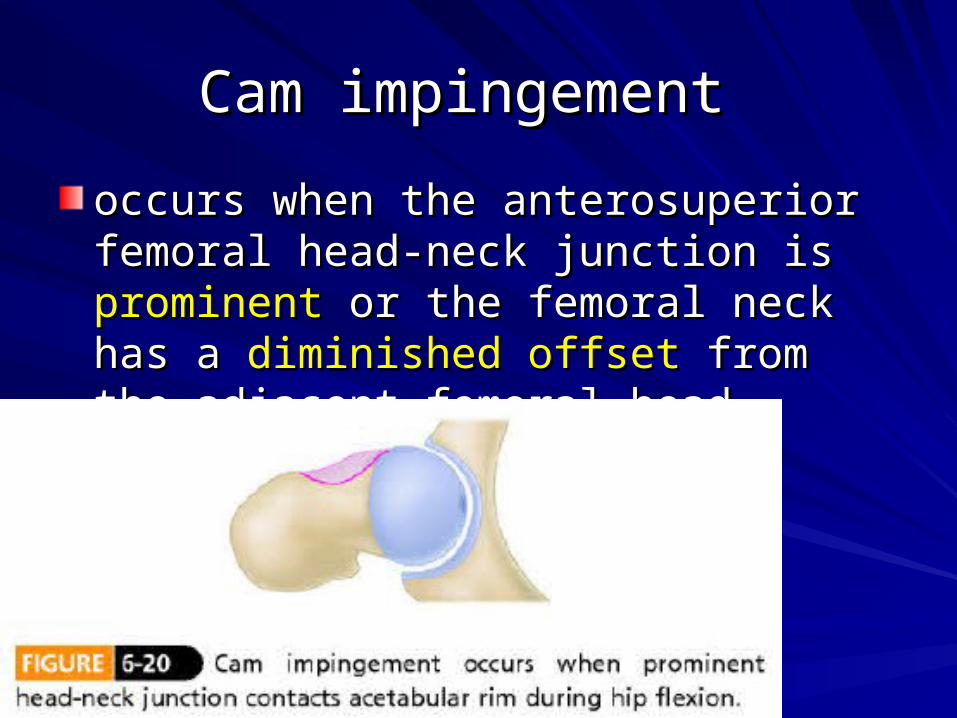
Cam impingement Cam impingement
occurs when the anterosuperior femoral occurs when the anterosuperior femoral head-neck junction is head-neck junction is prominentprominent or the or the femoral neck has a femoral neck has a diminished offset diminished offset from from the adjacent femoral headthe adjacent femoral head

A typical injury pattern A typical injury pattern a a tear at the tear at the base of the labrumbase of the labrum at the labral-chondral at the labral-chondral junction.junction.

The adjacent The adjacent articular cartilagearticular cartilage then then becomes injured because of becomes injured because of compressioncompression from the femoral head with its relatively from the femoral head with its relatively larger radius of curvature rotating into the larger radius of curvature rotating into the acetabulum. acetabulum. Frequently, the articular cartilage Frequently, the articular cartilage delaminatesdelaminates from the underlying from the underlying subchondral bone, progressing from the subchondral bone, progressing from the acetabular rimacetabular rim


Cam morphology is more common in Cam morphology is more common in young athletic malesyoung athletic males..
The The etiologyetiology of the deformity is of the deformity is unknownunknown, although some authors , although some authors may be a mild may be a mild variant of slipped capital variant of slipped capital femoral epiphysis femoral epiphysis OR OR developmental developmental abnormality of the lateral femoral abnormality of the lateral femoral physisphysis..

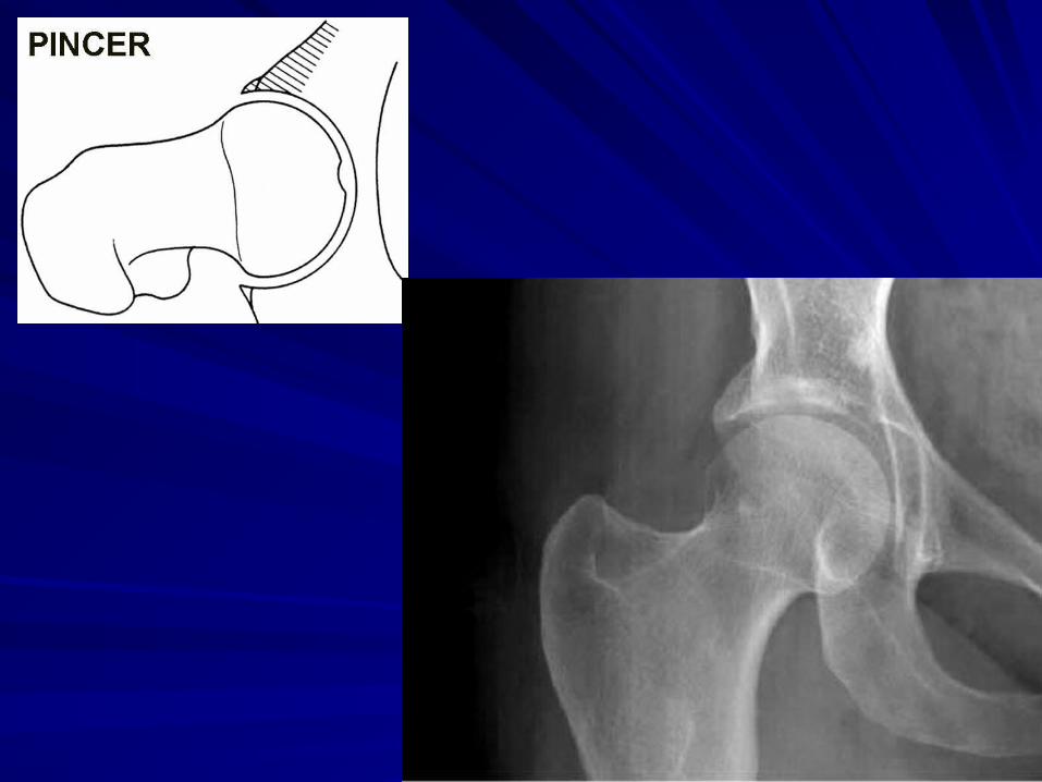
Pincer impingement Pincer impingement
occurs when the acetabular rim has an occurs when the acetabular rim has an area of area of overcoverageovercoverage causing causing impingement against the femoral neck with impingement against the femoral neck with functional motion functional motion

Overcoverage can be Overcoverage can be globalglobal, as in coxa , as in coxa profunda or protrusio acetabuli, or can be profunda or protrusio acetabuli, or can be localizedlocalized to the anterior acetabulum as to the anterior acetabulum as with acetabular retroversion.with acetabular retroversion.

Combined mechanism hip impingement Combined mechanism hip impingement occurs when cam and pincer morphology occurs when cam and pincer morphology coexist in the same hip.coexist in the same hip.

According to some authors, According to some authors, most hips most hips treatedtreated for femoroacetabular impingement for femoroacetabular impingement have have combinedcombined morphology. morphology.

Accurate diagnosis Accurate diagnosis of the source of pain in of the source of pain in young adults or adolescents is crucial in young adults or adolescents is crucial in obtaining optimal surgical outcomes with obtaining optimal surgical outcomes with FAI surgery. FAI surgery. The diagnosis of FAI is primarily made The diagnosis of FAI is primarily made clinicallyclinically from the patient's history and from the patient's history and physical examination and then correlated physical examination and then correlated with the with the radiographic findingsradiographic findings..
frog-leg lateral, frog-leg lateral, cross-table lateral, cross-table lateral, 45-degree modified Dunn view of the hip. 45-degree modified Dunn view of the hip. The modified Dunn view is obtained with The modified Dunn view is obtained with the the patient supinepatient supine with the with the hip in 45 hip in 45 degrees of flexiondegrees of flexion, , 20 degrees of 20 degrees of abductionabduction, and , and neutral rotationneutral rotation..anteroposterior pelvic viewanteroposterior pelvic view
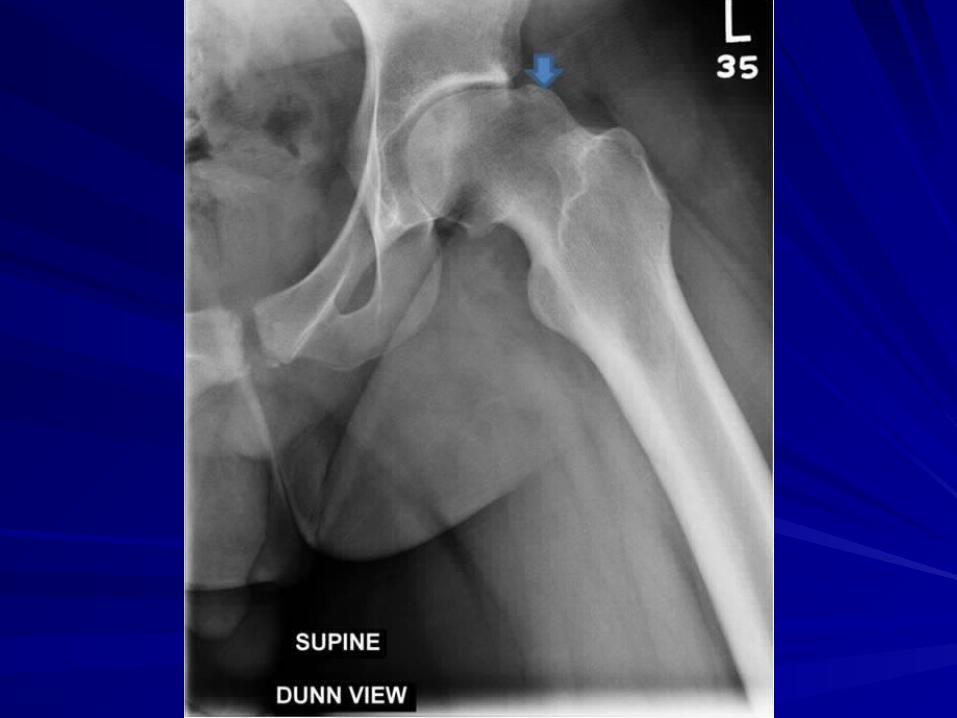
the the femoral head-neck junction femoral head-neck junction is is evaluated evaluated at different degrees of femoral at different degrees of femoral rotationrotation for the presence of for the presence of head-neck head-neck offset abnormalityoffset abnormality and and anterolateral anterolateral prominence of the femoral neckprominence of the femoral neck that can that can cause cam impingement.cause cam impingement.


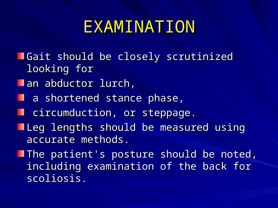
The Alpha AngleThe Alpha Angle
alpha angle is used to assess the femoral alpha angle is used to assess the femoral head-neck junction on the lateral and head-neck junction on the lateral and modified Dunn views. modified Dunn views.

Alfa angle Alfa angle 1.line drawn from the center 1.line drawn from the center of the femoral neck to the center of the of the femoral neck to the center of the femoral head 2.line drawn from the center femoral head 2.line drawn from the center of the femoral head of the femoral head to the point on the to the point on the anterior head-neck junction anterior head-neck junction where the where the contour of the contour of the femoral head diverges from femoral head diverges from the spherical contourthe spherical contour determined more determined more medially on the head.medially on the head.


Larger alpha angles do, however, appear Larger alpha angles do, however, appear to correlate with greater injury to the hip to correlate with greater injury to the hip observed at the time of surgery.observed at the time of surgery.correlated higher alpha angles with correlated higher alpha angles with increasing amounts of chondral injury at increasing amounts of chondral injury at the time of arthroscopic FAI surgery. the time of arthroscopic FAI surgery.
The alpha angle probably is related to its The alpha angle probably is related to its radial position on the femoral neck radial position on the femoral neck and the and the associated associated geometry of the adjacent geometry of the adjacent acetabulumacetabulum. . An alpha angle of more than 60 degrees An alpha angle of more than 60 degrees was a predictor of hip pain in another was a predictor of hip pain in another studystudy

Lateral center edge Lateral center edge (LCE) angle (LCE) angle < 20 degrees is < 20 degrees is indicative of hip indicative of hip dysplasiadysplasia with with inadequate coverage of inadequate coverage of the femoral head by the the femoral head by the lateral dome of the lateral dome of the acetabulum. acetabulum. 20 to 24 degrees have 20 to 24 degrees have borderline dysplasiaborderline dysplasia..>40 degrees display >40 degrees display overcoverageovercoverage..

The anterior head-The anterior head-neck offset ratio neck offset ratio the the cross-table lateral cross-table lateral view with the hip in 10 view with the hip in 10 degrees of internal degrees of internal rotation. rotation.

The The offset of the femoral head offset of the femoral head is determined by is determined by measuring the distance between two lines drawn measuring the distance between two lines drawn parallel to the axis of the femoral neck. The first line is parallel to the axis of the femoral neck. The first line is drawn through the most anterior portion of the femoral drawn through the most anterior portion of the femoral neck, and the second line is drawn through the neck, and the second line is drawn through the most most anterio portion of the femoral headanterio portion of the femoral head. The . The ratioratio is is determined by dividing this distance by the diameter of determined by dividing this distance by the diameter of the femoral head. the femoral head. A value of less than 0.15 has a 95% positive predictive A value of less than 0.15 has a 95% positive predictive value of diagnosing femor acetabular impingement.value of diagnosing femor acetabular impingement.

Special TestsFADIR impingement test: flexion, adduction, IR
Sensitivity=75%, specificity=43% in identifying patients with labral tears
FABER88% sensitive for intra-articular hip pathology
Resisted SLR – assesses labral loading Log Roll
Interrater reliability=0.63

Log Roll TestThe examiner passively
moves the patient’s lower extremity through the maximal available range of hip external (A) and internal rotation (B).
Eliciting a clicking or popping sensation may indicate an acetabular labral tear, while increased total range of motion when compared to the opposite side may indicate ligament or capsular laxity

TREATMENT OPTIONSTREATMENT OPTIONS
SURGICAL DISLOCATION OF THE HIPSURGICAL DISLOCATION OF THE HIPCOMBINED HIP ARTHROSCOPY AND COMBINED HIP ARTHROSCOPY AND LIMITED OPEN OSTEOCHONDROPLASTYLIMITED OPEN OSTEOCHONDROPLASTYPERIACETABULAR OSTEOTOMYPERIACETABULAR OSTEOTOMYHIP ARTHROSCOPYHIP ARTHROSCOPY


Activities CAUSING FAIActivities CAUSING FAIIce HockeyIce HockeyHorseback RidingHorseback RidingYogaYogaFootball (American)Football (American)SoccerSoccerBallet/Dance/AcrobaticsBallet/Dance/AcrobaticsGolfGolfTennisTennisBaseballBaseballField HockeyField HockeyRugbyRugbyBike Riding/CyclingBike Riding/CyclingMartial ArtsMartial ArtsDeep squatting activities such as power liftingDeep squatting activities such as power lifting

EtiologyEtiology Significant athletic activity before skeletal Significant athletic activity before skeletal maturity increases the risk of FAI.maturity increases the risk of FAI.
Prior Biomechanical theories suggest that Prior Biomechanical theories suggest that cartilage damage is initiated by Concentric or cartilage damage is initiated by Concentric or Eccentric overload.Eccentric overload.
- Eccentric overload - Eccentric overload - easily explained by - easily explained by
non-congruent articulations caused by non-congruent articulations caused by developmental dysplasias and post-traumatic developmental dysplasias and post-traumatic anatomy anatomy
- Concentric overload - Concentric overload - not as easy to explain - not as easy to explain
Ganz et al. CORR. 2003Ganz et al. CORR. 2003- - Summarized the concept of FAI Summarized the concept of FAI
- Mechanism for development of - Mechanism for development of osteoarthritis based on subtle aberrant bony morphology osteoarthritis based on subtle aberrant bony morphology
- - Acetabular Retroversion / Coxa Acetabular Retroversion / Coxa Profunda Profunda - - Femoral Head non-sphericityFemoral Head non-sphericity - - Abnormal ContactAbnormal Contact in Normal / Near in Normal / Near Normal Appearing Hips Normal Appearing Hips - - Abutment of the Proximal FemurAbutment of the Proximal Femur on the Acetabular Rim during terminal motion of the hip on the Acetabular Rim during terminal motion of the hip leading to lesions of the labrum and/or the adjacent leading to lesions of the labrum and/or the adjacent cartilage cartilage - - Chondral and Labral lesionsChondral and Labral lesions progress progress and result in degenerative disease and result in degenerative disease

FAI FormsFAI Forms
Generally occurs as two Cam and Pincer. Generally occurs as two Cam and Pincer. The The Cam form (Cam comes from the (Cam comes from the Dutch word meaning “cog”) describes the Dutch word meaning “cog”) describes the femoral head and neck relationship as femoral head and neck relationship as aspherical. aspherical. This loss of roundness contributes to This loss of roundness contributes to abnormal contact between the head and abnormal contact between the head and socket. socket.

CAM ImpingementCAM Impingement - - Anatomy Anatomy - Abnormal Femoral Head/Neck - Abnormal Femoral Head/Neck junction with increased radius at the waist junction with increased radius at the waist - Motion - Motion - Impingement occurs primarily during - Impingement occurs primarily during flexion, adduction, IR flexion, adduction, IR - - Mechanics Mechanics - Contact between the femoral neck - Contact between the femoral neck and acetabular rim induces compression and acetabular rim induces compression - Shear stress generated at the junction - Shear stress generated at the junction between the labrum and the cartilage and at the between the labrum and the cartilage and at the subchondral tidemark subchondral tidemark - Outward avulsion of the labrum - Outward avulsion of the labrum and/or an inward compression of the articular cartilage and/or an inward compression of the articular cartilage at Anterosuperior Rim at Anterosuperior Rim - -
Etiology of CAM ImpingementEtiology of CAM Impingement - Elliptical Femoral Head - Elliptical Femoral Head - Slipped Capital Femoral - Slipped Capital Femoral Epiphysis - Epiphysis - SCFESCFE - - Legg Calve PerthesLegg Calve Perthes - - Adult OsteonecrosisAdult Osteonecrosis - Malunited Femoral Neck - Malunited Femoral Neck Fractures Fractures

The Pincer FormThe Pincer FormPincer comes from the French word meaning “to Pincer comes from the French word meaning “to pinch” pinch” describes the situation where the socket or describes the situation where the socket or acetabulum has too much coverage of femoral acetabulum has too much coverage of femoral head. head. This over-coverage typically exists along the This over-coverage typically exists along the front-top rim of the acetabulum,front-top rim of the acetabulum,results in the labral cartilage being “pinched” results in the labral cartilage being “pinched” between the rim and the anterior femoral head-between the rim and the anterior femoral head-neck junction. neck junction.

Pincer form of the ImpingementPincer form of the Impingement
Secondary to “retroversion”, or “profunda”,Secondary to “retroversion”, or “profunda”, Most of the time, the Cam and Pincer Most of the time, the Cam and Pincer forms exist together. forms exist together.

Pincer ImpingementPincer Impingement - - AnatomyAnatomy
- Excessive Acetabular Coverage - Excessive Acetabular Coverage - Motion - Motion - Dependent on acetabular morphology - Dependent on acetabular morphology - - MechanicsMechanics - Linear Contact between the labrum and femoral - Linear Contact between the labrum and femoral head/neck junction head/neck junction - Anterior = Acetabular Retroversion - Anterior = Acetabular Retroversion
- Circumferential = Coxa Profunda - Circumferential = Coxa Profunda
- Force from the femoral neck is transferred through the - Force from the femoral neck is transferred through the labrum to the acetabular cartilage labrum to the acetabular cartilage Results is chronic degeneration of Results is chronic degeneration of anterior labrum and subsequent ossificationanterior labrum and subsequent ossification Further deepens the Further deepens the cup.cup.
Resultant leverage of head in acetabulum with excessive Resultant leverage of head in acetabulum with excessive ROM can result in contre-coup lesion in posteroinferior acetabulum and ROM can result in contre-coup lesion in posteroinferior acetabulum and posteromedial femoral head posteromedial femoral head
- Leading to Circumferential involvement - Leading to Circumferential involvement
Etiology Etiology - Acetabular Retroversion - Acetabular Retroversion - Coxa Profunda - Coxa Profunda - Protrusio Acetabuli - Protrusio Acetabuli - Iatrogenic overcorrection - Iatrogenic overcorrection for retroversion/dysplasia for retroversion/dysplasia - Coxa Vara - Coxa Vara - Os Acetabuli - Os Acetabuli

ASSOCIATED CARTILAGEASSOCIATED CARTILAGE
FAI is associated with cartilage damage, FAI is associated with cartilage damage, labral tears, labral tears, early hip arthritis, early hip arthritis, hyperlaxity, hyperlaxity, sports hernias, and sports hernias, and low back pain.low back pain.FAI is common in high level athletes, but FAI is common in high level athletes, but also occurs in active individualsalso occurs in active individuals

DiagnosisDiagnosis
Most patients can be diagnosed with a Most patients can be diagnosed with a good history,good history,A A patient’s historypatient’s history . . The The physical examphysical exam The The plain x-rayplain x-ray films films
EXAMINATIONEXAMINATION
Gait should be closely scrutinized looking for Gait should be closely scrutinized looking for an abductor lurch,an abductor lurch, a shortened stance phase,a shortened stance phase, circumduction, or steppage. circumduction, or steppage. Leg lengths should be measured using accurate Leg lengths should be measured using accurate methods. methods. The patient's posture should be noted, including The patient's posture should be noted, including examination of the back for scoliosis. examination of the back for scoliosis.

Anterior femoral-acetabular Anterior femoral-acetabular impingementimpingement
the affected hip is flexed to ninty degrees and the leg is the affected hip is flexed to ninty degrees and the leg is internally rotated and adducted. internally rotated and adducted. If there is abnormal contact between the anterior-If there is abnormal contact between the anterior-superior acetabular rim and femoral neck, pain may be superior acetabular rim and femoral neck, pain may be elicited. elicited. Posterior impingement may be tested by having the Posterior impingement may be tested by having the patient dangle their legs off the end of an examination patient dangle their legs off the end of an examination table, table, with the affected leg externally rotated by the examiner, with the affected leg externally rotated by the examiner, and the opposite limb held flexed by the patient. In a and the opposite limb held flexed by the patient. In a positive exam, the femur contacts the posterior positive exam, the femur contacts the posterior acetabular rim eliciting pain. acetabular rim eliciting pain.

EXAMINATIONEXAMINATIONThe range of motionThe range of motion A decreased range of motion of the hip, A decreased range of motion of the hip, especially in cases of an external rotation especially in cases of an external rotation contracture, can point to an intra-articular cause contracture, can point to an intra-articular cause of the pain. of the pain. Patients with hip pathology often also develop a Patients with hip pathology often also develop a flexion contracture. The Thomas test is a helpful flexion contracture. The Thomas test is a helpful maneuver. maneuver. A thorough neurovascular exam should be A thorough neurovascular exam should be conducted to rule out spine and other neural conducted to rule out spine and other neural causes of the patient's pain. causes of the patient's pain.

RADIOLOGYRADIOLOGY
The AP pelvis X-ray must be well centered The AP pelvis X-ray must be well centered and well developed as to show a clear and well developed as to show a clear outline of the acetabulum. outline of the acetabulum. The coccyx should point toward the The coccyx should point toward the symphysis pubis, and there should be symphysis pubis, and there should be about 1-2 cm between them. about 1-2 cm between them. The anterior and posterior walls, the tear The anterior and posterior walls, the tear drop and the lateral edge of the drop and the lateral edge of the acetabulum should be noted. acetabulum should be noted.

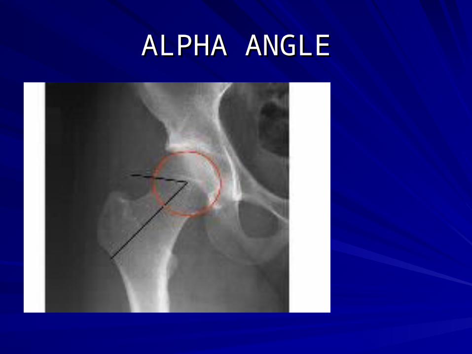
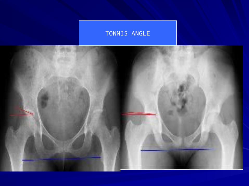
MeasurementsMeasurementsmay be taken to evaluate for hip dysplasia may be taken to evaluate for hip dysplasia including the including the Tönnis angleTönnis angle (abnormal < 10 (abnormal < 10 degrees),degrees), the lateral center-edge angle of Wibergthe lateral center-edge angle of Wiberg (abnormal < 25 degrees), and (abnormal < 25 degrees), and the the Anterior center-edge angle of LequesneAnterior center-edge angle of Lequesne (abnormal < 25 degrees) as measured on a (abnormal < 25 degrees) as measured on a false-profile radiograph. false-profile radiograph. The neck shaft angleThe neck shaft angle of the proximal femur is of the proximal femur is considered normal between 120 and 140 considered normal between 120 and 140 degrees.degrees.

Coxa ProfundaCoxa ProfundaWhen the floor of the acetabular fossa is in When the floor of the acetabular fossa is in line with the ilioischial line; line with the ilioischial line; Protrusio is present when the medial most Protrusio is present when the medial most femoral head overlaps the ilioischial line. femoral head overlaps the ilioischial line. The crossover signThe crossover sign is a sensitive and specific is a sensitive and specific indicator of native acetabular version. indicator of native acetabular version. On an On an AP pelvis radiographAP pelvis radiograph, the outlines of , the outlines of the edges of the anterior and posterior walls the edges of the anterior and posterior walls of the acetabulum should meet superiorly of the acetabulum should meet superiorly and laterally. and laterally.

COXA PROFUNDACOXA PROFUNDA
In cases of In cases of acetabular retroversionacetabular retroversion, this , this crossover of the anterior and posterior crossover of the anterior and posterior acetabular wall outlines is more distal. acetabular wall outlines is more distal. Changes in the acetabular rim may also Changes in the acetabular rim may also be noted. be noted. A A 'double line'double line' is seen in labral ' is seen in labral ossification. ossification. An An os acetabulios acetabuli may also be an indicator may also be an indicator of pathology of pathology

Alterations of the proximal femoral anatomy, such as Alterations of the proximal femoral anatomy, such as head neck offset and bump formation can be observed in head neck offset and bump formation can be observed in addition to acetabular and labral pathology.addition to acetabular and labral pathology. A pistol grip deformity of the femoral head is often seen A pistol grip deformity of the femoral head is often seen in Cam Type impingement. in Cam Type impingement. In this situation the superior-lateral head neck junction is In this situation the superior-lateral head neck junction is convex instead of concave. convex instead of concave. A high fovea can also indicate asphericity of the femoral A high fovea can also indicate asphericity of the femoral head that is not able to be appreciated on the AP films.head that is not able to be appreciated on the AP films. A cross table lateral may show an abnormality in the A cross table lateral may show an abnormality in the anterior head neck junction in cases of impingement. anterior head neck junction in cases of impingement.
ALPHA ANGLEALPHA ANGLE
TONNIS ANGLE

LCA
CROSSOVER
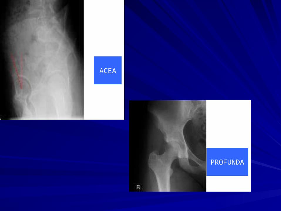
PROFUNDA
ACEA
MRIMRI
Often an MRI of the hip is used to confirm Often an MRI of the hip is used to confirm a labral tear or damage to the joint a labral tear or damage to the joint surface. surface. The MRI is most helpful in eliminating The MRI is most helpful in eliminating certain causes of non FAI hip pain certain causes of non FAI hip pain including avascular necrosis (dead bone) including avascular necrosis (dead bone) and tumors.and tumors.

DIFFRENTIAL DIAGNOSISDIFFRENTIAL DIAGNOSISHip Dysplasia (Adult Form)Hip Dysplasia (Adult Form)Lumbar Spine Pain (Low Back Pain)Lumbar Spine Pain (Low Back Pain)Lumbar Radiculopathy Low Back Facet Disease)Lumbar Radiculopathy Low Back Facet Disease)Sacroiliitis Sacroiliitis Trochanteric BursitisTrochanteric BursitisPiriformis SyndromePiriformis SyndromePsychosomatic Pain DisorderPsychosomatic Pain DisorderIliopsoas Tendinitis/Tendonitis/TendinosisIliopsoas Tendinitis/Tendonitis/TendinosisGroin Pull Groin Pull Sports Hernia (abdominal muscle strain)Sports Hernia (abdominal muscle strain)Iliac ApophysitisIliac ApophysitisQuadriceps Hernia/Strain Chronic Pain Syndromes Quadriceps Hernia/Strain Chronic Pain Syndromes

TreatmentTreatmentNon-operativeNon-operative
A course of non-operative treatment for A course of non-operative treatment for most hip pathology may be tried first. most hip pathology may be tried first. Patients presenting with femoroacetabular Patients presenting with femoroacetabular impingement or labral disease may try impingement or labral disease may try modification of activity modification of activity avoiding excessive hip movement avoiding excessive hip movement regular non-steroidal anti-inflammatory regular non-steroidal anti-inflammatory medicationmedication

ArthroscopyArthroscopy
Arthroscopic assessment of the hip can include Arthroscopic assessment of the hip can include examination of both the central and peripheral examination of both the central and peripheral compartments. compartments. The central compartment includes the labrum and all The central compartment includes the labrum and all structures located further medially. structures located further medially. Tearing of the labrum anterolaterally and damage to the Tearing of the labrum anterolaterally and damage to the acetabular cartilage is characteristic. acetabular cartilage is characteristic. The lesions of the labrum and any areas of chondral The lesions of the labrum and any areas of chondral damage are debrided. damage are debrided. Labral repair may be possible for specific tears. Labral repair may be possible for specific tears. For areas of exposed subchondral bone a microfracture For areas of exposed subchondral bone a microfracture technique may be performed.technique may be performed.
SAFE Surgical Hip Dislocation- SAFE Surgical Hip Dislocation- OsteoplastyOsteoplasty
Surgical dislocation of the hip has been described. Surgical dislocation of the hip has been described. A posterior incision (Kocher-Langenbeck) as this approach A posterior incision (Kocher-Langenbeck) as this approach usually provides better access to posterior parts of the joint usually provides better access to posterior parts of the joint after the hip is dislocated. after the hip is dislocated. A trochanteric flip osteotomy is performed. The trochanter is A trochanteric flip osteotomy is performed. The trochanter is osteotomized from a posterior to anterior direction, the cut osteotomized from a posterior to anterior direction, the cut exits superficial to the piriformis fossa superiorly and at the exits superficial to the piriformis fossa superiorly and at the vastus ridge inferiorly. vastus ridge inferiorly. The gluteus minimus muscle is dissected carefully off the The gluteus minimus muscle is dissected carefully off the capsule starting at the piriformis interval. capsule starting at the piriformis interval. The capsulotomy is Z-shaped (for the right hip), with the The capsulotomy is Z-shaped (for the right hip), with the superior limb located along the posterior acetabular rim, and superior limb located along the posterior acetabular rim, and the inferior limb located at the level of the anteromedial the inferior limb located at the level of the anteromedial femoral neckfemoral neck
After dislocation of the hip, the acetabular labrum and After dislocation of the hip, the acetabular labrum and the adjacent articular cartilage are assessed, and the the adjacent articular cartilage are assessed, and the identified lesions are tested for partial or complete identified lesions are tested for partial or complete avulsions from the acetabular rim. avulsions from the acetabular rim. The severity, extent, and location of these lesions should The severity, extent, and location of these lesions should be defined, and their association with FAI should be be defined, and their association with FAI should be confirmed by provocative maneuvers in flexion and confirmed by provocative maneuvers in flexion and internal rotation with the head relocated. internal rotation with the head relocated. The combination of anterior over coverage and the The combination of anterior over coverage and the status of the labrum and the acetabular articular cartilage status of the labrum and the acetabular articular cartilage will determine the type of treatment of the acetabular rim.will determine the type of treatment of the acetabular rim. In cases of anterior over coverage contributing to FAI, In cases of anterior over coverage contributing to FAI, as is frequent with acetabular retroversion, a resection as is frequent with acetabular retroversion, a resection osteoplasty of the anterosuperior rim is done. osteoplasty of the anterosuperior rim is done.

Periacetabular OsteotomyPeriacetabular Osteotomy
Reorientation of the articulating surfaces of the Reorientation of the articulating surfaces of the hip joint is an attractive procedure in the patient hip joint is an attractive procedure in the patient with hip dysplasia.with hip dysplasia.Increased joint congruity after reorientation of Increased joint congruity after reorientation of the osteotomized fragment allows load the osteotomized fragment allows load transmission through a broader area subjected transmission through a broader area subjected to less pressure. to less pressure. These changes can be expected to reduce pain These changes can be expected to reduce pain and possibly protect the articular cartilage from and possibly protect the articular cartilage from degenerative changesdegenerative changes

Pelvic OsteotomyPelvic Osteotomy
Corrects the major anatomic abnormality Corrects the major anatomic abnormality and has the further advantage over and has the further advantage over femoral osteotomy of not creating a femoral osteotomy of not creating a secondary femoral deformity. secondary femoral deformity. Femoral osteotomy may be added to Femoral osteotomy may be added to pelvic osteotomy when coexistent femoral pelvic osteotomy when coexistent femoral anatomic abnormalities are significant. anatomic abnormalities are significant.
Bernese Periacetabular OsteotomyBernese Periacetabular Osteotomy
The is indicated for patients with hip The is indicated for patients with hip symptoms of mechanical overload, symptoms of mechanical overload, impingement, or hip instability as a result impingement, or hip instability as a result of insufficient acetabular coverage. of insufficient acetabular coverage.