fetal death in mice lacking 5{alpha}-reductase type 1 ... death... · pairs of wild type or...
TRANSCRIPT

Mol. Endocrinol. 1997 11: 917-927, doi: 10.1210/me.11.7.917
Mala S. Mahendroo, Kristine M. Cala, Charles P. Landrum and David W. Russell
Fetal Death in Mice Lacking 5{alpha}-Reductase Type 1 Caused by Estrogen Excess
Society please go to: http://mend.endojournals.org//subscriptions/ or any of the other journals published by The EndocrineMolecular EndocrinologyTo subscribe to
Copyright © The Endocrine Society. All rights reserved. Print ISSN: 0021-972X. Online

Fetal Death in Mice Lacking 5a-Reductase Type 1 Caused byEstrogen Excess
Mala S. Mahendroo, Kristine M. Cala, Charles P. Landrum, andDavid W. Russell
Department of Molecular GeneticsUniversity of Texas Southwestern Medical CenterDallas, Texas 75235-9046
Female mice deficient in steroid 5a-reductase type1 have a decreased litter size. The average litter inhomozygous deficient females is 2.7 pups vs. 8.0pups in wild type controls. Oogenesis, fertilization,implantation, and placental morphology appearnormal in the mutant animals. Fetal loss occursbetween gestation days 10.75 and 11.0 commen-surate with a midpregnancy surge in placental an-drogen production and an induction of 5a-reduc-tase type 1 expression in the decidua of wild typemice. Plasma levels of androstenedione and tes-tosterone are 2- to 3-fold higher on gestation day 9,and estradiol levels are chronically elevated by 2-to 3-fold throughout early and midgestation in theknockout mice. Administration of an estrogen re-ceptor antagonist or inhibitors of aromatase re-verse the high rate of fetal death in the mutantmice, and estradiol treatment of wild type pregnantmice causes fetal wastage. The results suggestthat in the deficient mice, a failure to 5a-reduceandrogens leads to their conversion to estrogens,which in turn causes fetal death in midgestation.These findings indicate that the 5a-reduction ofandrogens in female animals plays a crucial role inguarding against estrogen toxicity during preg-nancy. (Molecular Endocrinology 11: 917–927,1997)
INTRODUCTION
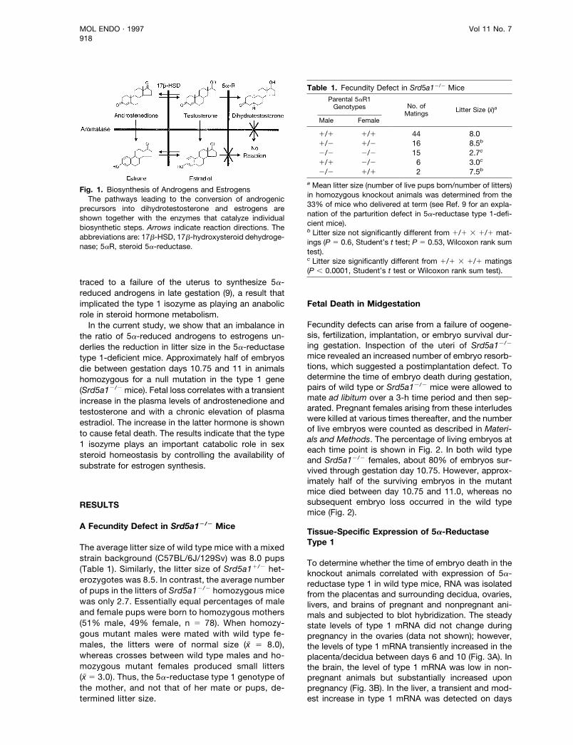
The androgens androstenedione and testosterone aresubstrates for two metabolic pathways that producepotent and antagonistic sex steroids (Fig. 1). In onepathway, they are converted into 5a-reduced andro-gens by steroid 5a-reductase isozymes (1). In theother, they are converted into estrogens by the aro-matase enzyme, a cytochrome P450 of the endoplas-mic reticulum (2). Conversion to these end products isirreversible and mutually exclusive in that 5a-reducedandrogens are not aromatase substrates and estro-
gens are not 5a-reductase substrates (Fig. 1). Theactions of 5a-reduced androgens and estrogens op-pose one another: 5a-reduced androgens masculinizewhile estrogens feminize. Under physiological condi-tions in each sex, a delicate balance is established inwhich appropriate amounts of androstenedione andtestosterone are converted into one or the other classof hormones. The set point of this fulcrum is differen-tially sustained in the two sexes by regulating theproduction of testicular androgens in males and theexpression of the aromatase enzyme in females.
Occasional individuals have an imbalance in the5a-reduced androgen-estrogen ratio due to geneticdefects in the enzymes that determine these metabolicfates (Fig. 1). For example, mutations in the aromatasegene cause an increase in the androgen-estrogen ratioleading to virilization and polycystic ovarian disease inaffected women (3, 4). Mutations in the 17b-hydrox-ysteroid dehydrogenase type 3 gene decrease theandrogen-estrogen ratio and cause gynecomastia inaffected men (5). Two different 5a-reductase genesencode the type 1 and type 2 isozymes in severalmammalian species (2). Mutations in the human 5a-reductase type 2 gene, which is normally expressed inthe urogenital tract and liver, cause male pseudoher-maphroditism but do not alter the 5a-reduced andro-gen-estrogen ratio in men or women (6, 7). This out-come is presumably due to the ability of the type 1isozyme, which is expressed in the liver and skin (8), tocompensate for the absence of the type 2 isozyme inaffected individuals.
No mutations in the 5a-reductase type 1 gene haveyet been identified in humans, perhaps because of thepresence of the type 2 isozyme in multiple tissues.However, the female mouse is an ideal animal model inwhich to study the type 1 isozyme because very little5a-reductase type 2 is expressed in the tissues of thismammal (9). To take advantage of this expressionpattern, and to explore the physiological role of thetype 1 isozyme, a line of mice with a mutation in the5a-reductase type 1 gene was developed (9). Theabsence of this isozyme had no obvious effect inmales but caused a parturition defect and a reductionin litter size in females. The parturition defect was
0888-8809/97/$3.00/0Molecular EndocrinologyCopyright © 1997 by The Endocrine Society
917
traced to a failure of the uterus to synthesize 5a-reduced androgens in late gestation (9), a result thatimplicated the type 1 isozyme as playing an anabolicrole in steroid hormone metabolism.
In the current study, we show that an imbalance inthe ratio of 5a-reduced androgens to estrogens un-derlies the reduction in litter size in the 5a-reductasetype 1-deficient mice. Approximately half of embryosdie between gestation days 10.75 and 11 in animalshomozygous for a null mutation in the type 1 gene(Srd5a12/2 mice). Fetal loss correlates with a transientincrease in the plasma levels of androstenedione andtestosterone and with a chronic elevation of plasmaestradiol. The increase in the latter hormone is shownto cause fetal death. The results indicate that the type1 isozyme plays an important catabolic role in sexsteroid homeostasis by controlling the availability ofsubstrate for estrogen synthesis.
RESULTS
A Fecundity Defect in Srd5a12/2 Mice
The average litter size of wild type mice with a mixedstrain background (C57BL/6J/129Sv) was 8.0 pups(Table 1). Similarly, the litter size of Srd5a11/2 het-erozygotes was 8.5. In contrast, the average numberof pups in the litters of Srd5a12/2 homozygous micewas only 2.7. Essentially equal percentages of maleand female pups were born to homozygous mothers(51% male, 49% female, n 5 78). When homozy-gous mutant males were mated with wild type fe-males, the litters were of normal size (x̄ 5 8.0),whereas crosses between wild type males and ho-mozygous mutant females produced small litters(x̄ 5 3.0). Thus, the 5a-reductase type 1 genotype ofthe mother, and not that of her mate or pups, de-termined litter size.
Fetal Death in Midgestation
Fecundity defects can arise from a failure of oogene-sis, fertilization, implantation, or embryo survival dur-ing gestation. Inspection of the uteri of Srd5a12/2
mice revealed an increased number of embryo resorb-tions, which suggested a postimplantation defect. Todetermine the time of embryo death during gestation,pairs of wild type or Srd5a12/2 mice were allowed tomate ad libitum over a 3-h time period and then sep-arated. Pregnant females arising from these interludeswere killed at various times thereafter, and the numberof live embryos were counted as described in Materi-als and Methods. The percentage of living embryos ateach time point is shown in Fig. 2. In both wild typeand Srd5a12/2 females, about 80% of embryos sur-vived through gestation day 10.75. However, approx-imately half of the surviving embryos in the mutantmice died between day 10.75 and 11.0, whereas nosubsequent embryo loss occurred in the wild typemice (Fig. 2).
Tissue-Specific Expression of 5a-ReductaseType 1
To determine whether the time of embryo death in theknockout animals correlated with expression of 5a-reductase type 1 in wild type mice, RNA was isolatedfrom the placentas and surrounding decidua, ovaries,livers, and brains of pregnant and nonpregnant ani-mals and subjected to blot hybridization. The steadystate levels of type 1 mRNA did not change duringpregnancy in the ovaries (data not shown); however,the levels of type 1 mRNA transiently increased in theplacenta/decidua between days 6 and 10 (Fig. 3A). Inthe brain, the level of type 1 mRNA was low in non-pregnant animals but substantially increased uponpregnancy (Fig. 3B). In the liver, a transient and mod-est increase in type 1 mRNA was detected on days
Fig. 1. Biosynthesis of Androgens and EstrogensThe pathways leading to the conversion of androgenic
precursors into dihydrotestosterone and estrogens areshown together with the enzymes that catalyze individualbiosynthetic steps. Arrows indicate reaction directions. Theabbreviations are: 17b-HSD, 17b-hydroxysteroid dehydroge-nase; 5aR, steroid 5a-reductase.
Table 1. Fecundity Defect in Srd5a12/2 Mice
Parental 5aR1Genotypes No. of
Matings Litter Size (x̄)a
Male Female
1/1 1/1 44 8.01/2 1/2 16 8.5b
2/2 2/2 15 2.7c
1/1 2/2 6 3.0c
2/2 1/1 2 7.5b
a Mean litter size (number of live pups born/number of litters)in homozygous knockout animals was determined from the33% of mice who delivered at term (see Ref. 9 for an expla-nation of the parturition defect in 5a-reductase type 1-defi-cient mice).b Litter size not significantly different from 1/1 3 1/1 mat-ings (P 5 0.6, Student’s t test; P 5 0.53, Wilcoxon rank sumtest).c Litter size significantly different from 1/1 3 1/1 matings(P , 0.0001, Student’s t test or Wilcoxon rank sum test).
MOL ENDO · 1997 Vol 11 No. 7918
9–14 of gestation (Fig. 3C). The levels of controlmRNAs (b-actin in the placenta/decidua, CRH in thebrain, and cyclophilin in the liver) remained constantduring these times (Fig. 3).
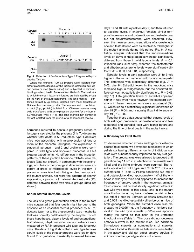
To confirm that the observed induction of 5a-reduc-tase type 1 mRNA in the placenta/decidua led to anincrease in enzyme mass, immunoblotting assayswere performed (Fig. 4). A protein of approximately 22kDa that comigrated with a recombinant 5a-reductasetype 1 standard was induced in the tissue on gestationdays 6 through 11 coincident with the observed in-crease in mRNA. The induction of 5a-reductase type 1protein in the uterus was monitored as a control. Inagreement with previous results (9), the content ofuterine 5a-reductase type 1 increased in late but notmidgestation (Fig. 4).
5a-Reductase Type 1 Expression in the Decidua
In situ mRNA hybridization was used to determinewhich cell types in the placenta/decidua express 5a-reductase type 1 on gestation day 8. An antisenseprobe revealed type 1 transcripts in decidual cells ofthe tissue (Fig. 5, A and B), whereas a sense probeproduced no specific hybridization pattern (Fig. 5, Cand D). Cells containing the type 1 mRNA were con-centrated in the decidua basalis, which is the region ofthe decidua opposed to the mesometrial aspect of theuterus. No specific hybridization was detected in thedecidua capsularis, which is located on the antime-sometrial side of the uterus, or in any of the extraem-bryonic membranes of the fetal-placental unit (Fig. 5, Aand B). Cells of the decidua basalis differentiate fromendometrial connective tissue cells of the uterus (10).Thus, the finding that maternal decidual cells express
5a-reductase type 1 is consistent with results from thebreeding experiments (Table 1), which showed that thefecundity defect was maternal in origin.
Expression of Placental Markers
The time of embryo death in gravid Srd5a12/2 femalescorrelates with a switch in the source of lactogenichormones required for pregnancy maintenance. Inearly gestation, PRL secretion from the anterior pitu-itary subserves both lactogenic and luteotrophic func-tion in the establishment and maintenance of preg-nancy (11). During midgestation, the sources and
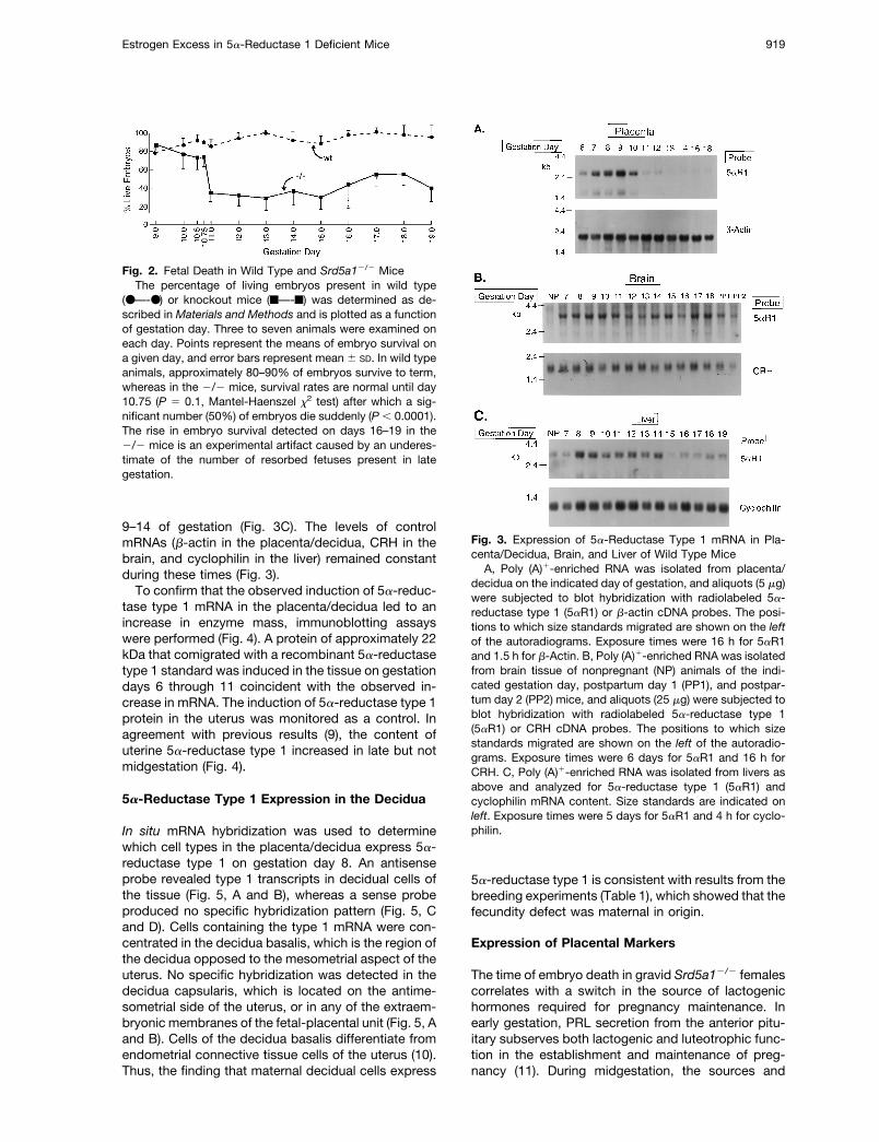
Fig. 2. Fetal Death in Wild Type and Srd5a12/2 MiceThe percentage of living embryos present in wild type
(●—-●) or knockout mice (f—-f) was determined as de-scribed in Materials and Methods and is plotted as a functionof gestation day. Three to seven animals were examined oneach day. Points represent the means of embryo survival ona given day, and error bars represent mean 6 SD. In wild typeanimals, approximately 80–90% of embryos survive to term,whereas in the 2/2 mice, survival rates are normal until day10.75 (P 5 0.1, Mantel-Haenszel x2 test) after which a sig-nificant number (50%) of embryos die suddenly (P , 0.0001).The rise in embryo survival detected on days 16–19 in the2/2 mice is an experimental artifact caused by an underes-timate of the number of resorbed fetuses present in lategestation.
Fig. 3. Expression of 5a-Reductase Type 1 mRNA in Pla-centa/Decidua, Brain, and Liver of Wild Type Mice
A, Poly (A)1-enriched RNA was isolated from placenta/decidua on the indicated day of gestation, and aliquots (5 mg)were subjected to blot hybridization with radiolabeled 5a-reductase type 1 (5aR1) or b-actin cDNA probes. The posi-tions to which size standards migrated are shown on the leftof the autoradiograms. Exposure times were 16 h for 5aR1and 1.5 h for b-Actin. B, Poly (A)1-enriched RNA was isolatedfrom brain tissue of nonpregnant (NP) animals of the indi-cated gestation day, postpartum day 1 (PP1), and postpar-tum day 2 (PP2) mice, and aliquots (25 mg) were subjected toblot hybridization with radiolabeled 5a-reductase type 1(5aR1) or CRH cDNA probes. The positions to which sizestandards migrated are shown on the left of the autoradio-grams. Exposure times were 6 days for 5aR1 and 16 h forCRH. C, Poly (A)1-enriched RNA was isolated from livers asabove and analyzed for 5a-reductase type 1 (5aR1) andcyclophilin mRNA content. Size standards are indicated onleft. Exposure times were 5 days for 5aR1 and 4 h for cyclo-philin.
Estrogen Excess in 5a-Reductase 1 Deficient Mice 919
hormones required to continue pregnancy switch tolactogens secreted by the placenta (11). To determinewhether fetal death in 5a-reductase type 1-deficientmice was associated with misexpression of one ormore of the placental lactogens, the expression ofplacental lactogen 1 and 2 and proliferin were com-pared in wild type and knockout placentas in RNAblotting experiments. No differences in the inductionpatterns of these peptide hormone mRNAs were de-tected (data not shown). In agreement with these find-ings, no obvious morphological differences were ap-parent at gross or microscopic levels in the day 11placentas associated with living or dead embryos inthe mutant animals, nor were the patterns of desminexpression, a product of maternal decidual cells (12),different between these two tissue groups (data notshown).
Serum Steroid Hormone Levels
The lack of a gross placentation defect in the mutantmice suggested that fetal death might be due to theabsence of an essential steroid produced by 5a-re-ductase type 1 or to the presence of a toxic metabolitethat was normally catabolized by the enzyme. To testthese hypotheses, plasma levels of androstenedione,testosterone, dihydrotestosterone, and estradiol weremeasured by RIA in pregnant wild type and knockoutmice. The data of Fig. 6 show that in wild type femalesserum levels of the three androgens were low on days6 and 7 of gestation, transiently increased between
days 8 and 10, with a peak on day 9, and then returnedto baseline levels. In knockout females, similar tem-poral increases in androstenedione and testosterone,but not dihydrotestosterone, were observed. How-ever, the mean serum concentrations of androstenedi-one and testosterone were as much as 5-fold higher inthe mutant animals during this period (Fig. 6). A sta-tistical analysis indicated that the androstenedionelevels on day 9 in knockout mice were not significantlydifferent from those in wild type animals (P 5 0.1,Wilcoxon rank sum test), whereas the testosteroneand dihydrotestosterone levels were significantly dif-ferent (P 5 0.03 and 0.01, respectively).
Estradiol levels in early gestation were 2- to 3-foldhigher in the mutant mice vs. wild type counterparts.This difference was statistically different (e.g. P 50.02, day 8). Estradiol levels in the knockout miceremained high in midgestation, but the observed dif-ference was not statistically significant (e.g. P 5 0.05,day 13). Late gestation levels of estradiol appearedhigher in wild type mice; however, the standard devi-ations in these measurements were substantial (Fig.6), which led to a statistically significant difference onday 18 (P 5 0.04) and a nonsignificant difference onday 19 (P 5 0.8).
Together these data suggested that plasma levels ofboth estrogen precursors (androstenedione and tes-tosterone) and estradiol itself were higher before andduring the time of fetal death in the mutant mice.
A Bioassay for Fetal Death
To determine whether excess androgens or estradiolcaused fetal death, we developed a bioassay in whichpellets containing different amounts of individual ste-roids were subcutaneously implanted on day 5 of ges-tation. The pregnancies were allowed to proceed untilgestation day 11 or 12, at which time the animals werekilled and the living embryos were counted. The re-sults obtained in wild type and Srd5a12/2 mice aresummarized in Table 2. Pellets containing 0.5 mg ofandrostenedione killed approximately half of the em-bryos in wild type mice and appeared to increase thefrequency of embryo death in the knockout animals.Testosterone had no statistically significant effects intwo wild type mice in this assay, and in the mutantmice this hormone may have conferred a mild protec-tive effect (Table 2). Estradiol at doses between 0.5and 0.005 mg killed essentially all embryos in mice ofboth genotypes. When the estradiol dose was de-creased to 0.0025 mg, the frequency of embryo sur-vival in the wild type animals (;50%) was approxi-mately the same as that seen in the untreatedknockout mice (Table 1). This dose did not decreaseembryo survival in the mutant mice. Ten additional5a-reduced steroids or combinations of steroids,which are listed in Materials and Methods, were testedin the assay and did not affect embryo survival inanimals of either genotype (data not shown).
Fig. 4. Detection of 5a-Reductase Type 1 Enzyme in Repro-ductive Tissues
Whole cell extracts (100 mg protein) were isolated fromeither placentae/decidua of the indicated gestation day (up-per panel) or uteri (lower panel) and subjected to immuno-blotting as described in Materials and Methods. The positionsto which the type 1 isozyme migrated are indicated by arrowson the right of the autoradiograms. The lane marked 2 con-tained extract (5 mg protein) isolated from mock-transfectedChinese hamster ovary cells. The lane marked 1 containedextract (5 mg protein) isolated from Chinese hamster ovarycells transfected with an expression plasmid for the human5a-reductase type 1 (41). The lane marked NP containedextract isolated from the uterus of a nonpregnant mouse.
MOL ENDO · 1997 Vol 11 No. 7920

In a preliminary attempt to determine the cause ofembryo death in estrogen-treated mothers, uteri fromcontrol and experimental animals were examined at agross morphological level. These experiments re-vealed that estrogen treatment caused hemorrhagingin the uteri of gravid wild type and knockout females(Fig. 7). Bleeding did not occur in the abdominalspace, but rather was limited to the uterine lumen andthe encircled fetal-placental units.
Estrogen Antagonists Increase Embryo SurvivalIn Srd5a12/2 Mice
The results from the steroid pellet studies suggestedthat excess androstenedione or estradiol caused fetaldeath in both wild type and knockout mice. Sinceandrostenedione is readily converted to estrone by thearomatase enzyme and thereafter to estradiol (Fig. 1),estrogen was thought to be the more likely hormoneresponsible for fetal death. If this interpretation is cor-rect, then compounds that inhibit aromatase or blockestrogen binding to the estrogen receptor should re-store normal embryo survival to Srd5a12/2 mice. The
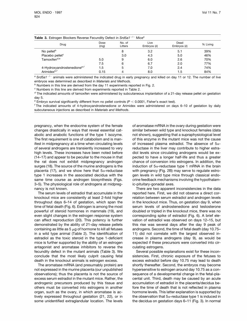
data of Table 3 show that injection of aromatase in-hibitors (4-hydroxyandrostenedione or Arimidex) ongestation days 6 through 10 prevented excess fetaldeath in the mutant mice. Approximately 79% of theknockout embryos survived in these experiments,which was a frequency identical to that observed inuntreated wild type mice (Fig. 2). Similar results wereobtained when pellets containing either 5.0 or 7.5 mgof the estrogen receptor antagonist tamoxifen wereimplanted on day 5 of gestation: administration of thisdrug led to more than 70% embryo survival frequen-cies (Table 3).
DISCUSSION
The absence of 5a-reductase type 1 in mice causes afecundity defect of maternal origin. The pregnancies ofthese animals are normal until day 10.75, but betweengestation day 10.75 and 11, approximately half of theembryos die. In wild type mice, expression of 5a-reductase type 1 increases to high levels in the de-cidua between gestation days 6 and 11. This increase
Fig. 5. Detection of 5a-Reductase Type 1 mRNA in Decidual CellsPlacenta/decidua tissue from gestation day 8 wild type mice was isolated and subjected to mRNA in situ hybridization analyses
using an antisense strand probe to detect 5a-reductase type 1 transcripts (A and B) or a sense strand probe as a negative control(C and D). Exposure times were 21 days. Hybridized sections were stained with hematoxylin and eosin and photographed usinglightfield and darkfield optics on a Leitz microscope. The fold-magnification was 93. A, Lightfield photograph, antisense probe.B, Darkfield photograph of panel A, antisense probe. C, Lightfield photograph, sense probe. D, Darkfield photograph of panel C,sense probe. Labels are: a, allantois; db, decidua basalis; dc, decidua capsularis; e, embryo; ys, yolk sac. Red blood cells markingmaternal and fetal blood vessels exhibit a yellow-green birefringence in the darkfield exposures of panels B and D.
Estrogen Excess in 5a-Reductase 1 Deficient Mice 921

correlates with a burst of androgen synthesis by theplacenta that leads to elevated plasma levels ofandrostenedione, testosterone, and dihydrotestos-terone. In the knockout mice, serum levels of andro-stenedione and testosterone increase during thissame time period, whereas plasma dihydrotestos-terone levels remain unchanged. The serum estra-diol levels are elevated throughout early and midg-estation in the Srd5a12/2 mice. Antagonists ofestrogen synthesis and action correct the fecunditydefect in knockout animals, a finding consistent withthe concept that fetal death in midpregnancy iscaused by estradiol toxicity. This scenario is furthersupported by the observation that exogenous estra-diol causes fetal death in the offspring of wild typemothers. Given the known pathways of androgenand estrogen biosynthesis (Fig. 1), the simplest in-terpretation of these results is that 5a-reductasetype 1 normally plays an important catabolic role in
pregnant females by converting androgens to theirnonaromatizable forms and that prevention of thisaction causes estradiol levels to increase and causefetal demise.
Steroid 5a-reductase occurs in two isozymic formscalled type 1 and type 2 that were initially postulated tofulfill distinct physiological roles. The type 1 isozymewas predicted to play a catabolic role in steroid hor-mone metabolism based on a high level of expressionin tissues that break down steroids such as the liverand kidney and a micromolar affinity for steroid sub-strates (13). Conversely, a preferential expression ofthe type 2 isozyme in androgen target tissues of themale reproductive tract, coupled with a nanomolaraffinity for steroid substrates, implied an anabolic rolefor the type 2 isozyme. Men that lack the type 2isozyme exhibit symptoms of androgen deficiency,which confirms the anabolic role of this isozyme inhumans (6).
Fig. 6. Steroid Hormone Levels in Wild Type and Srd5a12/2 MiceThe plasma levels of the indicated steroid hormones were measured in a single RIA assay in wild type (wt) and 5a-reductase
type 1 knockout mice (2/2) on the indicated days of gestation. Days 19 and 20 in wild type mice refers to values measured inpostpartum animals. Day 19 and 20 values in the mutant mice were measured in animals exhibiting delayed parturition (9).Hormone levels were measured in three to seven animals for each day except for day 20, wild type, for which only a single animalwas used. Points indicate mean hormone concentrations on a given day. Error bars represent mean 6 SD.
MOL ENDO · 1997 Vol 11 No. 7922

The physiological role of the type 1 isozyme hasbeen assessed in mice containing a null allele at theSrd5a1 locus. The analysis of these animals revealsthat the function of the type 1 isozyme is more com-plex than originally proposed in that it plays both an-abolic and catabolic roles in pregnant female mice. Inan anabolic capacity, the type 1 isozyme synthesizesa 5a-reduced androgen, most likely 5a-androstane-3a,17b-diol, which is required for the delivery of young
at term (9). In a catabolic capacity, the current studiesshow that the type 1 isozyme breaks down androgensand, in so doing, prevents their conversion toestrogens.
Male and female mice do not appear to require5a-reductase type 1 for phenotypic sexual differenti-ation or for the maintenance of steroid hormone ho-meostasis under normal conditions (9). The adverseconsequences of enzyme loss are not realized until
Table 2. Effect of Steroid Hormones on Embryo Survival
Steroida Dose (mg)bWild Type Females Srd5a12/2 Females
No. ofLitters
LiveEmbryos (x̄)
DeadEmbryos (x̄)
No. ofLitters
LiveEmbryos (x̄)
DeadEmbryos (x̄)
No pelletc 4 8.0 0.4 8 3.2 5.1Placebo pelletd 3 9.6 0.67 3 4.3 5.0Androstenedionee 0.5 4 3.3 4.5 5 1.0 7.8Testosteronef 0.5 2 8.8 1.5 5 6.2 3.2Estradiolg 0.5 2 0 7.0 5 0 8.4
0.08 5 0 7.4 6 0 7.80.02 2 0 5.0 6 0.3 6.30.01 2 0 11.0 6 0 6.80.005 2 0 8.5 5 0 5.00.0025 2 4.5 4.0 4 2.8 5.5
a A single pellet containing the indicated amount of steroid was subcutaneously implanted in wild type or knockout females onday 5 of gestation. Animals were killed on day 11 or 12 of gestation, and the number of live and dead embryos was determinedas described in Materials and Methods.b Total amount of steroid per pellet. All pellets were 21-day release.c Numbers in this line are derived from the day 11 experiments reported in Fig. 2.d Animals received placebo pellets containing vehicle alone.e For wild type animals, embryo survival significantly different from no pellet controls (P 5 0.009, Fisher’s exact test). For knockoutanimals, embryo survival significantly different from no pellet controls (P 5 0.018).f For wild type animals, embryo survival not significantly different from no pellet controls (P 5 1.0). For knockout animals, embryosurvival significantly different from no pellet controls (P 5 0.001).g For 0.0025 mg dose, embryo survival in knockout animals not significantly different from no pellet controls (P 5 1.0).
Fig. 7. Uteri of Control and Estrogen-Treated MiceA, Gestation day 12 uterus from wild type control mouse containing 10 embryos. Note pink color and turgid fetal-placental units
within both uterine horns. Labels are: o, ovary; b/c, bladder/cervix. B, Gestation day 11 uterus from Srd5a12/2 mouse treated with10 mg estradiol pellet. Note sectors of hemorrhage corresponding to fetal-placental units. Some are black, one is a deep redindicative of more recent bleeding. Labels are same as panel A. Pronounced bleeding was observed in both wild type andknockout females at all tested doses of estradiol with the exception of wild type mice treated with 2.5 mg estradiol pellets.
Estrogen Excess in 5a-Reductase 1 Deficient Mice 923
pregnancy, when the endocrine system of the femalechanges drastically in ways that reveal essential cat-abolic and anabolic functions of the type 1 isozyme.The first requirement is one of catabolism and is man-ifest in midpregnancy at a time when circulating levelsof several androgens are transiently increased to veryhigh levels. These increases have been noted before(14–17) and appear to be peculiar to the mouse in thatthe rat does not exhibit midpregnancy androgensurges (18). The source of the murine androgens is theplacenta (17), and we show here that 5a-reductasetype 1 increases in the associated decidua with thesame time course as androgen biosynthesis (Figs.3–5). The physiological role of androgens at midpreg-nancy is not known.
The serum levels of estradiol that accumulate in theknockout mice are consistently at least 2-fold higherthroughout days 6–14 of gestation, which span thetime of fetal death (Fig. 6). Estrogen is among the mostpowerful of steroid hormones in mammals (19), andeven slight changes in the estrogen response systemcan affect reproduction (20). This potency is furtherdemonstrated by the ability of 21-day release pelletscontaining as little as 5 mg of hormone to kill all fetusesin a wild type animal (Table 2). The identification ofestradiol as the toxic steroid in the type 1-deficientmice is further supported by the ability of an estrogenantagonist and aromatase inhibitors to reverse thefecundity defect in the mutant animals (Table 3). Weconclude that the most likely culprit causing fetaldeath in the knockout animals is estrogen excess.
The aromatase mRNA (and presumably protein) arenot expressed in the murine placenta (our unpublishedobservations); thus the placenta is not the source ofexcess serum estradiol in the mutant mice. Rather, theandrogenic precursors produced by this tissue andothers must be converted into estrogens in anotherorgan, such as the ovary, in which aromatase is ac-tively expressed throughout gestation (21, 22), or insome unidentified extraglandular location. The levels
of aromatase mRNA in the ovary during gestation weresimilar between wild type and knockout females (datanot shown), suggesting that a supraphysiological levelof this enzyme in the mutant mice was not the causeof increased plasma estradiol. The absence of 5a-reductase in the liver may contribute to higher estra-diol levels since circulating androgens would be ex-pected to have a longer half-life and thus a greaterchance of conversion into estrogens. In addition, theinduction of 5a-reductase type 1 mRNA in the brainwith pregnancy (Fig. 2B) may serve to regulate estro-gen levels in wild type mice through classical endo-crine feedback mechanisms involving the hypothalam-ic-pituitary-gonadal axes.
There are two apparent inconsistencies in the datareported here. First, we did not observe a direct cor-relation between serum estradiol and androgen levelsin the knockout mice. Thus, on gestation day 9, whenserum levels of androstenedione and testosteronedoubled or tripled in the knockout mice, there was nocorresponding spike of estradiol (Fig. 6). A brief ele-vation of estradiol was observed on days 12–15, butthis rise was several days after the day 9 peak ofandrogens. Second, the time of fetal death (day 10.75–11) did not correlate with the largest observed in-crease in plasma androgens (day 9), as would beexpected if these precursors were converted into cir-culating estrogens.
Several possible explanations exist for these incon-sistencies. First, chronic exposure of the fetuses toexcess estradiol before day 10.75 may lead to deathshortly thereafter. Second, the embryos may becomehypersensitive to estrogen around day 10.75 as a con-sequence of a developmental change in the fetal-pla-cental unit. Third, death may be caused by an acuteaccumulation of estradiol in the placenta/decidua be-fore the time of death that is not reflected in plasmahormone levels. This latter explanation is supported bythe observation that 5a-reductase type 1 is induced inthe decidua on gestation days 6–11 (Fig. 3). In normal
Table 3. Estrogen Blockers Reverse Fecundity Defect in Srd5a12/2 Micea
Drug Dose(mg)
No. ofLitters
LiveEmbryos (x̄)
DeadEmbryos (x̄) % Living
No pelletb 8 3.2 5.1 39%Placebo pelletc 3.0 4.3 5.0 46%Tamoxifend,e 5.0 9 6.0 2.6 70%
7.5 6 6.7 2.0 77%4-Hydroxyandrostenedionee,f 1.5 5 7.0 2.4 74%Arimidexe,f 0.15 4 8.0 1.5 84%
a Srd5a12/2 animals were administered the indicated drug in early pregnancy and killed on day 11 or 12. The number of liveembryos was determined as described in Materials and Methods.b Numbers in this line are derived from the day 11 experiments reported in Fig. 2.c Numbers in this line are derived from experiments reported in Table 2.d The indicated amounts of tamoxifen were administered by subcutaneous implantation of a 21-day release pellet on gestationday 5.e Embryo survival significantly different from no pellet controls (P , 0.0001, Fisher’s exact test).f The indicated amounts of 4-hydroxyandrostenedione or Arimidex were administered on days 6–10 of gestation by dailysubcutaneous injections as described in Materials and Methods.
MOL ENDO · 1997 Vol 11 No. 7924

mice, this induction would protect the fetus from per-ilous estrogen accumulation. Little is known concern-ing the turnover of estrogens and androgens in wildtype mice, much less in mutant animals; thus we cannot interpret these results from the standpoint of ki-netics. Despite these overall uncertainties, the abilityof excess estradiol to recapitulate the knockout phe-notype in wild type mice and of estrogen antagoniststo restore fecundity in the mutant mice argues in favorof an estrogen excess theory of fetal death. Finally, amild protective effect was observed upon testosteroneadministration to knockout mice (Table 2). This resultmay be a consequence of the ability of activated an-drogen receptor to oppose the deleterious effects ofexcess estradiol.
The mechanism by which estradiol brings about lossof fetal life in midgestation has not been explored.Inspection of the uteri of estrogen-treated animals re-vealed evidence of hemorrhage, which was limited tothe fetal-placental units within the organ (Fig. 7). Sim-ilar observations were made in some, but not all, un-treated knockout mice. Estradiol may thus alter thepermeability or development of the maternal and fetalblood vessels, causing the fetuses to bleed to death.Estradiol affects the production of nitric oxide by theendothelium (reviewed in Ref. 23), which suggests thetestable hypothesis that aberrant production of thispotent vasodilator may lead to excess bleeding andfetal death in the Srd5a12/2 mice. The observationthat tamoxifen blocks estrogen action in the knockoutmice (Table 3) suggests that the hormone is actingthrough a receptor-based mechanism; however, thetarget tissue and the mechanism by which tamoxifenexerts this protective effect remain to be determined.No differences in the level of estrogen receptor mRNAwere detected in the uteri and ovaries of mutant micevs. wild type controls (data not shown). Thus, it isunlikely that enhanced estrogen sensitivity bringsabout fetal death in the mutant animals.
The administration of estrogens during early, mid,and late gestation has previously been shown to dis-rupt implantation (24), cause fetal death and resorb-tion (25), or delay parturition (26). In the knockout micestudied here, implantation appears normal; however,fetal death occurs in midgestation as reported (25).The delay in parturition seen when ovarian extractswere administered late in gestation (26) is similar to theparturition defect described in the type 1-deficientmice (9). However, tamoxifen did not reverse the par-turition defect in these animals (data not shown),whereas 5a-reduced androgens did (9).
Finally, we can ask whether the current results haveany bearing on reproduction in women. There are well-documented cases of recurrent miscarriage occurringin midgestation (27), which ostensibly could be due toestrogen excess caused by the absence of the 5a-reductase type 1 isozyme. However, unlike the rodentplacenta, the human placenta is laden with aromatase,which produces very high levels of estrogen in theamniotic fluid (28). Thus, the human fetal-placental
unit must have developed a mechanism that protectsthe fetus from the toxic effects of estrogen that weobserve in mice.
MATERIALS AND METHODS
Mice
Animals were housed under a 12-h light cycle (0400–1600 h)at 22 C. All mice were of mixed strain (C57BL/6J/129Sv) andwere either wild type at the Srd5a1 locus on chromosome 13(29) or contained an induced null allele at this locus [deletionof proximal promoter and exon 1 (9)]. Timed matings werecarried out by placing one male with four female mice in acage from 0900–1200 h, after which the male was removedand females were checked for the presence of vaginal plugs.Gestation day 0 was defined by the presence of a plug.
Embryo survival in wild type and homozygous knockoutmice was determined as follows. Pregnant females arisingfrom timed matings were killed on gestation days 9–19 andtheir uteri were removed. The number of fetuses was deter-mined by counting implantation sites. Fetal-placental unitswere dissected from the uteri and scored for the presence orabsence of a beating heart in the embryo with the aid of alow-power microscope.
Organs were dissected from timed pregnant females onthe indicated days of gestation. Placental tissues removed ongestation days 6–8 refer to fetal-placental units and the im-mediately opposed decidua. After gestation day 8, embryoswere removed from this tissue before RNA or proteinisolation.
All animal experiments were carried out using protocolsapproved by the University of Texas Southwestern MedicalCenter Institutional Animal Care and Research AdvisoryCommittee.
RNA Blotting
RNA isolation and blotting were performed as previouslydescribed (9). Radiolabeled cDNA probes were prepared byrandom hexanucleotide priming or by the PCR (30). Comple-mentary DNA probes included mouse 5a-reductase type 1(9), human b-actin (31), mouse CRH (32), mouse placentallactogen 1 (33), mouse placental lactogen 2 (34), mouseproliferin (35), mouse estrogen receptor (36), rat aromatase(37), and rat cyclophilin (38). These were obtained from indi-vidual investigators (b-actin, CRH, proliferin, aromatase) orby the PCR using published cDNA sequences. Each RNAblotting experiment was repeated two to three times withsamples isolated from different sets of animals.
Immunoblotting
Immunoblotting of 5a-reductase type 1 protein was carriedout as previously described (8). The primary antibody used inthese experiments was raised against a multiantigen peptide(Bio-Synthesis, Lewisville, TX) composed of the amino acidsequence RAKEHHEWYLRKFEEYPKSRKILI, which corre-sponds to residues 233–256 of the human type 1 isozyme (2).Before use, the antiserum was affinity-purified on a Sepha-rose 4B column to which a peptide (LRKFEEYPKFRKIIIP) wascoupled (39). This sequence is a hybrid derived from thecarboxy termini of the rat and human type 1 isozymes (2). Thepurified antiserum was used at a concentration of 2 mg/ml inthe blotting reactions. Antigen-antibody complexes were de-tected by enhanced chemiluminescence. The placental ex-pression of the intermediate filament protein desmin wasfollowed by immunoblotting with an antibody from Sigma
Estrogen Excess in 5a-Reductase 1 Deficient Mice 925

(D-8281, St. Louis, MO). Each immunoblotting experimentwas repeated two or more times using tissues isolated fromdifferent animals.
In Situ mRNA Hybridization
Transcripts of the 5a-reductase type 1 gene were detectedby in situ hybridization in 5 mm sections of day 8 placenta/decidua as described previously (40). [33P]-RadiolabeledRNA probes in sense and antisense orientation were tran-scribed in vitro from a cDNA encoding amino acids 1–94 ofthe murine type 1 enzyme. Exposure times were 21 days.After development, tissue sections were photographed underlightfield and darkfield illumination on a Leitz Labrolux SPhotomicroscope (Rockleigh, NJ) outfitted with a Bunton lowmagnification darkfield illuminating condenser.
Hormone Measurements
Blood was drawn from the inferior vena cava of pregnantanimals between days 6 and 19 of gestation and 1 daypostpartum (labeled as d20). Blood samples were collectedfrom three to seven wild type or Srd5a12/2 females for eachtime point with the exception of day 20, wild type, for whichsteroids in only one animal were measured. Serum was col-lected and stored at 220 C until steroid analyses wereperformed.
Estradiol, androstenedione, testosterone, and dihydrotes-tosterone levels were quantified in serum by RIA after chro-matographic separation of steroids on Sephadex LH-20columns (Pharmacia, Inc., Piscataway, NJ). Steroid measure-ments were performed at the Oregon Regional PrimateResearch Center (Beaverton, OR). All samples were analyzedin a single, large experiment. The intraassay coefficientswere: estradiol, 5.8%; androstenedione, 12%; testosterone,1.0%; dihydrotestosterone, 9.8%. The average blank valueswere 1.2 pg for estradiol, 3.8 pg for androstenedione, 2.8 pgfor testosterone, and 7.0 pg for dihydrotestosterone.
Steroid Pellet Studies
On day 5 of pregnancy (plug day 5 0) animals were anes-thetized by intraperitoneal injection of 1.6 mg Nembutal (Ab-bott Laboratories, North Chicago, IL) dissolved in approxi-mately 100 ml saline. A 1-cm incision was made through theback skin, one or more steroid pellets (Innovative Research ofAmerica, Sarasota, FL) were inserted, and the incision wasclosed with wound clips. Animals were thereafter killed onday 11 or 12 of gestation, and embryo survival was deter-mined by the presence of a beating heart. Twenty-one-daytime release pellets were employed that contained the ste-roids or drugs indicated in Tables 2 and 3.
Several additional 5a-reduced steroids were tested fortheir ability to reverse the fecundity defect in Srd5a12/2 mice.Pellets were inserted as described above on gestation day 5.Pregnant females were then killed on gestation day 17 or 18,and the number of live and resorbed fetuses was determined.None of the following 5a-reduced steroids were effective inreversing the fecundity defect: dihydroprogesterone, 1.5 or25 mg, 30-day release; dihydrotestosterone, 0.5 or 1.5 mg,21-day release; 5a-androstan-3a,17b-diol, 0.13 or 0.91 mg,14-day release; 5a-androstan-3b,17b-diol, 0.13 mg, 14-dayrelease; androstanedione, 0.13 mg, 14-day release; andros-terone, 0.22 mg, 21-day release; 5a-androstan-3a,17b-dioltogether with 5a-androstan-3b,17b-diol, 0.13 mg each, 14-day release; epiandrosterone, 1 mg, 14-day release; allodi-hydrocortisone, 7.5 mg, 14-day release.
Aromatase Inhibitor Studies
The aromatase inhibitors 4-hydroxyandrostenedione (Sigma,St. Louis, MO) and Arimidex [ZD1033, 2,29-(5-(IH-1,2,4-tria-
zol-1-ylmethyl)-1,3-phenylene)-bis(2-methylpropiononitrile),Zeneca Pharmaceuticals, Maccelsfield, England], were ad-ministered on days 6–10 of gestation by daily subcutaneousinjections. 4-Hydroxy-androstenedione (1.5 mg) was injecteddaily in 75 ml propylene glycol. Arimidex (0.15 mg) was in-jected daily in 50 ml triolene (Sigma, St. Louis, MO). Ongestation day 11 or 12, the treated female was killed, and thenumber of live and dead embryos was determined by obser-vation of a heartbeat.
Statistical Analysis
The Students t test and Wilcoxon rank sum test were used todetermine significant differences in litter sizes reported inTable 1. The significance of changes in embryo survival withdrug/hormone treatments (Tables 2 and 3) was determinedusing Fisher’s exact test. The Mantel-Haenszel x2 test withcontinuity correction was used to compare significance ofembryo survival data in wild type vs. Srd5a12/2 animals fromday 9 to day 10.75 and from days 11 through 19 (Fig. 2).Steroid hormone profiles of wild type and Srd5a12/2 animalswere compared using the Wilcoxon rank sum test. For allstatistical tests employed, P , 0.05 indicates significance.
Acknowledgments
We thank Kevin Anderson and Daphne Davis for excellenttechnical assistance, David Hess (Oregon Primate Center,Beaverton, OR) for steroid hormone measurements, B. M.Vose (Zeneca Pharmaceuticals) for Arimidex, Dan Linzer(Northwestern University, Chicago, IL) for a mouse proliferincDNA, Audrey Seasholtz (University of Michigan, Ann Arbor,MI) for a mouse CRF cDNA, Evan Simpson (University ofTexas Southwestern, Dallas, TX) for a rat aromatase cDNA, R.Anne Word and Susan Davis for helpful discussions, andHelen Hobbs and Jean Wilson for critical reading of themanuscript. Jonathan Cohen and Jinping Wang providedinvaluable assistance with statistical analyses.
This research was supported by grants from the NIH (GM-43753), the Perot Family Foundation, and a postdoctoralfellowship awarded to M.S.M. from the Lalor Foundation.
Received December 5, 1996. Revision received January31, 1997. Accepted February 25, 1997.
Address requests for reprints to: David W. Russell, Depart-ment of Molecular Genetics, University of Texas Southwest-ern Medical Center, 5323 Harry Hines Boulevard, Dallas,Texas 75235-9046.
REFERENCES
1. Russell DW, Wilson JD 1994 Steroid 5a-reductase: twogenes/two enzymes. Annu Rev Biochem 63:25–61
2. Simpson ER, Mahendroo MS, Means GD, Kilgore MW,Hinshelwood MM, Graham-Lorence S, Amarneh B, Ito Y,Fisher CR, Michael MD, Mendelson CR, Bulun SE 1994Aromatase cytochrome P450, the enzyme responsiblefor estrogen biosynthesis. Endocr Rev 15:342–355
3. Harada N, Ogawa H, Shozu M, Yamada K, Suhara K,Nishida E, Takagi Y 1992 Biochemical and moleculargenetic analyses on placental aromatase (P450AROM) de-ficiency. J Biol Chem 267:4781–5
4. Ito Y, Fisher CR, Conte FA, Grumbach MM, Simpson ER1993 Molecular basis of aromatase deficiency in an adultfemale with sexual infantilism and polycystic ovaries.Proc Natl Acad Sci USA 90:11673–11677
5. Andersson S, Russell DW, Wilson JD 1996 17 b-Hy-
MOL ENDO · 1997 Vol 11 No. 7926

droxysteroid dehydrogenase 3 deficiency. TrendsEndocrinol Metab 7:121–126
6. Wilson JD, Griffin JE, Russell DW 1993 Steroid 5a-re-ductase 2 deficiency. Endocr Rev 14:577–593
7. Milewich L, Mendonca BB, Arnhold I, Wallace AM,Donaldson MDC, Wilson JD, Russell DW 1995 Womenwith steroid 5a-reductase 2 deficiency have normal con-centrations of plasma 5a-dihydroprogesterone duringthe luteal phase. J Clin Endocrin Metab 80:3136–3139
8. Thigpen AE, Silver RI, Guileyardo JM, Casey ML,McConnell JD, Russell DW 1993 Tissue distribution andontogeny of steroid 5a-reductase isozyme expression.J Clin Invest 92:903–910
9. Mahendroo MS, Cala KM, Russell DW 1996 5a-Reducedandrogens play a key role in murine parturition. Mol En-docrinol 10:380–392
10. Abrahamsohn PA, Zorn, TMT 1993 Implantation and de-cidualization in rodents. J Exp Zool 266:603–628
11. Soares MJ, Faria TN, Roby KF, Deb S 1991 Pregnancyand the prolactin family of hormones: coordination ofanterior pituitary, uterine, and placental expression. En-docr Rev 12:402–423
12. Glasser SR, Julian J 1986 Intermediate filament proteinas a marker of uterine stromal cell decidualization. BiolReprod 35:463–474
13. Normington K, Russell DW 1992 Tissue distribution andkinetic characteristics of rat steroid 5a-reductaseisozymes. Evidence for distinct physiological functions.J Biol Chem 267:19548–19554
14. Barkley MS, Michael SD, Geschwind II, Bradford GE1977 Plasma testosterone during pregnancy in themouse. Endocrinology 100:1472–1475
15. Barkley MS, Geschwind II, Bradford GE 1979 The ges-tational pattern of estradiol, testosterone and progester-one secretion in selected strains of mice. Biol Reprod20:733–738
16. Soares MJ, Talamantes F 1982 Gestational effects onplacental and serum androgen, progesterone and pro-lactin-like activity in the mouse. J Endocrinol 95:29–36
17. Soares MJ, Talamantes F 1983 Midpregnancy elevationof serum androstenedione levels in the C3H/HeN mouse:placental origin. Endocrinology 113:1408–1412
18. Gibori G, Chatterton RT, Chien JL 1979 Ovarian andserum concentrations of androgen throughout preg-nancy in the rat. Biol Reprod 21:53–56
19. Wilson JD 1994 A new beginning for estrogen physiol-ogy. J Clin Invest 94:2176
20. Davis VL, Couse JF, Goulding EH, Power SGA, Eddy EM,Korach KS 1994 Aberrant reproductive phenotypes evi-dent in transgenic mice expressing the wild-type mouseestrogen receptor. Endocrinology 135:379–386
21. Hickey GJ, Chen SA, Besman MJ, Shively JE, Hall PF,Gaddy-Kurten D, Richards JS 1988 Hormonal regulation,tissue distribution, and content of aromatase cyto-chrome P450 messenger ribonucleic acid and enzyme inrat ovarian follicles and corpora lutea: relationship toestradiol biosynthesis. Endocrinology 122:1426–1436
22. Doody KJ, Lephart ED, Stirling D, Lorence MC, MagnessRR, McPhaul MJ, Simpson ER 1991 Expression of mRNAspecies encoding steroidogenic enzymes in the ratovary. J Mol Endocrinol 6:153–162
23. Collins P 1996 Vascular aspects of oestrogen. Maturitas23:217–226
24. Smith MG 1926 On the interruption of pregnancy in therat by the injection of ovarian follicular extract. J HopkinsHosp Bull 39:203–214
25. Huggett ASG, Pritchard JJ 1945 Experimental foetaldeath: the surviving placenta. Proc R Soc Med38:261–266
26. Hain AM 1935 The physiology of pregnancy in the rat: Anhormonal investigation into the mechanism of parturition.Effect on the female rat of the ante-natal administrationof oestrin to the mother. J Exp Physiol 25:131–143
27. Kutteh WH, Carr BR 1993 Recurrent pregnancy loss. In:Carr BR, Blackwell RE (eds) Textbook of ReproductiveMedicine. Appleton and Lange, Norwalk, CT, pp559–579
28. Cunningham FG, MacDonald PC, Leveno KJ, Gant NF,Gilstrap LC 1993 The placental hormones. In: WilliamsObstetrics, ed 19. Appleton and Lange, Norwalk, CT, pp139–164
29. Jenkins EP, Hsieh C, Milatovich A, Normington K, Ber-man DM, Francke U, Russell DW 1991 Characterizationand chromosomal mapping of a human steroid 5a-re-ductase gene and pseudogene and mapping of themouse homologue. Genomics 11:1102–1112
30. Sambrook J, Fritsch EF, Maniatis T 1989 Molecular Clon-ing: A Laboratory Manual. Cold Spring Harbor Labora-tory Press, Cold spring Harbor, NY
31. Gunning P, Ponte P, Okayama H, Engel J, Blau H, KedesL 1983 Isolation and characterization of full-length cDNAclones for human a-, b-, and g-actin mRNAs: skeletal butnot cytoplasmic actins have an amino-terminal cysteinethat is subsequently removed. Mol Cell Biol 3:787–795
32. Keegan CE, Herman JP, Karolyi IJ, O’Shea KS, CamperSA, Seasholtz AF 1994 Differential expression of corti-cotropin-releasing hormone in developing mouse em-bryos and adult brain. Endocrinology 134:2547–2555
33. Colosi P, Talamantes F, Linzer DIH 1987 Molecular clon-ing and expression of mouse placental lactogen I com-plementary deoxyribonucleic acid. Mol Endocrinol1:767–776
34. Jackson LL, Colosi P, Talamantes F, Linzer DIH 1986Molecular cloning of mouse placental lactogen cDNA.Proc Natl Acad Sci USA 83:8496–8500
35. Linzer DI, Nathans D 1984 Nucleotide sequence of agrowth-related mRNA encoding a member of the prolac-tin-growth hormone family. Proc Natl Acad Sci USA81:4255–4259
36. White R, Lees JA, Needham M, Ham J, Parker MG 1987Structural organization and expression of the mouse es-trogen receptor. Mol Endocrinol 1:735–744
37. Hickey GJ, Krasnow JS, Beattie, WG, Richards, JS 1990Aromatase cytochrome P450 in rat ovarian granulosacells before and after luteinization: adenosine 39,59monophosphate-dependent and independent regula-tion. Mol Endocrinol 4:3–12
38. Danielson PE, Forss-Peter S, Brow MA, Calavetta L,Douglass J, Milner RJ, Sutcliffe JG 1988 p1B15: a cDNAclone of the rat mRNA encoding cyclophilin. DNA7:261–267
39. Harlow E, Lane D 1988 Antibodies, A Laboratory Manual.Cold Spring Harbor Laboratory Press, Cold Spring Har-bor, NY
40. Berman DM, Tian H, Russell DW 1995 Expression andregulation of steroid 5a-reductase in the urogenital tractof the fetal mice. Mol Endocrinol 9:1561–1570
41. Thigpen AE, Cala KM, Russell DW 1993 Characterizationof chinese hamster ovary cell lines expressing humansteroid 5a-reductase isozymes. J Biol Chem 268:17404–17412
Estrogen Excess in 5a-Reductase 1 Deficient Mice 927