fetal heart rate monitoring - mother baby university · fetal heart rate monitoring developed by -...
TRANSCRIPT

FETAL HEART RATE MONITORING
Developed by -
Stuart Shelton, MD Lisa Fikac, MSN, RNC-NIC
Expiration Date - 6/1/2016

This continuing education activity is provided by Cape Fear Valley Health System, Training and Development Department, which is an approved provider of Continuing Nursing Education by the North Carolina Nurses Association, an accredited approver by the American Nurses Credentialing Center’s Commission on Accreditation.
0.8 Contact hours will be awarded upon completion of the following criteria:
• Completion of the entire activity • Submission of a completed evaluation form • Completion a post-test with a grade of at least 85%.
The planning committee members and content experts have declared no financial relationships which would influence the planning of this activity.
Microsoft Office Clip Art is the source for all graphics unless otherwise noted.
• Discuss purpose and components of monitoring fetal heart rate patterns • Describe the National Institutes of Child Health and Human Development
(NICHD) 3-tier fetal heart rate (FHR) interpretation system • Discuss ways to identify and manage non-reassuring FHR patterns according to
NICHD categories
Throughout this Pea Pod and for the quiz, you may access larger monitor strip illustrations. Within the module, just click on the strip, and a larger illustration will open in a new window.
When you are ready to take the quiz, click on the "Test" icon at the end of the "Reference" section, you will be taken to the page where you may click the icon for access to the larger illustrations.
The purpose of fetal heart rate (FHR) monitoring is to -
• Assess fetal well-being and response to stress (labor) • Determine if the fetus is well oxygenated • Assist the healthcare provider in planning appropriate interventions to maintain
fetal well-being
There are several different methods of FHR monitoring. They include -
• Auscultation using - o Fetoscope o Ultrasound doppler
• Intermittent electronic fetal monitoring (EFM) o Should only be used in low-risk patients and in situations where there is
adequate staff for effective monitoring. • Continuous EFM
In order for FHR monitoring to be a useful tool, staff need to be able to -
• Accurately identify FHR patterns from tracings. • Communicate with other healthcare team members to plan appropriate
interventions. • Begin prompt and appropriate interventions as needed.
In order to promote safe care, staff need to be able to interpret and respond appropriately to FHR patterns. Standardized terminology needs to be used so all team members communicate effectively.
In 1997, the National Institutes of Child Health and Human Development (NICHD) sponsored a workshop to develop standardization in FHR terminology.

In 2008, NICHD held another workshop to -
• Update standardized definitions for FHR patterns. • Assess the 3-tier system for interpreting FHR patterns. • Recommend the 3-tier system for use in the United States.
Participants in this process included -
• NICHD • American Congress of Obstetricians and Gynecologists (ACOG) • Society for Maternal-Fetal Medicine
When using NICHD terminology, there are basic assumptions to keep in mind -
• Definitions are to be used for visual interpretation of FHR patterns. • Definitions apply to both external and internal monitoring. • Although definitions are applicable to antepartum observations, the emphasis is
on intrapartum patterns. • There is no differentiation made between short- and long-term variability. • FHR patterns are interpreted in the context of other factors, such as -
o Gestational age o Medications used o Maternal medical conditions o Fetal conditions

When describing FHR Patterns, there are five components assessed. These components are -
• Uterine contractions • Baseline FHR • Variability • Acceleration • Deceleration
Uterine Contractions
Assessment of uterine contractions looks at the number of contractions during a 10-minute window, averaged over 30 minutes.
• Normal: < 5 in 10 minutes • Tachysystole: > 5 in 10 minutes, also commenting on +/- decelerations
o Hyperstimulation should not be used
Baseline FHR
Baseline FHR is the average FHR rounded to 5 bpm during a 10-minute window.
• This does not include accelerations or decelerations. • There must be at least 2 minutes of identifiable baseline.
Terms to describe baseline FHR include -
• Bradycardia: FHR < 110 bpm • Normal: FHR 110-160 bpm • Tachycardia: FHR > 160 bpm
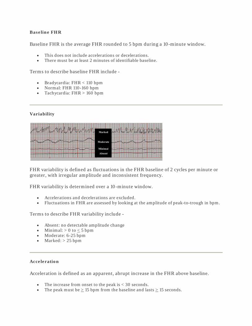
Variability
FHR variability is defined as fluctuations in the FHR baseline of 2 cycles per minute or greater, with irregular amplitude and inconsistent frequency.
FHR variability is determined over a 10-minute window.
• Accelerations and decelerations are excluded. • Fluctuations in FHR are assessed by looking at the amplitude of peak-to-trough in bpm.
Terms to describe FHR variability include -
• Absent: no detectable amplitude change • Minimal: > 0 to < 5 bpm • Moderate: 6-25 bpm • Marked: > 25 bpm
Acceleration
Acceleration is defined as an apparent, abrupt increase in the FHR above baseline.
• The increase from onset to the peak is < 30 seconds. • The peak must be > 15 bpm from the baseline and lasts > 15 seconds.
o For the fetus under 32 weeks gestation, the peak must be > 10 bpm from the baseline and lasts > 10 seconds.
Prolonged acceleration is > 2 minutes or longer and lasts < 10 minutes.
Any acceleration lasting > 10 minutes constitutes a baseline change.
Deceleration
Decelerations are described as -
• Early • Late • Variable • Prolonged
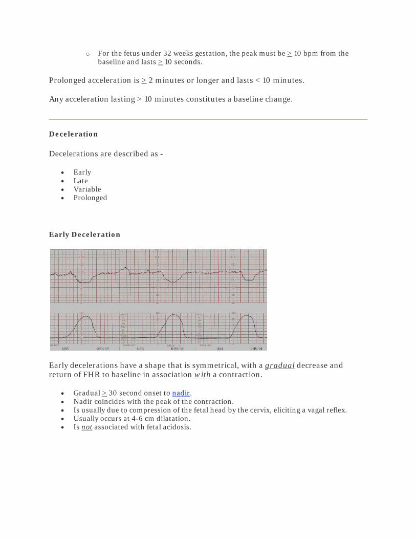
Early Deceleration
Early decelerations have a shape that is symmetrical, with a gradual decrease and return of FHR to baseline in association with a contraction.
• Gradual > 30 second onset to nadir. • Nadir coincides with the peak of the contraction. • Is usually due to compression of the fetal head by the cervix, eliciting a vagal reflex. • Usually occurs at 4-6 cm dilatation. • Is not associated with fetal acidosis.

Late Deceleration
Late decelerations have a shape that is symmetrical, with a gradual decrease and return of FHR to baseline in association with a contraction.
• Gradual > 30 second onset to nadir. • Nadir occurs after the peak of the contraction. • Is usually due to uteroplacental insufficiency and fetal hypoxia. • Although acidosis is not always present, late decelerations may be associated with fetal
acidosis.
Variable Deceleration
Variable decelerations are an abrupt onset of decreased FHR below baseline that may occur with or after a contraction
• Nadir occurs > 15 bpm and lasts for > 15 bpm. • Is usually due to cord compression and may be associated with fetal acidosis.
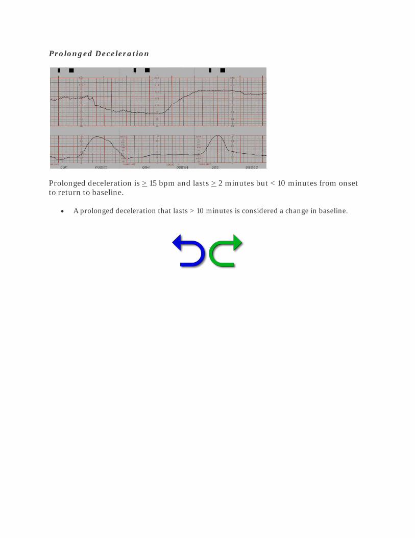
Prolonged Deceleration
Prolonged deceleration is > 15 bpm and lasts > 2 minutes but < 10 minutes from onset to return to baseline.
• A prolonged deceleration that lasts > 10 minutes is considered a change in baseline.
The quantitation of FHR patterns is based on -
• Magnitude - depth of nadir in bpm below the FHR baseline • Duration - time from the beginning to the end
Recurrent decelerations occur with > 50% of contractions in any 20-minute segment.
Intermittent decelerations occur with < 50% of contractions.
• Most common labor FHR abnormality • Usually does not require intervention • Associated with normal perinatal outcomes
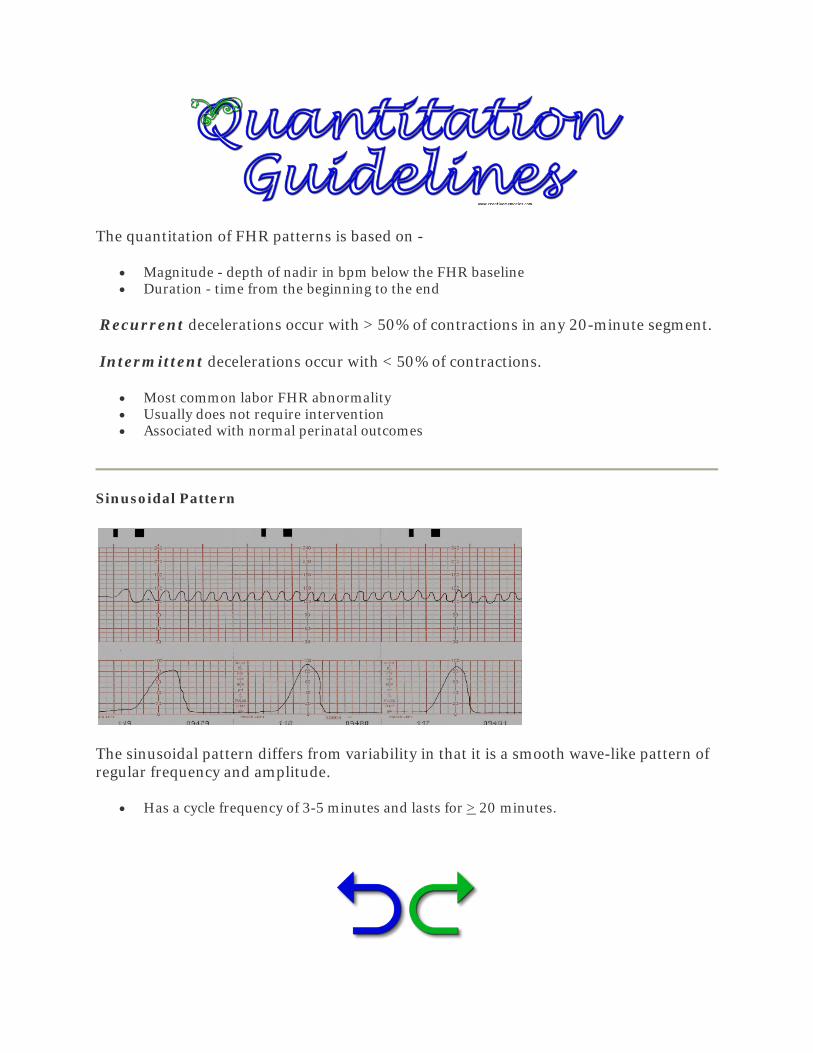
Sinusoidal Pattern
The sinusoidal pattern differs from variability in that it is a smooth wave-like pattern of regular frequency and amplitude.
• Has a cycle frequency of 3-5 minutes and lasts for > 20 minutes.

Category I
Category I is considered a "normal" tracing with -
• Baseline FHR 110-160 bpm • Moderate variability • No late or variable decelerations • Strongly predictive or normal fetal acid-base status • No intervention required
Category II
Category II is also known as an "indeterminate" tracing because it does not meet criteria for Category I or III.
• Covers a wide range of FHR patterns • Some patterns are concerning for fetal acidosis • Some patterns require intrauterine resuscitation • Requires continued monitoring and re-evaluation
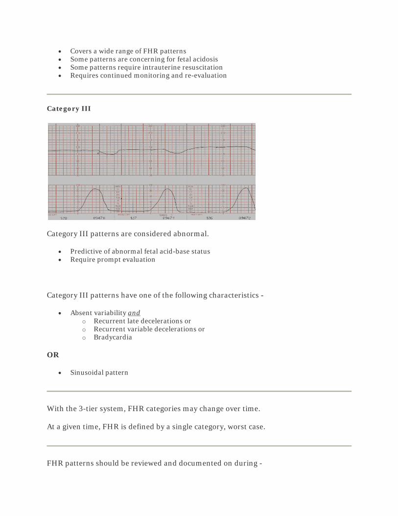
Category III
Category III patterns are considered abnormal.
• Predictive of abnormal fetal acid-base status • Require prompt evaluation
Category III patterns have one of the following characteristics -
• Absent variability and o Recurrent late decelerations or o Recurrent variable decelerations or o Bradycardia
OR
• Sinusoidal pattern
With the 3-tier system, FHR categories may change over time.
At a given time, FHR is defined by a single category, worst case.
FHR patterns should be reviewed and documented on during -

• First stage of labor o Low risk - every 30 minutes o High risk - every 15 minutes
• Second stage of labor o Low risk - every 15 minutes o High risk - every 5 minutes
The 3-tier system provides information about the current acid-base status of the fetus, but cannot predict the development of cerebral palsy
So, what FHR tracings necessitate a Cesarean delivery?

Category I - No intervention is required
• Reassess periodically and document
Category II - Management is guided by the presence/absence of -
• Accelerations - spontaneous or induced • Moderate variability
o Accelerations and/or moderate variability are highly predictive of normal pH
Category III pattern is highly predictive of fetal acidosis.
• Prepare for delivery • Begin intrauterine resuscitation measures • If the pattern remains persistent -> deliver
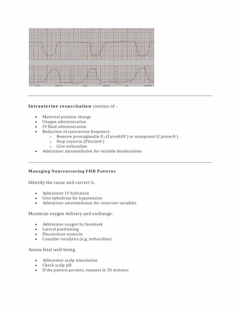
Intrauterine resuscitation consists of -
• Maternal position change • Oxygen administration • IV fluid administration • Reduction of contraction frequency
o Remove prostaglandin E2 (Cervidil®) or misoprotol (Cytotec®) o Stop oxytocin (Pitocin®) o Give terbutaline
• Administer amnioinfusion for variable decelerations
Managing Nonreassuring FHR Patterns
Identify the cause and correct it.
• Administer IV hydration • Give ephedrine for hypotension • Administer amnioinfusion for recurrent variables
Maximize oxygen delivery and exchange.
• Administer oxygen by facemask • Lateral positioning • Discontinue oxytocin • Consider tocolytics (e.g. terbutaline)
Assess fetal well-being.
• Administer scalp stimulation • Check scalp pH • If the pattern persists, reassess in 30 minutes

If acidosis cannot be excluded, deliver the fetus
So, let's practice!

FHR Pattern #1
What is the baseline FHR? Variability? Accelerations? Decelerations? Category? Interventions?
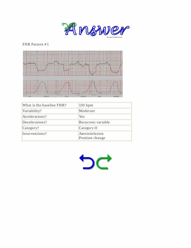
FHR Pattern #1
What is the baseline FHR? 130 bpm
Variability? Moderate
Accelerations? Yes
Decelerations? Recurrent variable
Category? Category II
Interventions? Amnioinfusion Position change

FHR Pattern #2
What is the baseline FHR? Variability? Accelerations? Decelerations? Category? Interventions?
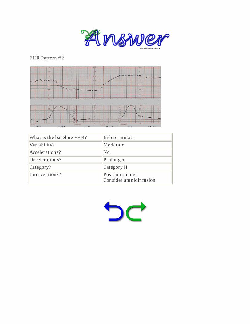
FHR Pattern #2
What is the baseline FHR? Indeterminate
Variability? Moderate
Accelerations? No
Decelerations? Prolonged
Category? Category II
Interventions? Position change Consider amnioinfusion

FHR Pattern #3
What is the baseline FHR? Variability? Accelerations? Decelerations? Category? Interventions?

FHR Pattern #3
What is the baseline FHR? 135 bpm
Variability? Moderate
Accelerations? Present
Decelerations? Absent
Category? Category I
Interventions? None

FHR Pattern #4
What is the baseline FHR? Variability? Accelerations? Decelerations? Category? Interventions?

FHR Pattern #4
What is the baseline FHR? 150 bpm
Variability? Moderate
Accelerations? Yes
Decelerations? Early
Category? Category I
Interventions? None

FHR Pattern #5
What is the baseline FHR? Variability? Accelerations? Decelerations? Category? Interventions?
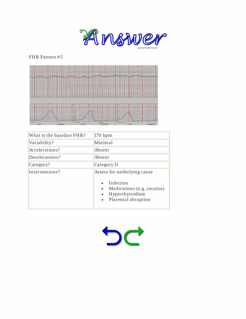
FHR Pattern #5
What is the baseline FHR? 170 bpm
Variability? Minimal
Accelerations? Absent
Decelerations? Absent
Category? Category II
Interventions? Assess for underlying cause
• Infection • Medications (e.g. cocaine) • Hyperthyroidism • Placental abruption
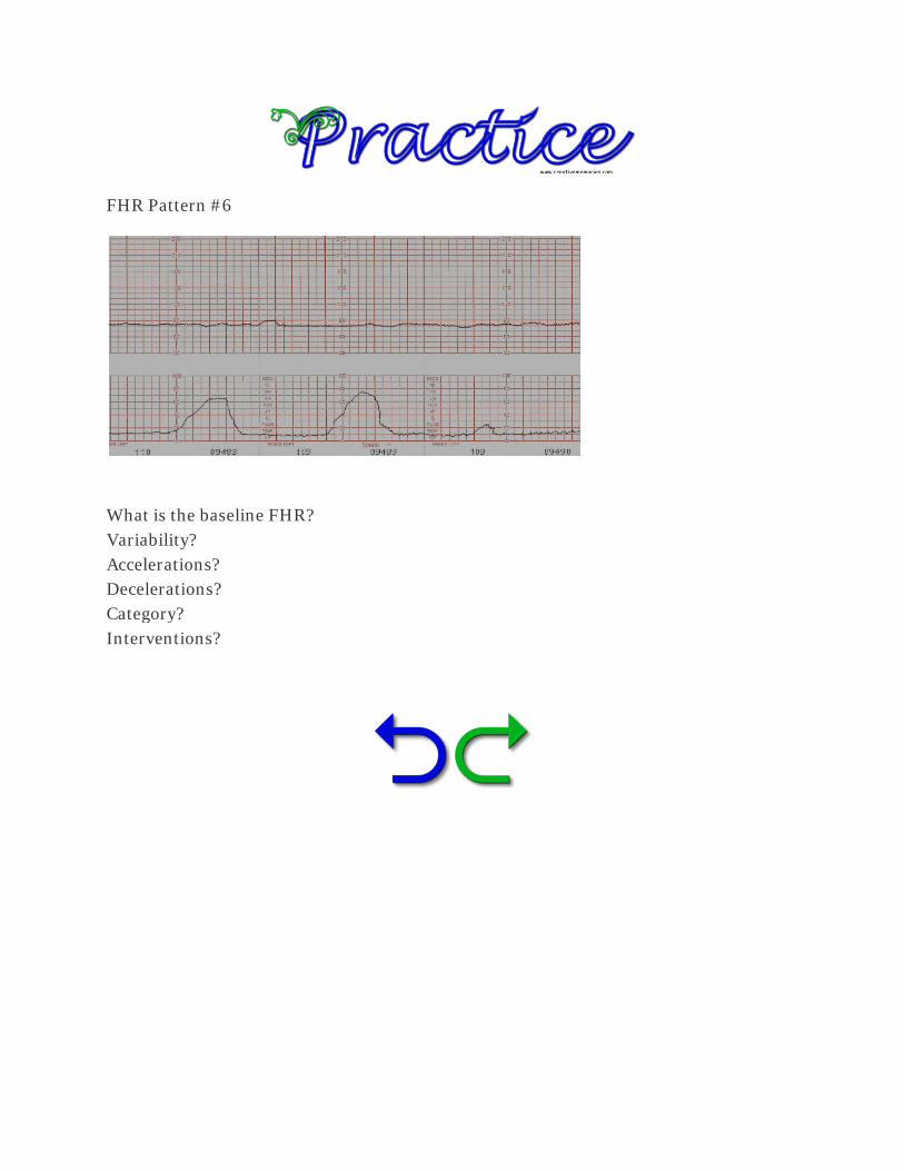
FHR Pattern #6
What is the baseline FHR? Variability? Accelerations? Decelerations? Category? Interventions?

FHR Pattern #6
What is the baseline FHR? 80 bpm
Variability? Minimal
Accelerations? Absent
Decelerations? Absent
Category? Category II
Interventions? Position change Oxygen IV fluids Reduce contraction frequency

FHR Pattern #7
What is the baseline FHR? Variability? Accelerations? Decelerations? Category? Interventions?

FHR Pattern #7
What is the baseline FHR? Indeterminate
Variability? Marked
Accelerations? Absent
Decelerations? Absent
Category? Category II
Interventions? Not addressed in bulletin Consider -
• Oxygen • IV fluids • Discontinue oxytocin
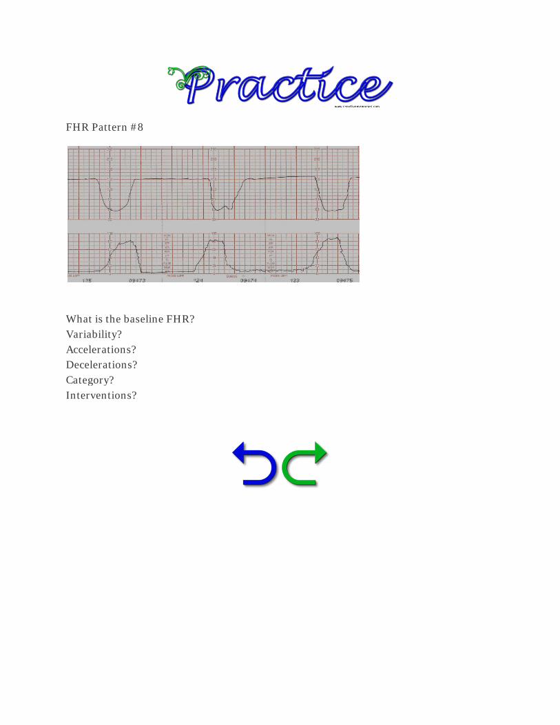
FHR Pattern #8
What is the baseline FHR? Variability? Accelerations? Decelerations? Category? Interventions?

FHR Pattern #8
What is the baseline FHR? 150 bpm
Variability? Absent
Accelerations? Absent
Decelerations? Recurrent variables
Category? Category III
Interventions? PREPARE for delivery Position change Oxygen IV fluids Reduce contraction frequency Amnioinfusion

FHR Pattern #9
What is the baseline FHR? Variability? Accelerations? Decelerations? Category? Interventions?
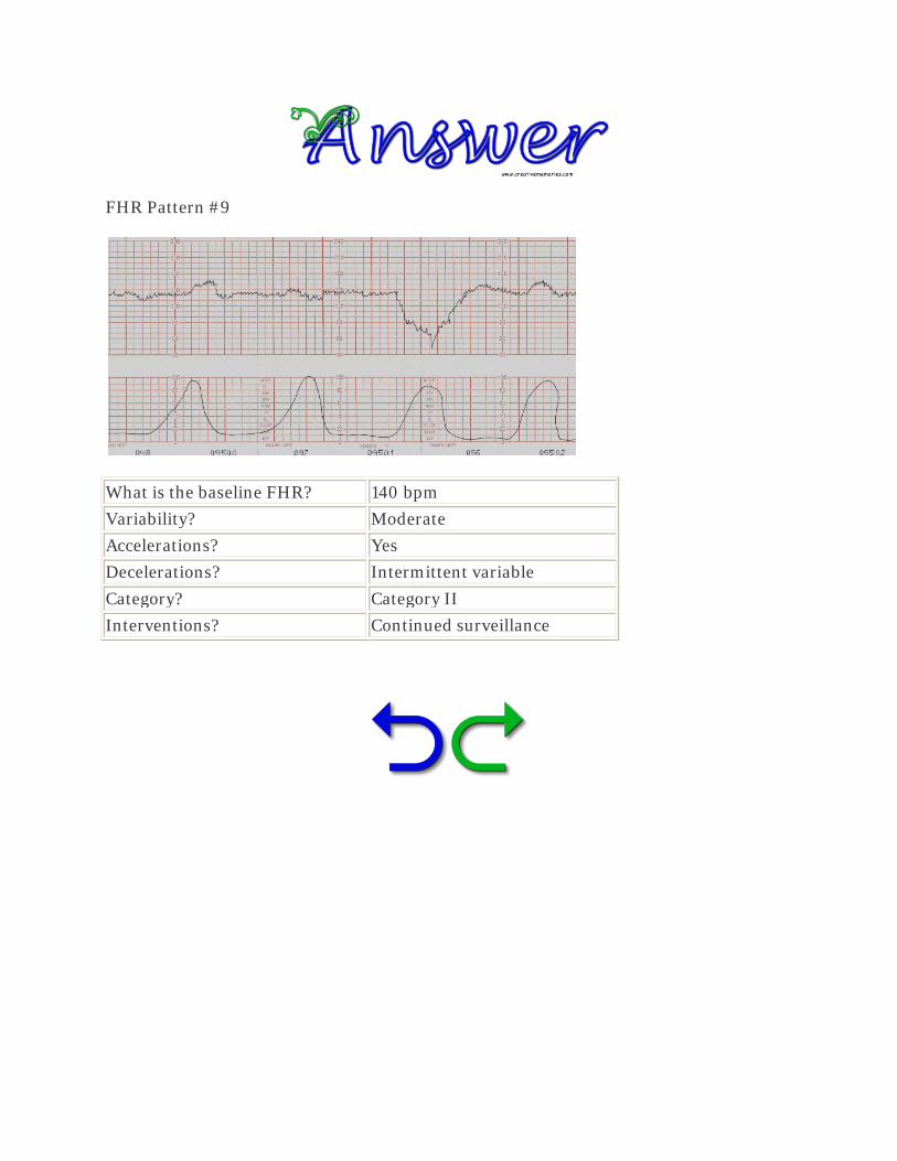
FHR Pattern #9
What is the baseline FHR? 140 bpm
Variability? Moderate
Accelerations? Yes
Decelerations? Intermittent variable
Category? Category II
Interventions? Continued surveillance

FHR Pattern #10
What is the baseline FHR? Variability? Accelerations? Decelerations? Category? Interventions?

FHR Pattern #10
What is the baseline FHR? 100 bpm
Variability? Moderate
Accelerations? Present
Decelerations? Absent
Category? Category II
Interventions? Continued surveillance

FHR Pattern #11
What is the baseline FHR? Variability? Accelerations? Decelerations? Category? Interventions?
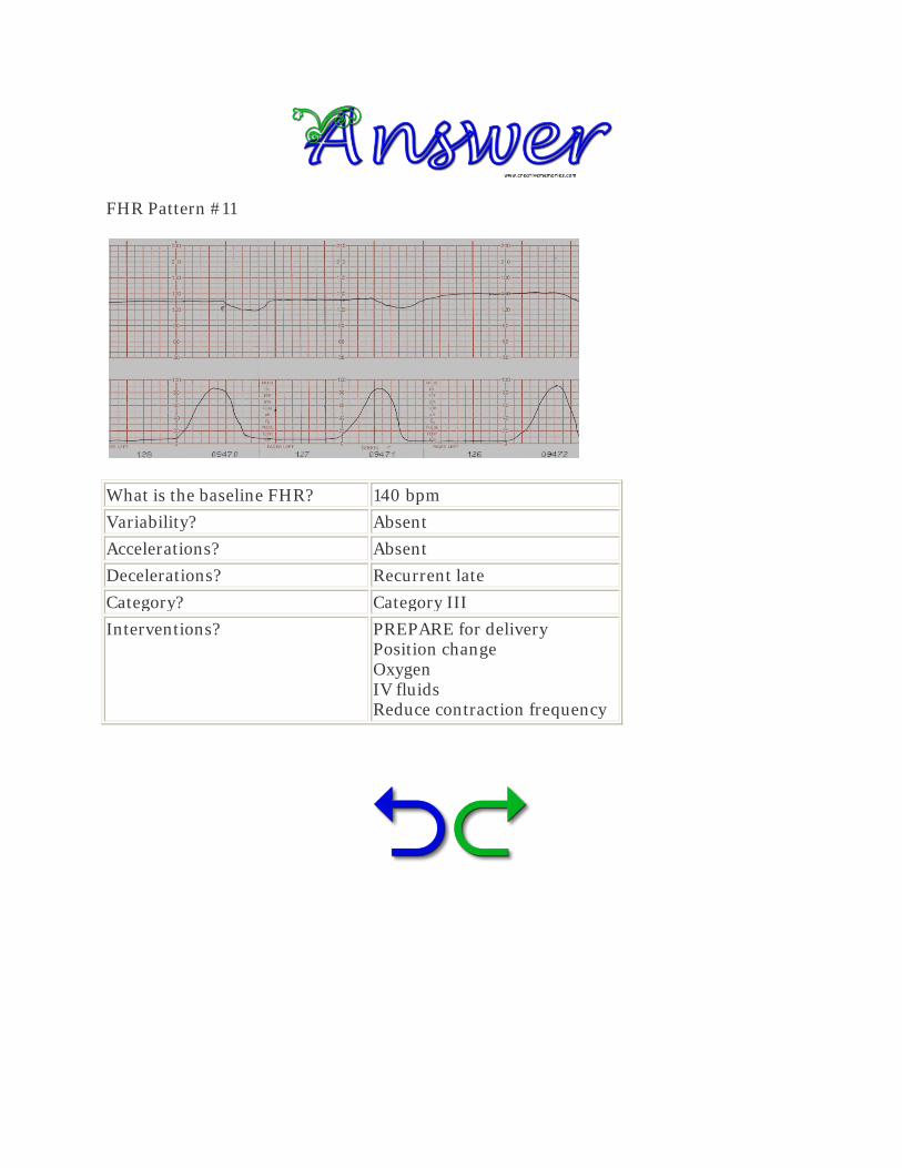
FHR Pattern #11
What is the baseline FHR? 140 bpm
Variability? Absent
Accelerations? Absent
Decelerations? Recurrent late
Category? Category III
Interventions? PREPARE for delivery Position change Oxygen IV fluids Reduce contraction frequency

FHR Pattern #12
What is the baseline FHR? Variability? Accelerations? Decelerations? Category? Interventions?

FHR Pattern #12
What is the baseline FHR? 150 bpm
Variability? Moderate
Accelerations? Absent
Decelerations? Absent
Category? Category I
Interventions? None

FHR Pattern #13
What is the baseline FHR? Variability? Accelerations? Decelerations? Category? Interventions?
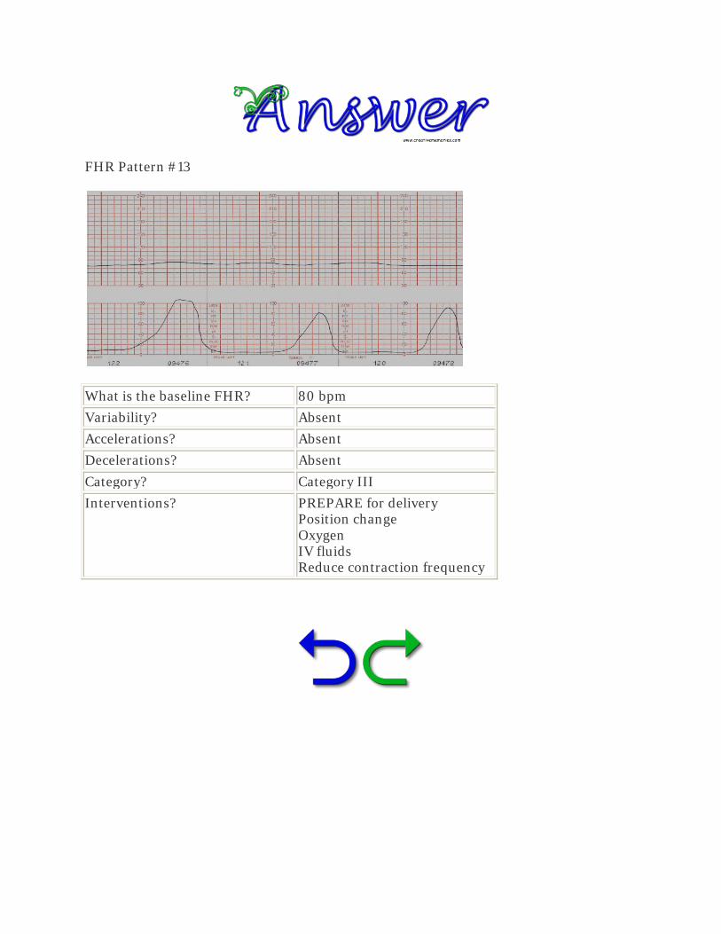
FHR Pattern #13
What is the baseline FHR? 80 bpm
Variability? Absent
Accelerations? Absent
Decelerations? Absent
Category? Category III
Interventions? PREPARE for delivery Position change Oxygen IV fluids Reduce contraction frequency

FHR Pattern #14
What is the baseline FHR? Variability? Accelerations? Decelerations? Category? Interventions?

FHR Pattern #14
What is the baseline FHR? 130 bpm
Variability? Absent
Accelerations? Absent
Decelerations? Absent
Category? Category II
Interventions? PREPARE for delivery Position change Oxygen IV fluids Reduce contraction frequency

American College of Obstetricians and Gynecologists. (2010). Practice bulletin no. 116: Management of intrapartum fetal heart rate tracings. Obstet Gynecol, 116(5), 1232-1240.
American Psychological Association. (2010). Publication Manual of the American Psychological Association, 6th Edition. Washington, DC: Author.
F.A. Davis Company. (2009). Taber’s Cyclopedic Medical Dictionary, 21st Edition Online. http://www.tabers.com/tabersonline/ub (Retrieved February 28, 2011).
Macones, G.A., Hankins, G.D., Spong, C.Y., Hauth, J., & Moore, T. (2008). The 2008 National Institute of Child Health and Human Development workshop report on electronic fetal monitoring: Update on definitions, interpretation, and research guidelines. JOGNN, 37(5), 510-515.
Macones, G.A., Hankins, G.D., Spong, C.Y., Hauth, J., & Moore, T. (2008). The 2008 National Institute of Child Health and Human Development workshop report on electronic fetal monitoring: Update on definitions, interpretation, and research guidelines. Obstet Gynecol, 112(3), 661-666.
Robinson, B. (2008). A review of NICHD standardized nomenclature for cardiotocography: The importance of speaking a common language when describing electronic fetal monitoring. Reviews in Obstetrics and Gynecology, 56(1), 56-60.

For access to larger monitor strip illustrations, please click on the image to the right. You may then click on each strip for a larger view.