fetal nucleic acids in maternal plasma: from biology to
TRANSCRIPT

397
Fetal nucleic acids in maternal plasma: from biology to clinical translation
Neha Bunkar1, Arpit Bhargava1,2, Koel Chaudhury3, Radhey Shyam Sharma4, Nirmal Kumar Lohiya5, Pradyumna Kumar Mishra1,2
1Translational Research Laboratory, School of Biological Sciences, Dr. Harisingh Gour Central University, Sagar, India, 2Department of Molecular Biology, ICMR-National Institute for Research in Environmental Health, Bhopal, India, 3School of Medical Science & Technology, Indian Institute of Technology, Kharagpur, India, 4Division of Reproductive Biology, Maternal and Child Health, Indian Council of Medical Research, New Delhi, India, 5Centre for Advanced Studies in Zoology, School of Life Sciences, University of Rajasthan, Jaipur, India
TABLE OF CONTENTS
1. Abstract2. Introduction3. Gene-environmental interaction - Epigenetics a primary determinant
3.1. Epigenetics3.1.1. DNA methylation3.1.2. Histone Modifications
3.1.2.1. Histone acetylation3.1.2.2. Histone methylation3.1.2.3. Histone phosphorylation
3.1.3. microRNAs4. Transgenerational inheritance of Epigenetic traits5. Fetal anomalies
5.1. Central nervous system5.2. Eye5.3. Cardiovascular5.4. Craniofacial5.5. Gastrointestinal5.6. Musculoskeletal
6. Next generation biomarkers6.1. Circulating cell free fetal DNA6.2. Circulating methylated DNA6.3. Circulating cell-free fetal miRNA
7. Isolation of circulating nucleic acids and sample processing8. Screening technologies for detection of Next Generation biomarkers
8.1. Multiplex Ligation-dependent Probe Amplification (MLPA)8.2. DNA Methylation-MLPA8.3. Microarray-based comparative genomic hybridization8.4. SNP array8.5. Amplification refractory mutation system-polymerase chain (ARMS-PCR)8.6. Methylation assay8.7. EpiTyper8.8. Sanger sequencing8.9. Next generation sequencing (NGS)8.10. Exome sequencing8.11. Nanopore sequencing8.12. Droplet digital PCR8.13. Quantitative reverse transcription PCR
[Frontiers In Bioscience, Landmark, 23, 397-431, January 1, 2018]

Fetal cell-free nucleic acids in maternal circulation
398 © 1996-2018
1. ABSTRACT
Exposure to environmental contaminants during the critical window of pregnancy results in deregulation of highly coordinated genetic and epigenetic mechanisms involved in prenatal growth. Such disturbances significantly alter the fetal programming, and lead to various developmental disorders immediately, over the lifetime, or transgenerationally. During the process of placental development, fetal nucleic acids enter maternal plasma as a result of necrotic, apoptotic, and inflammatory mechanisms. These nucleic acids reflect normal or abnormal ongoing cellular changes during prenatal fetal development. Here, we critically review the utility of maternally circulating cell free fetal nucleic acids towards developing reliable biomarkers for widespread screening of environmentally-associated fetal abnormalities. We further discuss the most recent developments in the fetal nucleic acid analysis, quantification methodologies, challenges involved in their accurate detection and their potential applications in fetomaternal medicine.
2. INTRODUCTION
Environmental factors have a profound and life-long impact on human health, which begins from conception. A pregnant woman is often exposed to a variety of physical, chemical, or biological factors which disturbs fetal genomic and epigenomic machinery. This disturbance results in the generation of different diseases and in rare cases, a malformed child. Although, it is difficult to identify the precise mechanistic cause of >50% anomalies, most of these cases are attributed to the combination of gene-environmental interaction, and nutritional factors (1, 2). Naturally, these cases attract wide attention and generate compassion as they contribute to long-term disability, which may have significant impacts on individuals, families, health-care systems, and societies. Worldwide, an estimated 3,03,000 newborns die within 4 weeks of birth every year due to birth defects, congenital disorders or congenital malformations (3). Genetic mutations such as Cystic Fibrosis and Haemophilia C present in some ethnic communities and consanguinity (when parents are related by blood) increases the prevalence of rare birth anomalies. It also nearly doubles the risk for
neonatal and childhood death, intellectual disability and other abnormalities (4, 5). A significant occurrence of these deformities (>94%) is associated with resource-constrained families in low- and middle-income countries and this higher risk relate to a possible lack of access to adequate, nutritious foods by pregnant women (6). While, deficiency in folate consumption by pregnant mothers increases the risk of having a baby with a neural tube defect, disproportionate vitamin A intake influences the regular fetal development (7). This demands the generation of an effective prenatal screening methodology with minimum or no risk to the growing fetus. Placenta acts as a mediator for the transport of nutrients from maternal circulation to the fetus (8). However, it is now well known that there is a significant two way fetomaternal transplacental transport in pregnancies which increases the flow of fetal biomaterials such as nucleated cells, apoptotic bodies and nucleic acids in maternal circulation. The theory of such transport was initially documented in 1893 by Schmorl who showed the existence of fetal-trophoblast in the pulmonary area of a woman. The findings gained noticed after a long time in 1969 when Walknowska observed X and Y chromosomes in the peripheral blood lymphocytes of a pregnant women. Since then, significant scientific interest was generated and various methods for the isolation and analysis of circulating fetal biomaterials in maternal blood have been discovered. The examination of fetal cells in maternal blood has been termed as “holy grail” for noninvasive prenatal diagnosis and there is a scientific surge to identify a circulating biomarker in maternal blood capable of early diagnosis and prognosis. The present review provides an updated and critical account of the significance of cell-free nucleic acids in maternal circulation towards developing a next-generation biomarker for environmental-associated fetal anomalies (Figure 1).
3. GENE-ENVIRONMENTAL INTERACTION - EPIGENETICS A PRIMARY DETERMINANT
The proof of concept that gene-environmental interactions play a crucial role in embryonic and fetal development has primarily emerged from retrospective data published over the last decades where several
9. Bio-informative approach analysis of circulating cffDNA9.1. Estimation of fractional fetal DNA concentration9.2. Fetal aneuploidy detection9.3. Monogenic disorders detection9.4. Methylomic analysis of circulation cffDNA
10. Future directions11. Acknowledgement12. References

Fetal cell-free nucleic acids in maternal circulation
399 © 1996-2018
human tragedies demonstrated the sensitivity of the developmental period to environmental exposures (9–12). The contamination of mercury resulted in severe brain dysfunction in children due to high mercury exposure of pregnant women in Minamata Bay of Japan (13); PCB contamination of rice oil in Japan where exposed mothers have lower IQ children as compared to the unexposed ones (14); the Seveso explosion had trans-generational effect in the offspring of the mothers exposed to high concentrations of dioxin during pregnancy at a manufacturing plant near Italy (15); in utero MIC exposure during the Bhopal gas tragedy which caused a persistently hyper-responsive cellular and humoral immune state in affected individuals with increased cytokine secretions, higher immunoglobulin levels and anti-nuclear antibodies (16–18). This posits that there are sensitive developmental phases during which tissue growth and functions can be significantly altered by different environmental stressors, increasing susceptibility to adverse health pregnancy outcomes (Table 1). Therefore, gene-environmental factors comprising the dynamic interaction between the genome, epigenome, and environmental factors immensely contribute to the fate of individual cells for
maintenance of a critical balance between cell death, proliferation, regeneration, and repair during embryonic and fetal development.
3.1. Epigenetics
Epigenetic is a term used to refer the processes which occur above the genetic level such as methylation-mediated regulation of genetic DNA, covalent modifications of histone tails going outward from the nucleosome, and transcriptional and posttranscriptional regulations by microRNA (miRNA) expression. Epigenetic marks are dynamic, vital encrypted sets of unique DNA methylation and particular covalent modified distinct amino acid residue of nucleosomal histone tails (19, 20). These marks can influence the set of given cue during the developmental stages, throughout the lifetime and passed on to the later generations through germs cells (developmental origins of health and disease (DOHaD) hypothesis) (21– 23). Moreover, the epigenome is rewritable throughout the developmental phase of a fetus to the newborn, toddler, adult, and aged, according to the internal and external cues. This intergenerational inheritance of
Figure 1. A scheme showing the importance of maternally circulating cell-free nucleic acids towards development of next-generation biomarkers for detection of environmental-associated fetal anomalies.

Fetal cell-free nucleic acids in maternal circulation
400 © 1996-2018
dynamic epigenetic signatures are not only linked with the parental and acquired influence of disease but also offers the opportunity to overcome disease circumstances by dealing with the epigenetic modulating interventions. Observations suggest that consumption of drugs, alcohol and tobacco by the male or female can alter certain epigenetic signatures which are able to have an impact on their own life and definitely up to the third generation, through sperm and ova epigenome, and on later generations that yet to be verified. Basically, epigenetic processes include the addition of the substrate for modification by the writers (DNMT, HAT, HMTs), deduction by the erasers (TET, HDAC, HDM,
KMT) and reading by the readers (MeCP, bromodomain, chromodomain, PHD) (19). These sets of biochemical interacting processes found abrupt in various diseases (24–26). Furthermore, DNA methylation and histone modifications mainly acetylation have been observed interconnected, crossing downstream affects one over other, requiring attention towards evaluating role in fetal anomalies (27).
3.1.1. DNA methylation
Cytosine is the only pyrimidine which gets methyl modification at carbon-5 involved in the
Table 1. Effects of maternal exposure to environmental hazards on gene-environmental modulators causingfetal abnormalities
Modulators Agents Underlying mechanisms Related abnormality Reference
Smoking In-utero exposure Mitochondrial damage; In-utero environment affect CDK in adult life
Chronic kidney diseaseRenal fibrosis
(156)
Maternal exposure Increased apoptosis causes germ cell depletion in the seminiferous tubules of neonatal and juvenile offspring
Impairs male offspring fertility (157)
Alcohol Prenatal alcohol exposure Gene alteration; genetic factors and epigenetic mechanisms
Birth defects, Neuro-developmental deficits (fetal alcohol spectrum disorders -FASD)
(158)
Air pollution Ambient air pollution during preconception and during early gestation
Case-control study Offspring congenital orofacial defects (159)
PM10 and SO2 during the second and third trimester
Case-control study Birth Defects (160)
SO2 and TSPM Case-control study Congenital heart disease (CHD) and ventricular septal defects (VSDs)
(161)
Volatile organic compounds -trichloroethylene and tetra-chloro-ethylene or per-chloro-ethylene in indoor air
Case-control study Low birth weightPreterm birthFetal growth restrictionConotruncal defects
(162)
Maternal peri-conceptional toxicants exposure
ATP Binding Cassette Subfamily B Member 1 (ABCB1) Gene C3435T polymorphism
Congenital heart defects in children (163)
Endocrine disruptors
Diethylstilbestrol, a estrogen mimetic Case-control study Sub-fertilityInfertilityCancer of reproductive tissues
(164)(165)
PAH Bioinformatic analysis of benzo-α-pyrene
Damage to the human placental insulin-like growth factor-1 gene
Disordered development and fetal programming
(166)
Air PAH pollutants Case-control study Neurobehavioral deficitsAttention-deficit/hyperactivity disorder symptomsLower processing speedDisrupted development of left hemisphere white matter
(167)
Heavy metals Toxic metals including arsenic, cadmium, manganese, and lead
Assess the association between metal concentrations in private well water and birth defect prevalence
Prevalence of conotruncal heart defects
(168)
Prenatal lead and cadmium coexposure
Synergistic effect Neuro-developmental effects (169)
Pesticide Chlorpyrifos (an organophosphate insecticide)
frontal and parietal cortical thinning
Neurobehavioral deficits (170)

Fetal cell-free nucleic acids in maternal circulation
401 © 1996-2018
epigenetic regulation. The DNA methylation process requires methyl group donor S-adenosylmethionine (SAM), writers - DNA methyltransferases (DNMT) like maintenance DNMT1, DNMT2, de novo DNMT3a/b and DNMT3L, erasers - ten-eleven translocase (TET), and readers - methyl-binding proteins like MeCP2, MBD1–4, and Kaiso (19). Other than the 5-methylcytosine, 5-hydroxymethylcytosine, 5-formylcytosine, and 5-carboxylcytosine are newly observed forms of modified cytosine residues produced by the action of DNA hydroxylases - TET1–3 during the process of DNA demethylation. Global DNA methylation marks transcriptional suppression while non-methylated genes tend to express frequently (28–31). The methylation of promoter sequences largely suppresses expression of tumor suppressor genes associated with the many cancers whereas growth-promoting (oncogene) observed to be hypomethylated (32). Moreover, the abrupt catalytic activity of DNMTs adds aberrant de novo methylation marks leading to atypical global methylation levels affecting overall gene expression (33). These aberrant DNMT expressions are not only limited to cancer itself but also present in other diseases such as anxiety, autism, cardiovascular disease, depression, dementia, obesity, and type 2 diabetes (34, 35). There are several other ways by which DNA methylation influence vital cellular processes such as gene silencing as they are able to exclude protein binding that is imprinting, block binding of several transcription factors, recruit specialized repressor complexes with HDACs, however, their downstream consequences are not fully acknowledged.
3.1.2. Histone modifications
Billions of DNA segments are assembled in a chromosome like structure using basic histone proteins which attract negatively charged phosphorylated groups of DNA. Five types of histone work as histone core includes dimers of four H2A, H2B, H3 and H4, and linker H1/H5 together to make structural unit i.e. nucleosome of supra-molecule chromatin. N-terminus of the core histones coming out from the nucleosome is known as histone tails (15–38 amino acids long). Histone tails are the primary site of specific covalent histone modifications such as methylation, acetylation, phosphorylation, ubiquitination, ribosylation, etc on the specific amino acid residues which depending upon the overall charge provides specific nucleosomal orientation. Hence, with DNA methylation, histone modifications (histone codes) and nucleosome positioning are the determinants that significantly influence the gene expression (36). Specificity of epigenetic regulation influenced by the numbers of specific covalent modifications made at the particular amino acid residues of histone tails. Histone acetylation, methylation, phosphorylation, and ubiquitination are the major process having notable
roles in the regulation of DNA repair, transcription, and chromatin structure condensation/decondensation.
3.1.2.1. Histone acetylation
Histone acetylation is the modification of lysine (K) and arginine (R) residues situated in the tails of histone core protrude out of nucleosome. Acetyl CoA is the main supplier of the acetyl group for this modification (acetylation). To date, two classes of writers including lysine acetyltransferases type-A, nuclear - GNAT, MYST, CBP/p300 families, and type-B cytoplasmic that modify free histones have been identified. Eraser including 18 histone deacetylases (HDAC) are further divided into four major classes consist of class I (HDAC 1–3 and 8), class II (HDAC 4–7 and 9–10), class III (sirtuin 1–7) and class IV (HDAC11). Unlike nicotinamide adenine dinucleotide (NAD+)-dependent sirtuins of class III, class I, II and IV HDACs have structural and functional similarity and require zinc ion for catalytic activity. Readers comprise of Bromodomain, BET family domain (BRD2, BRD3, BRD4, and BRDt) and plant homeodomain (PHD) fingers (37–40). Altered activity of HDACs and HATs resulting in aberrant gene silencing has been reported among patients of different diseases (41–42).
3.1.2.2. Histone methylation
Methylation of histones occurs at lysine (K) and arginine (R) residues and found to regulate DNA repair and transcription. In this process, S-adenosylmethionine and methionine are the prime sources of the methyl group. Moreover, writers for this modification are called as lysine and arginine methyltransferases depending on type of the modified residue i.e. lysine and arginine. Classes of lysine methyltransferases comprise of KMT2A/B/C, KMT3A/B, NSD2/3, and KMT6. Deduction of the methyl group is performed by the group of enzymes collectively known as demethylase and are categorized into two classes. Class I demethylase executes function via amine oxidation reaction with cofactor flavin adenine dinucleotide while class II- Jumonji demethylase carry out demethylation via oxidative mechanism and radical attack (involving Fe(II) and α-ketoglutarate). The most prominent demethylases for the process are KDM5A (JARID1A), KDM5C (JARID1C) and KDM6A (UTX) (43). Royal family including Tudor domains and chromo domains, malignant brain tumor (MBT) domains, PHD fingers are the readers for methylated histones. Tudor domain is well known to recognize the methylated arginine residues of histone tails (19, 44, 45). On the other hand, histone arginine methylation executed by arginine methyltransferase and arginine demethylases, are less studied (46). Evidences have significantly linked aberrant expression of histone methyltransferases with different life threatening diseases (19, 45).

Fetal cell-free nucleic acids in maternal circulation
402 © 1996-2018
3.1.2.3. Histone phosphorylation
It is the epigenetic covalent phosphorylation type modification of histone tails at serine (S), threonine (T) and tyrosine (Y) residues important for the proper regulation of different vital processes like DNA repair, replication, transcription and condensation. Adenosine triphosphate (ATP) acts as a phosphate group providing moiety for this modification. The genomic phosphorylation levels are primarily controlled by writers such as ATM, non-receptor tyrosine kinase-JAK2 and PIM1 and erasers like phosphatases. Phosphorylated amino acid moieties are distinguished by readers including 14–3-3 and BRCA1 C-terminus domain (BRCA1) and SH2 domain (47–49). Phosphorylation of histone and non-histone proteins is considered as an integral regulatory epigenetic and post-translational modification important for controlling the subsequent enzymes actions and several signaling routes (50).
3.1.2.4. Histone ubiquitination
Apart from the protein degradation, ubiquitination is a crucial process for managing DNA repair, condensation and transcription, via working at the epigenomic level. It mainly involves ubiquitin to modify lysine (K) of histones tails. The writers for this process include E1-ATP-dependent ubiquitin-activating enzyme, E2-Intermediate ubiquitin-conjugating enzyme e.g. HR6A/B, UbcH6, and E3-Terminal ubiquitin-protein ligase e.g. RNF20, RNF40, MDM2. While, Deubiquitinases (DUBs) are the deducting group of enzymes as erasers of this modification and few examples are USP7, USP22, USP44, and HAUSP. Reader for this includes the protein that has ubiquitination reading motif such as inverted ubiquitin interaction motif (IUIM) (51, 52).
3.1.3. microRNAs
Regulation at transcriptional and post-transcriptional level is primarily governed by a group of small non-coding RNAs of approximately 21–23 nucleotides in length which transcribe but not translate into the proteins. Partial complimentary binding of miRNA to its target mRNA leads to translation suppression, while perfect binding causes degradation of mRNA, thus regulate gene expression at post-transcription level. miRNAs are disseminated throughout the genome and mainly reside between independent transcription units (intergenic) or found in the intronic sequences of protein-coding genes or in intronic or exonic regions of noncoding RNAs (53,54). Primarily, the pri-miRNA is processed within the nuclear compartment to a precursor miRNA (pre-miRNA) by Drosha, a class 2 RNase III enzyme. Subsequently, precursor miRNA (pre-miRNA) sequences are exported to the cytoplasm by Exportin-5 and processed
by Dicer into mature miRNAs, and incorporated into the RNA-induced silencing complex (RISC). RISC forms a multi-protein complex by binding to mRNA on which miRNAs mediate sequence-specific recognition and binding, that either degrades or silences the target mRNA depending on the sequence complementarity (55). Whereas it is believed that miRNAs restrain translation, evidence also suggests that miRNAs can led changes in the RISC Argo component and enhances the process of translation (56). Therefore, while miRNAs appear to police translation in an inhibitory fashion, they may also enhance translation in defined biological settings.
4. TRANSGENERATIONAL INHERITANCE OF EPIGENETIC TRAITS
Environmental factors possess the potential to induce epigenetic alterations which persuades through downstream generations and may lead a significant impact on the etiology of different fetal diseases (57). Such transgenerational inheritance of altered epigenetic factors requires their germ-line transmission between different generations and has gained significant interest due to their ability to transfer the disease and phenotypic variation across generations without further environmental exposures. Exposures initiate a series of genetic or metabolic disturbances which interact and disturb parallel epigenetic mechanisms at some point during the early developmental stages (58–60). The majority of such environmental exposures directly affects the growth of somatic cells and influences the later life physiology of the exposed individual. However, if the exposure influences permanent epigenetic variations in the germline cells (egg/sperm) which sustains during the process of fertilization, it will result in transgenerational transmission of altered epigenetic traits without continued environmental exposure. This happens when cells are profoundly exposed to environmental contaminants during critical windows of development (61). The development of gonads and sex determination period during fetal growth is considered as the first decisive window, while the process of gametogenesis which leads to development of differentiated sperm or egg is the second critical window of exposure. In adult females the oogonia which are not actively developing offers a potential target for environmental exposures, while in adult males the actively differentiating sperms during the process of spermatogenesis are the soft targets for environmental exposures induced epigenetic changes. Moreover, studies have now clearly shown that the period of gonadal sex determination during fetus growth is a critical window for the induction of environmentally induced epigenetic transgenerational inheritance. It has been also reported that consequences of environmental exposure on adult male’s spermatogenesis can prop up transgenerational inheritance of altered epigenetic

Fetal cell-free nucleic acids in maternal circulation
403 © 1996-2018
traits (2, 62). During early development the germline stem cells migrate to colonize the fetal gonad prior to gonadal sex determination. Importantly, during this process almost all methylated DNA is demethylated and the process of re-methylation is initiated by germline DNA at the beginning of gonadal sex determination in a sex-specific manner. In males the re-methylation process gets completed later during fetal gonadal development, while in the female germ cell it is completed after birth. The exposure of environmental contaminants during this period significantly modifies the germ cells epigenetic programming to promote the transgenerational inheritance of abnormal epigenetic traits (2, 9).
5. FETAL ANOMALIES
The term fetal anomaly clearly refers to the structural abnormalities present at birth and is broadly classified into two categories: (i) major: any structural abnormality that requires medical and/or surgical intervention; (ii) minor: any structural deformity that does not require immediate medical or surgical treatment but might be an indicator of some development defects (63, 64). The spectrums of common fetal problems diagnosed among various regulatory systems of human body are discussed below.
5.1. Central nervous system
In a growing fetus, the brain and spinal cord develop from a simple structure called the neural tube. The neural tube protects the brain and spinal cord by zipping up along its length (65). A neural tube defect originates when the neural tube doesn’t close at any part along its length. The more frequently observed neural tube defects that are: (i) anencephalus; (ii) spina bifida without anencephalus; (iii) hydrocephalus without spina bifida; (iv) encephalocele; and (v) microcephalus (66).
5.2. Eye
The mature eye is a complex organ that develops through a delicate and complex process during embryogenesis. Alterations in molecular programming can lead to several disorders that become apparent at birth or shortly afterward. These defects are influenced by a combination of genetic factors and environmental conditions and the common conditions include anophthalmia and congenital cataract (67).
5.3. Cardiovascular
Cardiovascular defects are one of the most common type of birth anomalies. The defects can involve the walls of the heart, the valves of the heart,
and the arteries and veins near the heart causing disruption of the normal flow of blood through the heart. The most common environmental factor influencing cardiovascular tract include maternal intake of teratogenic agents (68, 69). The physiologic consequences of these anomalies greatly vary and range from a heart murmur or discrepancy in pulses in an asymptomatic child to severe cyanosis, heart failure (HF), or circulatory collapse. Broadly there are several types of common congenital heart defects which include: (i) septal defects - presence of a hole between two of the heart’s chambers; (ii) coarctation of the aorta - the main large artery of the body, called the aorta, is narrower than normal; (iii) pulmonary valve stenosis - the pulmonary valve that controls the flow of blood out of the lower right chamber of the heart to the lungs, is slender than normal; (iv) transposition of the great arteries - where the pulmonary and aortic valves and the arteries are connected to have swapped positions (70).
5.4. Craniofacial
Although the effect of environmental exposures on the development of craniofacial abnormalities is not fully deciphered, environmental exposures might play an important role, particularly in combination with genetic abnormalities. This diverse group of deformities involves abnormal growth of the head and facial bones and the two of the most common craniofacial defects are orofacial clefts (improper development of lip and mouth), and craniosynostosis (bones in the baby’s skull fuse too early). Besides, two other craniofacial defects include microtia, (improper growth of external portion of the ear); and anotia, which occurs when the external portion of the ear is missing (71).
5.5. Gastrointestinal
Gastrointestinal birth defects are another type of structural defect which originates due to the complex nature of the growth, elongation, and folding of the tract. These defects can occur anywhere along the gastrointestinal tract, which includes the esophagus, stomach, small and large intestine, rectum and anus. Some of the most common gastrointestinal birth defects include pyloric stenosis, Hirschsprung’s disease, diaphragmatic hernia, omphalocele, esophageal atresia, gastroschisis and, anal atresia and biliary atresia (72).
5.6. Musculoskeletal
The musculoskeletal defect is also one of the common congenital malformations present at birth in a newborn. The most prevalent types include upper limb reduction, lower limb reduction, gastroschisis and omphalocele (73).

Fetal cell-free nucleic acids in maternal circulation
404 © 1996-2018
6. NEXT GENERATION BIOMARKERS
The use of cell-free nucleic acids circulating in maternal plasma provides a unique opportunity to develop non-invasive diagnostic strategies for the early identification of different inherent and environment associated fetal diseases (Figure 2). Recent technological advancements offered the more rapid, accurate detection not only of cell-free fetal DNA (cffDNA), but also of methylated cffDNA, miRNA or mRNA circulating in the maternal connective tissues (74, 75). Thus both circulating fetal genetic and epigenetic biomolecules including DNA, methylated DNA, and miRNA in the maternal biopsy sample are being explored for the promising next-generation non-invasive biomarker detection (76). Moreover, the incorporation of different sophisticated bioinformatics tools will further improve the diagnostic application circulating cell free nucleic acids in prenatal testing of different fetal anomalies (77).
6.1. Circulating cell free fetal DNA
The clinical utility of this circulating DNA remained unnoticed for a long time after initial reports of Mandel and Metais in 1948. The DNA in circulation may vary from 0.5 to 5.0 kb and are quickly removed to maintain the balance (≤ 5ng/ml plasma) however, their concentration may be influenced (5 to 10 times) by different physiological conditions (78). Circulating DNA in human plasma primarily arises from dying cells (apoptotic/apo-necrotic/necrotic), metabolic secretion, nuclei expulsion during erythrocyte maturation, or during various immunological processes (79). In pregnant females, the continuous trophoblast turnover results in the release of apoptotic bodies comprising DNA into the maternal blood. This process starts from cytotrophoblast proliferation and is pursued by syncytiotrophoblast fusion, differentiation and release of apoptotic bodies (80). The percentage of circulating fetal DNA may be around 3–6% of the total circulating
Figure 2. Representative figure showing an overview of various basic steps involved in development and validation of cell-free nucleic acids based detection methodologies.

Fetal cell-free nucleic acids in maternal circulation
405 © 1996-2018
maternal DNA (81). It mainly comprises short fragments (≤ 193 bp) and can be diagnosed after a gestation age of 4 weeks (82). The DNA concentration increases with the increasing gestation time and is quickly removed from the maternal circulation (half-life = 16 min) (83). However, the total circulating DNA concentrations (placental and maternal) in the plasma may be affected by different health factors like obesity preeclampsia, gestational age, diabetes etc. Although known as fetal DNA this circulating DNA is of placental origin and strongly reflects placental morphogenesis and related abnormalities. Studies have also suggested a well-built relation of DNA release with oxidative stress (84, 85). Moreover, changes at the genomic and epigenomic levels are considered as the primary reason for generation of most of the diseases. These changes result in disrupted gene expression patterns and abnormal protein synthesis thereby affecting the veracity of the cellular system. The detection of patterns of different genomic imprints on circulating cffDNA may provide vital information about the mother and growing fetus.
Rapid advancements in non-invasive prenatal testing (NIPT) are led by the discovery of fetal-derived cell-free DNA in maternal circulation which is long associated with the detection of fetal complications (Table 2). Though, studies have indicated the role of cffDNA for prenatal analysis of preterm labor, preeclampsia, invasive placenta, monogenic disorders and fetal chromosomal aneuploidies. Basically, detection of fetal DNA from maternal DNA is governed by the unique fetal biological properties. Several methods are now available to quantify cffDNA fraction in maternal plasma that includes previous PCR assays to advanced next generation sequencing for non-invasive prenatal diagnosis (86, 87).
6.2. Circulating methylated DNA
In addition to the cffDNA, methylated cffDNA are also detected in the maternal plasma derived from trophoblast cells. Sequencing advancements have led identify methylated DNA separately from the unmethylated DNA. This also resulted in recognition of various candidate fetal epigenetic markers (88). One such non-invasive biomarker detected is fetal-specific placental-derived maspin which is a mammary serine protease inhibitor belonging to serpin (serpin peptidase inhibitor) family encoded by SERPINB5 gene (located on chromosome 18q21.3.3). Maspin functions as a tumor suppressor gene and the maspin knockout mice observed to be non-viable and died in early embryogenesis. Moreover, bisulfite sequencing of maspin promoter & real-time quantitative methylation-specific PCR revealed that this gene is differentially methylated in placental development and maternal blood (89). Maspin gene reported to be hypo-methylated in placental development but
hypermethylated in maternal circulation (90). Such observations of unmethylated and methylated gene status may provide potential next generation marker for noninvasive prenatal detection. Earlier it has been also shown that analysis of fetal-specific differential methylated regions may be used as an approach for the accurate diagnosis of Down’s syndrome (91).
6.3. Circulating cell-free fetal miRNA
RNA molecules previously considered as unstable moieties due to the presence of ribonucleases, are now known to exist and stably circulate in body fluids in different forms (92). These circulating RNAs are known to play a important role in patho-physiology of different diseases. Extracellular noncoding RNAs, like miRNAs are broadly engaged in vital signaling mechanisms, including those related to fetal development (93–95). miRNAs are transcribed and processed in the nucleus and then exported in the other sub-cellular compartments. The biological significance of miRNAs profoundly lies within their ability to regulate post-transcription and translation mechanisms via sequence specific complementarity. Under certain abnormal circumstances such as diseases, a higher concentration of miRNAs are observed in different biological fluids including plasma, and saliva. miRNAs enter circulation as a result of cellular demise (apoptotic/necrosis) through a ceramide-dependent secretory mechanism (96, 97). These miRNAs are known to have an important role in different regulatory mechanisms like trophoblast proliferation and syncytialization. Cell-free circulating miRNA derived from trophoblast cells are either present in vesicles (exosomes) or bound to the proteins like Ago2, nucleophosmin, ribonucleoprotein and high-density lipoproteins. The miRNAs in maternal circulation are secreted as 60–80nm sized exosomal vesicles through placental syncytiotrophoblasts. Moreover, trophoblasts miRNAs can also exist as apoptotic bodies which are considered as a significant source for the extracellular release of trophoblastic miRNA, and/or as protein-bound miRNAs. The protein complexes attached with the circulating miRNAs provide stability to these molecules against RNase digestion. The inconsistent concentration of circulating miRNAs observed throughout the embryonic development influences various fetoplacental-maternal interconnections (98). The improper expression of circulating placental miRNAs in maternal blood has been significantly observed during different pregnancy related complications (99, 100). miR144, a regulator of ischemia and hypoxia is reported to be significantly associated with early pathological preeclampsia related changes (101). Similarly, miR-206 is also considerably up-regulated in the placenta of pathological preeclampsia (102, 103). Studies have suggested that the presence of circulating miRNAs in body fluids and their association

Fetal cell-free nucleic acids in maternal circulation
406 © 1996-2018
Table 2. Use of cell free circulating DNA as biomarker based detection of fetal anomalies
Cell-free fetal DNA mediated analysis
Test population Mode of DNA isolation Downstream technique used
Reference
Twin zygosity assessment
8 twin and 11 singleton pregnancies Not specified Massively parallel sequencing (172)
Sex chromosomal aneuploidies
564 pregnant women with known fetal karyotype
QIAamp Circulating Nucleic Acid Kit (QIAGEN Inc., Valencia, CA, USA
Massively parallel sequencing (173)
Intrahepatic cholestasis
26 Intrahepatic cholestasis + 30 control pregnancy of third trimester
QIAamp DNA Blood Mini kit (Qiagen; Hilden, Germany)
Real time PCR (174)
Fetal aneuploidy detection
Not specified QIAamp Circulating Nucleic Acid Kit (Qiagen)
Massively parallel sequencingSemi-automated sequencing library preparation
(175)
Detection of RHD status and sex
120 (10.6.–13.9. weeks) + 118 (16–20.9. weeks) + 113 (27.9.–33.9. weeks) pregnancy with RhD negative nonalloimmunized women
QIAmpDNA Blood Minit Kit (Qiagen, Inc.; Valencia, CA)
Allele-specific primer extensions
(176)
Determination of fetal gender
202 singleton pregnancies at 4 to 13 weeks of gestation
QIAamp DSP Virus Kit (Qiagen, Hilden,Germany)
Quantitative fluorescent-polymerase chain reaction (QF-PCR)
(177)
Y-chromosome identification
26 pregnant women at different gestational ages
QIAamp® DSP Virus Spin Kit(Qiagen, Hilden, Germany)
Surface plasmon resonanceSRY-specific probes immobilized on the sensor chip
(178)
Non-invasive fetal RHD genotyping
10 non-pregnant and 35 pregnant women Qiagen QIAamp Circulating NucleicAcid kit (Qiagen, Germany)
Droplet digital PCRReal-time PCR
(179)
Down syndrome and other trisomies
2 euploid triplet pregnancies + 25 twin pregnancies (17 euploid, 5 discordant and 2 concordant for Down syndrome; one discordant for trisomy 13) pregnancies
NOT specified Massively parallel shotgun sequencing (MPSS)
(180)
Trisomy 21 Women at risk for trisomy 21 (trisomy 21 risk if more than one positive in 250 [> 1/250]);Total 976 eligible cases, 225 were processed: 8 were used for pretesting phase and 23 to build a reference set
NucleoSpin®Plasma XS Kit (Macherey-Nagel, Düren Germany)
Quantitative Real-Time PCR amplificationLibrary preparation
(181)
Prenatal chromosomal aneuploidy detection.
2,063 pregnant women with fetuses who were diagnosed as high risk of fetal defects
GenMagCirculating DNA from Plasma Kit
Next generation sequencingLibrary preparation
(87)
Subchromosomal abnormalities
1,476 pregnant women with fetal structural abnormalities detected on ultrasound
Not specified Semiconductor sequencing Acgh
(182)
Detection of microdeletion
2 patients having 22q11.2. deletion syndrome + 14 women at low risk for fetal chromosomal abnormalities
QIAamp Circulating Nucleic Acid Kit (Qiagen)
Next-generation sequencingLibrary preparation
(183)
Balanced fetal translocation
Collected plasma of a donor known to be carrying a fetus with a balanced translocation (t(8;11))
Not specified Illumina sequencing technology
(184)
Preeclampsia 107 pregnancies (51 with mild PE and 56 with severe PE) + 93 normotensive pregnancies during third trimester
QIAamp DNA Blood MiniKit (Qiagen, Valencia, CA, USA)
Real-time PCR together with the measurement of serum soluble endoglin using ELISA
(185)
Preeclampsia 16 women; 8 with preeclampsia and 8 normotensive control cohorts with singleton male pregnancy between 28 and 32 gestational weeks
Not specified Real-time PCR analysis (186)

Fetal cell-free nucleic acids in maternal circulation
407 © 1996-2018
with pregnancy outcomes, offer novel biomarkers and opens opportunities for non-invasive diagnosis of various fetal anomalies (104, 105). A detail about the feasibility of circulating miRNAs as noninvasive biomarkers for feto-maternal complications has been shown in Table 3.
7. ISOLATION OF CIRCULATING NUCLEIC ACIDS AND SAMPLE PROCESSING
cffDNA has increasingly gained importance as a significant clinical analyte for disease diagnosis and monitoring. However, utilizing an appropriate extraction protocol and internal quality controls is important to ensure the clinical utility of the cffDNA based analytical methods specifically for those which requires assessing minor nucleic acid fractions, like fetal-derived genomic sequences in maternally circulating cffDNA (106). Though, lack of dedicated extraction protocols and appropriate quality controls is among the major obstacles in successful utilization of cffDNA as a strong analyte (107, 108). Although, recent studies have evidenced the development towards defining the factors which influence pre-analytical sample processing and storage, unpredictability between different methods of isolation and quantification still represents a major cause of experimental inaccuracy (109–112). Studies have also revealed significant variability among the different cffDNA extraction methods which is mainly attributed to the low concentration and fragmented profile of circulating cffDNA (110, 113–116). The fragmented profile of fetal DNA results in the noted inefficiency of the extraction methods therefore suggesting the necessity of a dedicated separation approaches for improved cffDNA recovery (117). Importantly, some earlier studies on cffDNA mainly utilized commercial kits QIAamp® DNA blood mini kit and the QIAamp® DSP virus kit (both from QIAGEN). As these methods were initially developed to extract high-integrity genomic DNA from blood cells, they were observed to be less effective for their highly fragmented counterparts (113, 118–119). However, the emergence of certain dedicated commercial kits specifically developed for cffDNA isolation improved the robustness and
reproducibility of cffDNA based experiments. Some of these kits include NucleoSpin® Plasma XS (NS) kit (Macherey-Nagel), QIAamp circulating nucleic acid (CNA) kit (QIAGEN) and FitAmp™ plasma/serum DNA isolation (FA) kit (Epigentek). Following its isolation, quantification of cffDNA is another vital to perform further downstream analysis. Similar to the isolation, the low concentration and fragmented profile of cffDNA restricts its quantification through UV spectroscopy or fluorescence spectroscopy. Though, emergence of certain PCR and qPCR-based techniques had certainly improved the cffDNA quantification (120).
Similar to its DNA counterpart isolation and quantification of circulating RNAs is a herculean task. As RNAs circulate as protein/lipid complexes, it is very important to clear these and other contaminating agents like intact cells and/or apoptotic cells from the initial sample. Therefore, prior to further processing the samples must be centrifuged and treated with RNase inhibitor to protect specific RNA prototype specifically in the case of miRNAs. A number of protocols exist however, the most commonly used in past was the trizol based isolation method. The limited quality and quantity of the isolated RNA from such methods led to the development of certain membrane-based technologies. The RNA bounds to the membrane and gets eluted following quantification through RT-qPCR (121, 122). Different commercial kits are available for isolating circulating RNAs however, to name a few, NucleoSpin miRNA Plasma Kit, Macherey-Nagel, Germany and miRNeasy Serum/Plasma Kit, Qiagen, Germany. Following isolation, spectrophotometric quantification is done up to 2ng/µl. For levels below 2 ng/µl, such methods deem not fit and require PCR-based quantification.
8. SCREENING TECHNOLOGIES FOR DETECTION OF NEXT GENERATION BIOMARKERS
The emergence of novel technologies has made it possible to utilize circulating fetal nucleic acids in maternal plasma for early prediction of different paternally inherited and environmentally
Monogenic disorders 135 samples categorized in 4 groups (57 sera and 43 plasma samples from pregnancies range 8–38 weeks’ gestation)
MagNa PureLarge volume isolation kit according to the total nucleic acid plasmaextraction protocol of the MagNa Pure Compact instrument (RocheDiagnostics, Basel, Switzerland).
Approach based on mutant enrichment with 3’-modified oligonucleotides (MEMO) PCRReal-time PCR
(187)
Congenital adrenal hyperplasia
14 fetuses from 14 families Not specified Targeted massively parallel sequencing and haplotype analysis
(188)

Fetal cell-free nucleic acids in maternal circulation
408 © 1996-2018
Table 3. Feasibility of circulating microRNA as noninvasive biomarkers for feto-maternal complications
miRNA mediated analysis
Test population miRNA Mode of miRNA isolation
Downstream technique used
Status Outcome Reference
Preeclampsia First Phase4 mild + 4 severe preeclampsia + 4 controlSecond Phase16 mild + 22 severe preeclampsia + 32 controls
miR-144 Blood; mirVanaTM miRNA Isolation Kit (Ambion, Austin, TX, USA)
Real-time PCRmiRNA profiling by using SOLiD SequencingSmall RNA libraries preparation by using Small RNA Expression Kit, (Applied Biosystems)
Downregulated Associated with the early pathological preeclampsia related changes
(189)
Preeclampsia 40 pregnancy with preeclampsia, gestational hypertension and hypertensive disorders + 33 controls
miR-210 Blood/serum; phenol/chloroform extraction and column-based purification via Qiagen’s miRNeasy kit (Qiagen, Valencia, CA)
Real time PCR Upregulated Linked to the trophoblast invasion
(190)
Preeclampsia 18 women (16 and 28 weeks of gestation ) who later developed preeclampsia + 18 controls
miR-206 Plasma; miRNeasy mini kit (Qiagen, Hilden, Germany)
RT PCRReal-Time PCR MicroRNA profiling via OpenArray real-time PCR system (Applied Biosystems/Life Technologies, Paisley, UK)
Differential Involved inthe pathology of preeclampsia
(102)
Preeclampsia 20 pregnancy with severe preeclampsia (27–34 weeks of gestation ) + 20 controls
Chromosome 19 miRNA cluster C19MC miRNAs: miR-518b, miR-1323, miR-516b, miR-516a-5p, miR-525–5p, miR-515–5p, miR-520h, miR-520a-5p, miR-519d miR-526b
Blood: mirVana miRNA Isolation Kit (Ambion, Austin, TX,USA
Quantitative reverse transcription PCR
Upregulated in severe preeclampsia
Upregulation of C19MC miRNAs occurred as a consequence of severe preeclampsia
(191)
Preeclampsia, gestational hypertension and fetal growth restriction
63 pregnancy having preeclampsia with or without fetal growth restriction + 27 fetal growth restriction + 23 gestational hypertension + 55 controls
Chromosome 19 miRNA cluster C19MC microRNA: miR-516–5p, miR-517, miR-518b, miR-520a, miR-520h, miR-525, miR-526a
Blood: mirVana microRNA Isolation kit (Ambion, Austin, USA)
Real-time PCR Upregulated except miR-518b
Involvement in pathogenesis of preeclampsia
(192)
Fetal alcohol spectrum disorders (FASD)
68 pregnancy 11 miRNAs RNeasy Mini kit (Qiagen, #74106)
Quantitative reverse transcription PCR
Upregulated Prediction of infant outcome and classification of difficult-to-diagnose FASD subpopulations
(193)
Pre-gestational and gestational obesity
70 pregnant Caucasian women with control
miR-29c, miR-99b, miR-103, miR-221, miR-340, miR-30a-5p, miR-130a, miR-150, miR-122, miR-324–3p, miR-375, miR-652 miR-625
Plasma; mirVana PARIS isolation kit (Applied Biosystems)
Real-time PCR Differential Identification of novel altered regulatory miRNAs in gestational obesity and early markers for pre- and postnatal growth
(194)
Fetal cell-free nucleic acids in maternal circulation
409 © 1996-2018
induced diseases. For instance, multiple PCR-based assays were the earlier tool utilized to demonstrate the sequence of fetal cell-free DNA in the maternal plasma for fetal sex determination and RhD status determination. Real-time PCR was further used as advanced technologies for real-time quantification of circulatory nucleotide segments, however, screening with high precision requires more sensitive tools capable to provide accurate quantitative analysis of copy number variations, chromosomal aberrations, and aneuploidies. Nowadays, use of next-generation sequencing has greatly increased due to the more efficient parallel analysis of millions of cffDNA molecules in a single run (123). This provides the opportunity to characterize the fetal cffDNA in maternal circulation at a single-base resolution on a genome-wide scale. The further advent of bio-informative analysis facilitates large scale comprehensible screening and identification of cffDNAs in a range of pregnancy-associated diseases by their sequencing platforms (124). The generation and clearance of fetal DNA disturbs the baseline levels of circulating genetic materials and results in generation of a new homeostasis mechanism, which can be detected by coding/decoding of systemic scientific algorithms
through various bioinformatics tools. These algorithms mainly rely on the analysis of normal allelic ratio, the relative proportion of regional genomic representation or normal cffDNA size distribution. The screening of fetal cffDNA in maternal plasma as a next-generation non-invasive prenatal biomarker will not only provide information about pregnancy-associated complications but will also help in management of blood-based personalized medicinal care (125–127).
8.1. Multiplex ligation-dependent probe amplification (MLPA)
It is an easy, less costly, uncomplicated multiplex PCR-based semi-quantitative detection technique to screen DNA copy number (Figure 3). It utilizes hybridization, ligation and amplification of MLPA probes. The set of MLPA probes are of two types and have two oligonucleotide complementary sequences including an immediate adjacent sequence of target DNA and a universal primer (that doesn’t bind to target). One of the probes contains probe specific stuffer sequence that is different for each probe and doesn’t bind to target but their unique size allows electrophoretic resolution of the amplified fragments.
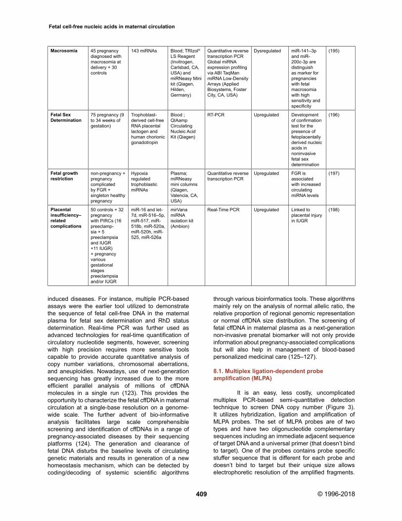
Macrosomia 45 pregnancy diagnosed with macrosomia at delivery + 30 controls
143 miRNAs Blood; TRIzol® LS Reagent (Invitrogen, Carlsbad, CA, USA) and miRNeasy Mini kit (Qiagen, Hilden, Germany)
Quantitative reverse transcription PCR Global miRNA expression profiling via ABI TaqMan miRNA Low-Density Arrays (Applied Biosystems, Foster City, CA, USA)
Dysregulated miR-141–3p and miR-200c-3p are distinguish as marker for pregnancies with fetal macrosomia with high sensitivity and specificity
(195)
Fetal Sex Determination
75 pregnancy (9 to 34 weeks of gestation)
Trophoblast-derived cell-free RNA placental lactogen and human chorionic gonadotropin
Blood ; QIAamp CirculatingNucleic Acid Kit (Qiagen)
RT-PCR Upregulated Development of confirmation test for the presence of fetoplacentally derived nucleic acids in noninvasivefetal sex determination
(196)
Fetal growth restriction
non-pregnancy + pregnancycomplicated by FGR + singleton healthy pregnancy
Hypoxia regulated trophoblastic miRNAs
Plasma; miRNeasy mini columns (Qiagen, Valencia, CA, USA)
Quantitative reverse transcription PCR
Upregulated FGR is associated with increased circulating miRNA levels
(197)
Placental insufficiency–related complications
50 controls + 32 pregnancywith PIRCs (16 preeclamp-sia + 5 preeclampsia and IUGR +11 IUGR) + pregnancy various gestationalstages preeclampsia and/or IUGR
miR-16 and let-7d, miR-516–5p, miR-517, miR-518b, miR-520a, miR-520h, miR-525, miR-526a
mirVana miRNA isolation kit (Ambion)
Real-Time PCR Upregulated Linked to placental injury in IUGR
(198)

Fetal cell-free nucleic acids in maternal circulation
410 © 1996-2018
Amplification of MLPA probes takes place only after ligation of both probes by thermostable ligase. This provides an advantage that unligated probe unable to amplify and to avoid nonspecific results. A different set of a universal primer pair is employed to amplify all the ligated probes, this provides the basis of amplification of MLPA probes that hybridize to the target sequence. The amplicons ranging between 130 and 481 nucleotides are usually separated using capillary electrophoresis. The whole process involves binding of each probe to target DNA, ligation of hybridized probes and subsequent amplification and electrophoresis, subsequent identification of DNA copy number of a specific genomic region (128).
8.2. DNA methylation-MLPA
This biomarker detection technique allows the study of DNA methylation based on the standard MLPA technique (Figure 4). Methylation profiling based on the MLPA technique uses methylation sensitive restriction enzyme and provides information about the methylation levels in target regions. Normally two
tubes are used, one with and other without restriction enzymes (RE). Initially, RE cleave at specific restriction sites on unmethylated DNA to discriminate them with their methylated counterparts. Later, undigested segments are amplified through thermocycler, leaving digested products. Capillary electrophoresis of PCR products generates data about the proportion of digested probes and, consequently, the proportion of methylated/unmethylated DNA is quantified (129).
8.3. Microarray-based comparative genomic hybridization
Array comparative genomic hybridization (aCGH) is the combination of microarray and comparative genomic hybridization and has many advantages over conventional cytogenetic methods such as high-throughput simultaneous detection, high resolution, attendant savings in labor and expense (Figure 5). This method has an ability to simultaneously detect multiple abnormalities such as deletions, duplications, aneuploidies, and/or amplifications of any locus represented on the array. aCGH possess the
Figure 3. A simplified figure illustrating different steps of multiplex ligation-dependent probe amplification (MLPA) method.

Fetal cell-free nucleic acids in maternal circulation
411 © 1996-2018
Figure 4. A sketch showing the multiple steps involved in the methylation specific multiplex ligation-dependent probe amplification method.
Figure 5. Representative demonstration of microarray-based comparative genomic hybridization for simultaneous detection of multiple abnormalities.

Fetal cell-free nucleic acids in maternal circulation
412 © 1996-2018
ability to assess all 46 chromosomes in a single test. The process of aCGH includes DNA extraction from test and reference, labeling with different fluorescent dye, mixing of both labeled DNA and then the application on the microarray slide, which are then arrayed with small segments of DNA as the targets. Hybridization of target DNA to its complementary DNA in test or reference is measured by digital imaging systems and quantifies the relative fluorescence intensities. A difference in the relative fluorescence of test and reference hybridized segments along the genome provides clue about the relative copy number of specific DNA region (130). aCGH ensures the quantitative assessment of individual exons also known as exon-by-exon or exon focused coverage which is beneficial to detect exonic duplications/deletions within genes (131).
8.4. SNP array
About 99.5% DNA sequence is approximately identical in two unrelated individuals. One of the most common differences is due to single nucleotide change where one has T but other has A at specific sites of the same genomic segment. These sites are named as single nucleotide polymorphism (SNP) and the SNP with a different base (A or T) are named as an allele. Most
common and less common alleles are called A or B, respectively. SNP array is the detection of the presence of a specific number of SNPs present at alleles of each individual (Figure 6). Analysis of intensity of each of the alleles of the SNPs facilitates the detection of deletions/duplications and heterozygosity and uniparental disomy status. Deletions reduce the fluorescent intensity while duplications increase the fluorescent intensity compared to the other. The relative intensity between the two alleles in each nucleotide tested confers heterozygous or homozygous positions (132).
8.5. Amplification refractory mutation system-polymerase chain (ARMS-PCR)
It is a simple and economical method which utilizes tetra-primer to genotype SNP and allows the detection of any mutation involving single base changes or small deletions. PCR followed by gel electrophoresis steps of this technique offers the analysis of SNPs in a fast, reliable, and low-cost way (133).
8.6. Methylation assay
Methylation assays are Infinium BeadChip based comprehensive genome-wide profiling of
Figure 6. A schematic representation of SNP array to detect heterozygous or homozygous positions and copy number imbalance.

Fetal cell-free nucleic acids in maternal circulation
413 © 1996-2018
human DNA methylation and allow interrogation of more than 485,000 DNA methylation sites per sample at single-base resolution (Figure 7). Methylation assay includes bisulfite treatment that converts cytosine into uracil but leaves methylated cytosine as it is. These bisulfite treated fragments are amplified by whole genomic amplification (WGA) using random hexamer primer and DNA polymerase. Amplified products are then fragmented enzymatically and applied on a microarray chip. The microarray chip contains two
unlike bead-bound probes for each interrogated methylated and unmethylated DNA. Probes attached to chip works as an allele-specific primer that differs only at the free end and used for single base extension in the next step. Dideoxynucleotide provides single base extension and their labeling with hapten emits fluorescence discrimination. Analysis of methylation data by software decodes fluorescent intensity ratio between two beads and provides detailed profiling of DNA methylation (134).
Figure 7. An outline of methylation assay for genome-wide profiling of DNA methylation.
Fetal cell-free nucleic acids in maternal circulation
414 © 1996-2018
8.7. Epityper
This tool provides quantitative analysis of DNA methylation by combining two methods, i.e. cleavage of bisulfite treated amplified DNA and matrix-assisted laser desorption/ionization time-of-flight mass spectrometry (MALDI-TOF MS) to reveal DNA methylation ratio. The whole process includes bisulfite treatment of genomic DNA followed by amplification that introduces a T7 promoter to be utilized for in vitro RNA transcription, the next step. RNase cleaves RNA specifically at cytosine and uracil for base specific cleavage. Cleaved products are then analyzed with MALDI-TOF MS analysis as a distinct signal pair pattern due to distinct methylated and non-methylated template DNA (135).
8.8. Sanger sequencing
This helps to reveal nucleotide content for a target sequence based on PCR. Sanger sequencing can read up to 1000 bases with a high accuracy. Simple PCR reaction utilized here has two kinds of nucleotide for PCR reaction - four deoxynucleotide and four differently labeled dideoxynucleotides. Dideoxynucleotides terminate the polymerization whenever added and allow size based separation. Amplified fragments are size separated by electrophoresis and the color is detected in order to predict the sequence. Automated Sanger sequencing is named as automated DNA sequencing that is the rapid advancement in sequencing technique aided by the use of capillary electrophoresis. Pyrosequencing allows real-time sequencing that during polymerization reaction one can get sequence up to 300–500 nucleotides (136).
8.9. Next generation sequencing
The advent of sequencing methods has allowed designing approaches for noninvasive diagnosis of fetal anomalies (137, 138). Importantly, these massively parallel sequencing strategies have gained significant importance specifically for clinical applications such as non invasive prenatal testing. This is mainly done to detect sub-chromosomal deletions/duplications and single-gene disorders. In addition, the deep sequencing of maternal plasma possesses the ability to reveal whole fetal genome and transcriptome which may help in delineating the critical information about fetal and maternal health. Interestingly, high-throughput DNA sequencing platforms also termed as next-generation sequencing (NGS) permits simultaneous sequencing of a huge amount of DNA molecules. NGS is fast, inexpensive, and accurate DNA sequencing at large-scale that it is capable to sequence millions of DNA fragments in a single reaction (Figure 8). All NGS methods are similar in measurement as they monitor the sequential
addition of nucleotides to immobilized and spatially arrayed DNA templates but have different method of template generation and analysis. Overview of NGS process includes sequential library preparation of double strand DNA extracted from the source. Sequential library preparation is done by fragmentation and size selection in smaller sequence-able segment depending upon sequencing platform’s specifications. Adapters are ligated to the ends of library fragments that meant to act as a primer in subsequent reactions. The prepared library is sequenced either through direct sequencing (single molecule template) or amplified and then sequence via clonally amplified templates (139). However, these sequencing methodologies are restricted by the availability of the right library.
8.10. Exome sequencing
Exome sequencing or whole exome sequencing allows identification of small insertions or deletions (indels), single-nucleotide variants (SNVs), and rare de novo mutations take place at all expressed genes in the genome that may generate a disease phenotype (140).
8.11. Nanopore sequencing
It is a low cost and relatively fast new developing DNA sequencing tool with single base precision. In this technique, a nanopore hole is utilized to channelize DNA through the hole containing sensitive optics (Figure 9). The channelization of DNA through hole is driven by the membrane potential to drive DNA from one side to another. All four unique size and charge of nucleotides provide a unique electrical signal that indicates the order of nucleotide to generate data (141).
8.12. Droplet digital PCR
This method utilizes the principles of microfluidics along with surfactant chemistries to split the PCR templates into a water-oil emulsion droplet system. These droplets functions identical to the wells/test tube of a PCR system and the amplification is done inside each droplet. In comparison to other PCR systems, the method requires less amount of starting sample, hence, minimizes the cost and loss of samples (142).
8.13. Quantitative reverse transcription PCR
Reverse transcriptase (RT) based PCR methodology is utilized when the target is to quantify RNA. The method exploits the ability of RT enzyme to form complementary DNA (cDNA) from RNA. Thus formed cDNA act as a template and gets amplified through regular PCR-based assay and gets detected. This can be done either in a single step (one step PCR) i.e. reverse transcription and PCR amplification

Fetal cell-free nucleic acids in maternal circulation
415 © 1996-2018
is done in one tube by utilizing sequence-specific primers or in two divided steps (two-step PCR) i.e cDNA formation and amplification in two different tubes (143).
9. BIO-INFORMATICS APPROACH FOR ANALYSIS OF CIRCULATING cffDNA
9.1. Estimation of fractional fetal DNA concentration
This can be analyzed by a polymorphism dependent or an independent approach. The dependent method either precisely involve paternal and maternal genotype to assess the concentration of fetal DNA i.e. parental-genotype dependent or can be performed in an independent mode which does not engage any parental genotype but depends on the allelic distribution in maternal plasma to assume fetal DNA concentration. Moreover, the polymorphism independent method is free from the dependency of parental genotype and assesses the fetal DNA concentration by utilizing the biological dissimilarity among maternal and cffDNA bio-molecules (137, 144, 145).
9.2. Fetal aneuploidy detection
Fetal aneuploidy in singleton pregnancies can be detected by two methods, i.e. whole-genome approach and the targeted approach (137, 146). The whole-genome approach can be either based on Tag-counting method in which each sequenced read is specifically mapped to the human genome or it utilizes size differences among maternal and fetal DNA (size based analysis) (137). Another method for aneuploidy analysis is the targeted approach, which is performed to enhance the sequencing depth at a specific targeted point with decrease in the genome-wide coverage. This method can be done either by allelic ratio analysis (ratio between the fetal specific alleles and shared alleles), dosage-type analysis (utilizes z-test to evaluate the chromosome over-representation) or by Bayesian-based maximal likelihood method (relies in PS algorithm based combinations).
9.3. Monogenic disorders detection
Increased ability to sequence whole fetal genome circulating in the maternal plasma
Figure 8. Figure showing an overview of the next generation sequencing technique.

Fetal cell-free nucleic acids in maternal circulation
416 © 1996-2018
has enabled researchers to cross-examine the complete fetal genome to diagnose the probability of possible monogenic diseases. This can be done by implementing different canonical algorithms i.e. relative haplotype dosage analysis (RHDO), haplotype counting approach and hidden Markov model (HMM). The RHDO analyses the allelic imbalance among the two maternal haplotypes and statistical inference of the feto-maternal inheritance while HMM relies on the determination of the relative expression of the parent haplotype pairs and is based on the latent state (144, 147).
9.4. Methylomic analysis of circulation cffDNA
Analysis of whole-genome methylome has facilitated the detection of disease specific methylated regions which can be used for disease diagnosis and monitoring. Assessment can be done by bisulfite treatment which converts cytosine residues to uracil without disturbing the methylated cytosines. These uracils are then amplified through PCR cycles thereby revealing the sequence methylation status (148). Different bisulfite treatment based sequence
alignment tools to have been developed and some of them also possess the ability to recognize differentiated methylated regions (DMRs) (149–151). Among these, BSmooth (152) and Methy-Pipe (153) have the ability to carry out sequence alignment, determination of methylation level, identification of DMR identification, and annotation of DMR. However, Methy-Pipe possesses the faster alignment ability than other packages including Bismark (154). The integrative nature of these two bioinformatics packages renders them well-suited for comprehensive analysis of bisulfite sequencing data. Importantly, available bioinformatics tools had also enabled noninvasive analysis of prenatal methylome (155). This can be approached either relying on the examination of fetal-specific polymorphic alleles or by assessing the fractional fetal DNA concentration and blood cells methylome to back analyze the placental methylome. Such prenatal analyses are used for noninvasive detection of trisomy 21. These methods may not only assist to know the placental- or fetal-specific methylation at individual CpG residues but also help to understand the associated fundamental mechanisms.
Figure 9. An outline of the basic steps involved in nanopore sequencing methodology.

Fetal cell-free nucleic acids in maternal circulation
417 © 1996-2018
10. FUTURE DIRECTIONS
The study of extracellular nucleic acids in body fluids such as serum, plasma, saliva, urine, milk, seminal plasma, tears, and amniotic fluids, as circulating entities capable of predicting the course of a wide range of diseases is now considered as the “holy grail” of non-invasive diagnosis. Since the discovery of cffDNA in maternal plasma in 1997 by Professor Dennis Lo, Director, Li Ka Shing Institute of Health Sciences at the Chinese University of Hong Kong, there has been significant progress in exploring these novel signatures as a source of fetal genetic material for prenatal diagnosis. Lo and colleagues demonstrated the presence of Y-chromosome-specific sequences in plasma of women who were carrying male fetuses using a real-time quantitative PCR-based approach. This discovery led to the opening of enormous opportunities to consider other applications of cffDNA in reproductive health research. The cffDNA originates in the maternal circulation when normal placental cell apoptosis causes the chromosomes to break into short fragments. Detectable in the maternal circulation from around 5 weeks’ gestation, the majority of cffDNA is of fetal origin, most of which are under 300 bp in length. As pregnancy advances, the proportion of cffDNA in maternal blood also increase that constitutes 3 - 13% of the total cell-free maternal DNA pool during the first and second trimester. cffDNA can be detected reliably as early as the seventh week of gestation and is primarily considered to be derived from apoptotic and trophoblastic cells in the placenta. As opposed to the earlier search for fetal cells in maternal circulation, methods for isolation and molecular characterization of cffDNA proved to be both specific and sensitive in various laboratories with high reproducibility. In addition, cffDNA had the advantage of being rapidly cleared from the maternal circulation after delivery, which essentially means, none remaining in circulation after infant’s birth except for cases where small amounts stay behind, including cells from previous pregnancies. Initially, because of the technological limitations to precisely identify cffDNA within the high pool of maternal cffDNA, application of this technology was specifically focused on settings in which the paternal, and potentially, the fetal genotypes differed from the mother’s. So, first clinical utilization of this phenomenon focused on the detection or exclusion of alleles that were ‘not’ present in the mother but were present in the fetus because they were paternally inherited or emerged de novo during conception. By the late ’90s, this technology was largely employed for fetal sex determination using Y-chromosome alleles, fetal Rhesus D (RhD) genotyping in RhD-negative mothers and gene mutations of paternal origin. The advent of omics-based technologies such as NGS enabled the precise identification of cffDNA sequences associated with specific chromosomes present in maternal blood. In addition, this technology proved to
be an extremely useful aid for quantification of cffDNA in the early identification of pregnancies at risk of other adverse outcomes, such as preeclampsia, HELLP syndrome (hemolysis elevated liver enzymes, and low platelet count) and intra-uterine growth retardation (IUGR). At the moment, it seems that massively parallel sequencing of fetal DNA from maternal blood using NGS technologies has enormous potential, not only for increasing our understanding of the causes of prenatal genetic disorders in the fetus but also for designing non-invasive clinical diagnostic tests. Despite being highly accurate and sensitive, NGS-based non-invasive prenatal diagnosis technologies have several limitations: (i) longer turnaround time of the test; (ii) high reagent and equipment costs; and (iii) significant percentage of cases, where the diagnosis cannot be made due to insufficient cffDNA content. Therefore, while deciding, these additional factors must be carefully considered in addition to the diagnostic specificity, sensitivity, and scalability. The idea of clinical efficacy may comprise elements of whether the clinical outcomes are effective and whether its implementation offers an economically efficient solution compared to alternative methods. Moreover, there are some discrepancies associated with NGS technologies that may be worth further pursuit in depth. One of the major bottlenecks is the ligation of cell-free DNA fragments to specific linkers before amplification and sequencing which might display a bias against large or very small fragments. Therefore, perhaps it may be necessary to use a couple of technologies together to unequivocally address this complex issue. Unlike cffDNA, knowledge about the diagnostic utility of circulating epigenetic signatures: methylated DNA; miRNA and post translationally modified histones are deficient. Moreover, categorized understanding of these novel entities through omics-based molecular technologies might also prompt development of a range of laboratory-based approaches, thus improving their broader translational utility for early fetal disease diagnosis. Largest opportunity for innovation lies in developing low-cost and acceptable platform technologies with accurate diagnostic and higher prognostic score. Ideally, the test must be: (i) facile; (ii) robust; (iii) rapid; (iv) technically uncomplicated; (v) reasonably priced; (vi) sensitive; (vii) specific; and (viii) applicable to specimens that are readily acquired. Such transformative tests might greatly reduce the disease burden; however, it is pertinent to indicate some of the pragmatic impediments associated with development and deployment of these investigative strategies. Besides successful validation in different clinical resource settings, regulatory hurdles required for approval or clearance of such tests might pose an integral challenge. Moreover, none of the omics-based molecular technologies are ideal and compatible as portable point-of-care devices. Therefore, to achieve rapid, highly sensitive, selective and reproducible results in a complex matrix such as plasma or serum,
Fetal cell-free nucleic acids in maternal circulation
418 © 1996-2018
“nano-biosensor” based detection methods may offer significant advantages, as these coalesces the use of a selective nano-material with biomolecular recognition species. These will allow rapid, economical, and repeat sampling features that permit their use in screening programs as well. Successful validation in different clinical resource settings; higher cost; and regulatory hurdles required for approval or clearance of such tests are the three major bottlenecks of the approach. The challenges are enormous but a cutting-edge nano-engineered approach may help to set priorities for establishing cffDNA in maternal circulation as a next generation diagnostic marker in fetal medicine.
11. ACKNOWLEDGEMENT
NB and AB contributed equally in this manuscript. The authors thank the Indian Council of Medical Research, Department of Science & Technology, Department of Biotechnology, and the Ministry of Human Resource & Development, Government of India, New Delhi for providing necessary financial support to the laboratory of Prof. (Dr.) Pradyumna Kumar Mishra.
12. REFERENCES
1. L. V. Speybroeck: From epigenesis to epigenetics. Ann NY Acad Sci 981, 61–81 (2002) DOI: 10.1111/j.1749-6632.2002.tb04912.x
2. M. K. Skinner: Environmental stress and epigenetic transgenerational inheritance. Mol Cell Endocrinol 398(1–2), 4–12 (2014) DOI: 10.1016/j.mce.2014.07.019
3. World Health Organization, 2016: Accessed on March 2, 2017
http://www.who.int/mediacentre/factsheets/fs370/en/
4. A. Kar, S. Phadnis, S. Dharmarajan, J. Nakade: Epidemiology & social costs of haemophilia in India. Indian J Med Res 140, 19–31 (2014)
PMID:PMC418115
5. N. Cordeiro, M. Tsimis, I. Burd: Infections and brain development. Obstet Gynecol Surv 70(10), 644–655 (2015)DOI: 10.1097/OGX.0000000000000236
6. A. Das, M. Sarkar: Pregnancy-related health information-seeking behaviors among rural pregnant women in India: validating the Wilson model in the Indian context. Yale J Biol Med 87(3), 251–62 (2014)
PMCID:PMC4144280
7. D. Chitayat, D. Matsui, Y. Amitai, D. Kennedy, S. Vohra, M. Rieder, G. Koren: Folic acid supplementation for pregnant women and those planning pregnancy: 2015 update. J Clin Pharmacol 56(2), 170–175 (2016)DOI: 10.1002/jcph.616
8. H. N. Munro, S. J. Pilistine, M. E. Fant: The placenta in nutrition. Ann Rev Nutr 3, 97–124 (1983)DOI: 10.1146/annurev.nu.03.070183. 000525
9. R. L. Jirtle, M. K. Skinner: Environmental epigenomics and disease susceptibility. Nat Rev Genet 8, 253–262 (2007)DOI: 10.1038/nrg2045
10. F. Perera, J. Herbstman: Prenatal environmental exposures, epigenetics, and disease. Repro Toxicol 31 (3), 363–373 (2011)DOI: 10.1016/j.reprotox.2010.12.055
11. A. Bhargava, R. P. Punde, N. Pathak, S. Dabadghao, P. Desikan, A. Jain, K. K. Maudar, P. K. Mishra: Status of inflammatory biomarkers in the population that survived the Bhopal gas tragedy: a study after two decades. Ind Health 48(2), 204–208 (2010)DOI: 10.13075/ijomeh.1896.00313
12. P. K. Mishra, G.V. Raghuram, N. Bunkar, A. Bhargava, N. K. Khare: Molecular bio-dosimetry for carcinogenic risk assessment in survivors of Bhopal gas tragedy. Int J Occup Med Environ Health 28(6), 921–939 (2015)
13. S. Bose-O’Reilly, K. M. McCarty, N. Steckling, B. Lettmeier: Mercury exposure and children’s health. Curr Probl Pediatr Adolesc Health Care 40(8), 186–215 (2010)DOI: 10.1016/j.cppeds.2010.07.002
14. S. L. Schantz: Developmental neurotoxicity of PCBs in humans: what do we know and where do we go from here? Neurotoxicol Teratol 18(3), 217–327; discussion 229–276 (1996)DOI: 10.1016/S0892-0362(96)90001-X
15. A. Wesselink, M. Warner, S. Samuels, A. Parigi, P. Brambilla, P. Mocarelli, B. Eskenazi: Maternal dioxin exposure and pregnancy outcomes over 30 years of follow-up in Seveso. Environ Int 63, 143–148 (2014)DOI: 10.1016/j.envint.2013.11.005
16. P. K. Mishra, S. Dabadghao, G. K. Modi, P. Desikan, A. Jain, I. Mittra, D. Gupta,

Fetal cell-free nucleic acids in maternal circulation
419 © 1996-2018
C. Chauhan, S. K. Jain, K. K. Maudar: In utero exposure to methyl isocyanate in the Bhopal gas disaster: evidence of persisting hyperactivation of immune system two decades later. Occup Environ Med 66, 279 (2009a)DOI: 10.1136/oem.2008.041517
17. P. K. Mishra, R. M. Samarth, N. Pathak, S. K. Jain, S. Banerjee, K. K. Maudar: Bhopal Gas Tragedy: review of clinical and experimental findings after 25 years. Int J Occup Med Environ Health 22(3), 193–202 (2009b)DOI: 10.2478/v10001-009-0028-1
18. P. K. Mishra: A pragmatic & translational approach of human biomonitoring to methyl isocyanate exposure in Bhopal. Indian J Med Res 135 (4), 479–484 (2012)
19. M. A. Dawson, T. Kouzarides: Cancer epigenetics: from mechanism to therapy. Cell 150(1), 12–27 (2012)DOI: 10.1016/j.cell.2012.06.013
20. N. Bunkar, N. Pathak, N.K. Lohiya, P.K. Mishra: Epigenetics: A key paradigm in reproductive health. Clin Exp Reprod Med 43(2), 59–81 (2016)DOI: 10.5653/cerm.2016.43.2.59
21. R. A. Waterland, K. B. Michels: Epigenetic Epidemiology of the Developmental Origins Hypothesis. Annu Rev Nutr 27, 363–88 (2007)DOI: 10.1146/annurev.nutr.27.061406.093705
22. H. Fukuoka, F. Sata: Molecular mechanism of developmental origins of health and disease (DOHaD). Nihon Eiseigaku Zasshi 71(3), 185–187 (2016)DOI: 10.1265/jjh.71.185
23. P. K. Mishra, N. Bunkar, G. V. Raghuram, N. K. Khare, N. Pathak, A. Bhargava: Epigenetic dimension of oxygen radical injury in spermatogonial epithelial cells. Reprod Toxicol 52, 40–56 (2015)DOI: 10.1016/j.reprotox.2015.02.006
24. K. D. Robertson: DNA methylation and human disease. Nat Rev Genet 6, 597–610 (2005)DOI: 10.1038/nrg1655
25. R. G. Urdinguio, J. V. Sanchez-Mut, M. Esteller: Epigenetic mechanisms in neurological diseases: genes, syndromes, and therapies. Lancet Neurol 8, 1056–1072 (2009)DOI: 10.1016/S1474-4422(09)70262-5
26. A. Portela, M. Esteller: Epigenetic modifications and human disease. Nat Biotechnol 28, 1057–1068 (2010)DOI: 10.1038/nbt.1685
27. T. Vaissière, C. Sawan, Z. Herceg: Epigenetic interplay between histone modifications and DNA methylation in gene silencing. Mutat Res 659(1–2), 40–48 (2008)DOI: 10.1016/j.mrrev.2008.02.004
28. T. H. Bestor: The DNA methyltransferases of mammals. Hum Mol Genet 9(16), 2395–2402 (2000)DOI: 10.1093/hmg/9.16.2395
29. W. Reik, W. Dean: DNA methylation and mammalian epigenetics. Electrophoresis 22, 2838–2843 (2001)DOI: 10.1002/1522-2683(200108)22:14< 2838::AID-ELPS2838>3.0.CO;2-M
30. O. Bogdanovic, G. J. Veenstra: DNA methylation and methyl-CpG binding proteins: developmental requirements and function. Chromosoma 118, 549–565 (2009)DOI: 10.1007/s00412-009-0221-9
31. M. Tahiliani, K. P. Koh, Y. Shen, W. A. Pastor, H. Bandukwala, Y. Brudno, S. Agarwal, L. M. Iyer, D. R. Liu, L. Aravind, A. Rao: Conversion of 5-methylcytosine to 5-hydroxymethylcytosine in mammalian DNA by MLL partner TET1. Science 324, 930–935 (2009)DOI: 10.1126/Science.1170116
32. F. Gaudet, J. G. Hodgson, A. Eden, L. Jackson-Grusby, J. Dausman, J. W. Gray, H. Leonhardt, R. Jaenisch: Induction of tumors in mice by genomic hypomethylation. Science 300(5618), 489–492 (2003)DOI: 10.1126/Science.1083558
33. K. W. Jair, K. E. Bachman, H. Suzuki, A. H. Ting, I. Rhee, R. W. Yen, S. B. Baylin, K. E. Schuebel: De novo CpG island methylation in human cancer cells. Cancer Res 66(2), 682–692 (2006)DOI: 10.1158/0008-5472.CAN-05-1980
34. S. Peleg, F. Sananbenesi, A. Zovoilis, S. Burkhardt, S. Bahari-Javan, R.C. Agis-Balboa, P. Cota, J.L. Wittnam, A. Gogol-Doering, L. Opitz, G. Salinas-Riester, M. Dettenhofer, H. Kang, L. Farinelli, W. Chen, A. Fischer:. Altered histone acetylation is associated with age-dependent memory

Fetal cell-free nucleic acids in maternal circulation
420 © 1996-2018
impairment in mice. Science 328(5979), 753–756 (2010)DOI: 10.1126/Science.1186088
35. A.V. Murgatroyd, Y. Patchev, V. Wu, Y. Micale, D. Bockmuhl, F. Fischer, C. Holsboer, T. Wotjak, O. F. Almeida, D. Spengler: Dynamic DNA methylation programs persistent adverse effects of early-life stress. Nat Neurosci 12(12), 1559–1566 (2009)DOI: 10.1038/nn.2436
36. X. Dong, Z. Weng: The correlation between histone modifications and gene expression. Epigenomics 5(2), 113–116 (2013)DOI: 10.2217/epi.13.13
37. S. D. Taverna, H. Li, A. J. Ruthenburg, C. D. Allis, D. J. Patel: How chromatin-binding modules interpret histone modifications: lessons from professional pocket pickers. Nat Struct Mol Biol 14, 1025–1040 (2007)DOI: 10.1038/nsmb1338
38. C. Choudhary, C. Kumar, F. Gnad, M. L. Nielsen, M. Rehman, T. C. Walther, J. V. Olsen, M. Mann: Lysine acetylation targets protein complexes and co-regulates major cellular functions. Science 325, 834–840 (2009)DOI: 10.1126/Science.1175371
39. M. Bantscheff, C. Hopf, M. M. Savitski, A. Dittmann, P. Grandi, A. M. Michon, J. Schlegl, Y. Abraham, I. Becher, G. Bergamini, M. Boesche, M. Delling, B. Dümpelfeld, D. Eberhard, C. Huthmacher, T. Mathieson, D. Poeckel, V. Reader, K. Strunk, G. Sweetman, U. Kruse, G. Neubauer, N. G. Ramsden, G. Drewes: Chemoproteomics profiling of HDAC inhibitors reveals selective targeting of HDAC complexes. Nat Biotechnol 29, 255–265 (2011) DOI: 10.1038/nbt.1759
40. W. Chung, J. Witherington: Progress in the discovery of small molecule inhibitors of bromodomain histone interactions. J Biomol Screen 16, 1170–1185 (2011)DOI: 10.1177/1087057111421372
41. M. Federico, L. Bagella: Histone deacetylase inhibitors in the treatment of hematological malignancies and solid tumors. J Biomed Biotechnol 2011, 475641 (2011)DOI: 10.1155/2011/475641
42. L. Pasqualucci, D. Dominguez-Sola, A. Chiarenza, G. Fabbri, A. Grunn, V. Trifonov,
L.H. Kasper, S. Lerach, H. Tang, J. Ma, D. Rossi, A. Chadburn, V.V. Murty, C. G. Mullighan, G. Gaidano, R. Rabadan, P. K. Brindle, R. Dalla-Favera: Inactivating mutations of acetyltransferase genes in B-cell lymphoma. Nature 471(7337), 189–195 (2011)DOI: 10.1038/nature09730
43. Y. Shi: Histone lysine demethylases: emerging roles in development, physiology and disease. Nat Rev Genet 8, 829–833 (2007)DOI: 10.1038/nrg2218
44. A. Barski, S. Cuddapah, K. Cui, T.-Y. Roh, D. E. Schones, Z. Wang, G. Wei, I. Chepelev, K. Zhao: High-resolution profiling of histone methylations in the human genome. Cell 129, 823–837 (2007)DOI: 10.1016/j.cell.2007.05.009
45. N. Mosammaparast, Y. Shi: Reversal of histone methylation: biochemical and molecular mechanisms of histone demethylases. Annu Rev Biochem 79, 155–179 (2010)DOI: 10.1146/annurev.biochem.78.070907. 103946
46. A. Di Lorenzo, M. T. Bedford: Histone arginine methylation. FEBS Lett 585(13), 2024–2031 (2011)DOI: 10.1016/j.febslet.2010.11.010
47. M. Stucki, J. A. Clapperton, D. Mohammad, M. B. Yaffe, S. J. Smerdon, S. P. Jackson: MDC1 directly binds phosphorylated histone H2AX to regulate cellular responses to DNA double-strand breaks. Cell 123, 1213–1226 (2005)DOI: 10.1016/j.cell.2005.09.038
48. M. A. Dawson, A. J. Bannister, B. Go¨ ttgens, S. D. Foster, T. Bartke, A. R. Green, T. Kouzarides: JAK2 phosphorylates histone H3Y41 and excludes HP1alpha from chromatin. Nature 461, 819–822 (2009)DOI: 10.1038/nature08448
49. S. H. Baek: When signaling kinases meet histones and histone modifiers in the nucleus. Mol Cell 42, 274–284 (2011)DOI: 10.1016/j.molcel.2011.03.022
50. L. S. Trevi-o, Q. Wang, C. L. Walker: Phosphorylation of epigenetic “readers, writers and erasers”: Implications for developmental reprogramming and the

Fetal cell-free nucleic acids in maternal circulation
421 © 1996-2018
epigenetic basis for health and disease. Prog Biophys Mol Biol 118(1–2), 8–13 (2015)DOI: 10.1016/j.pbiomolbio.2015.02.013
51. U. L. McClurg, C. N. Robson: Deubiquitinating enzymes as oncotargets. Oncotarget 6(12), 9657–9668 (2015)DOI: 10.18632/oncotarget.3922
52. R. Meas, P. Mao: Histone ubiquitination and its role in transcription and DNA damage response. DNA Repair (Amst.) 36, 36–42 (2015)DOI: 10.1016/j.dnarep.2015.09.016
53. P. P. Amaral, M. E. Dinger, T. R. Mercer, J. S. Mattick: The eukaryotic genome as an RNA machine. Science 319, 1787–1789 (2008)DOI: 10.1126/Science.1155472
54. K. C. Wang, H. Y. Chang: Molecular mechanisms of long noncoding RNAs. Mol Cell 43, 904–914 (2011)DOI: 10.1016/j.molcel.2011.08.018
55. E. Huntzinger, E. Izaurralde: Gene silencing by microRNAs: contributions of translational repression and mRNA decay. Nat Rev Genet 12, 99–110 (2011)DOI: 10.1038/nrg2936
56. S. Jonas, E. Izaurralde: Towards a molecular understanding of microRNA-mediated gene silencing. Nat Rev Genet 16, 421–433 (2015)DOI: 10.1038/nrg3965
57. S. Seisenberger, J. R. Peat, T. A. Hore, F. Santos, W. Dean, W. Reik: Reprogramming DNA methylation in the mammalian life cycle: building and breaking epigenetic barriers. Philos Trans R Soc Lond B Biol Sci 368(1609), 20110330 (2013)DOI: 10.1098/rstb.2011.0330
58. P. K. Mishra, G. V. Raghuram, D. Jain, S. K. Jain, N. K. Khare, N. Pathak: Mitochondrial oxidative stress-induced epigenetic modifications in pancreatic epithelial cells. Int J Toxicol 33(2), 116–129 (2014)DOI: 10.1177/1091581814524064
59. G. V. Raghuram, N. Pathak, D. Jain, H. Pandey, H. Panwar, S. K. Jain, S. Banerjee, P. K. Mishra: Molecular characterization of isocyanate-induced male germ-line genomic instability. J Environ Pathol Toxicol Oncol 29(3), 213–234. (2010)DOI: 10.1615/JEnvironPatholToxicolOncol.v29.i3.50
60. G. V. Raghuram, N. Pathak, D. Jain, H. Panwar, H. Pandey, S. K. Jain, P. K. Mishra: Molecular mechanisms of isocyanate induced oncogenic transformation in ovarian epithelial cells. Reprod Toxicol 30(3), 377–386 (2010)DOI: 10.1016/j.reprotox.2010.05.087
61. M. A. Hanson, M. K. Skinner: Developmental origins of epigenetic transgenerational Inheritance. Environ Epigenet 2(1), dvw002 (2016)DOI: 10.1093/eep/dvw002
62. Y. Kawasaki, J. Lee, A. Matsuzawa, T. Kohda, T. Kaneko-Ishino, F. Ishino: Active DNA demethylation is required for complete imprint erasure in primordial germ cells. Sci Rep 4, 3658 (2014)DOI: 10.1038/srep03658
63. M. DeSilva, F. M. Munoz, M. Mcmillan, A. T. Kawai, H. Marshall, K. K. Macartney, J. Joshi, M. Oneko, A. E. Rose, H. Dolk, F. Trotta, H. Spiegel, S. Tomczyk, A. Shrestha, S. Kochhar, E. O. Kharbanda: Congenital anomalies: Case definition and guidelines for data collection, analysis, and presentation of immunization safety data. Vaccine 34(49), 6015–6026 (2016)DOI: 10.1016/j.vaccine.2016.03.047
64. E. Scheuerle, A. S. Aylsworth: Birth defects and neonatal morbidity caused by teratogen exposure after the embryonic period. Birth Defects Res A Clin Mol Teratol 106(11), 935–939 (2016)DOI: 10.1002/bdra.23555
65. L. A. Nielsen, L. L. Maroun, H. Broholm, H. Laursen, N. Graem: Neural tube defects and associated anomalies in a fetal and perinatal autopsy series. APMIS 114(4), 239–246 (2006)DOI: 10.1111/j.1600-0463.2006.apm_325.x
66. L. Csabay, I. Szabó, C. Papp, E. Tóth-Pál, Z. Papp: Central nervous system anomalies. Ann N Y Acad Sci 847, 21–45 (1998)DOI: 10.1111/j.1749-6632.1998.tb08924.x
67. D. Taylor: Developmental abnormalities of the optic nerve and chiasm. Eye 21, 1271–1284 (2007)DOI: 10.1038/sj.eye.6702851
68. T. E. O’Toole, D. J. Conklin, A. Bhatnagar: Environmental risk factors for heart disease. Rev Environ Health 23(3), 167–202 (2008)DOI: 10.1515/REVEH.2008.23.3.167

Fetal cell-free nucleic acids in maternal circulation
422 © 1996-2018
69. K. E. Cosselman, A. Navas-Acien, J. D. Kaufman: Environmental factors in cardiovascular disease. Nat Rev Cardiol 12, 627–642 (2015)DOI: 10.1038/nrcardio.2015.152
70. J. I. Hoffman: Natural history of congenital heart disease. Problems in its assessment with special reference to ventricular septal defects. Circulation 37(1), 97–125 (1968)DOI: 10.1161/01.CIR.37.1.97
71. P. A. Trainor: Craniofacial birth defects: The role of neural crest cells in the etiology and pathogenesis of Treacher Collins syndrome and the potential for prevention. Am J Med Genet A 152A (12), 2984–2994 (2010)DOI: 10.1002/ajmg.a.33454
72. J. Keller, D. Frederking, P. Layer: The spectrum and treatment of gastrointestinal disorders during pregnancy. Nat Clin Pract Gastroenterol Hepatol 5, 430–443 (2008)DOI: 10.1038/ncpgasthep1197
73. S. I. Borah, A. K. Bhattacharjee, R. Baruah: Musculoskeletal birth defects at a tertiary centre and associated maternal risk factors. IJSR 5(11), 1823–1826 (2016)
74. Y. M. Lo, N. Corbetta, P. F. Chamberlain, V. Rai, I. L. Sargent, C. W. Redman, J. S. Wainscoat: Presence of fetal DNA in maternal plasma and serum. Lancet 350(9076), 485–487 (1997)DOI: 10.1016/S0140-6736(97)02174-0
75. G. Tounta, A. Kolialexi, N. Papantoniou, G. T. Tsangaris, E. Kanavakis, A. Mavrou: Non-invasive prenatal diagnosis using cell-free fetal nucleic acids in maternal plasma: Progress overview beyond predictive and personalized diagnosis. EPMA J 2(2), 163–171 (2011)DOI: 10.1007/s13167-011-0085-y
76. J. O. Kitzman, M. W. Snyder, M. Ventura, A. P. Lewis, R. Qiu, L. E. Simmons, H. S. Gammill, C. E. Rubens, D. A. Santillan, J. C. Murray, H. K. Tabor, M. J. Bamshad, E. E. Eichler, J. Shendure: Noninvasive whole-genome sequencing of a human fetus. Sci Transl Med 4(137), 137ra76 (2012)DOI: 10.1126/scitranslmed.3004323
77. K. Sun, P. Jiang, K. C. Chan, J. Wong, Y. K. Cheng, R. H. Liang, W. K. Chan, E. S. Ma, S. L. Chan, S. H. Cheng, R. W. Chan, Y. K.
Tong, S. S. Ng, R. S. Wong, D. S. Hui, T. N. Leung, T. Y. Leung, P. B. Lai, R. W. Chiu, Y. M. Lo: Plasma DNA tissue mapping by genome-wide methylation sequencing for noninvasive prenatal, cancer, and transplantation assessments. Proc Natl Acad Sci U S A 112(40), E5503- E5512 (2015)DOI: 10.1073/pnas.1508736112
78. R. Thierry, F. Mouliere, C. Gongora, J. Ollier, B. Robert, M. Ychou, M. Del Rio, F. Molina: Origin and quantification of circulating DNA in mice with human colorectal cancer xenografts. Nucleic Acids Res 18, 6159–6175 (2010)DOI: 10.1093/nar/gkq421
79. A. Bhargava. N. K. Khare, N. Bunkar, K. Chaudhury, K. C. Pandey, S. K. Jain, P. K. Mishra: Cell-free circulating epigenomic signatures: Non-invasive biomarker for cardiovascular and other age-related chronic diseases. Curr Pharm Des 23(8), 1175–1187 (2017) DOI: 10.2174/1381612822666161027145359
80. M. L. Tjoa, T. Cindrova-Davies, O. Spasic-Boskovic, D. W. Bianchi, G. J. Burton: Trophoblastic oxidative stress and the release of cell-free feto-placental DNA. Am J Pathol 169(2), 400–404 (2006)DOI: 10.2353/ajpath.2006.060161
81. Y. M. D. Lo, M. S. C. Tein, T. K. Lau, C. J. Haines, T. N. Leung, P. M. K. Poon, J. S. Wainscoat, P. K. Johnson, A. M. Z. Chang, N. M. Hjelm: Quantitative analysis of fetal DNA in maternal plasma and serum: implications for noninvasive prenatal diagnosis. Am J Hum Genet 62, 768–775 (1998)DOI: 10.1086/301800
82. S. Illanes, M. Denbow, C. Kailasam, K. Finning, P. W. Soothill: Early detection of cell-free fetal DNA in maternal plasma. Early Hum Dev 83, 563–566 (2007)DOI: 10.1016/j.earlhumdev.2006.11.001
83. Y. M. D. Lo, J. Zhang, T. N. Leung, T. K. Lau, A. M. Z. Chang, N. M. Hjelm: Rapid clearance of fetal DNA from maternal plasma. Am J Hum Genet 64, 218–224 (1999)DOI: 10.1086/302205
84. R. J. Levine, C. Qian, E. S. Leshane, K. F. Yu, L. J. England, E. F. Schisterman, T. Wataganara, R. Romero, D. W. Bianchi: Two-stage elevation of cell-free fetal DNA in

Fetal cell-free nucleic acids in maternal circulation
423 © 1996-2018
maternal sera before onset of preeclampsia. Am J Obstet Gynecol 190, 707–713 (2004)DOI: 10.1016/j.ajog.2003.12.019
85. N. L. Vora, K. L. Johnson, S. Basu, P. M. Catalano, S. Hauguel-De Mouzon, D. W. Bianchi: A multifactorial relationship exists between total circulating cell-free DNA levels and maternal BMI. Prenat Diagn 32, 912–914 (2012)DOI: 10.1002/pd.3919
86. S. Perlado, A. Bustamante-Aragonés, M. Donas, I. Lorda-Sánchez, J. Plaza, M. Rodríguez de Alba: Fetal genotyping in maternal blood by digital PCR: towards NIPD of monogenic disorders independently of parental origin. PLoS One 11(4), e0153258 (2016)DOI: 10.1371/journal.pone.0153258
87. X. P. Xu, H. Y. Gan, F. X. Li, Q. Tian, J. Zhang, R. L. Liang, M. Li, X. X. Yang, Y. S. Wu: A method to quantify cell-free fetal DNA fraction in maternal plasma using next generation sequencing: its application in non-invasive prenatal chromosomal aneuploidy detection. PLoS One 11(1), e0146997 (2016)DOI: 10.1371/journal.pone.0146997
88. D. E. Lee, S. Y. Kim, J. H. Lim, S. Y. Park, H. M. Ryu: Non-invasive prenatal testing of trisomy 18 by an epigenetic marker in first trimester maternal plasma. PLoS One 8 (11), e78136 (2013)DOI: 10.1371/journal.pone.0078136
89. A. Dokras, L. M. Gardner, D. A. Kirschmann, E. A. Seftor, M. J. Hendrix: The tumour suppressor gene maspin is differentially regulated in cytotrophoblasts during human placental development. Placenta 23, 274–280. 2002DOI: 10.1053/plac.2001.0784
90. S. S. Chim, Y. K. Tong, R. W. Chiu, T. K. Lau, T. N. Leung, L. Y. Chan, C. B. Oudejans, C. Ding, Y. M. Lo: Detection of the placental epigenetic signature of the maspin gene in maternal plasma. Proc Natl Acad Sci U S A 102(41), 14753–14758 (2005)DOI: 10.1073/pnas.0503335102
91. E.A. Papageorgiou, A. Karagrigoriou, E. Tsaliki, V. Velissariou, N. P. Carter, P. C. Patsalis: Fetal-specific DNA methylation ratio permits noninvasive prenatal diagnosis
of trisomy 21. Nat Med 17(4), 510–513 (2011)DOI: 10.1038/nm.2312
92. L. L. Poon, T. N. Leung, T. K. Lau, Y. M. Lo: Presence of fetal RNA in maternal plasma. Clin Chem 46, 1832–1834 (2000)
93. E. K. Ng, N. B. Tsui, T. K. Lau, T. N. Leung, R. W. Chiu, N. S. Panesar, L. C. Lit, K. W. Chan, Y. M. Lo: mRNA of placental origin is readily detectable in maternal plasma. Proc Natl Acad Sci U S A 100, 4748–4753 (2003)DOI: 10.1073/pnas.0637450100
94. J. A. Weber, D. H. Baxter, S. Zhang, D. Y. Huang, K. H. Huang, M. J. Lee, D. J. Galas, K. Wang: The microRNA spectrum in 12 body fluids. Clin Chem 56, 1733–1741 (2010)DOI: 10.1373/clinchem.2010.147405
95. H. Valadi, K. Ekström, A. Bossios, M. Sjöstrand, J.J. Lee, J.O. Lötvall: Exosome-mediated transfer of mRNAs and microRNAs is a novel mechanism of genetic exchange between cells. Nat Cell Biol 9, 654–659 (2007)DOI: 10.1038/ncb1596
96. P. S Mitchell, R. K. Parkin, E. M. Kroh, B. R. Fritz, S. K. Wyman, Pogosova- E. L. Agadjanyan, A. Peterson, J. Noteboom, K. C. O’Briant, A. Allen, D. W. Lin, N. Urban, C. W. Drescher, B. S. Knudsen, D. L. Stirewalt, R. Gentleman, R. L. Vessella, P. S. Nelson, D. B. Martin, M. Tewari: Circulating microRNAs as stable blood-based markers for cancer detection. Proc Natl Acad Sci U S A 105, 10513–10518 (2008)DOI: 10.1073/pnas.0804549105
97. O. Karlsson, R. S. Rodosthenous, C. Jara, K. J. Brennan, R. O. Wright, A. A. Baccarelli, R. J. Wright: Detection of long non-coding RNAs in human breast milk extracellular vesicles: Implications for early child development. Epigenetics 11, 721–729 (2016)DOI: 10.1080/15592294.2016.1216285
98. P. J. Quesenberry, J. Aliotta, M. C. Deregibus, G. Camussi: Role of extracellular RNA-carrying vesicles in cell differentiation and reprogramming. Stem Cell Res Ther 6, 153 (2015)DOI: 10.1186/s13287-015-0150-x

Fetal cell-free nucleic acids in maternal circulation
424 © 1996-2018
99. V. Neudecker, K. S. Brodsky, S. Kreth, A. A. Ginde, H. K. Eltzschig: Emerging roles for microRNAs in perioperative medicine. Anesthesiology 124, 489–506 (2016)DOI: 10.1097/ALN.0000000000000969
100. K. Kotlabovaa, J. Douchab, I. Hromadnikova: Placental-specific microRNA in maternal circulation - identification of appropriate pregnancy-associated microRNAs with diagnostic potential. J Reprod Immunol 89, 185–191 (2011)DOI: 10.1016/j.jri.2011.02.006
101. Y. Ouyang, J. F. Mouillet, C. B. Coyne, Y. Sadovsky: Review: placenta-specific microRNAs in exosomes-good things come in nano-packages. Placenta 35, S69-S73 (2014)DOI: 10.1016/j.placenta.2013.11.002
102. B. Toth, C. A. R. Lok, A. Böing, M. Diamant, J. A. van der Post, K. Friese, R. Nieuwland: Microparticles and exosomes: impact on normal and complicated pregnancy. Am J Reprod Immunol 58(5), 389–402 (2007)DOI: 10.1111/j.1600-0897.2007.00532.x
103. H. Li, J. Zhou, X. Wei, R. Chen, J. Geng, R. Zheng, J. Chai, F. Li, S. Jiang: miR-144 and targets, c-fos and cyclooxygenase-2 (COX2), modulate synthesis of PGE2 in the amnion during pregnancy and labor. Sci Rep 6, 27914 (2016)DOI: 10.1038/srep27914
104. C. Akehurst, H. Y. Small, L. Sharafetdinova, R. Forrest, W. Beattie, C. E. Brown, S. W. Robinson, J. D. McClure, L. M. Work, D. M. Carty, M. W. McBride, D. J. Freeman, C. Delles: Differential expression of microRNA-206 and its target genes in preeclampsia. J Hypertens 33, 2068–2074 (2015)DOI: 10.1097/HJH.0000000000000656
105. I. Floris, J. D. Kraft, I. Altosaar: Roles of microRNA across prenatal and postnatal periods. Int J Mol Sci 17(12), pii: E1994 (2016)DOI: 10.3390/ijms17121994
106. S. Devonshire, A. S. Whale, A. Gutteridge, G. Jones, S. Cowen, C. A. Foy, J. F. Huggett: Towards standardisation of cell-free DNA measurement in plasma: controls for extraction efficiency, fragment size bias and quantification. Anal Bioanal Chem 406(26), 6499–6512 (2014)DOI: 10.1007/s00216-014-7835-3
107. E. Crowley, F. Di Nicolantonio, F. Loupakis, A. Bardelli: Liquid biopsy: monitoring cancer-genetics in the blood. Nat Rev Clin Oncol 10(8), 472–484 (2013)DOI: 10.1038/nrclinonc.2013.110
108. M. Marzese, H. Hirose, D. S. Hoon: Diagnostic and prognostic value of circulating tumor-related DNA in cancer patients. Expert Rev Mol Diagn 13(8), 827–844 (2013)DOI: 10.1586/14737159.2013.845088
109. N. Barrett, B. G. Zimmermann, D. Wang, A. Holloway, L. S. Chitty: Implementing prenatal diagnosis based on cell-free fetal DNA: accurate identification of factors affecting fetal DNA yield. PLoS One 6(10), e25202 (2011)DOI: 10.1371/journal.pone.0025202
110. M. Fleischhacker, B. Schmidt, S. Weickmann, D. M. Fersching, G. S. Leszinski, B. Siegele, O. J. Stotzer, D. Nagel, S. Holdenrieder: Methods for isolation of cell-free plasma DNA strongly affect DNA yield. Clin Chim Acta 412(23–24), 2085–2088 (2011)DOI: 10.1016/j.cca.2011.07.011
111. M. Hidestrand, R. Stokowski, K. Song, A. Oliphant, J. Deavers, M. Goetsch, P. Simpson, R. Kuhlman, M. Ames, M. Mitchell, A: Tomita-Mitchell: Influence of temperature during transportation on cell-free DNA analysis. Fetal Diagn Ther 31(2), 122–128 (2012)DOI: 10.1159/000335020
112. D. Wong, S. Moturi, V. Angkachatchai, R. Mueller, G. DeSantis, D. van den Boom, M. Ehrich: Optimizing blood collection, transport and storage conditions for cell free DNA increases access to prenatal testing. Clin Biochem 46(12), 1099–1104 (2013)DOI: 10.1016/j.clinbiochem.2013.04.023
113. S. L. Fong, J. T. Zhang, C. K. Lim, K. W. Eu, Y. Liu: Comparison of 7 methods for extracting cell-free DNA from serum samples of colorectal cancer patients. Clin Chem 55(3), 587–589 (2009)DOI: 10.1373/clinchem.2008.110122
114. T. J. Legler, Z. Liu, A. Mavrou, K. Finning, I. Hromadnikova, S. Galbiati, C. Meaney, M. A. Hulten, F. Crea, M. L. Olsson, D. G. Maddocks, D. Huang, S. A. Fisher, M. Sprenger-Haussels, A. A. Soussan, C. E. van der Schoot: Workshop report on the

Fetal cell-free nucleic acids in maternal circulation
425 © 1996-2018
extraction of foetal DNA from maternal plasma. Prenat Diagn 27(9), 824–829 (2007)DOI: 10.1002/pd.1783
115. K. Page, D. S. Guttery, N. Zahra, L. Primrose, S. R. Elshaw, J. H. Pringle, K. Blighe, S. D. Marchese, A. Hills, L. Woodley, J. Stebbing, R. C. Coombes, J. A. Shaw: Influence of plasma processing on recovery and analysis of circulating nucleic acids. PLoS One 8(10), e77963 (2013)DOI: 10.1371/journal.pone.0077963
116. D. J. Huang, S. Mergenthaler-Gatfield, S. Hahn, W. Holzgreve, X. Y. Zhong: Isolation of cell-free DNA from maternal plasma using manual and automated systems. Methods Mol Biol 444, 203–208 (2008)DOI: 10.1007/978-1-59745-066-9_15
117. J. Jorgez, F. Z. Bischoff: Improving enrichment of circulating fetal DNA for genetic testing: size fractionation followed by whole gene amplification. Fetal Diagn Ther 25(3), 314–319 (2009)DOI: 10.1159/000235877
118. B. Clausen, G. R. Krog, K. Rieneck, M. H. Dziegiel: Improvement in fetal DNA extraction from maternal plasma. Evaluation of the NucliSens magnetic extraction system and the QIAamp DSP virus kit in comparison with the QIAamp DNA blood mini kit. Prenat Diagn 27(1), 6–10 (2007)DOI: 10.1002/pd.1605
119. B. Schmidt, S. Weickmann, C. Witt, M. Fleischhacker: Improved method for isolating cell-free DNA. Clin Chem 51(8), 1561–1563 (2005)DOI: 10.1373/clinchem.2005.051003
120. P. Pinzani, F. Salvianti, R. Cascella, D. Massi, V. De Giorgi, M. Pazzagli, C. Orlando: Allele specific Taqman-based real-time PCR assay to quantify circulating BRAFV600E mutated DNA in plasma of melanoma patients. Clin Chim Acta 411(17–18), 1319–1324 (2010)DOI: 10.1016/j.cca.2010.05.024
121. T. Sedlackova, G. Repiska, G. Minarik: Selection of an optimal method for co-isolation of circulating DNA and miRNA from the plasma of pregnant women. Clin Chem Lab Med 52(11), 1543–1548 (2014)DOI: 10.1515/cclm-2014-0021
122. A. Lekchnov, I. A. Zaporozhchenko, E. S. Morozkin, O. E. Bryzgunova, V. V. Vlassov,
P. P. Laktionov: Protocol for miRNA isolation from biofluids. Anal Biochem 499, 78–84 (2016)DOI: 10.1016/j.ab.2016.01.025
123. R. Gregg, I. B. Van den Veyver, S. J. Gross, R. Madankumar, B. D. Rink, M. E. Norton: Noninvasive prenatal screening by next-generation sequencing. Annu Rev Genomics Hum Genet 15, 327–347 (2014)DOI: 10.1146/annurev-genom-090413- 025341
124. L. Chan, P. Jiang: Bioinformatics analysis of circulating cell-free DNA sequencing data. Clin Biochem 48(15), 962–975 (2015)DOI: 10.1016/j.clinbiochem.2015.04.022
125. W. Bianchi: From prenatal genomic diagnosis to fetal personalized medicine: progress and challenges. Nat Med 18(7), 1041–1051 (2012)DOI: 10.1038/nm.2829
126. R. W. Chan, P. Jiang, X. Peng, L. S. Tam, G. J. Liao, E. K. Li, P. C. Wong, H. Sun, K. C. Chan, R. W. Chiu, Y. M. Lo: Plasma DNA aberrations in systemic lupus erythematosus revealed by genomic and methylomic sequencing. Proc Natl Acad Sci U S A 111(49), E5302- E5311 (2014)DOI: 10.1073/pnas.1421126111
127. T. Hardy, M. Zeybel, C. P. Day, C. Dipper, S. Masson, S. McPherson, E. Henderson, D. Tiniakos, S. White, J. French, D. A. Mann, Q. M. Anstee, J. Mann: Plasma DNA methylation: a potential biomarker for stratification of liver fibrosis in non-alcoholic fatty liver disease. Gut pii: gutjnl-2016–311526 (2016)
128. J. P. Schouten, C. J. McElgunn, R. Waaijer, D. Zwijnenburg, F. Diepvens, G. Pals: Relative quantification of 40 nucleic acid sequences by multiplex ligation-dependent probe amplification. Nucleic Acids Res 30, e57 (2002)DOI: 10.1093/nar/gnf056
129. O. Nygren, N. Ameziane, H. M. Duarte, R. N. Vijzelaar, Q. Waisfisz, C. J. Hess, J. P. Schouten, A. Errami: Methylation-specific MLPA (MS-MLPA), simultaneous detection of CpG methylation and copy number changes of up to 40 sequences. Nucleic Acids Res 33(14), e128 (2005)DOI: 10.1093/nar/gni127

Fetal cell-free nucleic acids in maternal circulation
426 © 1996-2018
130. D. Pinkel, R. Segraves, D. Sudar, S. Clark, I. Poole, D. Kowbel, C. Collins, W. L. Kuo, C. Chen, Y. Zhai, S.H. Dairkee, B.M. Ljung, J.W. Gray, D.G. Albertson: High resolution analysis of DNA copy number variation using comparative genomic hybridization to microarrays. Nat Genet 20, 207–211 (1998)DOI: 10.1038/2524
131. P. M. Boone, C. A. Bacino, C. A. Shaw, P. A. Eng, P. M. Hixson, A. N. Pursley, S. H. Kang, Y. Yang, J. Wiszniewska, B. A. Nowakowska, D. del Gaudio, Z. Xia, G. Simpson-Patel, L. L. Immken, J. B. Gibson, A. C. Tsai, J. A. Bowers, T. E. Reimschisel, C. P. Schaaf, L. Potocki, F. Scaglia, T. Gambin, M. Sykulski, M. Bartnik, K. Derwinska, B. Wisniowiecka-Kowalnik, S. R. Lalani, F. J. Probst, W. Bi, A. L. Beaudet, A. Patel, J. R. Lupski, S. W. Cheung, P. Stankiewicz: Detection of clinically relevant exonic copy-number changes by array CGH. Hum Mutat 31, 1326–1342 (2010)DOI: 10.1002/humu.21360
132. P. Konings, E. Vanneste, S. Jackmaert, M. Ampe, G. Verbeke, Y. Moreau, J. R. Vermeesch, T. Voet: Microarray analysis of copy number variation in single cells. Nat Protoc 7(2), 281–310 (2012)DOI: 10.1038/nprot.2011.426
133. R. F. Medrano, C. A. de Oliveira: Guidelines for the tetra-primer ARMS-PCR technique development. Mol Biotechnol 56(7), 599–608 (2014)DOI: 10.1007/s12033-014-9734-4
134. S. D. Fouse, R. O. Nagarajan, J. F. Costello: Genome-scale DNA methylation analysis. Epigenomics 2(1), 105–117 (2010)DOI: 10.2217/epi.09.35
135. E. D. Suchiman, R. C. Slieker, D. Kremer, P. E. Slagboom, B. T. Heijmans, E. W. Tobi: Design, measurement and processing of region-specific DNA methylation assays : the mass spectrometry-based method EpiTYPER. Front Genet 6, 287 (2015)DOI: 10.3389/fgene.2015.00287
136. M. Ronaghi, M. Uhlén, P. Nyrén: A sequencing method based on real-time pyrophosphate. Science 281(5375), 363–365 (1998)DOI: 10.1126/Science.281.5375.363
137. R. W. Chiu, K. C. Chan, Y. Gao, V. Y. Lau, W. Zheng, T. Y. Leung, C. H. Foo, B. Xie, N. B. Tsui, F. M. Lun, B. C. Zee, T. K. Lau, C. R. Cantor, Y. M. Lo: Noninvasive prenatal
diagnosis of fetal chromosomal aneuploidy by massively parallel genomic sequencing of DNA in maternal plasma. Proc Natl Acad Sci U S A 105, 20458–20463 (2008)DOI: 10.1073/pnas.0810641105
138. C. Fan, Y. J. Blumenfeld, U. Chitkara, L. Hudgins, S. R. Quake: Noninvasive diagnosis of fetal aneuploidy by shotgun sequencing DNA from maternal blood. Proc Natl Acad Sci U S A 105, 16266–16271 (2008)DOI: 10.1073/pnas.0808319105
139. M. Rizzo, M. L. Buck: Key principles and clinical applications of “next-generation” DNA sequencing. Cancer Prev Res 5, 887–900 (2012)DOI: 10.1158/1940-6207.CAPR-11-0432
140. P. Medvedev, M. Stanciu, M. Brudno: Computational methods for discovering structural variation with next-generation sequencing. Nat Methods 6, S13-S20 (2009)DOI: 10.1038/nmeth.1374
141. S. H. Cheng, P. Jiang, K. Sun, Y. K. Cheng, K. C. Chan, T. Y. Leung, R. W. Chiu, Y. M. Lo: Noninvasive prenatal testing by nanopore sequencing of maternal plasma DNA: feasibility assessment. Clin Chem 61(10), 1305–1306 (2015)DOI: 10.1373/clinchem.2015.245076
142. B. J. Hindson, K. D. Ness, D. A. Masquelier, P. Belgrader, N. J. Heredia, A. J. Makarewicz, I. J. Bright, M. Y. Lucero, A. L. Hiddessen, T. C. Legler, T. K. Kitano, M. R. Hodel, J. F. Petersen, P. W. Wyatt, E. R. Steenblock, P. H. Shah, L. J. Bousse, C. B. Troup, J. C. Mellen, D. K. Wittmann, N. G. Erndt, T. H. Cauley. R. T. Koehler, A. P. So, S. Dube. K. A. Rose, L. Montesclaros, S. Wang, D. P. Stumbo, S. P. Hodges, S. Romine, F. P. Milanovich, H. E. White, J. F. Regan, G. A. Karlin-Neumann, C. M. Hindson, S. Saxonov, B. W. Colston: High-throughput droplet digital PCR system for absolute quantitation of DNA copy number. Anal Chem 83(22), 8604–8610 (2011)DOI: 10.1021/ac202028g
143. W.M. Pfaffl. A-Z of Quantitative PCR. IUL Biotechnology Series. In: Bustin, S.(Ed.), Quantification strategies in real-time PCR. International University Line, La Jolla, California (2004)
144. Y. M. Lo, K. C. Chan, H. Sun, E. Z. Chen, P. Jiang, F. M. Lun, Y. W. Zheng, T. Y. Leung, T.

Fetal cell-free nucleic acids in maternal circulation
427 © 1996-2018
K. Lau, C. R. Cantor, R. W. Chiu: Maternal plasma DNA sequencing reveals the genome-wide genetic and mutational profile of the fetus. Sci Transl Med 2(61), 61ra91 (2010)DOI: 10.1126/scitranslmed.3001720
145. S. C. Yu, K. C. A. Chan, Y. W. Zheng, P. Jiang, G. J. Liao, H. Sun, R. Akolekar, T. Y. Leung, A. T. Go, J. M. van Vugt, R. Minekawa, C. B. Oudejans, K. H. Nicolaides, R. W. Chiu, Y. M. Lo: Size-based molecular diagnostics using plasma DNA for noninvasive prenatal testing. Proc Natl Acad Sci U S A 111(23), 8583–8588 (2014)DOI: 10.1073/pnas.1406103111
146. B. Sparks, E. T. Wang, C. A. Struble, W. Barrett, R. Stokowski, C. McBride, J. Zahn, K. Lee, N. Shen, J. Doshi, M. Sun, J. Garrison, J. Sandler, D. Hollemon, P. Pattee, A. Tomita-Mitchell, M. Mitchell, J. Stuelpnagel, K. Song, A. Oliphant: Selective analysis of cell-free DNA in maternal blood for evaluation of fetal trisomy. Prenat Diagn 32, 3–9 (2012)DOI: 10.1002/pd.2922
147. H. C. Fan, W. Gu, J. Wang, Y. J. Blumenfeld, Y. Y. El-Sayed, S. R. Quake: Non-invasive prenatal measurement of the fetal genome. Nature 487, 320–324 (2012)DOI: 10.1038/nature11251
148. S. J. Cokus, S. Feng, X. Zhang, Z. Chen, B. Merriman, C. D. Haudenschild, S. Pradhan, S. F. Nelson, M. Pellegrini, S. E. Jacobsen: Shotgun bisulphate sequencing of the arabidopsis genome reveals DNA methylation patterning. Nature 452, 215–219 (2008)DOI: 10.1038/nature06745
149. Y. Xi, W. Li: Bsmap: Whole genome bisulfite sequence mapping program. BMC. Bioinformatics 10, 232 (2009)DOI: 10.1186/1471-2105-10-232
150. F. Krueger, B. Kreck, A. Franke, S. R. Andrews: DNA methylome analysis using short bisulfite sequencing data. Nature Methods 9, 145–151 (2012)DOI: 10.1038/nmeth.1828
151. Q. Lim, C. Tennakoon, G. Li, E. Wong, Y. Ruan, C. L. Wei, W. K. Sung: Batmeth: Improved mapper for bisulfite sequencing reads on DNA methylation. Genome Biol 13, R82 (2012)DOI: 10.1186/gb-2012-13-10-r82
152. D. Hansen, B. Langmead, R. A. Irizarry. Bsmooth: From whole genome bisulfite sequencing reads to differentially methylated regions. Genome Biol 13, R83 (2012)DOI: 10.1186/gb-2012-13-10-r83
153. P. Jiang, K. Sun, F. M. Lun, A. M. Guo, H. Wang, K. C. Chan, R. W. Chiu, Y. M. Lo, H. Sun: Methy-pipe: An integrated bioinformatics pipeline for whole genome bisulfite sequencing data analysis. PLoS One 9(6), e100360 (2014)DOI: 10.1371/journal.pone.0100360
154. F. Krueger, S. R. Andrews. Bismark: A flexible aligner and methylation caller for bisulfite-seq applications. Bioinformatics 27, 1571–1572 (2011)DOI: 10.1093/bioinformatics/btr167
155. F. M. Lun, R. W. Chiu, K. Sun, T. Y. Leung, P. Jiang, K. C. Chan, H. Sun, Y. M. Lo: Noninvasive prenatal methylomic analysis by genomewide bisulfite sequencing of maternal plasma DNA. Clin Chem 59, 1583–1594 (2013)DOI: 10.1373/clinchem.2013.
156. S. Stangenberg, L. T. Nguyen, H. Chen, I. Al-Odat, M. C. Killingsworth, M. E. Gosnell, A. G. Anwer, E. M. Goldys, C. A. Pollock, S. Saad: Oxidative stress, mitochondrial perturbations and fetal programming of renal disease induced by maternal smoking. Int J Biochem Cell Biol 64, 81–90 (2015)DOI: 10.1016/j.biocel.2015.03.017
157. P. Sobinoff, J. M. Sutherland, E. L. Beckett, S. J. Stanger, R. Johnson, A. G. Jarnicki, A. McCluskey, J. C. St John, P. M. Hansbro, E. A. McLaughlin: Damaging legacy: maternal cigarette smoking has long-term consequences for male offspring fertility. Hum Reprod 29(12), 2719–2735 (2014)DOI: 10.1093/humrep/deu235
158. V. R. Liyanage, K. Curtis, R. M. Zachariah, A. E. Chudley, M. Rastegar: Overview of the Genetic Basis and Epigenetic Mechanisms that Contribute to FASD Pathobiology. Curr Top Med Chem 17(7), 808–828 (2017)DOI: 10.2174/1568026616666160414124816
159. Y. Zhu, C. Zhang, D. Liu, K.L. Grantz, M. Wallace, P. Mendola: Maternal ambient air pollution exposure preconception and during early gestation and offspring congenital

Fetal cell-free nucleic acids in maternal circulation
428 © 1996-2018
orofacial defects. Environ Res 140, 714–720 (2015)DOI: 10.1016/j.envres.2015.06.002
160. Z. Liang, L. Wu, L. Fan, Q. Zhao: Ambient air pollution and birth defects in Haikou city, Hainan province. BMC Pediatr 14, 283 (2014)DOI: 10.1186/s12887-014-0283-6
161. E. A. Gianicolo, C. Mangia, M. Cervino, A. Bruni, M. G. Andreassi, G. Latini: Congenital anomalies among live births in a high environmental risk area--a case-control study in Brindisi (southern Italy). Environ Res 128, 9–14 (2014)DOI: 10.1016/j.envres.2013.11.002
162. S. P. Forand, E. L. Lewis-Michl, M. I. Gomez: Adverse birth outcomes and maternal exposure to trichloroethylene and tetrachloroethylene through soil vapor intrusion in New York State. Environ Health Perspect 120(4), 616–621 (2012)DOI: 10.1289/ehp.1103884
163. C. Wang, L. Xie, K. Zhou, Y. Zhan, Y. Li, H. Li, L. Qiao, F. Wang, Y. Hua: Increased risk for congenital heart defects in children carrying the ABCB1 Gene C3435T polymorphism and maternal periconceptional toxicants exposure. PLoS One 8(7), e68807 (2013)DOI: 10.1371/journal.pone.0068807
164. C. E. Reed, S. E. Fenton: Exposure to diethylstilbestrol during sensitive life stages: a legacy of heritable health effects. Birth Defects Res C Embryo Today 99(2), 134–146. (2013)DOI: 10.1002/bdrc.21035
165. M. Manikkam, R. Tracey, C. Guerrero-Bosagna, M. K. Skinner: Plastics derived endocrine disruptors (BPA, DEHP and DBP) induce epigenetic transgenerational inheritance of obesity, reproductive disease and sperm epimutations. PLoS One 8(1), e55387 (2013)DOI: 10.1371/journal.pone.0055387
166. A. Fadiel, B. Epperson, M. I. Shaw, A. Hamza, J. Petito, F. Naftolin: Bioinformatic analysis of benzo-α-pyrene-induced damage to the human placental insulin-like growth factor-1 gene. Reprod Sci 20(8), 917–928 (2013)DOI: 10.1177/1933719112468946
167. S. Peterson, V. A. Rauh, R. Bansal, X. Hao, Z. Toth, G. Nati, K. Walsh, R. L. Miller, F.
Arias, D. Semanek, F. Perera: Effects of prenatal exposure to air pollutants (polycyclic aromatic hydrocarbons) on the development of brain white matter, cognition, and behavior in later childhood. JAMA Psychiatry 72(6), 531–540 (2015)DOI: 10.1001/jamapsychiatry.2015.57
168. P. Sanders, T. A. Desrosiers, J. L. Warren, A. H. Herring, D. Enright, A. F. Olshan, R. E. Meyer, R. C. Fry: Association between arsenic, cadmium, manganese, and lead levels in private wells and birth defects prevalence in North Carolina: a semi-ecologic study. BMC Public Health 14, 955 (2014)DOI: 10.1186/1471-2458-14-955
169. Y. Kim, E. H. Ha, H. Park, M. Ha, Y. Kim, Y. C. Hong, E. J. Kim, B. N. Kim: Prenatal lead and cadmium co-exposure and infant neurodevelopment at 6 months of age: the Mothers and Children’s Environmental Health (MOCEH) study. Neurotoxicology 35, 15–22 (2013)DOI: 10.1016/j.neuro.2012.11.006
170. V. A. Rauh, F. P. Perera, M. K. Horton, R. M. Whyatt, R. Bansal, X. Hao, J. Liu, D. B. Barr, T. A. Slotkin, B. S. Peterson: Brain anomalies in children exposed prenatally to a common organophosphate pesticide. Proc Natl Acad Sci U S A 109(20), 7871–7876 (2012)DOI: 10.1073/pnas.1203396109
171. M. Petit, S. Blangiardo, F. Richardson, Coquet, C. Chevrier, S. Cordier: Association of environmental insecticide exposure and fetal growth with a Bayesian model including multiple exposure sources: the PELAGIE mother-child cohort. Am J Epidemiol 175(11), 1182–1190 (2012)DOI: 10.1093/aje/kwr422
172. T. Y. Leung, J. Z. Qu, G. J. Liao, P. Jiang, Y. K. Cheng, K. C. Chan, R. W. Chiu, Y. M. Lo: Noninvasive twin zygosity assessment and aneuploidy detection by maternal plasma DNA sequencing. Prenat Diagn 33(7) 675–681 (2013)DOI: 10.1002/pd.4132
173. R. Mazloom, Ž. Džakula, P. Oeth, H. Wang, T. Jensen, J. Tynan, R. McCullough, J. S. Saldivar, M. Ehrich, D. van den Boom, A. T. Bombard, M. Maeder, G. McLennan, W. Meschino, G. E. Palomaki, J. A. Canick, C. Deciu: Noninvasive prenatal detection of sex chromosomal aneuploidies by sequencing

Fetal cell-free nucleic acids in maternal circulation
429 © 1996-2018
circulating cell-free DNA from maternal plasma. Prenat Diagn 33(6), 591–597 (2013)DOI: 10.1002/pd.4127
174. P. Yi, N. Yin, Y. Zheng, H. Jiang, X. Yu, Y. Yan, Q. Liu, F. Xiao, L. Li: Elevated plasma levels of hypermethylated RASSF1A gene sequences in pregnant women with intrahepatic cholestasis. Cell Biochem Biophys 67(3), 977–981 (2013)DOI: 10.1007/s12013-013-9592-x
175. T. J. Jensen, T. Zwiefelhofer, R. C. Tim, Ž. Džakula, S. K. Kim, A. R. Mazloom. Z. Zhu, J. Tynan, T. Lu, G. McLennan, G. E. Palomaki, J. A. Canick, P. Oeth, C. Deciu, D. van den Boom, M. Ehrich: High-throughput massively parallel sequencing for fetal aneuploidy detection from maternal plasma. PLoS One 8(3), e57381 (2013) DOI: 10.1371/journal.pone.0057381
176. J. Moise Jr., N. H. Boring, R. O’Shaughnessy, L. L. Simpson, H. M. Wolfe, J. K. Baxter, W. Polzin, K. A. Eddleman, S. S. Hassan, D. Skupski, G. McLennan, T. Paladino, P. Oeth, A. Bombard: Circulating cell-free fetal DNA for the detection of RHD status and sex using reflex fetal identifiers. Prenat Diagn 33(1), 95–101 (2013DOI: 10.1002/pd.4018
177. S. Y. Kim, J. H. Lim, S. Y. Park, M. Y. Kim, J. S. Choi, H. M. Ryu: Non-invasive prenatal determination of fetal gender using QF-PCR analysis of cell-free fetal DNA in maternal plasma. Clin Chim Acta 413(5–6), 600–604 (2012)DOI: 10.1016/j.cca.2011.12.001
178. G. Breveglieri, E. Bassi, S. Carlassara, L. C. Cosenza, P. Pellegatti, G. Guerra, A. Finotti, R. Gambari, M. Borgatti: Y-chromosome identification in circulating cell-free fetal DNA using surface plasmon resonance. Prenat Diagn 36(4), 353–361 (2016)DOI: 10.1002/pd.4788
179. I. Svobodová. E. Pazourková, A. Hořínek, M. Novotná, P. Calda, M. Korabečná: Performance of droplet digital PCR in non-invasive fetal RHD genotyping - comparison with a routine real-time PCR based approach. PLoS One 10(11), e0142572 (2015)DOI: 10.1371/journal.pone.0142572
180. A. Canick, E. M. Kloza, G. M. Lambert-Messerlian, J. E. Haddow, M. Ehrich, D. van den Boom, A. T. Bombard, C. Deciu, G.
E. Palomaki: DNA sequencing of maternal plasma to identify Down syndrome and other trisomies in multiple gestations. Prenat Diagn 32(8), 730–734 (2012)DOI: 10.1002/pd.3892
181. A. Alberti, L. J. Salomon, M. Le Lorc’h, A. Couloux, L. Bussières, S. Goupil, V. Malan, E. Pelletier, C. Hyon, F. Vialard, P. Rozenberg, P. Bouhanna, J. F. Oury, T. Schmitz, S. Romana, J. Weissenbach, M. Vekemans, Y. Ville: Non-invasive prenatal testing for trisomy 21 based on analysis of cell-free fetal DNA circulating in the maternal plasma. Prenat Diagn 35(5), 471–476 (2015)DOI: 10.1002/pd.4561
182. H. Yin, C. F. Peng, X. Zhao, B. A. Caughey, J. X. Yang, J. Liu, W. W. Huang, C. Liu, D. H. Luo, H. L. Liu, Y. Y. Chen, J. Wu, R. Hou, M. Zhang, M. Ai, L. Zheng, R. Q. Xue, M. Q. Mai, F. F. Guo, Y. M. Qi. D. M. Wang. M. Krawczyk, D. Zhang, Y. N. Wang, Q. F. Huang, M. Karin, K. Zhang: Noninvasive detection of fetal subchromosomal abnormalities by semiconductor sequencing of maternal plasma DNA. Proc Natl Acad Sci U S A 112(47), 14670–14675 (2015)DOI: 10.1073/pnas.1518151112
183. T. J. Jensen, Z. Dzakula, C. Deciu, D. van den Boom, M. Ehrich: Detection of microdeletion 22q11.2 in a fetus by next-generation sequencing of maternal plasma. Clin Chem 58(7), 1148–1151 (2012)DOI: 10.1373/clinchem.2011.180794
184. T. J. Jensen, S. K. Kim, D. van den Boom, C. Deciu, M. Ehrich: Noninvasive detection of a balanced fetal translocation from maternal plasma. Clin Chem 60(10), 1298–1305 (2014)DOI: 10.1373/clinchem.2014.223198
185. R. M. AbdelHalim, D. I. Ramadan, R. Zeyada, A. S. Nasr, I. A. Mandour: Circulating maternal total cell-free DNA, cell-free fetal DNA and soluble Endoglin levels in preeclampsia: predictors of adverse fetal outcome? A cohort study. Mol Diagn Ther 20(2), 135–149 (2016)DOI: 10.1007/s40291-015-0184-x
186. M. Seval, H. G. Karabulut, A. Tükün, A. Koç: Cell free fetal DNA in the plasma of pregnant women with preeclampsia. Clin Exp Obstet Gynecol 42(6), 787–791 (2015)DOI: 10.12891/ceog1982.2015

Fetal cell-free nucleic acids in maternal circulation
430 © 1996-2018
187. C. Guissart, V. Debant, M. Desgeorges, C. Bareil, C. Raynal, C. Toga, V. Pritchard, M. Koenig, M. Claustres, M. C. Vincent: Non-invasive prenatal diagnosis of monogenic disorders: an optimized protocol using MEMO qPCR with miniSTR as internal control. Clin Chem Lab Med 53(2), 205–215 (2015)DOI: 10.1515/cclm-2014-0501
188. I. New, Y. K. Tong, T. Yuen, P. Jiang, C. Pina, K. C. Chan, A. Khattab. G. J. Liao, M. Yau, S. M. Kim, R. W. Chiu, L. Sun, M. Zaidi, Y. M. Lo: Noninvasive prenatal diagnosis of congenital adrenal hyperplasia using cell-free fetal DNA in maternal plasma. J Clin Endocrinol Metab 99(6), E1022- E1030 (2014)DOI: 10.1210/jc.2014-1118
189. H. Li, J. Zhou, X. Wei, R. Chen, J. Geng, R. Zheng, J. Chai, F. Li, S. Jiang: miR-144 and targets, c-fos and cyclooxygenase-2 (COX2), modulate synthesis of PGE2 in the amnion during pregnancy and labor. Sci Rep 6, 27914 (2016)DOI: 10.1038/srep27914
190. L. Anton, A. O. Olarerin-George, N. Schwartz, S. Srinivas, J. Bastek, J. B. Hogenesch, M. A. Elovitz: MiR-210 inhibits trophoblast invasion and is a serum biomarker for preeclampsia. Am J Pathol 183(5), 1437–1445 (2013) DOI: 10.1016/j.ajpath.2013.07.021
191. K. Miura, A. Higashijima, Y. Murakami, O. Tsukamoto, Y. Hasegawa, S. Abe, N. Fuchi, S. Miura, M. Kaneuchi, H. Masuzaki: Circulating chromosome 19 miRNA cluster microRNAs in pregnant women with severe pre-eclampsia. J Obstet Gynaecol Res 41(10), 1526–1532 (2015)DOI: 10.1111/jog.12749
192. I. Hromadnikova, K. Kotlabova, M. Ondrackova, A. Kestlerova, V. Novotna, L. Hympanova, J. Doucha, L. Krofta: Circulating C19MC microRNAs in preeclampsia, gestational hypertension, and fetal growth restriction. Mediators Inflamm 2013, 186041 (2013)DOI: 10.1155/2013/186041
193. S. Balaraman, J. J. Schafer, A. M. Tseng, W. Wertelecki, L. Yevtushok, N. Zymak-Zakutnya, C. D. Chambers, R. C. Miranda: Plasma miRNA profiles in pregnant women predict infant outcomes following prenatal
alcohol exposure. PLoS One 11(11), e0165081 (2016)DOI: 10.1371/journal.pone.0165081
194. G. Carreras-Badosa, A. Bonmatí, F. J. Ortega, J. M. Mercader, M. Guindo-Martínez, D. Torrents, A. Prats-Puig, J. M. Martinez-Calcerrada, E. Platero-Gutierrez, F. De Zegher, L. Ibá-ez, J. M. Fernandez-Real, A. Lopez-Bermejo, J. Bassols: Altered circulating miRNA expression profile in pregestational and gestational obesity. J Clin Endocrinol Metab 100(11), E1446- E1456 (2015)DOI: 10.1210/jc.2015-2872
195. Q. Ge, Y. Zhu, H. Li, F. Tian, X. Xie, Y. Bai: Differential expression of circulating miRNAs in maternal plasma in pregnancies with fetal macrosomia. Int J Mol Med 35(1), 81–91 (2015)
196. E. Mersy, H. W. B. Faas, S. Spierts, M. H. L. Houben, V. E. M. Macville, G. M. S. Frints, D. C.Paulussen, A. Joris, J. A. Veltman: Cell-free RNA is a reliable fetoplacental marker in noninvasive fetal sex determination. Clin Chem 61(12), 1515–1523 (2015)DOI: 10.1373/clinchem.2015.244962
197. J. F. Mouillet, T. Chu, C. A. Hubel, D. M. Nelson, W. T. Parks, Y. Sadovsky: The levels of hypoxia-regulated microRNAs in plasma of pregnant women with fetal growth restriction. Placenta 31(9), 781–784 (2010)DOI: 10.1016/j.placenta.2010.07.001
198. I. Hromadnikova, K. Kotlabova, J. Doucha, K. Dlouha, L. Krofta: Absolute and relative quantification of placenta-specific micrornas in maternal circulation with placental insufficiency-related complications. J Mol Diagn 14(2), 160–167 (2012)DOI: 10.1016/j.jmoldx.2011.11.003
Abbreviations: aCGH, array comparative genomic hybridization; Ago2,Argonaute 2; ARMS-PCR, amplification refractory mutation system-polymerase chain; ATM, ataxia-telangiectasia mutated; ATP, adenosine triphosphate; BRCA1, BRCA1 C-terminus domain; BRD, BET family domain; CBP/p300, CREB-binding protein/p300; cffDNA, cell-free fetal DNA; DMRs, differentiated methylated regions; DNA, deoxyribonucleic acid; DNMT, DNA methyltransferases; DNMT1, DNA methyltransferases 1; DNMT2, DNA methyltransferases 2; DNMT3a/b, DNA methyltransferases 3a/b; DNMT3L,

Fetal cell-free nucleic acids in maternal circulation
431 © 1996-2018
DNA methyltransferases 3L; DOHaD, developmental origins of health and disease; DUBs, deubiquitinases; E1, ATP-dependent ubiquitin-activating enzyme; E2, intermediate ubiquitin-conjugating enzyme; E3, terminal ubiquitin-protein ligase; GNAT, Gcn5-related N-acetyltransferases; HAT, histone acetyl transferase; HAUSP, herpesvirus-associated ubiquitin-specific protease; HDAC, histone deacetylases; HDM, histone demethylase; HELLP syndrome, hemolysis elevated liver enzymes, and low platelet count; HMM, haplotype counting approach and hidden Markov model; HMT, histone methyl transferase; hPL, human placental lactogen; HR6A/B, ubiquitin-conjugating DNA repair enzyme; Indels, insertions or deletions; IQ, intelligence quotient; IUGR, intra-uterine growth retardation; IUIM, inverted ubiquitin interaction motif; JAK2, Janus kinase 2; K, lysine; KDM5A, lysine demethylase 5A; KDM5C, lysine demethylase 5C; KDM6A, lysine demethylase 6A; KMT, lysine methyltransferase; KMT2A/B/C, lysine methyltransferases 2A/B/C; KMT3A/B, lysine methyltransferases 3A/B; KMT6, lysine methyltransferases 6; MALDI-TOF MS, matrix-assisted laser desorption/ionization time-of-flight mass spectrometry; MBD1–4, methyl-binding proteins 1–4; MBT, malignant brain tumor; MDM2, mouse double minute 2 homolog; MeCP, methyl CpG binding protein; MIC, methyl isocyanate; miRNA, microRNA; MLPA, multiplex ligation-dependent probe amplification; mRNA, messenger RNA; mtDNA, mitochondrial DNA; MYST, histone acetyltransferase MOZ, Ybf2/ Sas3, Sas2 and Tip60; NGS, next generation sequencing; NIPT, non-invasive prenatal testing; NSD2/3, nuclear receptor binding SET domain protein 2/3; PCB, polychlorinated biphenyls; PCR, polymerase chain reaction; PHD, plant homeodomain; PIM1, proto-oncogene serine/threonine-protein kinase Pim-1; Pre-miRNA, precursor miRNA; qPCR, quantitative polymerase chain reaction; R, arginine; RHDO, relative haplotype dosage analysis; RISC, RNA-induced silencing complex; RNA, ribonucleic acid; RNF20, ring finger protein 20; RNF40, ring finger protein 40; RT-qPCR, quantitative reverse transcription polymerase chain reaction; S, serine; SAM, S-adenosylmethionine; Serpin, serpin peptidase inhibitor; SH2 domain, Src Homology 2 domain; SNP, single nucleotide polymorphism; SNVs, single-nucleotide variants; T, threonine; TET, ten-eleven translocase; UbcH6, ubiquitin-conjugating enzyme E2; USP22, ubiquitin specific peptidase 22; USP44, ubiquitin specific peptidase 44; USP7, ubiquitin specific peptidase 7; UTX, ubiquitously transcribed X chromosome
tetratricopeptide repeat protein; WGA, whole genomic amplification; Y, tyrosine.
Key Words: Circulating DNA, Epigenetic modifications, Particulate matter, Environmental Health, Biomarkers, Review
Send Correspondence to: Prof. (Dr.) Pradyumna Kumar Mishra, Head, Department of Molecular Biology, ICMR-National Institute for Research in Environmental Health, Kamla Nehru Hospital Building, Gandhi Medical College Campus), Bhopal - 462001, India, Tel: 91-9479983943, Fax: 91-755-2533976, E-mail: [email protected]