fetal vascular obstruction/fetal thrombotic vasculopathy ... · fetal vascular obstruction/fetal...
TRANSCRIPT

Thrombi in fetal vasculature.
Umbilical cord Villous stem vessel
Fetal Vascular Obstruction/Fetal Thrombotic Vasculopathy
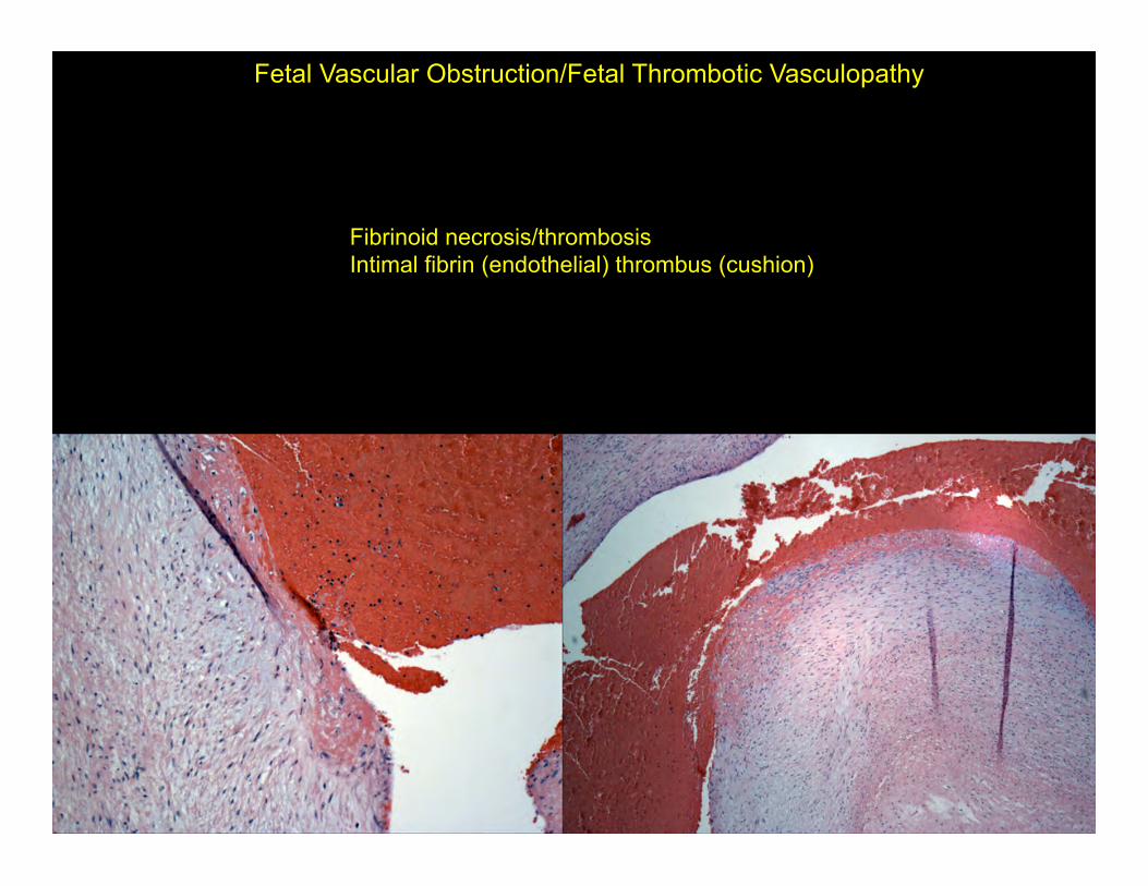
Fibrinoid necrosis/thrombosis Intimal fibrin (endothelial) thrombus (cushion)
Fetal Vascular Obstruction/Fetal Thrombotic Vasculopathy

Intimal mural fibrin thrombus (endothelial cushion)
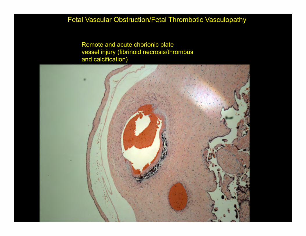
Remote and acute chorionic plate vessel injury (fibrinoid necrosis/thrombus and calcification)
Fetal Vascular Obstruction/Fetal Thrombotic Vasculopathy

Thrombus and vascular wall injury/repair (intimal fibrin cushion)
Fetal Vascular Obstruction/Fetal Thrombotic Vasculopathy

Early thrombus (hemorrhagic endovasculitis) and recanalization
Fetal Vascular Obstruction/Fetal Thrombotic Vasculopathy
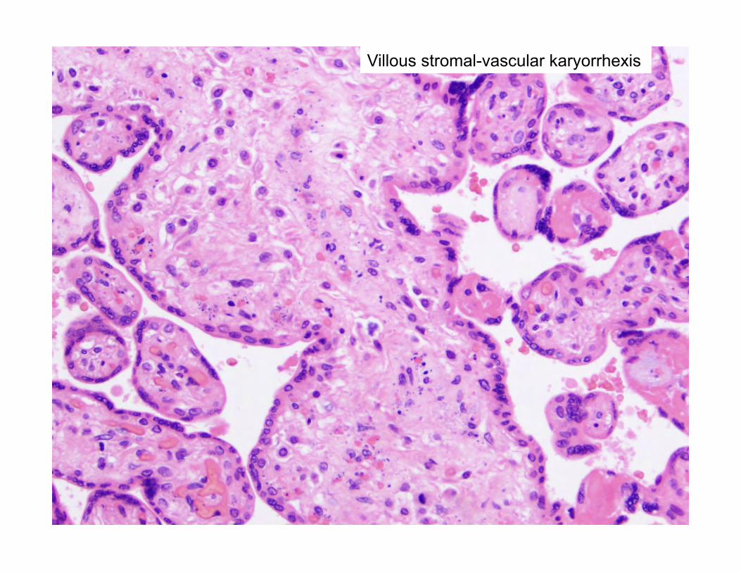
Villous stromal-vascular karyorrhexis
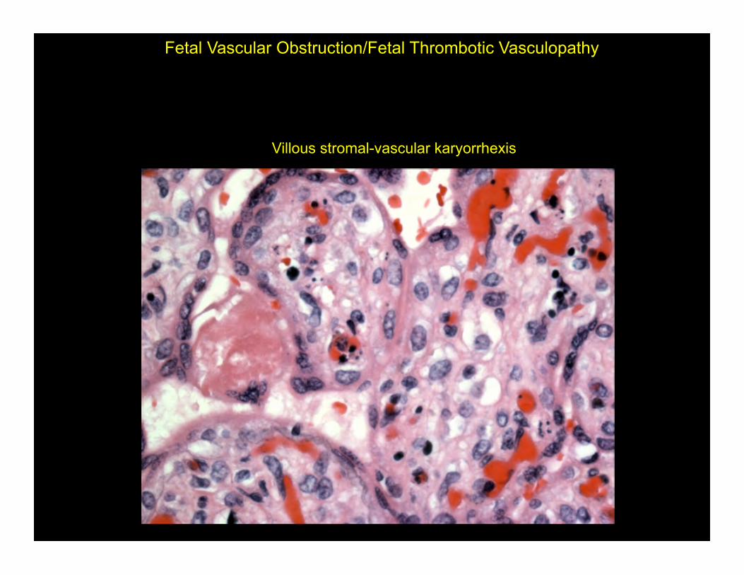
Villous stromal-vascular karyorrhexis
Fetal Vascular Obstruction/Fetal Thrombotic Vasculopathy

Avascular villi
Fetal Vascular Obstruction/ Fetal Thrombotic Vasculopathy

FINDINGS CONSISTENT WITH FETAL VASCULAR OBSTRUCTION (INCLUDING FETAL THROMBOTIC VASCULOPATHY) VASCULAR LESIONS:
-THROMBI INVOLVING CHORIONIC PLATE VESSELS/STEM VESSELS. -ACUTE/SUBACUTE/REMOTE MURAL INTIMAL FIBRIN THROMBI (ENDOTHELIAL CUSHIONS) INVOLVING CHORIONIC PLATE/STEM VESSELS. -RECANALIZED LUMENS INVOLVING CHORIONIC PLATE/STEM VESSELS.
VILLOUIS CHANGES:
-VILLOUS STROMAL-VASCULAR KARYORRHEXIS. -EXCLUSIVELY SMALL FOCI. -VARIABLY SIZED FOCI. -AVASCULAR VILLI. -EXCLUSIVELY SMALL FOCI. -VARIABLY SIZED FOCI. -GREATER THAN 45 AVASCULAR VILLI (3 FOCI OF MORE THAN 15 VILLI EACH)

Fetal Thrombotic Vasculopathy Criteria: Do not necessarily need to see the thrombi! I prefer to see some vascular injury unless the number of avascular villi is excessively high. Avascular villi or villous stromal-vascular karyorrhexis. (>15 avascular villi on 2 or 3 separate slides) (total 45 avascular villi). Remember to exclude “burned-out” chronic villitis as the cause for numerous avascular villi!

Saleemuddin A et al. Obstetric and Perinatal Complications in Placentas with FTV. Ped Devel Pathol 13:459-464, 2010. In placentas with FTV there is an increased risk of: PIH/preeclampsia Oligohydramnios Stillbirth Non-reassuring fetal heart tones IUGR Cardiac abnormalities (6-fold increase) ____________________________________________________________________ Gogia N and Machin GA Maternal thrombophilias are associated with specific placental lesions. Ped Devel Pathl 11:424-429, 2008. Placentas with the following pathologies showed acquired or genetic maternal thrombophilia in the following percentages: FTV (71%) MPVFD (23%) MFI (40%) FTV & MPVFD (50%)
Clinical implications of fetal thrombotic vasculopathy.

Fetal Vascular Obstruction and Umbilical Cord Accident in Stillbirths
Parast, Crum, Boyd Hum Pathol 39:948-953, 2008.
There should be an abnormal umbilical cord as well.

Take home points – 1. FTV causes include: infection, umbilical blood flow restriction, fetal/maternal thrombophilias (?), others. 2. Fetal thrombotic vasculopathy criteria:
3 foci of >15 avascular villi or with VSVK. +/- vascular lesion
3. Histologic changes associated with prolonged IUFD can mimic FTV. (hemorrhagic endovasculitis, karyorrhexis, avascular villi). Prolonged IUFD does not result in true “thrombi”! 4. FTV can result in a small placenta, but does not have to.

CHRONIC VILLITIS
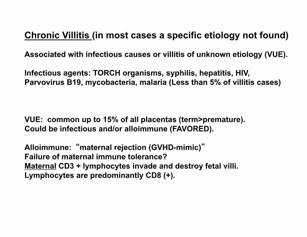
Chronic Villitis (in most cases a specific etiology not found) Associated with infectious causes or villitis of unknown etiology (VUE). Infectious agents: TORCH organisms, syphilis, hepatitis, HIV, Parvovirus B19, mycobacteria, malaria (Less than 5% of villitis cases) VUE: common up to 15% of all placentas (term>premature). Could be infectious and/or alloimmune (FAVORED). Alloimmune: “maternal rejection (GVHD-mimic)” Failure of maternal immune tolerance? Maternal CD3 + lymphocytes invade and destroy fetal villi. Lymphocytes are predominantly CD8 (+).

Histologic criteria for chronic villitis (reviewed Redline RW Human Pathol 38:1439-1446, 2007) : Lymphohistiocytic infiltrate. Avascular villi (not always present). Histiocytic giant cells not uncommon (no true granulomas). Plasma cells in villi – strongly consider infectious causes. Can see chronic deciduitis with plasma cells, especially if the infiltrate is localized to the basal plate villi. Histologic Classification: (high versus low grade chronic villitis) LOW GRADE: less than 10 villi per cluster Focal – only 1 slide involved. Multifocal - more than 1 slide involved. HIGH GRADE: more than 10 villi per focus Patchy – less than 5% of all distal villi involved. Diffuse – greater than 5% of all distal villi involved. HGCV associated with increased risk of adverse neurologic outcome and infectious etiologies.

High Grade Chronic Villitis (VUE)

High Grade Chronic Villitis (VUE)

High Grade Chronic Villitis (VUE) with obliterative vasculopathy

HGCV – chorionic plate with chronic chorionitis

High Grade Chronic Villitis (VUE) – histiocyte predominant with giant cells

CD3
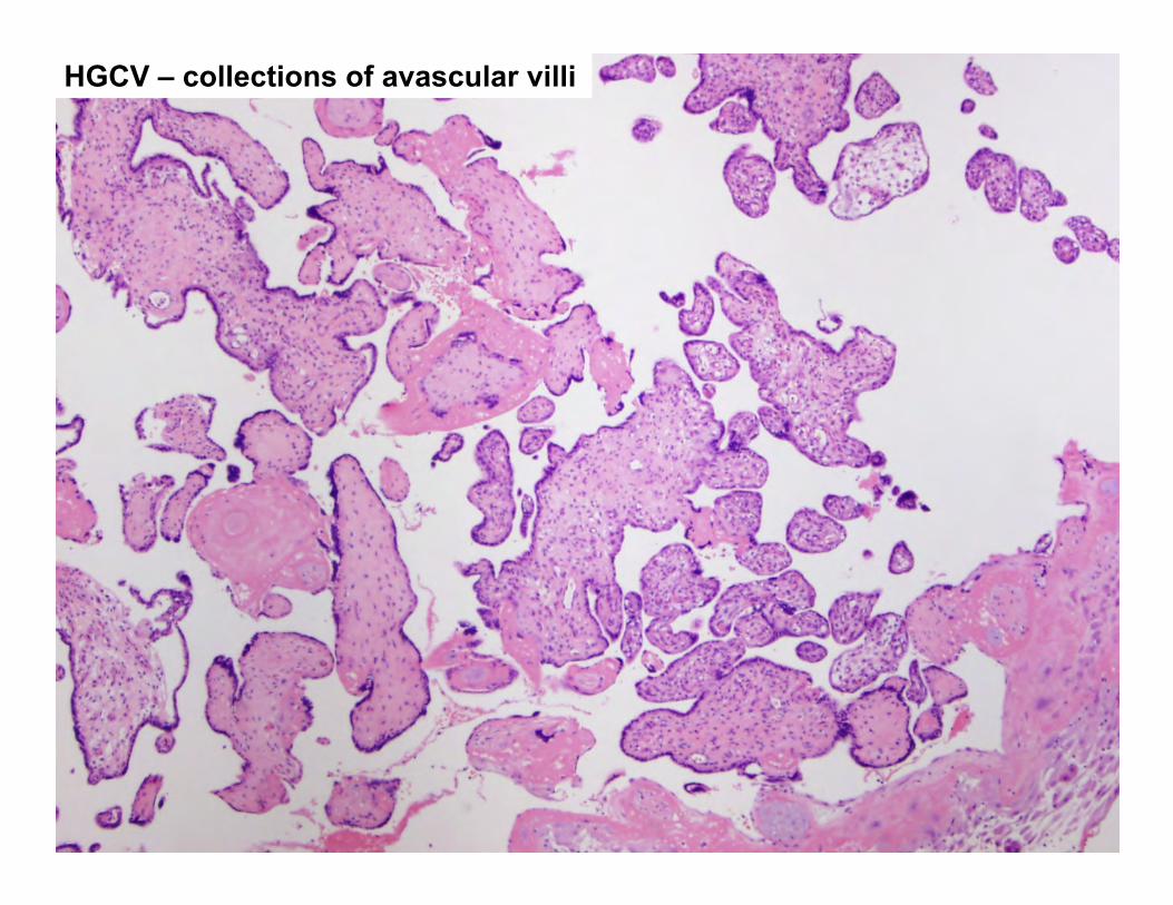
HGCV – collections of avascular villi

Chronic deciduitis – plasma cells in decidua. Plasma cells in the decidua are ALWAYS pathologic.
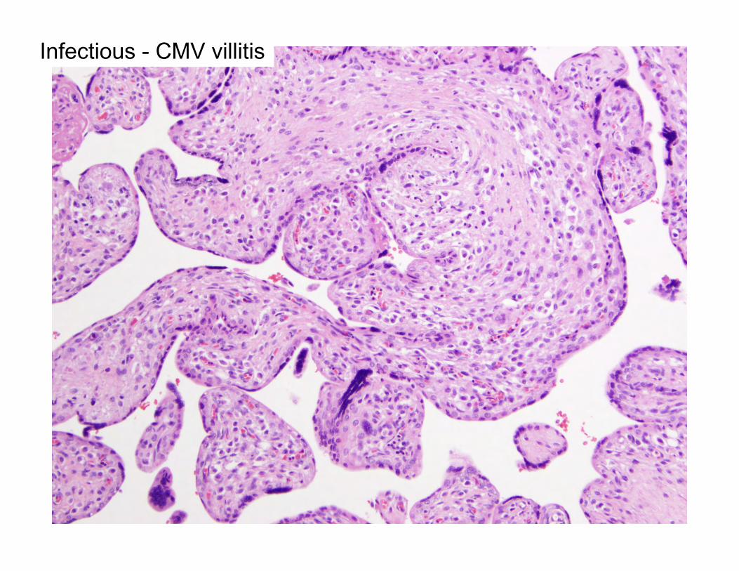
Infectious - CMV villitis
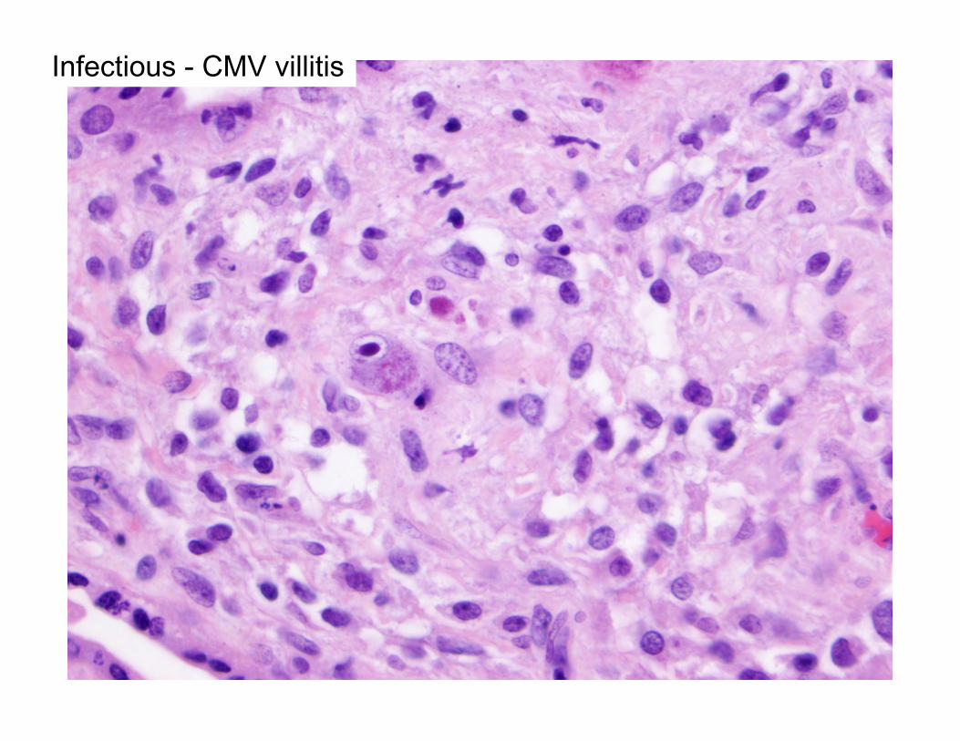
Infectious - CMV villitis
Other placenta histology occasionally seen with chronic villitis: AVASCULAR VILLI – BURNED OUT VILLITIS Increased perivillous fibrin Villous agglutination. Chronic deciduitis with plasma cells Chronic chorioamnionitis Fetal vascular thrombi Infarcts Villous dysmaturity Increased circulating nucleated fetal RBCs Hemosiderin deposition Chorangiosis

FOCAL/MULTIFOCAL LOW GRADE CHRONIC VILLITIS OR PATCHY/DIFFUSE HIGH GRADE CHRONIC VILLITIS. - NO DEFINITIVE VIRAL INCLUSIONS SEEN. - IMMUNOHISTOCHEMICAL STAIN FOR CYTOMEGALOVIRUS
(CMV) AND TOXOPLASMOSIS ARE NEGATIVE. (OPTIONAL) - AVASCULAR VILLI. - CHRONIC DECIDUITIS WITH PLASMA CELLS. - OBLITERATIVE VASCULITIS IN VESSELS OF TERMINAL/STEM
VILLI. - FOCAL CHRONIC CHORIONITIS. - CHRONIC DECIDUITIS WITH PLASMA CELLS. - ASSOCIATED PATCHY CHRONIC INTERVILLOSITIS. - SEE NOTE.
Note: Chronic villitis, although inflammatory, is associated with infection in only a small number of cases. In these instances, the most common infections are T. gondii, CMV, or syphilis. However, most of the time the inflammatory process is not associated with infection and is termed “villitis of unknown etiology.” High grade patterns of chronic villitis are associated with an increased risk of fetal growth restriction or fetal demise, and may recur in future pregnancies.

Clinical implications of chronic villitis. Infectious – depends on infection (stillbirth, chronic fetal infection, IUGR, small placenta, CNS injury, premature delivery) Villitis of unknown etiology- VUE – HGCV +/- obliterative vasculitis associated with: stillbirth, IUGR, prematurity, small placenta, recurrence in future pregnancies, oligohydramnios, fetal monitoring abnormalities, alloimmune thrombocytopenia, abnormal neurologic development. BP villitis not associated with poor outcomes. CV is more common in obese women.
From: Redline RW Human Pathol 38:1439-1446, 2007.