fiber based approaches as medicine delivery systems
TRANSCRIPT

Fiber Based Approaches as Medicine Delivery SystemsFarrokh Sharifi,†,∥ Avinash C. Sooriyarachchi,†,∥ Hayriye Altural,† Reza Montazami,†,‡
Marissa Nichole Rylander,§ and Nastaran Hashemi*,†,‡
†Department of Mechanical Engineering, Iowa State University, Ames, Iowa 50011, United States‡Center of Advanced Host Defense Immunobiotics and Translational Medicine, Iowa State University, Ames, Iowa 50011, UnitedStates§Department of Mechanical Engineering, University of Texas at Austin, Austin, Texas 78712, United States
ABSTRACT: The goal of drug delivery is to ensure thattherapeutic molecules reach the intended target organ or tissue,such that the effectiveness of the drug is maximized. Theefficiency of a drug delivery system greatly depends on thechoice of drug carrier. Recently, there has been growing interestin using micro- and nanofibers for this purpose. The reasons forthis growing interest include these materials’ high surface areato volume ratios, ease of fabrication, high mechanicalproperties, and desirable drug release profile. Here, we reviewdevelopments in using these materials made by the mostprevalent methods of fiber fabrication: electrospinning, micro-fluidics, wet spinning, rotary spinning, and self-assembly fordrug delivery purposes. Additionally, we discuss the potential touse these fiber based systems in research and clinicalapplications.
KEYWORDS: drug delivery, micro- and nanofiber fabrication, microfluidics, electrospinning, wet spinning, rotary spinning, self-assembly
1. INTRODUCTION
Since the beginning of the 20th century, efficient drug deliveryhas been a topic of continuous study and research. Regardlessof developments in areas such as cancer therapeutics and drugdelivery, progress in certain aspects of each area has beendampened by the inefficiency of the carriers used in these drugdelivery systems. The importance of the drug carrier can beattributed to four factors, which are (i) targeting of the drug tothe intended organ for maximum effect, (ii) evasion of theimmune system of the body to reach the final target, (iii)retention of the therapeutic molecules from the preparation andprocessing to the final target of the drug, and (iv) release of thedrug molecules at the destination organ such that the moleculescan exert the intended therapeutic effect. Therefore, the successof an extant or potential application of a drug delivery systeminnately depends on the choice of the drug carrier.1−4
Researchers have considered numerous carriers over the pastfew decades,5,6 and micro- and nanofibers have become anattractive prospective carrier option.7−13 This could beattributed to both the nature of the material used to fabricatethe fiber and the nature of this very fabrication process.Additionally, the low initial burst rate compared to sphericalvesicles and the controlled zero-order drug release profile, seenin drug loaded fibers, make them more suitable for drugdelivery applications.14−16 Materials such as alginate and gelatinused to fabricate these fibers are biocompatible,17,18 thuscausing no harm to the tissue of the host. Such fibers are also
biodegradable and do not accumulate in the human body,disrupting its biochemical and physiological processes.Furthermore, these materials exhibit low immunogenicity.This means that they do not provoke major immune responsesfrom the host’s immune system. As a result, fibers composed ofsuch biomaterials help create effective carriers of therapeuticmolecules.The morphology of the fibers makes them ideal candidates
for drug delivery systems. By virtue of the cylindrical shape ofthe fibers, they possess a high surface area to volume ratio.7,19
This provides the fibers the ability to release the drug into themedium over a large surface area. Furthermore, unlike sphericalvesicles, where the surface area to volume ratio can only becontrolled by varying the radius, both the length and the cross-sectional radius can be varied in fibers. This adjustability is vitalfor a drug delivery system in applications in which thefunctional parameters are to be precisely controlled. Addition-ally, several applications of drug delivery systems are seenwhere the drug delivery function occurs alongside a structuralrole. Tissue scaffolds, wound dressings, and other tissueengineering applications can be cited as prime examples ofsuch applications.7,20 For these roles, the shape and mechanicalproperties of micro- and nanofibers, composed of biomaterials,
Received: May 24, 2016Accepted: July 19, 2016Published: July 19, 2016
Review
pubs.acs.org/journal/abseba
© 2016 American Chemical Society 1411 DOI: 10.1021/acsbiomaterials.6b00281ACS Biomater. Sci. Eng. 2016, 2, 1411−1431

are essential. The shape and arrangement of such fibers couldbe easily changed, which makes them more suitable for drugdelivery systems than micro- and nanovesicles. Fibers withdifferent shapes and structures, such as hollow, flat, and ribbonshaped, can also be fabricated depending on the intendedapplication.21−23
Various methods have been used for the fabrication of thesemicro- and nanofibers over the past few decades. Microfluidicfiber fabrication, electrospinning, rotary spinning, self-assembly,and wet spinning are the most common methods. These fiveapproaches are schematically depicted in Figure 1. Moreover,variants of these methods have been developed to cater to theneeds of different applications. Among these methods,electrospinning and microfluidic fiber fabrication approachesare predominantly employed in research related to drugdelivery. Each of these methods have some advantages thatmake them suitable for specific drug delivery applications.The purpose of this review is to categorize research
developments in employing micro- and nanofibers for drugdelivery applications. The different fiber fabrication techniquesare reviewed, and further innovations in fiber based drugdelivery systems are discussed. For example, in discussingmicrofluidic fiber fabrication for drug delivery applications,structural developments, incorporation of stimuli-responsiveproperties, and innovations in the fabrication methodologywere explored in detail. Furthermore, special emphasis is givento the potential applications of these approaches in clinicalpractice. Finally, each of the applications are described in detailand tabulated to obtain a general overview of fibers utilized fordrug delivery purposes.
2. MICROFLUIDIC FIBER FABRICATION
Microfluidics is a promising approach that uses a small amountof materials for various applications such as biomedical areasand energy devices.29−35 This approach is the most recent offiber fabrication methods applied to research in utilizing fibersas drug carriers.36
In this method, fibers are fabricated by means of coaxial flowof prepolymer (core fluid) and sheath fluid in a microchannel.Microfluidic fiber fabrication methods are particularly useful fordrug delivery related applications due to the versatility,simplicity, and continuity of the process. There is no needfor high electric currents, pressures, and temperatures, whichimproves the biological compatibility of the fiber fabricationprocess. Additionally, a variety of biocompatible and non-cytotoxic substances can be used in this approach. The size andshape of the fiber can be tuned by simply changing theparameters involved in the process as well as a solidificationstrategy. Submethods of microfluidic fiber fabrication can becategorized based on the solidification strategy: photopolyme-rization, chemical reaction, and phase inversion approaches.Photopolymerization involves solidification of prepolymericmolecules by means of free-radical polymerization reactionwithin the microchannel. This method results in rapid and easygeneration of solid fibers while maintaining size and shape.24,37
In the chemical reaction method, the bonding of prepolymermolecules is facilitated by using small molecules or ions, such asCa2+.24,38 In the phase inversion method, the solvent in the corestream of the microfluidic chip is rapidly removed by means ofevaporation or extraction to expedite solidification of thepolymeric fiber.24,31,32,39
Figure 1. Most common methods used to fabricate fibers for drug delivery. (a) Illustration depicting the incorporation of drug molecules into amicrofiber. (b) Schematic of the microfluidic fiber fabrication approach. The manner in which the sheath and the core fluid flows are directed bymicrochannels to generate the fiber is depicted.24 (c) Schematic of fiber fabrication by electrospinning. The electrically ground plate collects thefiber.25 (d) Rotary spinning method where the solution is spun at high speeds and the centrifugal force causes the solvent to evaporate, which resultsin fiber fabrication.26 (e) Schematic of the building units which contribute to the formation of self-assembled nanofibers.27 (f) Wet spinning method,where the fibers are injected into a coagulation bath.28
ACS Biomaterials Science & Engineering Review
DOI: 10.1021/acsbiomaterials.6b00281ACS Biomater. Sci. Eng. 2016, 2, 1411−1431
1412

Each of these submethods have distinct advantages anddisadvantages. In particular, photopolymerization producesfibers in less than a second, and control over the fiber diameterover large spans of the fibers is significant. However, thedegradability of the fibers fabricated by photopolymerizationdecreases, which could damage living organisms. Biodegrad-ability and biocompatibility are the primary advantages of fibersfabricated using chemical cross-linking and phase inversionstrategies. However, there are some limitations and difficultiesassociated with shape formation of the fibers due to the slowersolidification rate of these two submethods.24
Microfluidic fiber fabrication is a promising platform tofabricate fibers with a wide range of characteristics. However,the production rate in this technique is relatively slow mainlybecause the flow has to be in the laminar regime. Otherwise,the fluids introduced in the microfluidic device become mixedtogether and the diffusion will not occur only at the fluid/fluidinterface. Moreover, the fluids, i.e. core and sheath fluids, haveto be matched in terms of viscosity in order to keep the shearforce at the fluid/fluid interface in the stable flow regime. Inaddition, there is always a possibility of the microchannelclogging due to undesirable shear force, which is originatedfrom the mismatch between the properties of the fluids or frominappropriate flow rate ratio. This issue can be minimized byadjusting the flow rate ratio between the fluids, theconcentration of polymer and cross-linking agent, and theUV intensity in a suitable range.31,34,40
2.1. Magnetic-Responsive and Photoresponsive DrugDelivery Systems. Incorporating stimuli-responsive proper-ties to micro- and nanofibers significantly increases the utility ofsuch materials within biological environments. Such stimuli-responsive nanoparticles have been successfully used in drug/gene delivery,41 bioseparation,42 and magnetic resonanceimaging,43 but rarely with micro- and nanofibers. This is
particularly true in drug delivery, as the environment itself andthe requirements for therapeutic material are constantlychanging. These stimuli-responsive characteristics, such asphotoresponsiveness,44 magnetic manipulation,45 and temper-ature sensitivity,46 are achieved by the incorporation of minuteparticles into the fiber structure during the fabrication process.The advantages of fabricating such stimuli-responsive ther-apeutic-incorporated materials and scaffolds are numerous. Thiswork provides controllability, response properties, attenuation,and rapid drug release to micro- and nanofibers. Theadvantages of such stimuli-responsive fibers are ease offabrication, high production rate, low cost, biocompatibility,and uniform distribution of magnetic particles in the fibers.47
However, there are unique challenges associated with utilizingthese fibers for actual drug release applications in vivo. Oneobvious practical difficulty would be implementing an optimumconfiguration of magnets when administering drugs usingmagnetic-responsive fibers.48 Photoresponsive drug loadedfibers are relatively easy to fabricate but challenging to deployas drug carriers. Nevertheless, once these challenges areovercome, stimuli-responsive drug incorporating fibers havethe potential to change the way that therapeutic substances aredelivered to various cells and tissues in the body.The use of magnetite nanoparticles in hydrogel spheres for
cell encapsulation related studies has been discussed as well.However, Lim et al. extended the method to microfibers.19 Thefocus of the study was to incorporate stimuli-responsivefeatures to hydrogel microfibers fabricated by a microfluidicapproach with controllable size and morphology. As shown inFigure 2(a), a microcapillary device was used to generate Ca-alginate templates. In this process, hydrogel monomers wereincluded and subjected to UV radiation (photopolymerization)to create poly(N-isopropylacrylamide) (PNIPAm) basedhydrogel microfibers. PNIPA was chosen for this study because
Figure 2. Different fabrication methods of magnetic stimuli-responsive micro- and nanofibers: (a) Use of magnetite nanoparticles in hydrogel cellencapsulation. A mixture of sodium alginate, N-iso-propylacrylamide (NIPAm), cross-linkers, and photoinitiator in aqueous form was inserted atinlet A. At inlet B, calcium ions were injected to diffuse into alginate and form calcium alginate.19 (b) The pneumatic microvalve structure used inmicrofiber fabrication.49 (c) Microfiber fiber fabrication system accompanied by photographs of the microfiber in the channel and two differentjunctions.45
ACS Biomaterials Science & Engineering Review
DOI: 10.1021/acsbiomaterials.6b00281ACS Biomater. Sci. Eng. 2016, 2, 1411−1431
1413

of its temperature-responsive nature. Furthermore, the resultingmicrofibers were photoresponsive due to the inclusion ofphotothermal magnetite nanoparticles. Ionic attractions wereobserved between the polymer and calcium ions, facilitated bythe capillary tubes and coaxial flows. This aided the uniformityand the small diameters required for the templates. Themicrofiber diameter was controlled by adjusting the capillarytube sizes. Hollow fibers were obtained by changing the orderof the inlets in microfiber fabrication. Functional materials wereeither blended into the core fluid or copolymerized. Theresponse of PNIPAm microfibers, with blended in magnetitenanoparticles responsive to light and temperature, was observedin the form of volume change.The controllability of the microfiber could also be achieved
by including magnetic nanoparticles in the alginate fiberfabricated with the microfluidic approach. Hu et al. obtainedthis result by evenly dispersing magnetic nanoparticles inalginate solution and subjecting it to a microfluidic fabricationprocess, illustrated in Figure 2(b).49 This process was enhancedby incorporating hemicylindrical channels of multilayeredpneumatic valves. The channels were capable of fully closingand regulating the flow rates of magnetic and nonmagneticalginate. Through this process, microfibers with differentmagnetic properties were produced. Importantly, there wasno aggregation of magnetic nanoparticles produced by aprecipitation method inside the alginate solution. This aidedthe uniformity of the final product during the fabricationprocess. The ability of the microfibers to be utilized in amagnetic field, to assist drug targeting, growth factor delivery,and possibly other kinds of therapeutics, makes themcandidates worthy of further investigation. This method couldpotentially address controllability, actuation, and responseproperties in drug delivery systems.Lin et al. also focused on the use of magnetic nanoparticles
for fabricating stimuli-responsive fibers for drug delivery. Their
emphasis was laid on utilizing the unique stimuli-responsivecharacteristics in drug release and cell encapsulation.45 Asshown in Figure 2(c), a microfluidic method that involvedmultiple inlets at different points along the center channel wasutilized. In this design, alginate solution was controlled atdifferent points along the channel in a laminar flow regime. Thediameter of the fibers was varied from 211 to 364 μm bychanging the alginate solution flow rate. The magnetic ironoxide and diclofenac, the model drug, were incorporated intothe alginate solution prior to the microfluidic fiber fabricationprocedure. The drug release profile of the model drugdemonstrated that the release process was linear and steadyand could be controlled externally by a magnetic field. Thiscontrol is achieved by the attraction of the magneticnanoparticles toward the magnetic field, making the microfibermore porous. The initial burst rate observed in microfibers wassmaller compared to spherical microvesicles. Furthermore, themicrofluidic method of microfiber fabrication results in a highencapsulation rate of 90% for both the magnetic iron oxidenanoparticles and the drug itself. Therefore, this is an efficient,convenient, and controllable method for drug delivery.The magnetic-responsive micro- and nanofibers generated by
the aforementioned means do not display morphologicalchanges that might adversely affect their function in drugdelivery. The cylindrical forms of the fibers fabricated by Lim etal. are depicted in Figure 3(a) and (b).19 Figure 3(c) and (d)show an alginate microfiber with diclofenac and magnetic ironoxide.45 These microfibers are shown in Figure 3(e).49
2.2. Other Innovative Approaches to Microfluidic-Spun Fiber for Drug Delivery. Certain alterations have beenintroduced to this conventional microfluidic fiber fabricationprocedure in order to ensure the enhanced functionality offibers. These alterations include implementing a rollermechanism to draw out thinner fibers,50 composing fibers oftriblock copolymers as opposed to conventional PLGA,51 and
Figure 3. (a) EDTA treated poly(N-isopropylacrylamide) (PNIPAm) microfibers fabricated by the microfluidic approach.19 (b) Optical micrographof PNIPAm microfiber with an inner capillary diameter of 500 μm.19 (c) Alginate microfiber for magnetic-responsive controlled drug release.45 (d)SEM image of the fiber shown in part (c).45 (e) 100% alginate hydrogel microfibers fabricated by a multilayered pneumatic microvalve basedfabrication methodology. The horizontal scale bar represents 50 μm.49
ACS Biomaterials Science & Engineering Review
DOI: 10.1021/acsbiomaterials.6b00281ACS Biomater. Sci. Eng. 2016, 2, 1411−1431
1414

enhanced continuous production methods.36 In addition,attempts have been made to use glass for making microfluidicchips, instead of PDMS, as glass has more desirable opticalproperties.52 Some research has been conducted on creatingspherical vesicles from microfibers by drawing them out in amicrofluidic arrangement. This method is unconventional inthe sense that the fibers do not carry the drug to the target. Infact, they act as an intermediate stage in the final drug carrierfabrication.53 Some studies focus on increasing the concen-tration of loaded drugs, which may be essential in curbingrapidly spread infections.7 Each of these innovative approachesalso have shortcomings, which shall be discussed on a case bycase basis.From a drug delivery system perspective, innovative methods
for microfiber fabrication are important. Toward this end, Kanget al. described a solely PDMS based method of fabricatingmicrofibers utilizing the cylindrical channel created by thedeflection of a thin PDMS layer.54 The principle was extendedfor creating rectangular molds, and an additional 12 rectangular“micromixers” were created on a single chip with 5 coaxial flowchannels. In these channels, microfibers with a differentchemical composition were fabricated. The process andresulting fibers are schematically depicted in Figure 4. The
diameters of the fibers fabricated were 75 to 115 μm. The coresolution was sodium alginate, and the sheath fluid was calciumchloride. Bovine serum albumin (BSA) was successfullyincluded in the fibers via the core fluid to demonstrate theability of the fibers to carry therapeutic agents. However, thedisadvantage of this method is the size limitation of the fibers,
which could potentially be addressed by adjusting the flow rateand channel size of the coaxial flow.A higher degree of control over fiber dimensions is important
in creating fibers for drug delivery applications. Su et al. haveinvestigated the possibility of using a roller assisted microfluidicsystem for generating alginate microfibers, where it is possibleto exert such a degree of control.50 The alginate solution withthe monomers and other particles, such as nanoparticles fordrug delivery or cells for cell based therapy, was passed througha single microchannel. Then, calcium chloride solution wasused to cross-link the alginate, which was collected with a roller,resulting in a microfiber. The roller was the critical componentthat reduced the diameter of the fiber to the order of 1 μm, andit also affected the shape of the fiber. Silver nanoparticles areimportant antimicrobial agents and possess wound healingproperties. These particles were included by diffusing silver ionsto the alginate microfiber and subjecting it to UV light. Suchnanoparticles could be potentially released in low concen-trations to create antimicrobial activity in wound careapplications. Biomaterials were included in the microfiber byblending with the alginate solution, which results in a successfulmethod of incorporating anticancer drugs in drug deliveryapplications. This method could be applied to other hydrogelfiber types, which may increase the range of microfibers capableof incorporating drug delivery systems into their composition.In the field of drug delivery systems, controlling the release
rate of an encapsulated drug or a protein in the microfiber is animportant factor. Marimuthu et al. successfully conducted astudy where microfibrous scaffolds composed of amphiphilictriblock copolymer were fabricated utilizing microfluidics andthe porosity was controlled.51 This controllable porosity wasobtained by combining the effects of immersion precipitationand solvent evaporation with the microfluidic generation offibers. It was demonstrated that the porosity of the fiber has anotable effect on the release of fibronectin, which was themodel protein used. In principle, the method could be extendedto use with gene therapy and other therapeutic agents in drugdelivery systems. The copolymer used was an amphiphilic ABAcopolymer (PPDO-co-PCL-b-PEG-b-PPDO-co-PCL) capableof facilitating drug delivery due to its hydrophobic (PPDO/PCL) and hydrophilic (PEG) segments. Along with theenhanced drug encapsulation and release characteristics, themethod is cost-effective, is robust, and can be modified forother therapeutic applications.56
As Berthier et al. have pointed out, there are advantagesspecific to both polystyrene and PDMS as materials formicrofluidic apparatus as well as disadvantages.52 Of the two,polystyrene is more often used in conventional biorelatedlaboratory activities, but it has proven to be a difficult materialto engineer. The same difficulties have been faced byresearchers intending to use polystyrene in microfluidics,which has led to more prevalent use of other materials forresearch. Johnson et al. developed an innovative method to usePDMS molds to make polystyrene based microfluidic devices.57
Powdered polystyrene in a Petri dish was melted against a moldmade out of PDMS to fabricate a microfluidic chip withchannels and grooves. The absorption levels of drugs byfabricated devices were clearly demonstrated and compared toconventional PDMS devices. The results indicated thatclopidogrel, the drug of choice for the study, is not absorbedby the PDMS material. Therefore, microfluidic devices could beused for laboratory drug delivery system related research, and inencapsulating drugs into microfibers.
Figure 4. (a) Microfluidic fiber fabrication platform in which 5 fibersof different composition are fabricated in parallel. A stepwise gradientacross 5 output solutions is achieved with 12 rectangular micromixers,and fibers were generated from these solutions through 5 coaxial flowchannels. (b) Illustration of the alligator teeth micromixer. (c) Coaxialflow channel with a single sheath inlet. (d) SEM image of alginatefiber.55
ACS Biomaterials Science & Engineering Review
DOI: 10.1021/acsbiomaterials.6b00281ACS Biomater. Sci. Eng. 2016, 2, 1411−1431
1415

The required concentration of a therapeutic substance has tobe relatively high to have any effect on certain tissues orpathogens. Microfluidic fiber fabrication allows for creation offibers with a higher capacity to carry drugs. The deviation fromthe conventional method does not necessarily have to bestructural, and it could be a change in the constituent materialsused. Ahn et al. investigated a process in which certain details ofthe conventional microfluidic approach were changed in orderto make the intended effect.7 Isopropyl alcohol was used as asheath flow to create alginate polymer chains packed in a highlyordered and tight manner to form the fiber. This minimizes theswelling of the fibers and makes the concentration of the drugloaded in the fiber as high as possible. This overall arrangementof polymers within the fibers led to delayed degradation of themicrofiber. The degradation was further slowed down byimmersing the drug loaded fibers in a calcium ion bath. Thiswas done to increase the cross-linking of the polymers, whichresults in a stronger polymer. The core fluid was alginatesolution with Ampicillin dissolved in it. Figure 5(a) describesthe fabrication of drug loaded alginate fibers with calciumchloride in deionized water as the sheath flow and in thecoagulation bath. Figure 5(b) depicts the fabrication procedurewith calcium chloride dissolved in isopropyl alcohol as both thesheath fluid and the coagulation bath.Ampicillin is a widely used antibiotic that is active against
Gram-positive and Gram-negative bacteria.58 It was demon-strated that the drug loaded fibers were successful based on theantimicrobial action of drug loaded fibers placed in a bacterialculture. Further fibers were embedded in a chitosan patch andsuccessfully used on a bacteria infected wound of a mouse. Thisexperimental application is schematically depicted in Figure6(a). The actual structure of the dressing scaffold and theexcision wound are shown in Figure 6(b) and (c), respectively.The essential details pertaining to separate cases where
micro- and nanofibers fabricated by the microfluidic approach
for drug delivery applications are given in Table 1. The corefluid, sheath fluid, diameter of the cross section, and drug type(if indicated) used in each instance are indicated.
3. ELECTROSPINNINGElectrospinning is a widely used and thoroughly investigatedmethod of micro- and nanofiber fabrication. In this method, thepolymer is subjected to an electric field applied between asyringe containing the polymer and a collector. The jet movesin the air, and during this time, it is either electrically deflected,collected on a grounded stationary metal screen,60 or collected
Figure 5. Schematic depicting the manner in which drugs are loaded into alginate fibers. The polymers aggregate and encapsulate the ampicillinmolecules along the direction of flow in each groove. (a) Fiber is fabricated with a solution of calcium chloride in deionized water as the sheath fluid;(b) fiber is fabricated with a solution of calcium chloride in isopropyl alcohol (IPA) as the sheath fluid. In either case, the resulting fiber is passedonto a calcium chloride solution for further coagulation.7
Figure 6. (a) Schematic of a bacterial wound with a fibrous scaffoldcontaining drugs. The dressing scaffold is composed of alginate fiberswith a high concentration of ampicillin capable of exerting thenecessary antibacterial action to control an infection. (b) SEM imageof the actual dressing scaffold with the chitosan matrix and theampicillin loaded fiber. (c) Excision wound.7
ACS Biomaterials Science & Engineering Review
DOI: 10.1021/acsbiomaterials.6b00281ACS Biomater. Sci. Eng. 2016, 2, 1411−1431
1416

on a grounded rotating or translating metal target.61 Differentvariants of this method exist, but the core concept is the same.By varying the conditions of the process, including the voltageand the material, fibers with a range of cross-sectional diameterswere obtained. The advantages of using electrospinning tofabricate macro- and nanofibers in drug delivery systemsinclude the ease of fabrication of fiber with diverse morphologyand dimensions, the ability to include the therapeutic moleculesin the polymer solution, and the ability to orient fibers eitherrandomly or in an ordered manner, for tissue engineeringrelated drug delivery applications.62
Although electrospinning is a common approach forfabricating fibers, it has some disadvantages that adverselyaffect the biocompatibility aspect of this method. The diameterof the electrospun fibers is typically smaller than a single cell,which makes it challenging to encapsulate living cells andmicroparticles into the fibers.28,63 Additionally, the commonsolvents that are used in this process, such as 2,2,2-trifluoroethanol (TFE), tetrahydrofuran (THF), dimethylfor-mamide (DMF), dichloromethane (DCM), chloroform, acrylicacid, and hexafluoroisopropanol (HFIP), have high toxicity.64
Due to the surface tension at the liquid/air interface, the crosssections of the electrospun fibers are mostly circular, whereas,in the microfluidic approach, the shape and surface of the fiberscan be simply tuned by changing the 3D hydrodynamicfocusing force.31,34,54,65 Besides the size and shape limitationand toxicity of the solvents, the utilization of high electric fields(∼1−2 kV/cm) in this technique makes electrospinningunsuitable for certain drug delivery systems where live cellsneed to be included in the fibers.28 However, due to thefunctionality of the electrospinning method, many studiesfocused on improving the shortcomings that exist in a typicalelectrospinning process.64,66−68 For example, some reportsshowed that by simultaneously elesctrospinning fibers andelectrospraying the cell suspension, the cells will have highviability.67,68 There are some studies in which drugs wereencapsulated into the fibers, whereas, in other reports, thecombination of both fiber and liposomes were used toencapsulate the drugs and control the rate of release.
3.1. Electrospinning Fiber Fabrication Technique inDrug Delivery Application. Usage of micro- and nanofibersin drug delivery is still an emerging topic in biomedical
Table 1. Details of Fibers Fabricated by the Microfluidic Approach for Drug Delivery Systems
Authors Drug TypeDiameter(μm) Core Sheath
Lim et al.19 N/A 10−600 A mixture of Na-alginate, NIPAm, cross-linkers and photoinitiator in aqueous form Ca2+ solutionHu et al.49 N/A 45−50 Alginate solution with magnetic nanoparticles CaCl2
solutionLin et al.45 Diclofenac 211−364 Alginate solution and model drug CaCl2
solutionMazzitelli etal.59
Drug delivery systemsCAM, ERSM, LS, andCOBE
180−500 Na-alginate through one inlet, Na-alginate and drug delivery system (CAM, ERSM, LS,or COBE) through one inlet, Na-alginate and cell suspensions through one inlet.
N/A
Kang et al.55 BSA 75−115 Na-alginate solution and BSA CaCl2solution
Su et al.50 Silver particles ∼1 Na-alginate solution CaCl2solution
Marimuthu etal.51
Fibronectin 2−200 10% PPDO-co-PCL-b-PEG-b-PPDO-co-PCL copolymer in CH2Cl2 DI water
Ahn et al.7 Ampicillin N/A Alginate and Ampicillin solution CaCl2 in IPAor DI water
Figure 7. (a) Schematic depicting the cross-linking of gelatin nanofibers with riboflavin which may potentially be used in the dentin, pericardium, oreye.78 (b) BUD-core/shell-nanofibers that could be potentially used for drug delivery.81 (c) PVP and PEO fibers containing HIV prophylactic drugMaraviroc.84 (d) Coaxial PCL/PEO hollow fibers which may be loaded with dexamethasone for inflammation treatment.82
ACS Biomaterials Science & Engineering Review
DOI: 10.1021/acsbiomaterials.6b00281ACS Biomater. Sci. Eng. 2016, 2, 1411−1431
1417

research. Due to the structural advantages fibers offer, theycould be used in specific sites in the body in conjunction withtissue. The range of molecules that could be delivered byelectrospun fibers extends from small biomolecules tomolecules as big as proteins and DNA plasmids.69−75 Hence,this offers a significant advantage over conventional carriers.76
Additionally, subcellular vesicles and stimuli-responsive time-variant morphological features could be incorporated into thefibers. However, further innovations could result in drugdelivery systems designed for specific applications. Woundhealing, for instance, is an area widely studied in applications ofdrug delivery systems which incorporate electrospun fiberstructures. However, a single material may fall short inmechanical strength or may have less than optimal drugloading capacity in wound dressing applications. Bai et al. havesucceeded in addressing both issues by fabricating a chitosan/polycaprolactone (CS/PCL) nonwoven mat utilizing theelectrospinning method.77 The arrangement of fabricated fibersprovides a greater area for active ingredients in the fiber tointeract with the surroundings. Tree oil and calcium ions wereincorporated into the chitosan component of the structure,leading to increased platelet aggregation and antibacterial aswell as anti-inflammatory effects, respectively.Releasing hydrophilic therapeutic compounds from hydro-
phobic fibers is a challenge. PCL-riboflavin nanofibers with radiiranging from 267 ± 37 nm and gelatin-riboflavin microfiberswith radii 2.18 ± 0.3 μm were fabricated by Sridhar et al.78
Riboflavin is commonly known as Vitamin B2. It performs arange of critical metabolic functions as well as contributing towound healing and maintaining tissue health.79,80 Cross-linkingwas caused by the oxygen radicals generated by UV irradiationof riboflavin, which leads to an increase in mechanical strengthof the fibers. This is schematically presented in Figure 7(a).The riboflavin release rate from these fibers was altered bytreating the fibers with plasma and changing the plasmatreatment duration. Therefore, noncytotoxic riboflavin canfunction as a cross-linker and a therapeutic agent whose releaserate can be controlled. Such riboflavin cross-linked fibers couldbe potentially used for fibrous scaffolds in tissue engineeringapplications such as dentin, parts of the eye, and thepericardium.Oral ingestion of drugs is the most prevalent form of drug
delivery in the pharmaceutical industry. Toward this end, Xu etal. created and investigated a drug delivery system consisting ofBudesonide (BUD) loaded core/shell nanofibers that followsthe aforementioned oral colonic pathway.81 Coaxial electro-spinning was performed to prepare Budesonide loadedethylcellulose(EC)-core/Euragit S100-shell nanofibers, shownin Figure 7(b). The diameter of these fibers averaged 190 nm,and the average core diameter, which is uniform throughout thelength, was 74 nm. Budesonide-loaded Eudragit S100/ethyl-cellulose nanofibers, which are regarded as composite nano-fibers, were fabricated utilizing blend electrospinning. Thecomposite fiber was used as a control, and both were subjectedto in vitro and in vivo tests, with the latter being carried out inrats. It was observed that the desired pH dependent behaviorwas observed from the Budesonide loaded core/shell nano-fibers, and the encapsulated drug was shielded from the acidiccontents of the stomach and small intestine until the colon wasreached. Budesonide in nanofiber encapsulated form couldpotentially be used for future applications in an oral colonicdrug delivery systems. This is a possibility due to the
advantages this particular method offers over the currentconventional method used in the pharmaceutical industry.Rubert et al. investigated fibroblast growth factor delivery
and tissue engineering applications of PEO/PCL electrospuncoaxial microfibers.82 Fibrous cartilage in the human body healsat a slower rate due to the lack of cells which produce collagen.Figure 7(c) shows a method in which PCL/PEO coaxialmicrofibers were loaded with fibroblast growth factor. Thefibers had an outer surface made of PCL, and the fiberdiameters ranged from 7.49 ± 0.835 μm to 7.489 ± 0.445 μm.As noted in previous reviewed work, electrospinningparameters could be adjusted to change the fiber morphologyto the desired form. The therapeutic substance wasencapsulated properly, and the release lasted over 9 days. Itwas observed that the growth factor was expressed more afterthe first day, but reduced in release after day 9, proving that themethod could be a potent form of growth factor delivery basedon microfibers. Lei et al. utilized electrospun PLGA microfibersto deliver designer RNAi plasmid, complexed with a genecarrier polyethylenimine, to treat brain tumor by suppressingthe expression of Matrix metalloproteinase-2 in tumor cells.83
Anticancer drug paclitaxel and the RNAi plasmid were dualencapsulated in disc shaped implants composed of electrospunPLGA microfibers. The microfiber implant was able to releasethe drug and the gene in a sustainable manner. The gene/drugdelivery was observed to be capable of greater tumor regressioncompared to the single delivery of a drug by a microfiber orcommercial drug treatment currently available. The microfibersthat constitute the disc shaped implant had diameters of 835 ±92 nm.Pre-exposure prophylaxis of HIV is an important step in
preventing the spread of the HIV virus. Implementing suchmethods in precoital vaginal settings have unique challenges.Rapidly releasing a large volume of the drug without discomfortis difficult to accomplish with conventional drug deliverysystems. Ball and Woodrow fabricated PVP and PEOmicrofibers loaded with maraviroc shown in Figure 7(d).Maraviroc is the prophylactic drug, while polysorbate 20 is thewetting agent which aids quick release of the drug.84 The fibersranged in diameter from 0.4 to 2 μm. The drug loading was upto 28 wt % and conditions of high moisture were facilitated toresult in rapid release of the whole 28 wt % of the drug under 6min under sink conditions. During the electrospinning process,the drug retained the original potency and had no side effects.This could form the basis of a novel prophylactic measure tocurb the spreading of HIV due to the efficiency of the drugrelease mechanism.
3.2. Drug Delivery in Conjunction with Liposomesand Electrospun Micro- and Nanofibers. Liposomes arespherical subcellular vesicles with at least a single lipid bilayer,and they have been extensively used for experimental andclinical studies in drug delivery.85 The ability to load the vesiclewith various molecules ranging from chlorine in woolchlorination86,87 to chemotherapeutic nanomedicines to treatcancer, e.g. Camptothecin and platinum(II) drugs,88 has madeliposomes an ideal nanocarrier, particularly in drug delivery.The reasons for the popularity of liposomes, for drug
delivery, include the longevity of liposomes in the bloodstream,the ability to penetrate cells in the delivery of substances, andthe stimuli-sensitive nature of liposomes. In research carried outby Mickova et al., horse radish peroxidase was used as themodel encapsulate, and its enzymatic activity was preserved.This lead to the conclusion that the mesenchymal stem cell
ACS Biomaterials Science & Engineering Review
DOI: 10.1021/acsbiomaterials.6b00281ACS Biomater. Sci. Eng. 2016, 2, 1411−1431
1418

proliferation was enhanced by the delivery of recombinantgrowth factors provided by this drug delivery system.89 Inaddition, there is potential for clinical applications based on theuse of stimuli-responsive liposomes in drug delivery.90
Liposomes, which are adherent to microfibers, are capable ofencapsulating a range of important biomaterials ranging fromdrugs to DNA. Here, we have a closer look at the variousfabrication methods in which liposomes have been incorpo-rated to fibers.Liposomes, blended with poly(vinyl alcohol)-core/poly-ε-
caprolactone-shell nanofibers, were fabricated by coaxialelectrospinning as well as controls with no embeddedliposomes.89 According to stereological measurements, thecontrols had nanofiber populations with peak diameters of 50and 150 nm, while the liposome embedded fibers had bulgesalong the length with diameters 233.4 ± 36.9 nm withembedded liposomes at places. The appearance of suchliposome embedded fibers is shown in Figure 8.Incorporation of magnetic nanoparticles is a common way of
introducing stimuli-responsive properties to drug deliverysystems.91,92 This is applicable to liposomes as well. For
example, fibers were electrospun from a core solutioncontaining polyvinylpyrrolidone K90, phosphatidyl choline,and Fe3O4 nanoparticles, and then subsequently added towater.90 It was observed that the fibers have the Fe3O4 particlesuniformly distributed, which is illustrated in Figure 9(a). Thefibers were observed to spontaneously undergo self-assembly toform liposomes. Additionally, the size was seen to vary with theFe3O4 concentration. The retention of sensitivity to magneticstimuli was observed to be retained throughout the electro-spinning and self-assembly processes. An actual view of theliposomes obtained is shown in Figure 9(b). Similar but non-stimuli-responsive liposomes were fabricated through molecularself-assembly by adding electrospun amphiphilic nanofiberscomposed of hydrophilic polymer polyvinylpyrrolidone K60(PVP) and soybean lecithin.93 The liposomes were phospha-tidyl choline (PC), and the diameter was varied by changing thePC content of the fibers.Rampichova et al. utilized the process of microfiber
fabrication by electrospinning to produce poly(2-hydroxyethylmethacrylate) (PHEMA) fibers to be used as a tissueengineering scaffold for cartilage. The fibers fabricated had
Figure 8. (a) FESEM image of coaxially electrospun PVA-core/PCL-shell nanofibers with liposomes encapsulated along the length of the fiber. (b)The bulge caused along the length of the nanofiber due to the liposome is clearly seen.89
Figure 9. (a) Schematic of magnetic liposome self-assembly. Fe3O4 nanoparticles are stirred into the polymer solution, and then subjected to anelectrospinning fabrication process. Once hydrated, liposomes form through self-assembly. (b) SEM micrograph of a magnetic liposome fabricatedfrom the fibers by a self-assembly process, which results in stimuli-responsiveness in addition to drug delivery capacity.90
ACS Biomaterials Science & Engineering Review
DOI: 10.1021/acsbiomaterials.6b00281ACS Biomater. Sci. Eng. 2016, 2, 1411−1431
1419

diameters in the submicron range.94 In addition, a relativelystraightforward drug delivery system was created in which fetalbovine serum loaded liposomes were anchored to the fabricatedfibers. This anchoring occurs as a result of the adhesion ofliposomes to PHEMA. An enhanced proliferation of chon-drocytes, in the vicinity of such liposomes attached to the fibers,was found. This observation showed the development ofcontrolled drug release mechanisms in conjunction withmicrofibers in tissue engineering applications.3.3. Miscellaneous Innovations in Electrospun Fiber
Based Drug Delivery. Programmable transdermal drugdelivery is one such exploratory application of micro- andnanofibers in drug delivery systems. Tran et al. incorporatedtwo types of ibuprofen (an anti-inflammatory drug) into themicrofibers using an electrospinning approach.95 One type ofmicrofibers were fabricated from polycaprolactone (PCL),while the other type was composed of poly(N-isopropylacry-lamide-co-methacrylic acid) (pNIPAM-co-MAA). The averagediameter of PCL fibers was 1237 nm, whereas that of pNIPAM-co-MAA fibers was 1608 nm. The drug release characteristicsunder different pH and temperature conditions, from bothtypes of fibers, were investigated. It was revealed that the drugrelease rate from PCL fibers is largely unresponsive to both pHand temperature changes whereas pNIPAM-co-MAA drugrelease rates were highly sensitive to changes in the above-mentioned conditions. The drug release profile from the latterwas linear and controllable at temperatures higher than thelower critical temperature of pNIPAM-co-MAA and at a pHhigher than the pKa of carboxylic acids. Furthermore, at roomtemperature, the drug release rate from pNIPAM-co-MAA wasshown to be ten times higher than the release rate at a highertemperature. Therefore, the highly stimuli-responsive nature ofibuprofen loaded pNIPAM-co-MAA fibers can be utilized infuture in vivo and potentially clinical applications.A nanofiber based drug delivery system, capable of exercising
precise control over drug delivery, by means of coating withnanotubes, has been investigated by Abidian et al.96
Biodegradable drug loaded poly(L-lactide) (PLLA) and poly-(lactide-co-glycolide) PLGA nanofibers with diameters rangingfrom 40 to 500 nm were fabricated by electrospinning. Thedrug loaded into the fibers in the study was dexamethasone.The nanofibers were coated with poly(3,4-ethylenedioxythio-phene) (PEDOT) nanotubes by means of electrodeposition.The process is detailed in Figure 10. This conducting
nanopolymer coating significantly reduces the impedance andincreases the charge capacity such that drug release from thefibers can be simulated to a desired extent, by means ofelectricity. Due to the controllability and precision of themechanism, this can be a potential drug delivery system.Zhou et al. devised a method to exert more control over drug
release by fabricating water-soluble drug loaded ultrafine fibersby means of microsol-electrospinning.97 Core−shell poly(L-lactic acid) microfibers loaded with chloroquine-hyaluronic acidnanoparticles were created by microsol electrospinning.Chloroquine phosphate, which is the therapeutic substanceused in the study, is historically noted for being used in thetreatment of falciparum malaria, vivax malaria,98,99 infectionscaused by protozoa,100 and certain other diseases. Thefabricated fibers had diameters ranging from 1.33 ± 0.32 μmto 1.48 ± 0.44 μm and cores ranging from 0.29 to 0.83 μmdepending on the composition of hyaluronic acid and the drugconstituent ratio in the fibers. The morphology and physicalfeatures of such fibers were no different than those of fibersfabricated from conventional electrospinning. The loadingefficiency of the microfibrous scaffolds was approximately80%. The burst release was lower than that of conventionallyprepared microfibers, and drug release occurred for 42 days.The rate of drug release could be adjusted by changing theconcentrations of microsol and drug in the fibers.Puhl et al. fabricated nonwoven PCL microfibers with
diameters ranging from 1.6 to 10 μm by electrospinning, andlysozyme crystals with diameters in the range of 0.7−2.1 μmwere encapsulated during the process.101 Lysozyme, an enzymein the crystalline form, is the most concentrated form of theprotein, which makes for increased drug loading.102 Theenzyme itself is used for wound treatment, and electrospunchitosan mats, which are loaded with it, have been used forwound healing.103 In the study, the fiber diameter and thelysozyme loading concentration were varied. Additionally,PCL/PEG and PCL/PLGA blends were used to enhance thehydrophilic nature, porosity, and degradability of the fibers,which in turn affects the drug release profile. It was observedthat the bioactivity of the encapsulant was preserved during theelectrospinning, and the burst release could be controlled bychanging the aforementioned fiber and crystal properties. Thesefactors did not affect the long-term drug release phase, but itwas extended to over 11 weeks by PLGA inclusion during theelectrospinning fabrication.
Figure 10. Schematic diagram of controlled release of dexamethasone. (a) Electrospun PLGA fiber loaded with dexamethasone. (b) Degradation ofPLGA fibers by hydrolysis, which results in releasing the drug. (c) Electrochemical deposition of PEDOT around the PLGA fiber. (d) This results inthe sustained release of the drug over time. (e) PEDOT nanotubes in a neutral electrical condition. (f) Applying a nonzero voltage allows exertion ofcontrol over the rate of dexamethasone release due to contraction or expansion of the PEDOT. (g) A PEDOT nanotube which was formed around aPLGA fiber. The PLGA solution of fiber was subsequently dissolved, resulting in this appearance.96
ACS Biomaterials Science & Engineering Review
DOI: 10.1021/acsbiomaterials.6b00281ACS Biomater. Sci. Eng. 2016, 2, 1411−1431
1420

Llorens et al. obtained a method in which both antimicrobialand anticancer drugs were released from the same scaffoldcomposed of microfibers fabricated using electrospinning.104 Inthis study, uniaxial microfibers were fabricated by varying core−shell compositions and distributions. This was accomplished bysubjecting ploy(ethylene glycol) (PEG) and poly(butylenesuccinate) (PBS) in a dichloromethane solution to coaxialelectrospinning. The diameters of the core/shell fibers rangedfrom 4.40 ± 0.92 μm to 4.83 ± 1.27 μm, and for the uniaxialblended fibers were in the range of 0.43 ± 0.10 μm to 0.76 ±0.18 μm. The core−shell microfibers were prepared such thatthe hydophobic drugs triclosan and curcumin were loaded intoPEG and PBS, respectively. The triclosan incorporated intoPEG lead to the rapid release often needed to combat microbialinfections. Moreover, a slower sustained release required foranticancer therapy was displayed by curcumin loaded in PBS.104
The drug loaded fibers were observed to be biocompatibleupon investigation, and the release profiles of drugs in differentmedia were compared. It was observed that the lowest releaserate was found in PEG-rich uniaxial fibers in a buffer salinemedium, whereas the highest release was obtained in coaxialmicrofibers in a buffer saline/ethanol mixture.Fibers have also been electrospun from biological molecules.
A prime example is the fabrication of hemoglobin andmyoglobin mats by means of an electrospinning approach.105
The fibers produced in this manner were ribbon-like. Thedimensions varied with the concentration of hemoglobin andmyoglobin in the electrospun solution. For hemoglobinconcentrations from 150 to 225 mg/L, the average width andthickness of fiber mats changed from 2.68 ± 0.83 μm to 3.55 ±
1.49 μm and from 0.49 ± 0.08 μm to 0.99 ± 0.41 μm,respectively. It was found that there is a correlation between theporosity of the fibers and the hemoglobin concentration. Thesefibrous structures could function as vehicles for drug delivery.
3.4. Controlled Drug Release with Electrospun Micro-and Nanofibers. Drug release profiles of micro- and nanofiberbased drug delivery systems are an important factor. Forexample, acute microbial infection control requires the releaseof a large dosage of drug in a specific location over a shortperiod of time. However, treatment of a chronic infection orcontaining the spread of a tumor requires a longer sustainedrelease of the drug in controlled quantities.106 The typicalrelease profile of a drug delivery system consists of a rapidinitial burst release followed by a longer period of controlledrelease.16 This is shown in schematic form in Figure 11(a). Thedrug has to be administered less frequently to the patientbecause of the sustained release of the drug.107 This sustainedrelease resembles the flat portion of the separate plots depictedin Figure 11(b).108 The release rates and the time at which therelease rate plateaus out depend on the drug molecule, theconcentration of the drug molecule in the fiber, and the natureof the fiber itself.69−72,74 Luu et al. incorporated DNA into afibrous scaffold made by PLGA and PLA−PEG blockcopolymers. They showed that the DNA release can becontrolled by changing the block copolymer content.69 Anotherexample, i.e. a study by Qi et al. in which drug-fibercombinations drastically affect the burst and sustained releaseprofiles for drug molecules, is shown in Figure 11(c).109 Theeffect of drug concentration on the release profile is clearlyobserved in Figure 11(d).10 These can be attained on a case by
Figure 11. General overview of drug release profiles. (a) A schematic depicting the burst-effect in a zero order controlled drug delivery.16 (b) Therelease profiles of protein and dye in DMSO (dimethyl sulfoxide) and dichloromethane from microfluidically fabricated fibers.108 (c) The releaseprofiles of tetracycline hydrochloride (TCH) from electrospun TH-1/PLGA, TH-2/PLGA, tetracycline hydrochloride loaded/poly(lactic-co-glycolicacid) composite (TCH/PLGA) nanofibers, and tetracycline hydrochloride loaded halloysite nanotubes (TCH/HNT) fibers.109 (d) Release profilesof chlorhexidine (CHX) in poly(hydroxybutyrate) (PHB) and poly(hydroxybutyrate)/poly(ethylene oxide) (PHB/PEO) electrospun fibers.10
ACS Biomaterials Science & Engineering Review
DOI: 10.1021/acsbiomaterials.6b00281ACS Biomater. Sci. Eng. 2016, 2, 1411−1431
1421

case basis just by adjusting the varying burst release propertiesof the encapsulated drug or the sustained release.When conventional methods of drug loading are followed
with electrospun fibers, the burst release effect, observed at theoutset, hampers the effectiveness of the blended or mixed drug.This is a major disadvantage in drug delivery systemapplications. Qi et al. addressed this issue by fabricatingtetracycline hydrochloride (TCH) loaded halloysite nanotubes(HNTs)/poly(lactic-co-glycolic acid) composite nanofibers(TCH/HNTs/PLGA) with improved tensile strength, sus-tained 3D form, and cytocompatibility.109 TCH, thepharmaceutical substance applied in the study, is an antibioticwhich is extensively used to treat periodontal infections110,111
and periodontal applications in veterinary medical applica-tions.112 Additionally, the composite fibers had the ability toovercome the initial burst release effect and release theantibiotic drug in a sustained manner for 42 days. Hence, theintended TCH induced therapeutic effect is sustained and lastslonger. The fabrication process involved TCH encapsulationwithin HNTs, and the composite fibers were electrospun fromthe solution with PLGA. The diameter of fabricated drugloaded composite fibers was 519 ± 133 nm.Li et al. took a similar comparative approach to study the
effects of binary fibers which were loaded with dexamethasonein anti-inflammatory applications.113 Dexamethasone is asynthetic glucocorticoid, which is used primarily as an anti-inflammatory drug in a clinical context;114 however, it may alsobe used for unconventional applications such as treatment ofbacterial meningitis115 and multiple myeloma.116 Immisciblepolymer blends of polycaprolactone (PCL) and poly(ethyleneoxide) (PEO) were prepared with different compositions of thetwo constituents in each case. The general physical character-istics of the fibers were not changed in the direct loading of thedrug during the electrospinning process. Depending on therelative composition of each component, the physical structureof the fibers varied from straight, straight with pores, to porouslamellar. The diameter of the fibers ranged from 0.92 ± 0.61μm to 4.782 ± 0.37 μm. The nature of dexamethasone releasewas a function of the wettability of the fiber, with highlyhydrophilic compositions leading to minimize the undesiredburst release. Regardless of the morphology and other chemicalproperties, all fibers were biocompatible. The dexamethasonewas effective in reducing inflammation caused by lipopolysac-charides.113
Inclusion of other nanostructures in microfibers generated byelectrospinning could assist in a desirable drug release profile.In a study, drug-loaded halloysite clay nanotubes, with 50 nmdiameter and length of 600 nm, were embedded intoelectrospun PCL/gelatin microfibers with mean diameter of400 nm.117 Prepared halloysite loaded microfibers were formedinto a membrane with tensile strength doubling in the directionof the rotation of the collector. The embedding of drug loadedhalloysite nanotubes into the fibers resulted in a drug releaseperiod of over 20 days compared to the control, in which thedrug was directly loaded to the PCL/gelatin fiber, which lastedfor a mere 96 h.109 This microfiber-halloysite membranousstructure had good biocompatibility, and eukaryotic cells wereable to grow along the membrane. Furthermore, the drug actedto prevent the colonization of anaerobic Fusobacteria.The same principle of including nanostructures in the micro-
and nanofiber structures has been explored by Valarezo et al. byincluding layered double hydroxide (LDH) nanoparticleswithin electrospun PCL microfibers.118 The drug Amoxicillin
was interspersed within the nanostructures. The interspersionof the drug in LDH was done by a coprecipitation process.These PCL fibers were organized into nonwoven mats withindividual fibers of an average diameter of 0.8 μm. The fiberswere randomly oriented in these structures, and the drugloading along with the nanoparticles appeared to slightlyincrease the average cross-sectional area of the fibers. The initialburst release, followed by sustained release of the drug, wasobserved in this case as well. However, the intercalation of thedrug within the LDH allowed for a slower sustained release.There is potential for successful use of this method for wounddressings, oral care, and dermatological treatment applications.Rubert et al. fabricated coaxial electrospun fibers with the sameconstituents in which dexamethasone was encapsulated.82 Thefibers were hollow fibers of the core−shell type with a highlyporous morphology conducive to drug release. The diameter ofthe fibers was 8.255 ± 0.838 μm and 8.7 ± 0.813 μm beforeand after dexamethasone encapsulation, respectively. Therelease of the drug occurs in two stages; the burst releaseoccurs in the first 0.5−3 h, and a late more stable release occursover more than 12 days. This dual stage drug release propertymakes this drug loaded electrospun microfiber structure idealfor implantation and acute inflammation as well as for chronicinflammation control. The drug loaded fibers were successful inreducing the proliferation of lipopolysaccharide (LPS)stimulated macrophages and at expressing inflammation relatedgenes.Paclitaxel is an antitumor drug that acts to inhibit tumor cell
growth by enhancing microtubule assembly resulting inblocking cell replication.119 This drug has been widely usedin clinical anticancer applications. Huang et al. successfullyfabricated paclitaxel loaded poly(L-lactic acid-co-ε-caprolactone)(P(LLA-CL)) microfibers utilizing coaxial electrospinning.120
The diameters of the fibers were 233 ± 68 nm for fibers with4% P(LLA-CL) by weight and 1459 ± 150 nm for 10% P(LLA-CL). The solvent used in electrospinning was 2,2,2- trifluoro-ethane (TFE). As in previous cases, there was burst releaseinitially for the first 24 h, and then sustained release for the next60 days. The drug release involved polymer degradation anddiffusion; however, this rate depended heavily on theconcentration of initial drug loading. The accumulation releaseof the drug and the release rate are inversely related to thelength of the hydrophobic polymeric chain and drug loadedconcentration. The cytotoxic nature of the drug loaded fiberssuccessfully inhibited HeLa cell growth, indicating the potentialsuccess of the method for anticancer drugs and othertherapeutic substances with a hydrophobic nature.Sabitha and Rajiv applied a different approach to include
drugs into electrospun fibers by facilitating imbibition oftigecycline in PCL urethane urea microfibers.121 This wasperformed by immersing the fibers in a tigecycline solution.The diameter of the fibers was 1.5−2 μm after drug imbibitionhas taken place. The imbibition of the antibiotic improved thethermal stability of the fiber. In addition, typical initial burstrelease followed by sustained release over a prolonged period ofthe drug was observed. The antibiotic activity was retained afterthe fabrication process. This method could potentially beutilized to create a scaffold to manage soft tissue infections andas a bandage to prevent such infections.Although a survey of the literature relevant to micro- and
nanofiber fabrication reveals the widespread use of certainmaterials for micro- and nanofiber fabrication, such as alginateand gelatin, other materials could be used, such as animal
ACS Biomaterials Science & Engineering Review
DOI: 10.1021/acsbiomaterials.6b00281ACS Biomater. Sci. Eng. 2016, 2, 1411−1431
1422

proteins. For example, micro- and nanofibers from cod (Gadusmorhua) sarcoplasmic proteins were electrospun by Stephansenet al. to investigate the bioactivity, the ability to encapsulatedrugs, and the nature of drug release from drug incorporatedfibers.122 The fiber diameter was dependent on the codsarcoplasmic protein concentration of the electrospun solution,with fiber diameters ranging from 100 nm for lowerconcentrations to 1000 nm and higher for higher concen-trations. The water insoluble fibers were hydrolyzed byproteinases to result in small peptides which were theimmediate product that successfully inhibited diabetes relatedenzyme DPP-IV, demonstrating its bioactive property.Dipeptide Ala-Trp encapsulated in the fibers was used tostudy the drug release profile from the fiber in both gastric andintestinal buffer. The drug release profile was different in thiscase, where the burst release phase took place within 1 min,resulting in release of 40% of the encapsulated substance, thenext 30% within 30 min (gastric buffer), and 15 min (intestinalbuffer) for gastric and intestinal buffer, respectively. The last
30% is assumed to be released in a sustained long-term manner,as it was not released within the duration of the experiment.The encapsulation of Rhodamine B was done to study themanner in which encapsulated compounds are distributed. Thisdemonstrated that the distribution is largely uniform, except forcertain regions, possibly due to the high density of the proteinfiber network. The study demonstrates the potential use of thecod sarcoplasmic fibers for encapsulating and carrying bioactivepeptides for therapeutic purposes.The clinical applications of matrices composed of electro-
spun nanofibers are numerous. In this regard, biodegradablepolydioxanone (PDS II) periodontal drug delivery systems,incorporated with either metronidazole (MET) or ciprofloxacin(CIP), have been electrospun. They were used to investigatethe effects of antibiotics on oral commensal bacteria as well aspathogenic bacterial species.123 The fibers were electrospunfrom PDS solutions containing 5 wt % MET, 25 wt % MET, 5wt % CIP, and 25 wt % CIP. In this study, pure PDS fibers werefabricated as a control. It was noticed that while the highest
Table 2. Details of Fibers Fabricated by Electrospinning for Drug Delivery Systems
Author Material Drug Function Diameter
Rampichova etal.94
PHEMA BSA Chondrocyte seeding andproliferation
2.1 ± 0.6 μm to 3.6 ± 1.3 μm
Bai et al.77 CS/PCL tree oil, calciumion
Wound healing 2.51 ± 0.69 μm
Qi et al.109 TCH/HNTs/PLGA TCH Antibacterial action 519 ± 133 nmLi et al.113 PCL and PEO Dexamethasone Anti-inflammatory action 0.92 ± 0.61 μm to 4.782 ± 0.37 μmRubert et al.82 PCL and PEO Dexamethasone Anti-inflammatory action 8.255 ± 0.838 μm and 8.7 ± 0.813 μmValarezo etal.118
PCL Amoxicillin Wound dressings, oral care anddermatological treatmentapplications
0.8 μm
Huang et al.120 P(LLA-CL) Paclitaxel Anticancer therapeutic 233 ± 68 nm 1459 ± 150 nmSabitha andRajiv121
PCL urethane urea Tigecycline Soft tissue infections and as abandage
1.5−2 μm
Stephansen etal.122
Cod sarcoplasmic protein Dipeptide Ala-Trp Bioactive peptides fortherapeutic purposes
100−1000 nm
Bottino et al.123 polydioxanone Metronidazole,ciproflaxin
Inhibiting peridontopathogenicactivity
765 ± 288 nm to 1158 ± 402 nm
Albuquerque etal.124
polydioxanone Metronidazole,ciprofloxacin,Minocycline
Inhibiting actinomycesnaeslundii activity
689 ± 312 nm to 718 ± 125 nm
Reise et al.125 polylactide metronidazole Peridontal disease treatment 0.64 to 1.2 μmRho et al.126 type I collagen Early stage wound healing 100−1200 nmXu et al.81 EC/Eudragit S100 Budesonide Drug dependent 74 nm-core 190 nm shellLlorens et al.104 PEG and PBS Triclosan
CurcurminTreatment of microbialinfection and cancer
core/shell- 4.40 ± 0.92 μm to 4.83 ± 1.27 μm uniaxialblended fibers-0.43 ± 0.10- 0.76 ± 0.18 μm
Tran et al.95 PCL/pNIPAM-co-MAA Ibuprofen Anti-inflammatory drug action pNIPAM-co-MAA fibers were 1608 nm, PCL was 1237 nmAbidian et al.96 PLLA/PGLA, coated
with PEDOTnanotubes
Dexamethasone Antibiotic/antifungal action 40−500 nm
Mickova et al.89 PVA/PCL Horseradishperoxidase/growth factor
Mesenchymal stem cellproliferation
50 and 150 nm, peak diameters of 50 and 150 nm
Zhou et al.97 Core−shell PLLAmicrofibers
Chloroquine Treatment of Malaria core:1.33 ± 0.32 μm to 1.48 ± 0.44 μm shell: 0.29−0.83 μm
Puhl et al.101 PCL Lysozyme Wound dressing 1.6−10 μmSridhar et al.78 PCL/Gelatin Riboflavin Therapeutic action 1.7−2.2 μmRubert et al.82 PEO/PCL Fibroblast growth
factorCartilage regeneration 7.49 ± 0.835 μm to 7.489 ± 0.445 μm
Ball andWoodrow84
PEO/PVP Maraviroc HIV prophylaxis 400−2000 nm
Xue et al.117 PCL/gelatin Metronidazole Antimicrobial action 400 nmLei et al.83 PLGA RNAi plasmid Brain tumor therapy (Matrix
metalloproteinase-2)835 ± 92 nm
Barnes et al.105 Hemoglobin andMyoglobin
Could Potentiallybe used
Could Potentially be used Width and thickness: 2.68 ± 0.83 to 3.55 ± 1.49 μm and from0.49 ± 0.08 to 0.99 ± 0.41 μm
ACS Biomaterials Science & Engineering Review
DOI: 10.1021/acsbiomaterials.6b00281ACS Biomater. Sci. Eng. 2016, 2, 1411−1431
1423

fiber diameters were observed in pure PDS fibers at 1,158 ±402 nm, the lowest diameters were observed in 25 wt % CIPincorporated fibers. Furthermore, fibers with higher weightpercentage of incorporated therapeutic molecules releaseddrugs for longer than others. As desired, CIP fiber matricessignificantly inhibited periodontopathogens without inhibitingthe activity of commensal oral bacteria. Thus, it wasrecommended to do in vivo tests using animal models inorder to test the viability of this method for clinicalapplications.Additional research has been done on utilizing electrospun
fibers for oral drug delivery applications.124 A triple antibioticpaste-mimic scaffold was fabricated by adding metronidazole,ciprofloxacin, and minocycline to a polydioxanone (PDS)solution and then subjecting it to electrospun fibers. Thediameter of the pure PDS scaffold was 689 ± 312 nm, whereasthat of the TAP mimic was 718 ± 125 nm. The purpose of thestudy was to study the effectiveness of the TAP-mimic scaffoldfor drug delivery to Actinomyces naeslundii, often seen intraumatized permanent teeth diagnosed with necrotic pulps.Toward this end, a rapid burst release was seen in the first 24 hof application, whereas a slow sustained release was observedfor the following 4 weeks of both drugs, save for minocycline.Compared to the negative control, which consisted of a 7-dayold biofilm infected dentine and pure PDS scaffolds, the TAP-mimic scaffold was successful in reducing viable bacterialpopulations. The sustained drug release and the cytocompat-ibility of metronidazole(MNA)- loaded electrospun fiber matsmade them significant candidates for clinical applications,particularly in the treatment of periodontal diseases. In researchconducted by Reise et al., polylactide (PLA) fibers integratedwith 0.1−40% (w/w) MNA were electrospun, and the releaseprofiles of MNA from these fibers were investigated.125 Thediameters of the fibers fabricated ranged from 0.64 to 1.2 μm.MNA yielded fibers with the smallest diameter and the highestsurface area to volume ratio, resulting in the highest initialrelease rate. This rate decreased over time until the third dayand then followed a linear trend beyond that period.Furthermore, 32−48% of the drug was released in the firstweek and release of MNA lasted until the end of 4 weeks. It wasobserved that mats with 48% MNA resulted in effective actionagainst F. nucleatum and P. gingivalis. Additionally, Rho et al.utilized electrospun collagen nanofibers for early stage woundhealing and interaction with human keratinocytes.126 Type Icollagen in 1,1,1,3,3,3-hexafluoro-2-propanol was embeddedinto electrospun fibers, and the diameter ranged from 100 to1200 nm. These nanofibers form a matrix that was cross-linkedby glutaraldehyde vapor with a saturated aqueous solution. The
cross-linked matrix was then treated with 0.1 M glycine. Thetensile strength and high cytocompatibility make thesestructures ideal for tissue engineering scaffolds. Furthermore,the interactions of keratinocytes and the nanofibers thatconstitute the matrix allow early stage wound healing.The material composition of the electrospun fiber, drug used,
and target organ or tissue and the diameter of each of the casesdiscussed in this section are detailed in Table 2.
4. WET SPINNING, ROTARY SPINNING, ANDMOLDING APPROACHES IN DRUG DELIVERY
Wet spinning and rotary spinning have been used less often infabricating fibers for drug delivery related research, but they areamong the older methods used in fabricating fibers. In wetspinning, the biomaterial is dissolved in deionized water andstirred at high angular velocities to generate a viscous solution.The solution is then extruded into a coagulation bath and driedto obtain the fiber for the desired application.127−130 Althoughthis method is simple, there is potential for long-term exposureof cells to cytotoxic chemicals used during the fabricationprocess.28 As a result, fibers used in drug delivery applications,with cell encapsulation involved, may not be fabricated by wetspinning. In rotary spinning, high-speed rotation of themicrofiber constituent solution results in high centrifugalforces, and the ensuing solvent evaporation generates micro-fibers.131 More novel methods such as mold based fiberfabrication for study of drug release characteristics have beenexplored recently. One such approach is fabricating hollowalginate fibers in a mold composed of a hollow fibermicrofiltration membrane.132
Wet spinning produces microfibers that could form scaffoldswith both tissue engineering and drug delivery applications.PLLA (poly(L-lactic acid)) and PLGA (poly(lactic-co-glycolicacid)) microfibers fabricated by wet spinning were loaded withinsulin, lysozyme, and bovine serum albumin by means of acryogenic emulsion technique (Figure 12(a)).133 The molecularweights of insulin, lysozyme, and bovine serum albumin were5.8 kDa, 14.3 kDa, and 66.0 kDa, respectively. It was apparentthat the mechanical and drug release rates were highlydependent on the molecular weights of the encapsulatedproteins for both the fiber types. Lighter hydrophilic proteins,such as insulin, remained encapsulated within the fibers longer(63 days) than the heavier molecules. This indicates thatsmaller therapeutic molecules are more suitable for applicationssuch as drug delivery and tissue regeneration, both of whichrequire long-term sustained delivery of drugs. Loading of BSAin PLGA resulted in a higher tensile strength compared toPLLA. It was concluded from the study that semicrystalline
Figure 12. (a) Scanning electron micrographs under 350-time magnification BSA loaded PLGA microfibers fabricated by wet spinning.133 (b)Surface and cross-sectional features of microfibers fabricated by wet spinning. Magnification value for the image is 1300-times.134
ACS Biomaterials Science & Engineering Review
DOI: 10.1021/acsbiomaterials.6b00281ACS Biomater. Sci. Eng. 2016, 2, 1411−1431
1424
fibers such as PLLA may be suitable for the encapsulation oflighter proteins, whereas amorphous porous microfiberscomposed of PLGA may be better candidates for heavierproteins such as BSA. The radius of fabricated fibers measuredapproximately 50 μm.Lavin et al. demonstrated again that encapsulation of drugs in
wetspun microfibers increases the mechanical properties ofmicrofibrous scaffolds.134 Hydrophobic anti-inflammatory drugdexamethasone was encapsulated in poly(L-lactic acid) (PLLA)wetspun fibers. In this research, the in vitro drug release kineticswas studied as well as the mechanical properties of the fibers.The cross section of PLLAfiber, shown in Figure 12(b), had anaverage diameter of 64.3 ± 7 μm. It was observed that the drugrelease profile was long-term and linear, releasing 28% of theencapsulated drug until the end of 56 days. Additionally, anincrease in crystallinity of the fibers was observed due toencapsulation. The drug loaded fibers retained 97% of thetensile strength present at the outset compared to a controlwithout the drug. It was concluded that loading therapeuticmolecules less than 600 kDa might have a positive effect on thetensile strength of the microfibers.In rotary spinning, the fiber morphology is primarily
controlled by adjusting the rotary speed, polymeric solutioncomposition, and inner diameter of the wall orifices.135 Szabo etal. fabricated hydroxypropyl cellulose fibers by rotary spinning,which were subjected to a preformulation study. This is a stagein drug development where the morphological and chemicalproperties of a therapeutic substance are characterized.136 Thisinvestigation is significant because microfiber based orodisper-sible tablets were prepared for in vitro dissolution enhancement.The drug used in the experiment was carvedilol. Theorodispersible tablets were produced by compressing fibersthat were milled, sieved, and mixed with excipients. Thediameter of the resulting fibers ranges from 12.6 ± 4.8 μm to13.5 ± 6.1 μm. The most interesting observation in the studywas that the drug dissolution rate from the microfiber basedformula was independent of the pH of the medium.Furthermore, drugs were found to be in an amorphous formin the fibers.Hollow and solid microfibers can be generated by mold
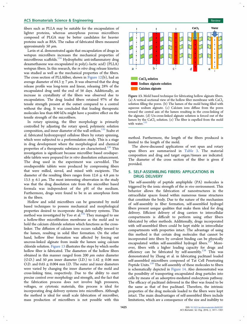
based techniques to possess mechanical and morphologicalproperties desired to incorporate drug delivery systems. Thismethod was investigated by Yoo et al.132 They managed to usea hollow-fiber microfiltration membrane as the mold and tohold the calcium chloride solution which functions as the cross-linker. The diffusion of calcium ions occurs radially inward tothe lumen, resulting in solid fiber formation. On the otherhand, hollow fiber formation was affected by forcing outuncross-linked alginate from inside the lumen using calciumchloride solution. Figure 13 illustrates the steps by which seethehollow fiber is fabricated. The diameters of the hollow fibersobtained in this manner ranged from 200 μm outer diameter(O.D.) and 50 μm inner diameter (I.D.) to 1.42 ± 0.06 mmO.D. and 0.65 ± 0.004 mm I.D. The outer and inner diameterswere varied by changing the inner diameter of the mold andcross-linking time, respectively. Due to the ability to exertprecise control over morphology and strength, and the fact thatthe fabrication process does not involve high pressures,voltages, or cytotoxic materials, this process is ideal forincorporating drug delivery systems into microfibers. Althoughthe method is ideal for small scale fabrication of microfiber,mass production of microfibers is not possible with this
method. Furthermore, the length of the fibers produced islimited to the length of the mold.The above-discussed applications of wet spun and rotary
spun fibers are summarized in Table 3. The materialcomposition and drug and target organ/tissues are indicated.The diameter of the cross section of the fiber is given ifprovided.
5. SELF-ASSEMBLING FIBERS: APPLICATIONS INDRUG DELIVERY
The self-assembly of peptide amphiphile (PA) molecules istriggered by the ionic strength of the in vivo environment. Thisbehavior allows the fabrication of nanostructures in theextracellular spaces found within numerous tissue structuresthat constitute the body. Due to the nature of the mechanismof self-assembly in fiber formation, self-assembled hydrogelfibers present unique qualities that make them ideal for drugdelivery. Efficient delivery of drug carriers to intercellularcompartments is difficult to perform using other fibersfabricated by other methods. Additionally, drugs incorporatedwith self-assembled fibers could be kept stable in intercellularcompartments with properties intact. The advantage of usingthis method is that certain drug molecules that cannot beincorporated into fibers by covalent binding can be physicallyencapsulated within self-assembled hydrogel fibers.137 More-over, fibers with a higher loading capacity for drugs andefficiency can be fabricated by self-assembly.138 This wasdemonstrated by Zhang et al. in fabricating paclitaxel loadedself-assembled microfibers composed of Tat Cell PenetratingPeptide Units.139 The self-assembly of these molecules to fibersis schematically depicted in Figure 14. Also demonstrated wasthe possibility of transporting encapsulated drug particles intocells by means of an adsorptive-mediated endocytosis pathway.The efficacy of paclitaxel delivered in the fiber was found to bethe same as that of free paclitaxel. Therefore, the intrinsicproperties of the drug molecule loaded to the fibers were keptintact. The main disadvantages of self-assembled fibers includelimitations, which are a consequence of the size and inability to
Figure 13. Mold based technique for fabricating hollow alginate fibers.(a) A vertical sectional view of the hollow fiber membrane with CaCl2solution filling the pores. (b) The lumen of the mold being filled withaqueous sodium alginate. (c) Calcium ions diffuse from the porestoward the central axis of the lumen resulting in the cross-linking ofthe alginate. (d) Un-cross-linked alginate solution is forced out of thelumen by the CaCl2 solution. (e) The fiber is expelled from the moldwith water.132
ACS Biomaterials Science & Engineering Review
DOI: 10.1021/acsbiomaterials.6b00281ACS Biomater. Sci. Eng. 2016, 2, 1411−1431
1425

incorporate certain molecules and structures in encapsulatedform in self-assembled fibers.The nature of self-assembly, to generate nanofibers, makes it
an ideal technique to be used in other minimally invasiveapplications as well. PA molecules designed to self-assemble tonanofibers were investigated by Shah et al.140 The nanofibersfabricated in this study were capable of cartilage regenerationthrough transforming growth factor β-1 (TGβ-1). This isaccomplished by displaying a higher density of binding epitopesto the growth factor. The slow release of TGβ-1 from suchfibers in vitro resulted in an increase in the survival rate andchondrogenic differentiation of human mesenchymal stem cells.The material is capable of articular cartilage generationregardless of the presence of the exogenous growth factor ina rabbit model. This means that the potential for the usage ofthis material for cartilage regeneration activities is significant.The application of self-assembling peptides to generatenanofibers capable of creating microenvironments within themyocardium was explored by Davis et al.141 The microenviron-ments capable of promoting vascular smooth muscle cellrecruitment were generated by injecting designer self-assembling peptide molecules. This enabled formation offunctional vascular structures, suggesting potential for injectabletissue regeneration strategies in the future. In a separate study,Heparin-binding peptide amphiphiles were used to generate
nanofibers that interact with heparin.142 A significant improve-ment in pancreatic islet engraftment was observed whenvascular endothelial growth factor (VEGF) and fibroblastgrowth factor-2 (FGF-2) were delivered from fabricated fibers.Hence, peptide amphiphile self-assembly based nanofiberscould potentially be used for drug delivery applications. Inaddition, investigations carried out by Nagai et al. on self-assembling peptide nanofibers suggested that the releaseprofiles of drugs, which are integrated to hydrogel scaffoldscomposed of self-assembled peptides, could be customized bycontrolling nanofiber-diffusant molecular interactions.143 Sin-gle-molecule fluorescence correlation spectroscopy was used tostudy the release kinetics of functional proteins. These proteinsspan a wide range of molecular mass, hydrodynamic radii, andisoelectric points, and create a nanofiber hydrogel scaffoldconsisting of designer self-assembling peptides by Koutsopou-los et al.144 The observations indicated that the proteinfunctionality was unaffected by the encapsulation and releasefrom the scaffold, demonstrating the suitability of such scaffoldsfor sustained delivery of drugs in a clinical context. Self-assembling peptide scaffolds, integrated with epidermal growthfactor, were investigated as a method to accelerate woundhealing by Schneider et al.145 The scaffold was tested on ahuman skin equivalent model, and it was observed that theepidermal growth factor was only released when the scaffoldwas in contact with the tissue model. Furthermore, the scaffolddemonstrated an accelerated rate of wound healing. The use ofself-assembling peptide amphiphile nanofiber scaffolds forreplacement of dental caries with dental stem cells demon-strates the versatility of the technique.146 Instead of replacingdecayed dental structures with inert materials, scaffolds of thisnature could prove to be an effective biological substitute.Similarly, electrospun scaffolds of self-assembling peptideshybridized with poly(ethylene oxide) have been explored as aviable biomaterial for bone tissue engineering due to thebiomimicry of self-assembled biological molecules147
Instances of self-assembled fibers with regard to specificapplications are summarized in Table 4. The materialcomposition and function are given.
Table 3. Details of Fibers Fabricated by Wet Spinning and Rotary Spinning Methods for Drug Delivery Systems
Author Material Drug Target Diameter
Lavin et al.133 PLLA, PLGA Insulin, Lysozyme, BSA pancreas N/ALavin et al.134 PLLA Dexamethasone Inflamed tissue 64.3 ± 7 μmSzabo et al.135 Hydroxypropyl cellulose Semisynthetic alkaloid N/A 12.6 ± 4.8 μm to 13.5 ± 6.1 μm
Figure 14. Schematic of self-assembly of Tat Cell Penetrating PeptideUnits to form nanofibers where paclitaxel is encapsulated.139
Table 4. Details of Fibers Fabricated by Self-Assembly
Author Material Function
Jiang et al.148 laminin, isoleucine-lysine-valine-alanine-valine Peptide amphiphile (IKVAVPA)
Promoting neurite outgrowth and inhibit glialdifferentiation
Tysseling-Mattiace et al.149 IKVAV PA regeneration of both sensory and motor neurons,reduction in cell mortality
Guo et al.150 RADA16-I peptide Reknitting the injured spinal cordShah et al.140 TGFBPA cartilage regenerationDavis et al.141 RAD16-II peptide Promote vascular cell recruitment in the myocardium.Stendahl et al.142 Heparin-binding peptide amphiphiles Improvement in pancreatic islet engraftmentSchneider et al.145 self-complementary peptides with 16 amino-acids (RADA16-I,
Ac-RADARADARADARADA-CONH2)Acceleration of wound healing
Galler et al.146 13-amino-acid peptide (GTAGLIGQERGDS) Dental carrie replacement with dental stem cellsBrun et al.147 Several peptides with varying sequences. Applications related to bone tissue engineering
ACS Biomaterials Science & Engineering Review
DOI: 10.1021/acsbiomaterials.6b00281ACS Biomater. Sci. Eng. 2016, 2, 1411−1431
1426

6. CONCLUSIONMicro- and nanofibers are promising candidates for carriers indrug delivery systems. The surface area to volume ratio,morphology, material properties, and drug release profile can beeasily tuned. In this review paper, we showed that electro-spinning, microfluidic, wet and dry spinning, and self-assemblyapproaches are commonly used for fabricating fibers applied fordrug delivery research. All methods are relatively straightfor-ward and feasible under laboratory settings. Among thesemethods, electrospinning was found to be the most prominentapproach, and has been used extensively to fabricate micro- andnanofibers. This method has demonstrated great controllabilityin creating ultrafine fibers, but the limitations in size, shape,biocompatible solvent, and process may render it unsuitable forcertain applications. The microfluidic platform is relatively newwith promising potential to fabricate fibers in a variety ofdimensions, cross-sectional shapes, and biocompatible materialswithout changing the environmental conditions. This makes it adesirable approach for cell incorporation in drug deliverysystems. Wet and rotary spinning were seen to be lesscommonly used for generating fibers for drug deliveryapplications. Micro- and nanofibers fabricated by these methodshave been incorporated with stimuli-responsive characteristics,such as responsiveness to magnetism and light. Such stimuli-responsive fibers along with the drug carrying capacity havenumerous potential applications in drug delivery. Additionally,the incorporation of liposomes with micro- and nanofibers wereshown to enhance the drug carrying capacity in drug deliverysystems. The use of self-assembly methods for fiber fabricationhas proved to be a novel and effective method to createstructures in the intercellular matrix while encapsulating drugs.For each of these fiber fabrication methods, the constituent
molecules are biocompatible. Thus, they are viable carriers fordrugs in vivo. For example, the incorporation of liposomes intoelectrospun fibers, utilization of the burst release for HIVprophylaxis, and stimuli-responsive fibers are all promisingprospects for drug delivery applications. A wide range ofmolecules could be delivered by these fibers. Additionally, it ispossible to exert precise control over drug release profiles fromfibers. There is a significant potential for fiber based drugdelivery systems in modern clinical medicine. The above-mentioned advantages offered by fiber based delivery systemsoutperform current conventional drug delivery systems.Methods such as orodispersible tablets and vaccines could beincorporated with fiber based drug delivery systems in order toimprove the efficiency of drug delivery. However, the majorityof developments detailed in this review involve in vitroinvestigations of the efficacy and efficiency of fibrous drugcarriers. Before clinical trials, in vivo studies must be performedextensively to identify the best candidates for drug deliverysystems incorporated with fibers. Once extensive in vivo studiesare carried out, manufacturing processes should be developedto commercialize these drug delivery systems in clinicalmedicine.
■ AUTHOR INFORMATIONCorresponding Author*E-mail: [email protected] Contributions∥F.S. and A.C.S. contributed equally.NotesThe authors declare no competing financial interest.
■ ACKNOWLEDGMENTS
This work was supported by the Office of Naval ResearchGrant N000141612246, the Iowa State University PresidentialInitiative for Interdisciplinary Research, and the U.S. Depart-ment of Energy Office of Science under the ScienceUndergraduate Laboratory Internship (SULI) program. H.A.was supported by the Scientific and Technological ResearchCouncil of Turkey (TUBITAK, Postdoctoral fellowship). Wewould like to thank Ashley Christopherson and JonathanVander Wiel for their assistance with this project.
■ REFERENCES(1) Mitra, S.; Gaur, U.; Ghosh, P. C.; Maitra, A. N. Tumour targeteddelivery of encapsulated dextran−doxorubicin conjugate usingchitosan nanoparticles as carrier. J. Controlled Release 2001, 74, 317−323.(2) Hans, M. L.; Lowman, A. M. Biodegradable nanoparticles fordrug delivery and targeting. Curr. Opin. Solid State Mater. Sci. 2002, 6,319−327.(3) Jabbari, E.; Kim, D.-H.; Lee, L. P.; Ghaemmaghami, A.;Khademhosseini, A. Handbook of Biomimetics and Bioinspiration;World Scientific: 2014.(4) Kumbar, S. G.; Nair, L. S.; Bhattacharyya, S.; Laurencin, C. T.Polymeric Nanofibers as Novel Carriers for the Delivery ofTherapeutic Molecules. J. Nanosci. Nanotechnol. 2006, 6, 2591−2607.(5) Korsmeyer, R. W.; Gurny, R.; Doelker, E.; Buri, P.; Peppas, N. A.Mechanisms of solute release from porous hydrophilic polymers. Int. J.Pharm. 1983, 15, 25−35.(6) Acar, H.; Banerjee, S.; Shi, H.; Jamshidi, R.; Hashemi, N.; Cho,M.; Montazami, R. Transient Biocompatible Polymeric Platforms forLong-Term Controlled Release of Therapeutic Proteins and Vaccines.Materials 2016, 9, 321.(7) Ahn, S. Y.; Mun, C. H.; Lee, S. H. Microfluidic spinning offibrous alginate carrier having highly enhanced drug loading capabilityand delayed release profile. RSC Adv. 2015, 5, 15172−15181.(8) Chen, D.; Singh, D.; Sirkar, K. K.; Pfeffer, R. ContinuousSynthesis of Polymer-Coated Drug Particles by Porous Hollow FiberMembrane-Based Antisolvent Crystallization. Langmuir 2015, 31,432−441.(9) Garg, T.; Rath, G.; Goyal, A. K. Biomaterials-based nanofiberscaffold: targeted and controlled carrier for cell and drug delivery. J.Drug Targeting 2015, 23, 202−221.(10) Fernandes, J. G.; Correia, D. M.; Botelho, G.; Padrao, J.;Dourado, F.; Ribeiro, C.; Lanceros-Mendez, S.; Sencadas, V. PHB-PEO electrospun fiber membranes containing chlorhexidine for drugdelivery applications. Polym. Test. 2014, 34, 64−71.(11) Fogaca, R.; Ouimet, M. A.; Catalani, L. H.; Uhrich, K. E.Bioactive-Based Poly(anhydride-esters) and Blends for ControlledDrug Delivery. Tailored Polymer Architectures for Pharmaceutical andBiomedical Applications; American Chemical Society: 2013; pp 27−37.(12) Manasco, J. L.; Tang, C.; Burns, N. A.; Saquing, C. D.; Khan, S.A. Rapidly dissolving poly(vinyl alcohol)/cyclodextrin electrospunnanofibrous membranes. RSC Adv. 2014, 4, 13274−13279.(13) Gaharwar, A. K.; Mihaila, S. M.; Kulkarni, A. A.; Patel, A.; DiLuca, A.; Reis, R. L.; Gomes, M. E.; van Blitterswijk, C.; Moroni, L.;Khademhosseini, A. Amphiphilic beads as depots for sustained drugrelease integrated into fibrillar scaffolds. J. Controlled Release 2014, 187,66−73.(14) Ulery, B. D.; Kumar, D.; Ramer-Tait, A. E.; Metzger, D. W.;Wannemuehler, M. J.; Narasimhan, B. Design of a Protective Single-Dose Intranasal Nanoparticle-Based Vaccine Platform for RespiratoryInfectious Diseases. PLoS One 2011, 6, e17642.(15) Allison, S. D. Analysis of initial burst in PLGA microparticles.Expert Opin. Drug Delivery 2008, 5, 615−628.(16) Huang, X.; Brazel, C. S. On the importance and mechanisms ofburst release in matrix-controlled drug delivery systems. J. ControlledRelease 2001, 73, 121−136.
ACS Biomaterials Science & Engineering Review
DOI: 10.1021/acsbiomaterials.6b00281ACS Biomater. Sci. Eng. 2016, 2, 1411−1431
1427

(17) Becker, T. A.; Kipke, D. R.; Brandon, T. Calcium alginate gel: Abiocompatible and mechanically stable polymer for endovascularembolization. J. Biomed. Mater. Res. 2001, 54, 76−86.(18) Orive, G.; Ponce, S.; Hernandez, R. M.; Gascon, A. R.; Igartua,M.; Pedraz, J. L. Biocompatibility of microcapsules for cellimmobilization elaborated with different type of alginates. Biomaterials2002, 23, 3825−3831.(19) Lim, D.; Lee, E.; Kim, H.; Park, S.; Baek, S.; Yoon, J. Multistimuli-responsive hydrogel microfibers containing magnetite nano-particles prepared using microcapillary devices. Soft Matter 2015, 11,1606−1613.(20) Mazzitelli, S.; Capretto, L.; Quinci, F.; Piva, R.; Nastruzzi, C.Preparation of cell-encapsulation devices in confined microenviron-ment. Adv. Drug Delivery Rev. 2013, 65, 1533−1555.(21) Mohammadi, M.; Alizadeh, P.; Clemens, F. J. Synthesis ofCaCu3Ti4O12 nanofibers by electrospinning. Ceram. Int. 2015, 41,13417−13424.(22) Cao, J.; Wang, Z.; Wang, R.; Liu, S.; Fei, T.; Wang, L.; Zhang, T.Synthesis of core-shell [small alpha]-Fe2O3@NiO nanofibers withhollow structures and their enhanced HCHO sensing properties. J.Mater. Chem. A 2015, 3, 5635−5641.(23) Garifullin, R.; Guler, M. O. Supramolecular chirality in self-assembled peptide amphiphile nanostructures. Chem. Commun. 2015,51, 12470−12473.(24) Jun, Y.; Kang, E.; Chae, S.; Lee, S.-H. Microfluidic spinning ofmicro- and nano-scale fibers for tissue engineering. Lab Chip 2014, 14,2145−2160.(25) Park, J.-S. Electrospinning and its applications. Adv. Nat. Sci.:Nanosci. Nanotechnol. 2010, 1, 043002.(26) Badrossamay, M. R.; McIlwee, H. A.; Goss, J. A.; Parker, K. K.Nanofiber Assembly by Rotary Jet-Spinning. Nano Lett. 2010, 10,2257−2261.(27) Higashi, N.; Koga, T. Self-Assembled Peptide Nanofibers. InSelf-Assembled Nanomaterials I: Nanofibers; Shimizu, T, Ed.; SpringerBerlin Heidelberg: Berlin, Heidelberg, 2008; pp 27−68.(28) Tamayol, A.; Akbari, M.; Annabi, N.; Paul, A.; Khademhosseini,A.; Juncker, D. Fiber-based tissue engineering: Progress, challenges,and opportunities. Biotechnol. Adv. 2013, 31, 669−687.(29) Whitesides, G. M. The origins and the future of microfluidics.Nature 2006, 442, 368−373.(30) Caplin, J. D.; Granados, N. G.; James, M. R.; Montazami, R.;Hashemi, N. Microfluidic Organ-on-a-Chip Technology for Advance-ment of Drug Development and Toxicology. Adv. Healthcare Mater.2015, 4, 1426−1450.(31) Sharifi, F.; Kurteshi, D.; Hashemi, N. Designing highlystructured polycaprolactone fibers using microfluidics. Journal of theMechanical Behavior of Biomedical Materials 2016, 61, 530−540.(32) Goodrich, P. J.; Sharifi, F.; Hashemi, N. Rapid prototyping ofmicrochannels with surface patterns for fabrication of polymer fibers.RSC Adv. 2015, 5, 71203−71209.(33) Sharifi, F.; Ghobadian, S.; Cavalcanti, F. R.; Hashemi, N. Paper-based devices for energy applications. Renewable Sustainable EnergyRev. 2015, 52, 1453−1472.(34) Sharifi, F.; Bai, Z.; Montazami, R.; Hashemi, N. Mechanical andphysical properties of poly(vinyl alcohol) microfibers fabricated by amicrofluidic approach. RSC Adv. 2016, 6, 55343−55353.(35) Hashemi, N.; Lackore, J. M.; Sharifi, F.; Goodrich, P. J.;Winchell, M. L.; Hashemi, N. A paper-based microbial fuel celloperating under continuous flow condition. Technology 2016, 04, 98−103.(36) Shin, S.-J.; Park, J.-Y.; Lee, J.-Y.; Park, H.; Park, Y.-D.; Lee, K.-B.;Whang, C.-M.; Lee, S.-H. On the Fly” Continuous Generation ofAlginate Fibers Using a Microfluidic Device. Langmuir 2007, 23,9104−9108.(37) Choi, C.-H.; Yi, H.; Hwang, S.; Weitz, D. A.; Lee, C.-S.Microfluidic fabrication of complex-shaped microfibers by liquidtemplate-aided multiphase microflow. Lab Chip 2011, 11, 1477−1483.
(38) Hu, M.; Deng, R.; Schumacher, K. M.; Kurisawa, M.; Ye, H.;Purnamawati, K.; Ying, J. Y. Hydrodynamic spinning of hydrogelfibers. Biomaterials 2010, 31, 863−869.(39) Bai, Z.; Mendoza Reyes, J. M.; Montazami, R.; Hashemi, N. On-chip development of hydrogel microfibers from round to square/ribbon shape. J. Mater. Chem. A 2014, 2, 4878−4884.(40) Chae, S.-K.; Kang, E.; Khademhosseini, A.; Lee, S.-H. Micro/Nanometer-Scale Fiber with Highly Ordered Structures by Mimickingthe Spinning Process of Silkworm. Adv. Mater. 2013, 25, 3071−3078.(41) Williams, P. S.; Carpino, F.; Zborowski, M. MagneticNanoparticle Drug Carriers and Their Study by Quadrupole MagneticField-Flow Fractionation. Mol. Pharmaceutics 2009, 6, 1290−1306.(42) Peter, J. F.; Otto, A. M. Magnetic particles as powerfulpurification tool for high sensitive mass spectrometric screeningprocedures. Proteomics 2010, 10, 628−633.(43) Xu, C.; Sun, S. Superparamagnetic nanoparticles as targetedprobes for diagnostic and therapeutic applications. Dalton Transactions2009, 5583−5591.(44) Yui, N.; Okano, T.; Sakurai, Y. Photo-responsive degradation ofheterogeneous hydrogels comprising crosslinked hyaluronic acid andlipid microspheres for temporal drug delivery. J. Controlled Release1993, 26, 141−145.(45) Lin, Y.-S.; Huang, K.-S.; Yang, C.-H.; Wang, C.-Y.; Yang, Y.-S.;Hsu, H.-C.; Liao, Y.-J.; Tsai, C.-W. Microfluidic Synthesis ofMicrofibers for Magnetic-Responsive Controlled Drug Release andCell Culture. PLoS One 2012, 7, e33184.(46) Sershen, S. R.; Westcott, S. L.; Halas, N. J.; West, J. L.Temperature-sensitive polymer−nanoshell composites for photo-thermally modulated drug delivery. J. Biomed. Mater. Res. 2000, 51,293−298.(47) Duffy, D. C.; McDonald, J. C.; Schueller, O. J. A.; Whitesides, G.M. Rapid Prototyping of Microfluidic Systems in Poly-(dimethylsiloxane). Anal. Chem. 1998, 70, 4974−4984.(48) Dobson, J.; Yiu, H. H. P.; McBain, S. C. Magnetic nanoparticlesfor gene and drug delivery. Int. J. Nanomed. 2008, 3, 169−180.(49) Hu, C.; Nakajima, M.; Yue, T.; Takeuchi, M.; Seki, M.; Huang,Q.; Fukuda, T. On-chip fabrication of magnetic alginate hydrogelmicrofibers by multilayered pneumatic microvalves. Microfluid. Nano-fluid. 2014, 17, 457−468.(50) Su, J.; Zheng, Y.; Wu, H. Generation of alginate microfibers witha roller-assisted microfluidic system. Lab Chip 2009, 9, 996−1001.(51) Marimuthu, M.; Kim, S.; An, J. Amphiphilic triblock copolymerand a microfluidic device for porous microfiber fabrication. Soft Matter2010, 6, 2200−2207.(52) Berthier, E.; Young, E. W. K.; Beebe, D. Engineers are fromPDMS-land, Biologists are from Polystyrenia. Lab Chip 2012, 12,1224−1237.(53) Kaufman, J. J.; Ottman, R.; Tao, G.; Shabahang, S.; Banaei, E.-H.; Liang, X.; Johnson, S. G.; Fink, Y.; Chakrabarti, R.; Abouraddy, A.F. In-fiber production of polymeric particles for biosensing andencapsulation. Proc. Natl. Acad. Sci. U. S. A. 2013, 110, 15549−15554.(54) Kang, E.; Choi, Y. Y.; Chae, S.-K.; Moon, J.-H.; Chang, J.-Y.;Lee, S.-H. Microfluidic Spinning of Flat Alginate Fibers with Groovesfor Cell-Aligning Scaffolds. Adv. Mater. 2012, 24, 4271−4277.(55) Kang, E.; Shin, S.-J.; Lee, K. H.; Lee, S.-H. Novel PDMScylindrical channels that generate coaxial flow, and application tofabrication of microfibers and particles. Lab Chip 2010, 10, 1856−1861.(56) Remant Bahadur, K. C.; Bhattarai, S. R.; Aryal, S.; Khil, M. S.;Dharmaraj, N.; Kim, H. Y. Novel amphiphilic triblock copolymerbased on PPDO, PCL, and PEG: Synthesis, characterization, andaqueous dispersion. Colloids Surf., A 2007, 292, 69−78.(57) Johnson, A. S.; Anderson, K. B.; Halpin, S. T.; Kirkpatrick, D.C.; Spence, D. M.; Martin, R. S. Integration of multiple components inpolystyrene-based microfluidic devices part I: fabrication andcharacterization. Analyst 2013, 138, 129−136.(58) Acred, P.; Brown, D. M.; Turner, D. H.; Wilson, M. J.Pharmacology and chemotherapy of ampicillina new broad-spectrum penicillin. Br. J. Pharmacol. Chemother. 1962, 18, 356−369.
ACS Biomaterials Science & Engineering Review
DOI: 10.1021/acsbiomaterials.6b00281ACS Biomater. Sci. Eng. 2016, 2, 1411−1431
1428

(59) Mazzitelli, S.; Capretto, L.; Carugo, D.; Zhang, X.; Piva, R.;Nastruzzi, C. Optimised production of multifunctional microfibres bymicrofluidic chip technology for tissue engineering applications. LabChip 2011, 11, 1776−1785.(60) Doshi, J.; Reneker, D. H. Electrospinning process andapplications of electrospun fibers. J. Electrost. 1995, 35, 151−160.(61) Kenawy, E.-R.; Abdel-Hay, F. I.; El-Newehy, M. H.; Wnek, G. E.Processing of polymer nanofibers through electrospinning as drugdelivery systems. Mater. Chem. Phys. 2009, 113, 296−302.(62) Sill, T. J.; von Recum, H. A. Electrospinning: Applications indrug delivery and tissue engineering. Biomaterials 2008, 29, 1989−2006.(63) Lopez-Rubio, A.; Sanchez, E.; Sanz, Y.; Lagaron, J. M.Encapsulation of Living Bifidobacteria in Ultrathin PVOH ElectrospunFibers. Biomacromolecules 2009, 10, 2823−2829.(64) Khorshidi, S.; Solouk, A.; Mirzadeh, H.; Mazinani, S.; Lagaron, J.M.; Sharifi, S.; Ramakrishna, S. A review of key challenges ofelectrospun scaffolds for tissue-engineering applications. J. Tissue Eng.Regener. Med. 2015, n/a.(65) Boyd, D. A.; Shields, A. R.; Naciri, J.; Ligler, F. S. HydrodynamicShaping, Polymerization, and Subsequent Modification of Thiol ClickFibers. ACS Appl. Mater. Interfaces 2013, 5, 114−119.(66) Rezaei, B.; Ghani, M.; Askari, M.; Shoushtari, A. M.; Malek, R.M. A. Fabrication of Thermal Intelligent Core/Shell Nanofibers by theSolution Coaxial Electrospinning Process. Adv. Polym. Technol. 2016,35, n/a.(67) Lu, Y.; Huang, J.; Yu, G.; Cardenas, R.; Wei, S.; Wujcik, E. K.;Guo, Z. Coaxial electrospun fibers: applications in drug delivery andtissue engineering. Wiley Interdisciplinary Reviews: Nanomedicine andNanobiotechnology 2016.(68) Townsend-Nicholson, A.; Jayasinghe, S. N. Cell Electro-spinning: a Unique Biotechnique for Encapsulating Living Organismsfor Generating Active Biological Microthreads/Scaffolds. Biomacromo-lecules 2006, 7, 3364−3369.(69) Luu, Y. K.; Kim, K.; Hsiao, B. S.; Chu, B.; Hadjiargyrou, M.Development of a nanostructured DNA delivery scaffold viaelectrospinning of PLGA and PLA−PEG block copolymers. J.Controlled Release 2003, 89, 341−353.(70) Kim, K.; Yu, M.; Zong, X.; Chiu, J.; Fang, D.; Seo, Y.-S.; Hsiao,B. S.; Chu, B.; Hadjiargyrou, M. Control of degradation rate andhydrophilicity in electrospun non-woven poly(d,l-lactide) nanofiberscaffolds for biomedical applications. Biomaterials 2003, 24, 4977−4985.(71) Kim, K.; Luu, Y. K.; Chang, C.; Fang, D.; Hsiao, B. S.; Chu, B.;Hadjiargyrou, M. Incorporation and controlled release of a hydrophilicantibiotic using poly(lactide-co-glycolide)-based electrospun nano-fibrous scaffolds. J. Controlled Release 2004, 98, 47−56.(72) Liang, D.; Luu, Y. K.; Kim, K.; Hsiao, B. S.; Hadjiargyrou, M.;Chu, B. In vitro non-viral gene delivery with nanofibrous scaffolds.Nucleic Acids Res. 2005, 33, e170−e170.(73) Chiu, J. B.; Liu, C.; Hsiao, B. S.; Chu, B.; Hadjiargyrou, M.Functionalization of poly(L-lactide) nanofibrous scaffolds withbioactive collagen molecules. J. Biomed. Mater. Res., Part A 2007,83A, 1117−1127.(74) Chew, S. Y.; Wen, J.; Yim, E. K. F.; Leong, K. W. SustainedRelease of Proteins from Electrospun Biodegradable Fibers. Bio-macromolecules 2005, 6, 2017−2024.(75) Park, S.; Son, Y. J.; Leong, K. W.; Yoo, H. S. Therapeuticnanorods with metallic multi-segments: Thermally inducible encapsu-lation of doxorubicin for anti-cancer therapy. Nano Today 2012, 7,76−84.(76) Goldberg, M.; Langer, R.; Jia, X. Nanostructured materials forapplications in drug delivery and tissue engineering. J. Biomater. Sci.,Polym. Ed. 2007, 18, 241−268.(77) Bai, M.-Y.; Chou, T.-C.; Tsai, J.-C.; Yang, H.-C. Activeingredient-containing chitosan/polycaprolactone nonwoven mats:Characterizations and their functional assays. Mater. Sci. Eng., C2013, 33, 224−233.
(78) Sridhar, R.; Madhaiyan, K.; Sundarrajan, S.; Gora, A.;Venugopal, J. R.; Ramakrishna, S. Cross-linking of protein scaffoldsfor therapeutic applications: PCL nanofibers delivering riboflavin forprotein cross-linking. J. Mater. Chem. B 2014, 2, 1626−1633.(79) Wollensak, G.; Spoerl, E.; Seiler, T. Riboflavin/ultraviolet-a−induced collagen crosslinking for the treatment of keratoconus. Am. J.Ophthalmol. 2003, 135, 620−627.(80) Braun, E.; Kanellopoulos, J.; Pe, L.; Jankov, M. Riboflavin/Ultraviolet A−Induced Collagen Cross−Linking in the Managementof Keratoconus. Investigative Ophthalmology & Visual Science 2005, 46,4964−4964.(81) Xu, Q.; Zhang, N.; Qin, W.; Liu, J.; Jia, Z.; Liu, H. Preparation, <i > In Vitro</i> and < i > In Vivo</i> Evaluation of BudesonideLoaded Core/Shell Nanofibers as Oral Colonic Drug Delivery System.J. Nanosci. Nanotechnol. 2013, 13, 149−156.(82) Rubert, M.; Li, Y.-F.; Dehli, J.; Taskin, M. B.; Besenbacher, F.;Chen, M. Dexamethasone encapsulated coaxial electrospun PCL/PEOhollow microfibers for inflammation regulation. RSC Adv. 2014, 4,51537−51543.(83) Lei, C.; Cui, Y.; Zheng, L.; Kah-Hoe Chow, P.; Wang, C.-H.Development of a gene/drug dual delivery system for brain tumortherapy: Potent inhibition via RNA interference and synergistic effects.Biomaterials 2013, 34, 7483−7494.(84) Ball, C.; Woodrow, K. A. Electrospun Solid Dispersions ofMaraviroc for Rapid Intravaginal Preexposure Prophylaxis of HIV.Antimicrob. Agents Chemother. 2014, 58, 4855−4865.(85) Torchilin, V. P. Multifunctional nanocarriers. Adv. Drug DeliveryRev. 2012, 64 (Supplement), 302−315.(86) Delamaza, A.; Parra, J. L.; Bosch, P. USING LIPOSOMES INWOOL CHLORINATION - STABILITY OF CHLORINE LIP-OSOMES AND THEIR APPLICATION ON WOOL FIBERS. Text.Res. J. 1991, 61, 357−362.(87) Delamaza, A.; Parra, J. L. UNILAMELLAR LIPID BILAYERSINCLUDING CHOLESTEROL IN WOOL CHLORINATION -STABILITY OF CHLORINE LIPOSOMES AND APPLICATIONON WOOL. Text. Res. J. 1993, 63, 44−50.(88) Kieler-Ferguson, H. M.; Frechet, J. M. J.; Szoka, F. C., Jr Clinicaldevelopments of chemotherapeutic nanomedicines: polymers andliposomes for delivery of camptothecins and platinum (II) drugs.WileyInterdisciplinary Reviews: Nanomedicine and Nanobiotechnology 2013, 5,130−138.(89) Mickova, A.; Buzgo, M.; Benada, O.; Rampichova, M.; Fisar, Z.;Filova, E.; Tesarova, M.; Lukas, D.; Amler, E. Core/Shell Nanofiberswith Embedded Liposomes as a Drug Delivery System. Biomacromo-lecules 2012, 13, 952−962.(90) Song, H.-h.; Gong, X.; Williams, G. R.; Quan, J.; Nie, H.-l.; Zhu,L.-m.; Nan, E.-l.; Shao, M. Self-assembled magnetic liposomes fromelectrospun fibers. Mater. Res. Bull. 2014, 53, 280−289.(91) Huang, J.; Shu, Q.; Wang, L.; Wu, H.; Wang, A. Y.; Mao, H.Layer-by-layer assembled milk protein coated magnetic nanoparticleenabled oral drug delivery with high stability in stomach and enzyme-responsive release in small intestine. Biomaterials 2015, 39, 105−113.(92) Mou, X.; Ali, Z.; Li, S.; He, N. Applications of MagneticNanoparticles in Targeted Drug Delivery System. J. Nanosci.Nanotechnol. 2015, 15, 54−62.(93) Yu, D.-G.; Branford-White, C.; Williams, G. R.; Bligh, S. W. A.;White, K.; Zhu, L.-M.; Chatterton, N. P. Self-assembled liposomesfrom amphiphilic electrospun nanofibers. Soft Matter 2011, 7, 8239−8247.(94) Rampichova, M.; Martinova, L.; Kost’akova, E.; Filova, E.;Míckova, A.; Buzgo, M.; Michalek, J.; Pradny, M.; Necas, A.; Lukas, D.;Amler, E. A simple drug anchoring microfiber scaffold for chondrocyteseeding and proliferation. J. Mater. Sci.: Mater. Med. 2012, 23, 555−563.(95) Tran, T.; Hernandez, M.; Patel, D.; Wu, J. Temperature and pHResponsive Microfibers for Controllable and Variable IbuprofenDelivery. Adv. Mater. Sci. Eng. 2015, 2015, 1−6.
ACS Biomaterials Science & Engineering Review
DOI: 10.1021/acsbiomaterials.6b00281ACS Biomater. Sci. Eng. 2016, 2, 1411−1431
1429

(96) Abidian, M. R.; Kim, D.-H.; Martin, D. C. Conducting-PolymerNanotubes for Controlled Drug Release. Adv. Mater. (Weinheim, Ger.)2006, 18, 405−409.(97) Zhou, L.; Zhu, C. H.; Edmonds, L.; Yang, H. L.; Cui, W. G.; Li,B. Microsol-electrospinning for controlled loading and release ofwater-soluble drugs in microfibrous membranes. RSC Adv. 2014, 4,43220−43226.(98) Gemma, S.; Travagli, V.; Savini, L.; Novellino, E.; Campiani, G.;Butini, S. Malaria Chemotherapy: Recent Advances in DrugDevelopment. Recent Pat. Anti-Infect. Drug Discovery 2010, 5, 195−225.(99) Bajpai, A. K.; Choubey, J. Design of gelatin nanoparticles asswelling controlled delivery system for chloroquine phosphate. J.Mater. Sci.: Mater. Med. 2006, 17, 345−358.(100) Grundy, R.; Blowers, R. A case of primary amoebicmeningoencephalitis treated with chloroquine. East African Medical J.1970, 47, 153−158.(101) Puhl, S.; Li, L.; Meinel, L.; Germershaus, O. ControlledProtein Delivery from Electrospun Non-Wovens: Novel Combinationof Protein Crystals and a Biodegradable Release Matrix. Mol.Pharmaceutics 2014, 11, 2372−2380.(102) Shenoy, B.; Wang, Y.; Shan, W.; Margolin, A. L. Stability ofcrystalline proteins. Biotechnol. Bioeng. 2001, 73, 358−369.(103) Charernsriwilaiwat, N.; Opanasopit, P.; Rojanarata, T.;Ngawhirunpat, T. Lysozyme-loaded, electrospun chitosan-based nano-fiber mats for wound healing. Int. J. Pharm. 2012, 427, 379−384.(104) Llorens, E.; Ibanez, H.; del Valle, L. J.; Puiggalí, J.Biocompatibility and drug release behavior of scaffolds prepared bycoaxial electrospinning of poly(butylene succinate) and polyethyleneglycol. Mater. Sci. Eng., C 2015, 49, 472−484.(105) Barnes, C. P.; Sell, S. A.; Boland, E. D.; Simpson, D. G.;Bowlin, G. L. Nanofiber technology: Designing the next generation oftissue engineering scaffolds. Adv. Drug Delivery Rev. 2007, 59, 1413−1433.(106) Griffith, L. G. Polymeric biomaterials. Acta Mater. 2000, 48,263−277.(107) Brazel, C. S.; Peppas, N. A. Temperature- and pH- SensitiveHydrogels for Controlled Release of Antithrombotic Agents. MRSOnline Proc. Libr. 1993, 331, 10.1557/PROC-331-211(108) Marimuthu, M.; Kim, S. Spontaneous extrusion of porousamphiphilic triblock copolymeric microfibers under microfluidicconditions. Polym. J. 2010, 42, 100−102.(109) Qi, R.; Guo, R.; Zheng, F.; Liu, H.; Yu, J.; Shi, X. Controlledrelease and antibacterial activity of antibiotic-loaded electrospunhalloysite/poly(lactic-co-glycolic acid) composite nanofibers. ColloidsSurf., B 2013, 110, 148−155.(110) Tonetti, M.; Cugini, M. A.; Goodson, J. M. Zero-order deliverywith periodontal placement of tetracycline-loaded ethylene vinylacetate fibers. J. Periodontal Res. 1990, 25, 243−249.(111) Alger, F. A.; Solt, C. W.; Vuddhakanok, S.; Miles, K. TheHistologic Evaluation of New Attachment in Periodontally DiseasedHuman Roots Treated With Tetracycline-Hydrochloride andFibronectin. J. Periodontol. 1990, 61, 447−455.(112) Wikesjo, U. M. E.; Claffey, N.; Christersson, L. A.; Franzetti, L.C.; Genco, R. J.; Terranova, V. P.; Egelberg, J. Repair of periodontalfurcation defects in beagle dogs following reconstructive surgeryincluding root surface demineralization with tetracycline hydrochlorideand topical fibronectin application. J. Clin. Periodontol. 1988, 15, 73−80.(113) Li, Y.-F.; Rubert, M.; Yu, Y.; Besenbacher, F.; Chen, M.Delivery of dexamethasone from electrospun PCL-PEO binary fibersand their effects on inflammation regulation. RSC Adv. 2015, 5,34166−34172.(114) Hickey, T.; Kreutzer, D.; Burgess, D. J.; Moussy, F.Dexamethasone/PLGA microspheres for continuous delivery of ananti-inflammatory drug for implantable medical devices. Biomaterials2002, 23, 1649−1656.(115) de Gans, J.; van de Beek, D. Dexamethasone in Adults withBacterial Meningitis. N. Engl. J. Med. 2002, 347, 1549−1556.
(116) Dimopoulos, M.; Spencer, A.; Attal, M.; Prince, H. M.;Harousseau, J.-L.; Dmoszynska, A.; Miguel, J. S.; Hellmann, A.; Facon,T.; Foa, R.; Corso, A.; Masliak, Z.; Olesnyckyj, M.; Yu, Z.; Patin, J.;Zeldis, J. B.; Knight, R. D. Lenalidomide plus Dexamethasone forRelapsed or Refractory Multiple Myeloma. N. Engl. J. Med. 2007, 357,2123−2132.(117) Xue, J. J.; Niu, Y. Z.; Gong, M.; Shi, R.; Chen, D. F.; Zhang, L.Q.; Lvov, Y. Electrospun Microfiber Membranes Embedded withDrug-Loaded Clay Nanotubes for Sustained Antimicrobial Protection.ACS Nano 2015, 9, 1600−1612.(118) Valarezo, E.; Tammaro, L.; Gonzalez, S.; Malagon, O.; Vittoria,V. Fabrication and sustained release properties of poly(ε-caprolactone)electrospun fibers loaded with layered double hydroxide nanoparticlesintercalated with amoxicillin. Appl. Clay Sci. 2013, 72, 104−109.(119) Peltier, S.; Oger, J.-M.; Lagarce, F.; Couet, W.; Benoît, J.-P.Enhanced Oral Paclitaxel Bioavailability After Administration ofPaclitaxel-Loaded Lipid Nanocapsules. Pharm. Res. 2006, 23, 1243−1250.(120) Huang, H.-H.; He, C.-L.; Wang, H.-S.; Mo, X.-M. Preparationof core-shell biodegradable microfibers for long-term drug delivery. J.Biomed. Mater. Res., Part A 2009, 90A, 1243−1251.(121) Sabitha, M.; Rajiv, S. Synthesis and characterization ofbiocompatible tigecycline imbibed electrospun poly ?-caprolactoneurethane urea fibers. RSC Adv. 2015, 5, 2249−2257.(122) Stephansen, K.; Chronakis, I. S.; Jessen, F. Bioactiveelectrospun fish sarcoplasmic proteins as a drug delivery system.Colloids Surf., B 2014, 122, 158−165.(123) Bottino, M. C.; Arthur, R. A.; Waeiss, R. A.; Kamocki, K.;Gregson, K. S.; Gregory, R. L. Biodegradable nanofibrous drug deliverysystems: effects of metronidazole and ciprofloxacin on periodonto-pathogens and commensal oral bacteria. Clinical Oral Investigations2014, 18, 2151−2158.(124) Albuquerque, M. T. P.; Ryan, S. J.; Munchow, E. A.; Kamocka,M. M.; Gregory, R. L.; Valera, M. C.; Bottino, M. C. AntimicrobialEffects of Novel Triple Antibiotic Paste−Mimic Scaffolds onActinomyces naeslundii Biofilm. Journal of Endodontics 2015, 41,1337−1343.(125) Reise, M.; Wyrwa, R.; Muller, U.; Zylinski, M.; Volpel, A.;Schnabelrauch, M.; Berg, A.; Jandt, K. D.; Watts, D. C.; Sigusch, B. W.Release of metronidazole from electrospun poly(l-lactide-co-d/l-lactide) fibers for local periodontitis treatment. Dent. Mater. 2012,28, 179−188.(126) Rho, K. S.; Jeong, L.; Lee, G.; Seo, B.-M.; Park, Y. J.; Hong, S.-D.; Roh, S.; Cho, J. J.; Park, W. H.; Min, B.-M. Electrospinning ofcollagen nanofibers: Effects on the behavior of normal humankeratinocytes and early-stage wound healing. Biomaterials 2006, 27,1452−1461.(127) Kauffman, J. S.; Ellerbrock, B. M.; Stevens, K. A.; Brown, P. J.;Pennington, W. T.; Hanks, T. W. Preparation, Characterization, andSensing Behavior of Polydiacetylene Liposomes Embedded in AlginateFibers. ACS Appl. Mater. Interfaces 2009, 1, 1287−1291.(128) Zhang, F.; Lu, Q.; Yue, X.; Zuo, B.; Qin, M.; Li, F.; Kaplan, D.L.; Zhang, X. Regeneration of high-quality silk fibroin fiber by wetspinning from CaCl2−formic acid solvent. Acta Biomater. 2015, 12,139−145.(129) Fang, B.; Peng, L.; Xu, Z.; Gao, C. Wet-Spinning ofContinuous Montmorillonite-Graphene Fibers for Fire-ResistantLightweight Conductors. ACS Nano 2015, 9, 5214−5222.(130) Arafat, M. T.; Tronci, G.; Yin, J.; Wood, D. J.; Russell, S. J.Biomimetic wet-stable fibres via wet spinning and diacid-basedcrosslinking of collagen triple helices. Polymer 2015, 77, 102−112.(131) Sebe, I.; Kallai-Szabo, B.; Kovacs, K. N.; Szabadi, E.; Zelko, R.Micro- and macrostructural characterization of polyvinylpirrolidonerotary-spun fibers. Drug Dev. Ind. Pharm. 2015, 41, 1829−1834.(132) Yoo, S. M.; Ghosh, R. Fabrication of alginate fibers using amicroporous membrane based molding technique. Biochem. Eng. J.2014, 91, 58−65.(133) Lavin, D. M.; Zhang, L.; Furtado, S.; Hopkins, R. A.;Mathiowitz, E. Effects of protein molecular weight on the intrinsic
ACS Biomaterials Science & Engineering Review
DOI: 10.1021/acsbiomaterials.6b00281ACS Biomater. Sci. Eng. 2016, 2, 1411−1431
1430

material properties and release kinetics of wet spun polymericmicrofiber delivery systems. Acta Biomater. 2013, 9, 4569−4578.(134) Lavin, D. M.; Stefani, R. M.; Zhang, L.; Furtado, S.; Hopkins,R. A.; Mathiowitz, E. Multifunctional polymeric microfibers withprolonged drug delivery and structural support capabilities. ActaBiomater. 2012, 8, 1891−1900.(135) Szabo, P.; Sebe, I.; Stiedl, B.; Kallai-Szabo, B.; Zelko, R.Tracking of crystalline-amorphous transition of carvedilol in rotaryspun microfibers and their formulation to orodispersible tablets for invitro dissolution enhancement. J. Pharm. Biomed. Anal. 2015, 115,359−367.(136) Szabo, P.; Kallai-Szabo, B.; Sebe, I.; Zelko, R. Preformulationstudy of fiber formation and formulation of drug-loaded microfiberbased orodispersible tablets for in vitro dissolution enhancement. Int. J.Pharm. 2014, 477, 643−649.(137) Vemula, P. K.; Kohler, J. E.; Blass, A.; Williams, M.; Xu, C.;Chen, L.; Jadhav, S. R.; John, G.; Soybel, D. I.; Karp, J. M. Self-assembled hydrogel fibers for sensing the multi-compartmentintracellular milieu. Sci. Rep. 2014, 4, 4466.(138) Liberato, M. S.; Kogikoski, S.; da Silva, E. R.; de Araujo, D. R.;Guha, S.; Alves, W. A. Polycaprolactone fibers with self-assembledpeptide micro/nanotubes: a practical route towards enhancedmechanical strength and drug delivery applications. J. Mater. Chem.B 2016, 4, 1405−1413.(139) Zhang, P.; Cheetham, A. G.; Lin, Y.-a.; Cui, H. Self-AssembledTat Nanofibers as Effective Drug Carrier and Transporter. ACS Nano2013, 7, 5965−5977.(140) Shah, R. N.; Shah, N. A.; Del Rosario Lim, M. M.; Hsieh, C.;Nuber, G.; Stupp, S. I. Supramolecular design of self-assemblingnanofibers for cartilage regeneration. Proc. Natl. Acad. Sci. U. S. A.2010, 107, 3293−3298.(141) Davis, M. E.; Motion, J. P. M.; Narmoneva, D. A.; Takahashi,T.; Hakuno, D.; Kamm, R. D.; Zhang, S.; Lee, R. T. Injectable Self-Assembling Peptide Nanofibers Create Intramyocardial Microenviron-ments for Endothelial Cells. Circulation 2005, 111, 442−450.(142) Stendahl, J. C.; Wang, L.-J.; Chow, L. W.; Kaufman, D. B.;Stupp, S. I. Growth factor delivery from self-assembling nanofibers tofacilitate islet transplantation. Transplantation 2008, 86, 478−481.(143) Nagai, Y.; Unsworth, L. D.; Koutsopoulos, S.; Zhang, S. Slowrelease of molecules in self-assembling peptide nanofiber scaffold. J.Controlled Release 2006, 115, 18−25.(144) Koutsopoulos, S.; Unsworth, L. D.; Nagai, Y.; Zhang, S.Controlled release of functional proteins through designer self-assembling peptide nanofiber hydrogel scaffold. Proc. Natl. Acad. Sci.U. S. A. 2009, 106, 4623−4628.(145) Schneider, A.; Garlick, J. A.; Egles, C. Self-Assembling PeptideNanofiber Scaffolds Accelerate Wound Healing. PLoS One 2008, 3,e1410.(146) Galler, K. M.; Cavender, A.; Yuwono, V.; Dong, H.; Shi, S. T.;Schmalz, G.; Hartgerink, J. D.; D’Souza, R. N. Self-Assembling PeptideAmphiphile Nanofibers as a Scaffold for Dental Stem Cells. Tissue Eng.,Part A 2008, 14, 2051−2058.(147) Brun, P.; Ghezzo, F.; Roso, M.; Danesin, R.; Palu, G.; Bagno,A.; Modesti, M.; Castagliuolo, I.; Dettin, M. Electrospun scaffolds ofself-assembling peptides with poly(ethylene oxide) for bone tissueengineering. Acta Biomater. 2011, 7, 2526−2532.(148) Jiang, H.; Guler, M. O.; Stupp, S. I. The internal structure ofself-assembled peptide amphiphiles nanofibers. Soft Matter 2007, 3,454−462.(149) Tysseling-Mattiace, V. M.; Sahni, V.; Niece, K. L.; Birch, D.;Czeisler, C.; Fehlings, M. G.; Stupp, S. I.; Kessler, J. A. Self-assemblingnanofibers inhibit glial scar formation and promote axon elongationafter spinal cord injury. J. Neurosci. 2008, 28, 3814−3823.(150) Guo, J.; Su, H.; Zeng, Y.; Liang, Y.-X.; Wong, W. M.; Ellis-Behnke, R. G.; So, K.-F.; Wu, W. Reknitting the injured spinal cord byself-assembling peptide nanofiber scaffold. Nanomedicine 2007, 3,311−321.
ACS Biomaterials Science & Engineering Review
DOI: 10.1021/acsbiomaterials.6b00281ACS Biomater. Sci. Eng. 2016, 2, 1411−1431
1431