fibroepithelioma of pinkus revisited - springer · review fibroepithelioma of pinkus revisited...
TRANSCRIPT
REVIEW
Fibroepithelioma of Pinkus Revisited
Ellen S. Haddock . Philip R. Cohen
Received: May 10, 2016 / Published online: June 21, 2016� The Author(s) 2016. This article is published with open access at Springerlink.com
ABSTRACT
Background: Fibroepithelioma of Pinkus (FeP)
is considered a variant of basal cell carcinoma
(BCC); however, in the past 20 years, some
researchers have argued for its classification as
a trichoblastoma. Recently, use of a new
immunostaining marker and further
dermoscopic characterization of FeP have
advanced the debate about its proper
classification.
Purpose: A review of the evidence for and
against classification of FeP as BCC or
trichoblastoma is presented.
Methods: Using PubMed, the term FeP was
searched and relevant citations were assessed.
Additional relevant articles were identified from
references of key papers.
Results: FeP shares characteristics of both
trichoblastoma and BCC.
Conclusion: Derived from the same cell type,
BCC and trichoblastoma may be best
considered as representing opposite ends of a
spectrum of differentiation, with FeP deserving
an intermediate classification.
Keywords: Basal; Carcinoma; Cell;
Fibroepithelioma; PHLDA1; Pinkus;
Trichoblastoma; Trichoepithelioma
INTRODUCTION
Fibroepithelioma of Pinkus (FeP) is an
uncommon skin lesion originally thought to
be a rare variant of basal cell carcinoma (BCC).
In the past couple decades, controversy has
developed over whether FeP is a type of BCC or a
trichoblastoma. Both trichoblastoma, a benign
neoplasm, and BCC, a malignant neoplasm, are
thought to be derived from follicular epithelial
stem cells in the bulge area of the hair follicle
[1–3]. Follicular stem cells in turn give rise to
follicular germinative cells, also known as
trichoblasts, which can develop into all the
components of the folliculo-sebaceous-apocrine
Enhanced content To view enhanced content for thisarticle go to http://www.medengine.com/Redeem/88D4F0600EB07454.
E. S. Haddock (&)School of Medicine, University of California SanDiego, San Diego, CA, USAe-mail: [email protected]
P. R. CohenDepartment of Dermatology, University ofCalifornia San Diego, San Diego, CA, USAe-mail: [email protected]
Dermatol Ther (Heidelb) (2016) 6:347–362
DOI 10.1007/s13555-016-0123-8
unit [4–7]. Trichoblasts that give rise to
trichoblastoma and basal cell carcinoma
express common epithelial keratins and are
differentiated toward the outer root sheath and
the companion layer of the hair follicle [8, 9].
Follicular cells that instead express hair keratins
give rise to hair fiber, while those expressing yet
another different set of keratins give rise to the
inner root sheath [9]. In this paper, FeP is
reviewed and the arguments for and against its
classification as a trichoblastoma or BCC are
presented.
METHODS
Using PubMed, the term FeP was searched and
relevant citations were assessed. Additional
relevant articles were identified from
references of key papers [1–66]. This article is
based on previously conducted studies and does
not involve any new studies of human or
animal subjects performed by any of the
authors.
HISTORY
FeP was first described in 1953 by Hermann
Pinkus as ‘‘an unusual variety of basal-cell
epithelioma’’, which he considered to be
premalignant [10]. In a 1965 paper, he
elaborated that ‘‘the epithelial part of
premalignant fibroepithelial tumors resembles
that of trichoepitheliomas in its degree of
differentiation, while the growth
characteristics of the entire tumor is closely
related to superficial basal cell epithelioma of
the trunk’’, in some cases transforming into an
ulcerating basal cell epithelioma [11], thereby
justifying its categorization as a type of BCC.
Following Pinkus’s description, FeP was
generally accepted to be a variant of BCC until
Hartschuh et al. in 1997 and Bowen et al. in
2005 suggested that FeP might be more closely
related to trichoblastoma than to BCC [12, 13],
unleashing a controversy.
NOMENCLATURE
Some authors suggest that the term
trichoepithelioma refers to a superficial
trichoblastoma [5, 14]. The World Health
Organization uses the terms synonymously
[15]. The terms are used interchangeably in
this paper.
INCIDENCE
FeP is relatively rare, with Dr. Pinkus identifying
only 4 cases among 900 epitheliomas [10]. In
series of BCCs, the frequency of FeP ranges from
0.2% to 1.4% [16–18]. However, FeP may be
underreported, as it can resemble other benign
tumors which may not be biopsied [19]. FeP
usually presents in adults older than age 50 [20],
and women may be affected slightly more often
than men (54% female in Bowen et al.’s series of
114 patients) [13]. Rare cases of children with
FeP have been reported [21, 22].
CLINICAL PRESENTATION
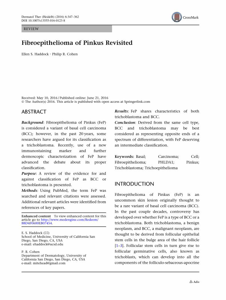
FeP typically presents as a skin-colored, firm,
dome-shaped, sessile, fleshy papule or plaque,
which can mimic a pedunculated fibroma,
acrochordon, seborrheic keratosis, or dermal
nevus (Fig. 1) [11, 23, 24]. It may be pigmented
[24–26], and patients can—albeit
uncommonly—present with multiple lesions
[25]. It most often occurs on the lumbosacral
area but may also occur on the abdomen, head,
axillae, thigh, groin, and plantar foot [19, 20,
27, 28]. It often develops in patients with a
348 Dermatol Ther (Heidelb) (2016) 6:347–362
history of BCC [26] and may have increased
prevalence in irradiated skin [12].
DERMOSCOPY
Dermoscopy reveals fine arborizing vessels
[19, 29] and white septal lines or streaks [19],
which are thought to be a consequence of
significant fibrosis. Some lesions have
distributed gray-brown structureless
pigmentation and gray-blue dots [19].
PATHOLOGY
The histopathology of FeP is, as Pinkus
described it, ‘‘peculiar and unmistakable’’ [10].
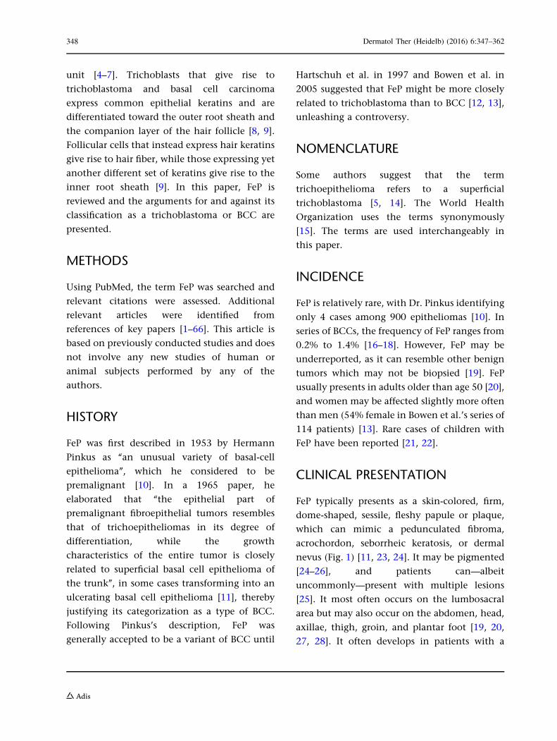
Thin anastomosing strands of basaloid or
squamous keratinocytes project downward
from the epidermis in a fenestrated pattern
[30]. The cords of epithelial cells are embedded
in abundant fibrous stroma, like window frames
separating panes of glass (Figs. 2, 3) [13, 19].
From a three-dimensional perspective, the
tumor resembles a honeycomb or sponge
composed of thin epithelial septa, with the
intervening spaces filled with stroma [11, 19].
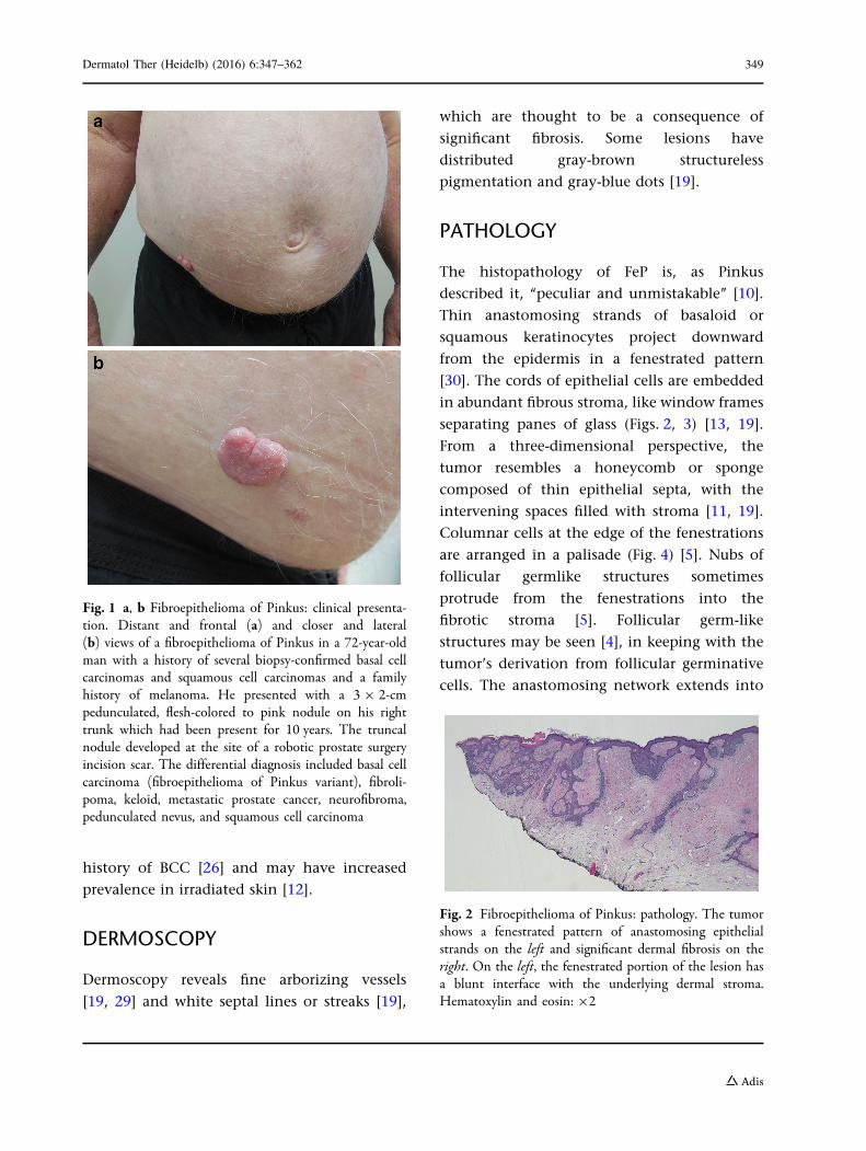
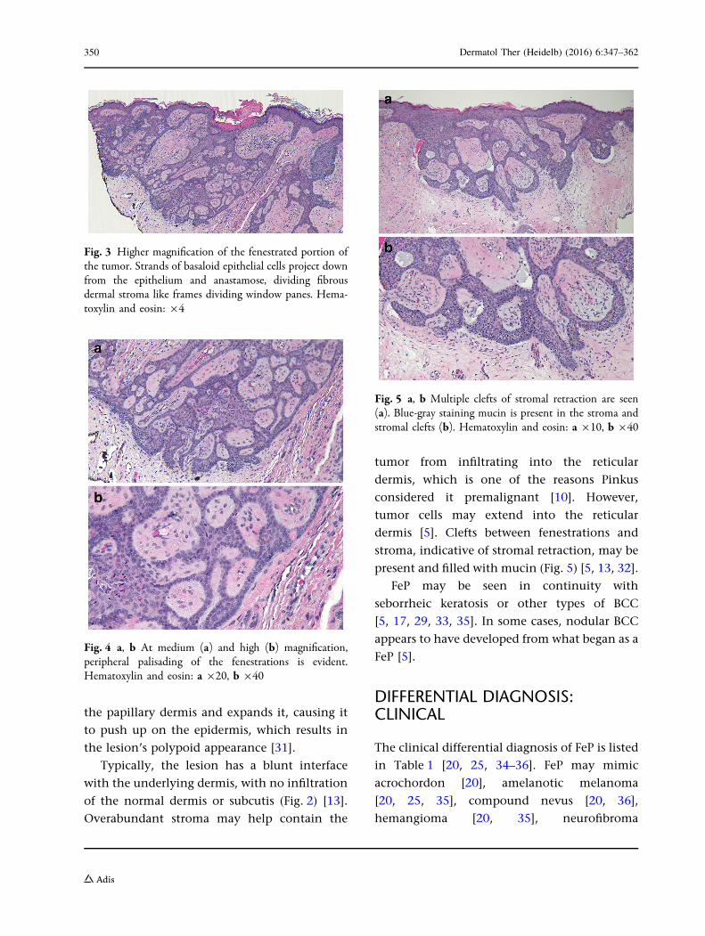
Columnar cells at the edge of the fenestrations
are arranged in a palisade (Fig. 4) [5]. Nubs of
follicular germlike structures sometimes
protrude from the fenestrations into the
fibrotic stroma [5]. Follicular germ-like
structures may be seen [4], in keeping with the
tumor’s derivation from follicular germinative
cells. The anastomosing network extends into
Fig. 2 Fibroepithelioma of Pinkus: pathology. The tumorshows a fenestrated pattern of anastomosing epithelialstrands on the left and significant dermal fibrosis on theright. On the left, the fenestrated portion of the lesion hasa blunt interface with the underlying dermal stroma.Hematoxylin and eosin: 92
Fig. 1 a, b Fibroepithelioma of Pinkus: clinical presenta-tion. Distant and frontal (a) and closer and lateral(b) views of a fibroepithelioma of Pinkus in a 72-year-oldman with a history of several biopsy-confirmed basal cellcarcinomas and squamous cell carcinomas and a familyhistory of melanoma. He presented with a 3 9 2-cmpedunculated, flesh-colored to pink nodule on his righttrunk which had been present for 10 years. The truncalnodule developed at the site of a robotic prostate surgeryincision scar. The differential diagnosis included basal cellcarcinoma (fibroepithelioma of Pinkus variant), fibroli-poma, keloid, metastatic prostate cancer, neurofibroma,pedunculated nevus, and squamous cell carcinoma
Dermatol Ther (Heidelb) (2016) 6:347–362 349
the papillary dermis and expands it, causing it
to push up on the epidermis, which results in
the lesion’s polypoid appearance [31].
Typically, the lesion has a blunt interface
with the underlying dermis, with no infiltration
of the normal dermis or subcutis (Fig. 2) [13].
Overabundant stroma may help contain the
tumor from infiltrating into the reticular
dermis, which is one of the reasons Pinkus
considered it premalignant [10]. However,
tumor cells may extend into the reticular
dermis [5]. Clefts between fenestrations and
stroma, indicative of stromal retraction, may be
present and filled with mucin (Fig. 5) [5, 13, 32].
FeP may be seen in continuity with
seborrheic keratosis or other types of BCC
[5, 17, 29, 33, 35]. In some cases, nodular BCC
appears to have developed from what began as a
FeP [5].
DIFFERENTIAL DIAGNOSIS:CLINICAL
The clinical differential diagnosis of FeP is listed
in Table 1 [20, 25, 34–36]. FeP may mimic
acrochordon [20], amelanotic melanoma
[20, 25, 35], compound nevus [20, 36],
hemangioma [20, 35], neurofibroma
Fig. 5 a, b Multiple clefts of stromal retraction are seen(a). Blue-gray staining mucin is present in the stroma andstromal clefts (b). Hematoxylin and eosin: a 910, b 940
Fig. 4 a, b At medium (a) and high (b) magnification,peripheral palisading of the fenestrations is evident.Hematoxylin and eosin: a 920, b 940
Fig. 3 Higher magnification of the fenestrated portion ofthe tumor. Strands of basaloid epithelial cells project downfrom the epithelium and anastamose, dividing fibrousdermal stroma like frames dividing window panes. Hema-toxylin and eosin: 94
350 Dermatol Ther (Heidelb) (2016) 6:347–362
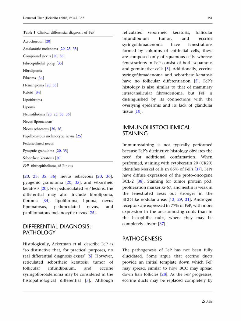
[20, 25, 35, 36], nevus sebaceous [20, 36],
pyogenic granuloma [20, 35], and seborrheic
keratosis [20]. For pedunculated FeP lesions, the
differential may also include fibrolipoma,
fibroma [34], lipofibroma, lipoma, nevus
lipomatosus, pedunculated nevus, and
papillomatous melanocytic nevus [25].
DIFFERENTIAL DIAGNOSIS:PATHOLOGY
Histologically, Ackerman et al. describe FeP as
‘‘so distinctive that, for practical purposes, no
real differential diagnosis exists’’ [5]. However,
reticulated seborrheic keratosis, tumor of
follicular infundibulum, and eccrine
syringofibroadenoma may be considered in the
histopathological differential [5]. Although
reticulated seborrheic keratosis, follicular
infundibulum tumor, and eccrine
syringofibroadenoma have fenestrations
formed by columns of epithelial cells, these
are composed only of squamous cells, whereas
fenestrations in FeP consist of both squamous
and germinative cells [5]. Additionally, eccrine
syringofibroadenoma and seborrheic keratosis
have no follicular differentiation [5]. FeP’s
histology is also similar to that of mammary
intracanalicular fibroadenoma, but FeP is
distinguished by its connections with the
overlying epidermis and its lack of glandular
tissue [10].
IMMUNOHISTOCHEMICALSTAINING
Immunostaining is not typically performed
because FeP’s distinctive histology obviates the
need for additional confirmation. When
performed, staining with cytokeratin 20 (CK20)
identifies Merkel cells in 85% of FePs [37]. FePs
have diffuse expression of the proto-oncogene
BCL-2 [38]. Staining for tumor protein p53,
proliferation marker Ki-67, and nestin is weak in
the fenestrated areas but stronger in the
BCC-like nodular areas [13, 29, 31]. Androgen
receptors are expressed in 77% of FeP, with more
expression in the anastomosing cords than in
the basophilic nubs, where they may be
completely absent [37].
PATHOGENESIS
The pathogenesis of FeP has not been fully
elucidated. Some argue that eccrine ducts
provide an initial template down which FeP
may spread, similar to how BCC may spread
down hair follicles [28]. As the FeP progresses,
eccrine ducts may be replaced completely by
Table 1 Clinical differential diagnosis of FeP
Acrochordon [20]
Amelanotic melanoma [20, 25, 35]
Compound nevus [20, 36]
Fibroepithelial polyp [35]
Fibrolipoma
Fibroma [34]
Hemangioma [20, 35]
Keloid [34]
Lipofibroma
Lipoma
Neurofibroma [20, 25, 35, 36]
Nevus lipomatosus
Nevus sebaceous [20, 36]
Papillomatous melanocytic nevus [25]
Pedunculated nevus
Pyogenic granuloma [20, 35]
Seborrheic keratosis [20]
FeP fibroepithelioma of Pinkus
Dermatol Ther (Heidelb) (2016) 6:347–362 351
solid strands of tumor [28]. Carcinoembryonic
antigen (CEA) is a glycoprotein found in sweat
glands as well as gastrointestinal tumors and
fetal tissues [39]. Stern et al. found that 9 of 12
FePs examined stained positive for CEA,
indicating the presence of eccrine ducts [28].
Lending support to the theory that eccrine
ducts provide a template for FeP growth is the
relatively high incidence of FeP on the glabrous
sole of the foot, which has many eccrine sweat
glands and few hair follicles [28]. Roth et al.
found that 6 of 20 (30%) BCCs on the sole were
FePs [40]. However, others have argued that
eccrine ducts do not anastomose and that the
eccrine duct foci in FePs may represent normal
eccrine ducts trapped within the tumor or
ductal differentiation [41].
Development of BCC and FeP on the
glabrous skin of the sole of the foot, which
lacks hair follicles [28, 40] and presumably lacks
follicular germinative cells, suggests that not
only follicular germinative cells but also eccrine
gland stem cells may give rise to BCC and FeP
[42].
Fibroepithelioma-like hyperplasia has been
associated with Paget disease, especially
anogenital Paget disease, which raises the
possibility that in some cases it may be a
reactive process [43]. Finally, mutations in
tumor suppressor genes p53 and patched-1
(ptch1) may predispose to the development of
FeP [26], but there is some evidence that the
level of p53 in FeP is lower than that in BCC
[13].
Ackerman et al. suggest that FeP may
develop from seborrheic keratosis, since FePs
often have infundibular tunnels filled with
corneocytes in lamellate array, which may be
the remnants of seborrheic keratosis [4]. Others
propose that cases of FeP in continuity with
nodular BCC indicate that trichoblastoma can
progress to BCC with the acquisition of
additional genetic mutations [17].
TREATMENT
Complete excision is the typical treatment for
FeP [20], and prognosis is good. Aggressive
biologic behavior with local destruction or
metastasis is extremely rare [44]. However,
there is at least one report of a metastatic BCC
with some FeP histopathology [45].
CLASSIFICATION OF FEP
The controversy about the proper classification
of FeP centers around its common locations and
histopathologic and immunohistochemical
features (Table 2) [4, 5, 10, 11, 13, 15, 17,
19, 28, 29, 31, 33, 34, 37, 38, 45–55, 61, 63,
65, 66].
Location
The argument that FeP should be characterized
as a trichoblastoma rather than a BCC has been
made most persuasively by Bowen et al. [13].
The authors point out that FePs tend to occur in
locations that are relatively uncommon for
BCC. While FeP occur most often on the
trunk, 80% of BCC occur on the head and
neck, with only 15% of BCC occurring on the
trunk [13], which receives less sun exposure.
Yet, trichoblastoma are also most common on
the head and neck [15, 46], so FeP’s relatively
low prevalence on the head and neck does not
clearly support either classification. Some argue
that the relatively high incidence of FeP on the
sole of the foot suggests that FeP is more closely
related to BCC than trichoblastoma, as
trichoblastoma rarely occurs on the sole of the
foot [47].
352 Dermatol Ther (Heidelb) (2016) 6:347–362
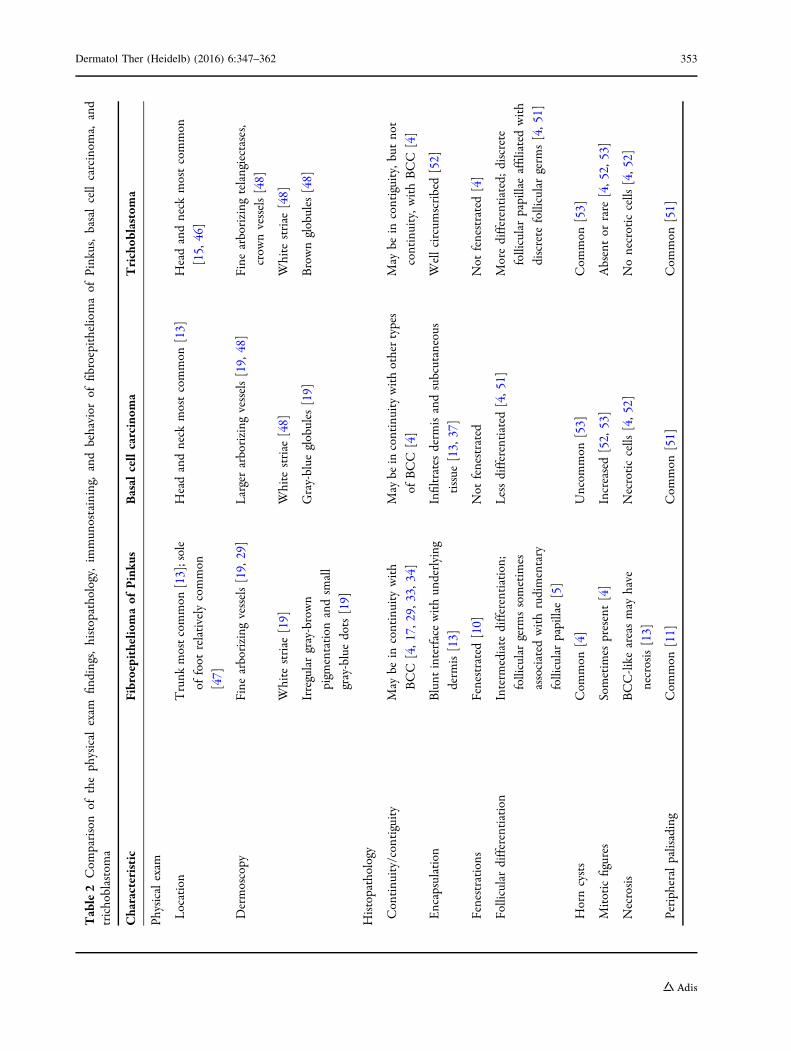
Table2
Com
parisonof
thephysical
exam
findings,histopathology,im
mun
ostaining,
andbehavior
offibroepitheliomaof
Pinkus,basalcellcarcinom
a,and
trichoblastoma
Characteristic
Fibroepitheliomaof
Pinku
sBasal
cellcarcinom
aTrichob
lastom
a
Physicalexam
Location
Trunk
mostcommon
[13];sole
offoot
relativelycommon
[47]
Headandneck
mostcommon
[13]
Headandneck
mostcommon
[15,
46]
Dermoscopy
Fine
arborizing
vessels[19,
29]
Largerarborizing
vessels[19,
48]
Fine
arborizing
telangiectases,
crow
nvessels[48]
White
striae
[19]
White
striae
[48]
White
striae
[48]
Irregulargray-brown
pigm
entation
andsm
all
gray-bluedots[19]
Gray-blue
globules
[19]
Brownglobules
[48]
Histopathology
Continu
ity/contiguity
May
bein
continuity
with
BCC
[4,1
7,29,3
3,34]
May
bein
continuitywithothertypes
ofBCC
[4]
May
bein
contiguity,b
utnot
continuity,w
ithBCC
[4]
Encapsulation
Blunt
interfacewithun
derlying
derm
is[13]
Infiltrates
derm
isandsubcutaneous
tissue
[13,
37]
Wellcircum
scribed[52]
Fenestrations
Fenestrated[10]
Not
fenestrated
Not
fenestrated[4]
Folliculardifferentiation
Interm
ediate
differentiation;
folliculargerm
ssometim
es
associated
withrudimentary
follicularpapillae[5]
Lessdifferentiated
[4,5
1]Moredifferentiated;discrete
follicularpapillaeaffiliatedwith
discrete
folliculargerm
s[4,5
1]
Horncysts
Com
mon
[4]
Uncom
mon
[53]
Com
mon
[53]
Mitoticfigures
Sometim
espresent[4]
Increased[52,
53]
Absentor
rare
[4,5
2,53]
Necrosis
BCC-like
areasmay
have
necrosis[13]
Necroticcells
[4,5
2]Nonecroticcells
[4,5
2]
Peripheralpalisading
Com
mon
[11]
Com
mon
[51]
Com
mon
[51]
Dermatol Ther (Heidelb) (2016) 6:347–362 353
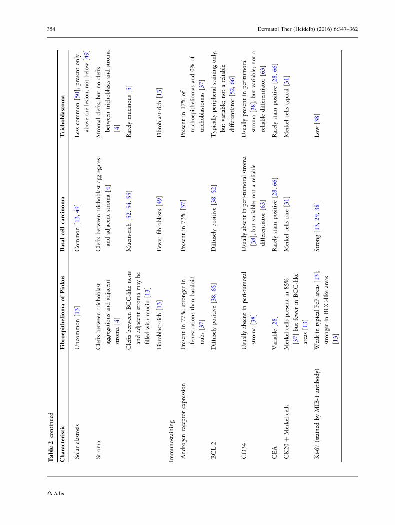
Table2
continued
Characteristic
Fibroepitheliomaof
Pinku
sBasal
cellcarcinom
aTrichob
lastom
a
Solarelastosis
Uncom
mon
[13]
Com
mon
[13,
49]
Lesscommon
[50];presentonly
abovethelesion,n
otbelow[49]
Stroma
Cleftsbetweentrichoblast
aggregations
andadjacent
stroma[4]
Cleftsbetweentrichoblastaggregates
andadjacent
stroma[4]
Stromalclefts,b
utno
clefts
betweentrichoblastsandstroma
[4]
CleftsbetweenBCC-like
nests
andadjacent
stromamay
be
filledwithmucin
[13]
Mucin-rich[52,
54,5
5]Rarelymucinous[5]
Fibroblast-rich[13]
Fewer
fibroblasts[49]
Fibroblast-rich[13]
Immun
ostaining
And
rogenreceptor
expression
Presentin
77%;stronger
in
fenestration
sthan
basaloid
nubs
[37]
Presentin
73%
[37]
Presentin
17%
of
trichoepitheliomas
and0%
of
trichoblastomas
[37]
BCL-2
Diffuselypositive
[38,
65]
Diffuselypositive
[38,
52]
Typicallyperipheralstaining
only,
butvariable;notareliable
differentiator
[52,
66]
CD34
Usuallyabsent
inperi-tum
oral
stroma[38]
Usuallyabsent
inperi-tum
oralstroma
[38],b
utvariable;notareliable
differentiator
[63]
Usuallypresentin
peritumoral
stroma[38],b
utvariable;nota
reliabledifferentiator
[63]
CEA
Variable[28]
Rarelystainpositive
[28,
66]
Rarelystainpositive
[28,
66]
CK20
?Merkelcells
Merkelcells
presentin
85%
[37]
butfewer
inBCC-like
areas[13]
Merkelcells
rare
[31]
Merkelcells
typical[31]
Ki-6
7(stained
byMIB-1
antibody)
Weakin
typicalF
ePareas[13];
stronger
inBCC-like
areas
[13]
Strong
[13,
29,3
8]Low
[38]
354 Dermatol Ther (Heidelb) (2016) 6:347–362

Table2
continued
Characteristic
Fibroepitheliomaof
Pinku
sBasal
cellcarcinom
aTrichob
lastom
a
Nestin
Nestinpresentin
stroma
surround
ingBCC-like
areas
butabsent
from
thin
anastomosingstrand
s[31]
Labelsstromaof
BCC
[31]
Expressed
inperi-tum
oralstroma
[61]
p53
Weakin
mostFePareas
[13,
29];stronger
in
BCC-like
areas[13]
Moderateto
strong
[13,
29]
Weak[13]
PHLDA1
Anastom
osingstrand
spositive;
basaloid
nubs
negative
[31]
Negative[31]
Positive
[31]
Behavior
Clin
icalbehavior
Typicallynotaggressive
[31],
butonereportof
metastasis
[45]
Moreaggressive
[31];locally
destructivewithraremetastasis[52]
Benign[52]
Treatmentresponse
toim
iquimod
Unresponsive[19]
Responsive[19]
Not
evaluated
BCCbasalcellcarcinom
a,FePfibroepitheliomaof
Pinkus
Dermatol Ther (Heidelb) (2016) 6:347–362 355
Dermoscopy
From the perspective of dermoscopy, FeP shares
similarities with both BCC and trichoblastoma.
FeP and trichoblastoma have small, fine
arborizing vessels [19, 29, 48] that are similar
to those seen in other BCC but smaller in caliber
and less branched [19, 48]. White striae, thought
to result from fibrosis, can be seen in FeP, BCC,
and trichoblastoma [19, 48]. The gray-blue dots
of FeP are suggestive of the gray-blue globules
described in BCC [19], while trichoblastoma has
been described as having brown globules [48].
Crown vessels may be present in trichoblastoma
[48] but are not typically seen in FeP or BCC.
Overall, FeP, trichoblastoma, and BCC possess
significant dermoscopic similarities and cannot
always be distinguished.
Pathology
With regard to histology, Bowen et al. argue
that the histology of FeP differs from BCC in
that FeP typically has a blunt interface between
the lesion and the underlying dermis; blunt
interface was seen in all 75 FeP lesions reviewed
by Bowen et al. [13]. In contrast, Ackerman
et al. show photographs of FeP extending into
the dermis and contained within subcutaneous
fat [5]. These lesions are well circumscribed,
however—a characteristic usually associated
with benign lesions. Additionally, FeP rarely
have significant dermal solar elastosis [13],
which is common in BCC [13, 49] and less
common in trichoblastoma [49, 50].
While trichoblastomas tend to be well
differentiated toward follicular papillae [4, 51],
and BCC tend to be less differentiated [4, 51],
FeP tend to have an intermediate amount of
differentiation; in FeP, follicular germs may or
may not be associated with rudimentary
follicular papillae [4, 5].
Similarly, the amount of mitotic figures [4]
and necrosis [13] in FeP is intermediate to that
of BCC, in which mitotic figures and necrosis
are common [4], and trichoepithelioma, in
which they are rare or absent [4, 52, 53].
However, horn cysts, which are common in
FeP [4], are often seen in trichoepithelioma but
rarely seen in BCC [53].
Bowen et al. argue that FeP’s stromal
composition is more consistent with
trichoblastoma than BCC, because
fibroblast-rich stroma is typically seen in
benign lesions like trichoblastoma [13].
However, the stromal retraction and mucin
deposition seen in FeP [13] is more consistent
with BCC [4, 5, 52, 54, 55].
Associated BCC
Multiple cases of FeP in continuity with nodular
BCC [4, 17, 29, 33, 34] suggest FeP’s close
relationship with BCC. From the perspective of
one who believes FeP is BCC, Ackerman
explains that it is not unusual for more than
one type of BCC to be in continuity, so it is not
surprising to find FeP in continuity with BCC;
in contrast, BCC and trichoblastoma occur only
in contiguity, not in continuity [4]. As
mentioned above, Misago et al. posit that
cases of FeP in continuity with nodular BCC
may illustrate how trichoblastoma can
transform into BCC as additional genetic
mutations are accumulated [17].
Immunohistochemistry Staining
CK20 Staining
Expression of cytokeratins, which are
well-established markers of epithelial
differentiation, may be used to differentiate
neoplasms and identify their cells of origin [8].
Kurzen et al. found that trichoblastomas and
356 Dermatol Ther (Heidelb) (2016) 6:347–362
BCCs express similar cytokeratin profiles. Both
consistently express CK6hf, CK14, and CK17,
while neither expresses hair keratins [8]. Of the
19 cytokeratins studied by Kurzen et al., only
CK20, which is found in Merkel cells, was useful
for differentiating BCC and trichoblastoma [8].
Merkel cells, the neuroendocrine cells of the
skin [12, 56], are commonly found in
trichoblastomas but are generally absent from
basal cell carcinoma [8, 12, 31].
Hartschuh et al. identified CK20-positive
Merkel cells in the fenestrated portion of FePs but
not in other types of basal cell carcinoma and
therefore suggested that FeP ismore closely related
to trichoblastoma than BCC [12]. Bowen et al.
reported similar findings of CK20-positive Merkel
cells in the fenestrated areas of FePs but not in
other types of basal cell carcinoma [13]. In the
‘‘BCC-like areas’’ of FePs they found fewer Merkel
cells (2 of 5 specimens) or an absence of Merkel
cells (3of 5 specimens) [13], apatterncorroborated
by Katona et al. [37]. However, LeBoit et al. point
out that this method of differentiation is not
absolute: cases of invasive neoplasms containing
Merkel cells have been documented [58].
PHLDA1 Staining
In 2006, Ohyama et al. characterized a follicular
stem cell marker, PHLDA1 (pleckstrin
homology-like domain, family A, member 1),
that was found to be more sensitive and specific
for differentiating trichoblastoma from BCC
than CK20-stained Merkel cells [31, 57, 59].
PHLDA1 is consistently positive in
trichoepitheliomas and negative in BCCs
[31, 57]. Sellheyer et al. found that all 19 of 19
trichoepitheliomas were immunoreactive for
PHLDA1 while all 11 of 11 basal cell
carcinomas lacked PHLDA1 expression [57].
In FePs, Sellheyer et al. found that PHLDA1
labels the fenestrations but not the basaloid nubs;
all nodular parts of the tumor were negative for
PHLDA1 [31]. Sellheyer et al. found that, as
basaloid sprouts develop from the fenestrations,
they lose their immunoreactivity for PHLDA1
[31]. This is not dissimilar from Bowen et al.’s
findings that the fenestrations stained positive
for CK20 but few CK20-positive Merkel cells were
found in the basaloid nubs; indeed, Sellheyer
et al. found that immunoreactivity for PHLDA1
and presence of Merkel cells are correlated in FePs
[31].
However, Sellheyer et al.’s interpretation is
completely different from Bowen’s. Sellheyer
et al. propose that the fenestrated network that
contains Merkel cells and stains positive for
PHLDA1 is a tumor-specific form of epidermal
hyperplasia that is part of the tumor but not
itself malignant [31]. Sellheyer et al. agree with
Pinkus’s original description that ‘‘the real BCC
in FeP are the basaloid nubbins’’ and view the
loss of PHLDA1 immunoreactivity as part of the
progression from benign epidermal hyperplasia
to malignant BCC [10, 31]. Therefore, they
argue that the presence of Merkel cells in the
epidermal hyperplasia of FeP does not disqualify
FeP from being a BCC [31].
Nestin Staining
Similarly, Sellheyer et al. found nestin
immunoreactivity in the BCC-like,
PHLDA1-negative portions of the FePs but no
nestin in the fenestrated, PHLDA1-positive
portion of the FeP [31]. Nestin is a
neuroepithelial stem cell protein that labels
the stroma of BCC [31, 60]. Since the stroma
of BCCs stains positive for nestin, Sellheyer
et al. posit that the presence of nestin in the
BCC-like areas of FePs indicates that they are
true BCCs [31]. However, this argument is
somewhat undermined by another paper by
Sellheyer et al. that reports that nestin is
also expressed trichoblastoma and
trichoepithelioma [61].
Dermatol Ther (Heidelb) (2016) 6:347–362 357
Ki-67 and p53 staining
The pattern of basaloid nubs staining more
similar to BCC and fenestrations staining more
similar to trichoblastoma is also seen in
staining for the protein product of tumor
suppressor gene p53 and the cellular
proliferation marker Ki-67. Bowen et al.
reported that staining for p53, which is
mutated and overexpressed in 56% of BCC,
was generally low in FePs. Similarly, staining
with MIB-1 antibody, which recognizes Ki-67
antigen in formalin-fixed, paraffin-embedded
tissue [62], was also lower than in typical BCCs
[13]. However, the ‘‘BCC-like areas’’ of the FeP
stained stronger than the rest of the FeP for
p53 and Ki-67 [13].
CD34 Staining
The amount of CD34, a hematopoietic
progenitor cell antigen, in peritumoral stromal
cells has been proposed as a differentiator
between trichoepithelioma and basal cell
carcinoma [63, 64]. The stroma immediately
surrounding trichoepitheliomas typically stains
positive for CD34, while stroma immediately
adjacent to basal cell carcinomas typically does
not [38], but there is some variability [63].
Naeyaert et al. found that CD34 expression in
FeP is generally absent in the peritumoral
stroma, as it is in BCC [38].
BCL-2 Staining
With regard to the staining for the
proto-oncogene BCL-2, Naeyaert et al. and
Marusic et al. found that FePs are diffusely
positive, similar to BCC [38, 52, 65], whereas in
trichoepitheliomas, BCL-2 positivity is typically
is limited to the periphery [42, 66]. However,
BCL-2 may also be diffusely positive in
trichoepithelioma and is not considered a
reliable differentiator [52, 66].
Carcinoembryonic Antigen (CEA) Staining
Despite the theory that FeP may spread down
eccrine ducts and the detection of CEA in some
FePs [28], Swanson et al. found that CEA is
usually absent in both BCC and
trichoepithelioma [66]. Therefore, the presence
of absence of CEA staining is not helpful for
categorizing FeP as one or the other.
Androgen Receptor Staining
The similarly high prevalence of androgen
receptors in FeP and BCC (77% vs. 73%) and
the absence or rarity of androgen receptors in
trichoepithelioma and trichoblastoma (17%
and 0%, respectively; p = 0.0007) supports
classification of FeP as BCC [37].
Clinical Behavior and Response
to Therapy
FeP’s non-aggressive clinical behavior seems
more similar to trichoblastoma than BCC.
Additionally, FeP may respond differently to
treatment than BCC; Zalaudek et al. reported a
man with a FeP that was resistant to 5%
imiquimod treatment, whereas his other BCCs
resolved with this therapy [19].
CONCLUSION
Review of the evidence for and against
classification of FeP as BCC or trichoblastoma
finds that FePs possess characteristics of both.
Dermoscopy findings and the amount of
mitotic figures, necrosis, and follicular
differentiation are intermediate between
trichoblastoma and BCC. Its mucin-rich
stroma and stromal retraction are more similar
to BCC, although its relative lack of solar
elastosis and relative prevalence of horn cells
are more consistent with trichoblastoma.
358 Dermatol Ther (Heidelb) (2016) 6:347–362
Immunohistologically, the fenestrated portion
of the tumor more closely resembles
trichoblastoma while the solid basaloid nubs
more closely resemble BCC, with regard to
PHLDA1, p53, and Ki-67 staining, and can
become full-fledged BCC. With regard to
CD34, BCL-2, and androgen receptor
expression, FeP is more similar to BCC. In its
behavior, it generally seems more similar to
trichoblastoma, despite one case of FeP
histopathology in metastatic BCC [45]. Some
characteristics of FeP are distinct from both BCC
and trichoblastoma, such as its predilection for
the trunk and its fenestrated histology.
To some extent, whether the lesion should
be considered a trichoblastoma until BCC
develops within it, or whether it should be
called BCC from the beginning, since it may
give rise to BCC, seems a semantic issue.
Perhaps Katona et al. make the most
reasonable proposal—that FeP may be a
trichoblastic tumor intermediate between
trichoblastoma and BCC [37]. Considering
that trichoblastoma and BCC share a common
cell of origin [37] and represent two different
points in the differentiation of that progenitor
cell [55], with similar cytokeratin profiles and
mutations in the tumor suppressor ptch1
[8, 37, 58], it makes more sense to consider
them on a spectrum from benign to malignant
rather than to attempt to draw an artificial line
between the two. In his 1965 paper, Pinkus said,
‘‘Having been able to collect a graded series
from the typical premalignant fibroepithelial
tumor to the plate-like or almost macular basal
cell epithelioma of the very superficial type, I
find it impossible to draw a distinct line
anywhere in this series’’ [11]. This review of
FeP, and the evidence to classify the tumor as
either a BCC or a trichoblastoma, finds the line
similarly difficult to draw, and perhaps doing so
is unnecessary.
ACKNOWLEDGMENTS
No funding or sponsorship was received for this
study or publication of this article. All named
authors meet the International Committee of
Medical Journal Editors (ICMJE) criteria for
authorship for this manuscript, take
responsibility for the integrity of the work as a
whole, and have given final approval for the
version to be published. The named authors,
Ellen S. Haddock, AB, MBA, and Philip R. Cohen,
MD, confirm that no deserving authors have
been omitted. There are no individuals to be
included in a Contributorship section. No
editorial assistance was used in the preparation
of the manuscript.
Disclosures. Ellen S. Haddock and Philip R.
Cohen have nothing to disclose.
Compliance with Ethics Guidelines. This
article is based on previously conducted
studies and does not involve any new studies
of human or animal subjects performed by any
of the authors.
Open Access. This article is distributed
under the terms of the Creative Commons
Attribution-NonCommercial 4.0 International
License (http://creativecommons.org/licenses/
by-nc/4.0/), which permits any noncommer-
cial use, distribution, and reproduction in any
medium, provided you give appropriate credit
to the original author(s) and the source, provide
a link to the Creative Commons license, and
indicate if changes were made.
REFERENCES
1. Cotsarelis G, Sun TT, Lavker RM. Label-retainingcells reside in the bulge area of pilosebaceous unit:
Dermatol Ther (Heidelb) (2016) 6:347–362 359
implications for follicular stem cells, hair cycle, andskin carcinogenesis. Cell. 1990;61:1329–37.
2. Cotsarelis G. Epithelial stem cells: a folliculocentricview. J Invest Dermatol. 2006;126:1459–68.
3. Peterson SC, Eberl M, Vagnozzi AN, Belkadi A,Veniaminova NA, Verhaegen ME, Bichakjian CK,Ward NL, Dlugosz AA, Wong SY. Basal cellcarcinoma preferentially arises from stem cellswithin hair follicle and mechanosensory niches.Cell Stem Cell. 2015;6:400–12.
4. Ackerman AB, Gottlieb GJ. Fibroepithelial tumor ofPinkus is trichoblastic (basal-cell) carcinoma. Am JDermatopathol. 2005;27:155–9.
5. Ackerman AB, Reddy VB, Soyer HP. Chapter 23—trichoblastic carcinoma. In: Ackerman AB, ReddyVB, Soyer HP, eds. Neoplasms with folliculardifferentiation. New York: Ardor Scribendi, 2001;625-1005
6. Lavker RM, Sun TT, Oshima H, Barrandon Y,Akiyama M, Ferraris C, Chevalier G, Favier B,Jahoda CA, Dhouailly D, Panteleyev AA,Christiano AM. Hair follicle stem cells. J InvestigDermatol Symp Proc. 2003;8:28–38.
7. Reya T, Clevers H. Wnt signalling in stem cells andcancer. Nature. 2005;434:843–50.
8. Kurzen H, Esposito L, Langbein L, Hartschuh W.Cytokeratins as markers of follicular differentiation:an immunohistochemical study of trichoblastomaand basal cell carcinoma. Am J Dermatopathol.2001;23:501–9.
9. Langbein L, Rogers MA, Praetzel S, Winter H,Schweizer J. K6irs1, K6irs2, K6irs3, and K6irs4represent the inner-root-sheath-specific type IIepithelial keratins of the human hair follicle.J Invest Dermatol. 2003;120:512–22.
10. Pinkus H. Premalignant fibroepithelial tumors ofskin. AMA Arch Derm Syphilol. 1953;67:598–616.
11. Pinkus H. Epithelial and fibroepithelial tumors. BullN Y Acad Med. 1965;41:176–89.
12. Hartschuh W, Schulz T. Merkel cell hyperplasia inchronic radiation-damaged skin: its possiblerelationship to fibroepithelioma of Pinkus. J CutanPathol. 1997;24:477–83.
13. Bowen AR, LeBoit PE. Fibroepithelioma of Pinkus isa fenestrated trichoblastoma. Am J Dermatopathol.2005;27:149–54.
14. Busam KJ. Chapter 11—adnexal tumors. In:Dermatopathology. Philadelphia: Saunders; 2009.p. 383.
15. Hurt MA, Cribier B, Kaddu S, Schulz T, Kutzner H,Hartschuh W. Chapter 3—appendageal tumors. In:LeBoit PE, Burg G, Weedon D, Sarasin A, editors.World Health Organization classification of tumors:pathology and genetics of skin tumors. Lyon: IARC;2006. p. 152–153.
16. Betti R, Inselvini E, Carducci M, Crosti C. Age andsite prevalence of histologic subtypes of basal cellcarcinomas. Int J Dermatol. 1995;34:174–6.
17. Misago N, Narisawa Y. Polypoid Basal cellcarcinoma on the perianal region: a case reportand review of the literature. J Dermatol.2004;31:51–5.
18. Rahbari H, Mehregan AH. Basal cell epitheliomas inusual and unusual sites. J Cutan Pathol.1979;6:425–31.
19. Zalaudek I, Ferrara G, Broganelli P, Moscarella E,Mordente I, Giacomel J, Argenziano G. Dermoscopypatterns of fibroepithelioma of Pinkus. ArchDermatol. 2006;142:1318–22.
20. Cohen PR, Tschen JA. Fibroepithelioma of Pinkuspresenting as a sessile thigh nodule. Skinmed.2003;2:385–7.
21. Pan Z, Huynh N, Sarma DP. Fibroepithelioma ofPinkus in a 9-year-old boy: a case report. Cases J.2008;1:21.
22. Scalvenzi M, Francia MG, Falleti J, Balato A. Basalcell carcinoma with fibroepithelioma-like histologyin a healthy child: report and review of theliterature. Pediatr Dermatol. 2008;25:359–63.
23. Reggiani C, Zalaudek I, Piana S, Longo C,Argenziano G, Lallas A, Pellacani G, MoscarellaE. Fibroepithelioma of Pinkus: case reports andreview of the literature. Dermatology.2013;226:207–11.
24. Strauss RM, Edwards S, Stables GI. Pigmentedfibroepithelioma of Pinkus. Br J Dermatol.2004;150:1208–9.
25. Attafi SS, Jones M, Fazaa B, Zermani R, RommanySR. Pinkus tumour: an unusual case. Pathologica.2013;105:140–1.
26. Tarallo M, Cigna E, Fino P, Lo Torto F, Corrias F,Scuderi N. Fibroepithelioma of Pinkus: variant ofbasal cell carcinoma or trichoblastoma? Casereport. G Chir. 2011;32:326–8.
27. Lin TC, Lee TL, Lin TY, Wu PY. An infrequent caseof neoplasm with fibroepithelioma of Pinkus andhidradenomatous features arising at theumbilicus: a rare finding. Am J Dermatopathol.2011;33:750–2.
360 Dermatol Ther (Heidelb) (2016) 6:347–362
28. Stern JB, Haupt HM, Smith RR. Fibroepithelioma ofPinkus: eccrine duct spread of basal cell carcinoma.Am J Dermatopathol. 1994;16:585–7.
29. Roldan-Marın R, Ramırez-Hobak L,Gonzalez-de-Cossio AC, Toussaint-Caire S.Fibroepithelioma of Pinkus in continuity with apigmented nodular basal cell carcinoma (BCC): adermoscopic and histologic correlation. J Am AcadDermatol. 2016;74:e91–3.
30. Soyer HP, Rigel DS, Wurm EMT: Chapter 108—actinic keratosis, basal cell carcinoma andsquamous cell carcinoma. In: Bolognia JL, LorizzoJL, Schaffer JV, editors. Dermatology. Philadelphia:Elsevier; 2012. p. 1773–1793.
31. Sellheyer K, Nelson P, Kutzner H. Fibroepitheliomaof Pinkus is a true basal cell carcinoma developingin association with a newly identifiedtumour-specific type of epidermal hyperplasia. Br JDermatol. 2012;166:88–97.
32. Schadt CR, Boyd AS. Eccrine syringofibroadenomawith co-existent squamous cell carcinoma. J CutanPathol. 2007;34(Suppl 1):71–4.
33. Ioannidis O, Papaemmanuil S, Kakoutis E,Papadopoulos G, Chatzopoulos S, Kotronis A,Makrantonakis N. Fibroepithelioma of Pinkus incontinuity with nodular basal cell carcinoma:supporting evidence of the malignant nature ofthe disease. Pathol Oncol Res. 2011;17:155–7.
34. Gellin GA, Bender B. Giant premalignantfibroepithelioma. Arch Dermatol. 1966;94:70–3.
35. Jones CC, Ansari SJ, Tschen JA. Cysticfibroepithelioma of Pinkus. J Cutan Pathol.1991;18:220–2.
36. Scherbenske JM, Kopeloff IH, Turiansky G, Sau P. Asolitary nodule on the chest. Fibroepithelioma ofPinkus. Arch Dermatol. 1990;126(955):958.
37. Katona TM, Ravis SM, Perkins SM, Moores WB,Billings SD. Expression of androgen receptor byfibroepithelioma of Pinkus: evidence supportingclassification as a basal cell carcinoma variant? Am JDermatopathol. 2007;29:7–12.
38. Naeyaert JM, Pauwels C, Geerts ML, Verplancke P.CD-34 and Ki-67 staining patterns of basaloidfollicular hamartoma are different from those infibroepithelioma of Pinkus and other variants ofbasal cell carcinoma. J Cutan Pathol.2001;28:538–41.
39. Saga K. Histochemical and immunohistochemicalmarkers for human eccrine and apocrine sweatglands: an aid for histopathologic differentiation of
sweat gland tumors. J Investig Dermatol Symp Proc.2001;6:49–53.
40. Roth MJ, Stern JB, Haupt HM, Smith RR, Berlin SJ.Basal cell carcinoma of the sole. J Cutan Pathol.1995;22:349–53.
41. Sina B, Kauffman CL. Fibroepithelioma of Pinkus:eccrine duct spread of basal cell carcinoma. Am JDermatopathol. 1995;17:634–6.
42. Biedermann T, Pontiggia L, Bottcher-Haberzeth S,Tharakan S, Braziulis E, Schiestl C, Meuli M,Reichmann E. Human eccrine sweat gland cellscan reconstitute a stratified epidermis. J InvestDermatol. 2010;130:1996–2009.
43. Ishida-Yamamoto A, Sato K, Wada T, Takahashi H,Toyota N, Shibaki T, Yamazaki K, Tokusashi Y,Miyokawa N, Iizuka H. Fibroepithelioma-likechanges occurring in perianal Paget’s disease withrectal mucinous carcinoma: case report and reviewof 49 cases of extramammary Paget’s disease.J Cutan Pathol. 2002;29:185–9.
44. Su MW, Fromer E, Fung MA. Fibroepithelioma ofPinkus. Dermatol Online J. 2006;12:2.
45. Colvett KT, Wilson FC, Stanton RA. Atypicalpresentation of metastatic basal cell carcinoma.South Med J. 2004;97:305–7.
46. Cowen EW, Helm KF, Billingsley EM. An unusuallyaggressive trichoblastoma. J Am Acad Dermatol.2000;42:374–7.
47. Stern JB, Haupt HM. Basal cell carcinoma of thesole: implications for fibroepithelioma of pinkus.Am J Dermatopathol. 2007;29:494.
48. Pitarch G, Botella-Estrada R. Dermoscopic findings intrichoblastoma.ActasDermosifiliogr. 2015;106:e45–8.
49. Costache M, Bresch M, Boer A. Desmoplastictrichoepithelioma versus morphoeic basal cellcarcinoma: a critical reappraisal ofhistomorphological and immunohistochemicalcriteria for differentiation. Histopathology.2008;52:865–76.
50. Rapini RP. Chapter 22—follicular neoplasms. In:Practical dermatopathology, 2nd ed. London:Elsevier; 2012. p. 311–321.
51. Brooke JD, Fitzpatrick JE, Golitz LE. Papillarymesenchymal bodies: a histologic finding usefulin differentiating trichoepitheliomas from basal cellcarcinomas. J Am Acad Dermatol. 1989;2:523–8.
52. Poniecka AW, Alexis JB. An immunohistochemicalstudy of basal cell carcinoma and
Dermatol Ther (Heidelb) (2016) 6:347–362 361
trichoepithelioma. Am J Dermatopathol.1999;21:332–6.
53. Alsaad KO, Obaidat NA, Ghazarian D. Skin adnexalneoplasms—part 1: an approach to tumours of thepilosebaceous unit. J Clin Pathol. 2007;60:129–44.
54. Crowson AN. Basal cell carcinoma: biology,morphology and clinical implications. ModPathol. 2006;19(Suppl 2):S127–47.
55. Tebcherani AJ, de Andrade HF, Jr Sotto MN.Diagnostic utility of immunohistochemistry indistinguishing trichoepithelioma and basal cellcarcinoma: evaluation using tissue microarraysamples. Mod Pathol. 2012;25:1345–53.
56. Schulz T, Hartschuh W. Merkel cells are absent inbasal cell carcinomas but frequently found intrichoblastomas. An immunohistochemical study.J Cutan Pathol. 1997;24:14–24.
57. Sellheyer K, Nelson P. Follicular stem cell markerPHLDA1 (TDAG51) is superior to cytokeratin-20 indifferentiating between trichoepithelioma andbasal cell carcinoma in small biopsy specimens.J Cutan Pathol. 2011;38:542–50.
58. LeBoit PE. Trichoblastoma, basal cell carcinoma,and follicular differentiation: what should we trust?Am J Dermatopathol. 2003;25:260–3.
59. Ohyama M, Terunuma A, Tock CL, Radonovich MF,Pise-Masison CA, Hopping SB, Brady JN, Udey MC,Vogel JC. Characterization and isolation of stemcell-enriched human hair follicle bulge cells. J ClinInvest. 2006;116:249–60.
60. Sellheyer K, Krahl D. Spatiotemporal expressionpattern of neuroepithelial stem cell marker nestin
suggests a role in dermal homeostasis,neovasculogenesis, and tumor stromadevelopment: a study on embryonic and adulthuman skin. J Am Acad Dermatol. 2010;63:93–113.
61. Sellheyer K, Krahl D. Does the peritumoral stromaof basal cell carcinoma recapitulate the follicularconnective tissue sheath? J Cutan Pathol.2011;38:551–9.
62. Matsuta M, Kimura S, Kosegawa G, Kon S, MatsutaM. Immunohistochemical detection of Ki-67 inepithelial skin tumors in formalin-fixedparaffin-embedded tissue sections using a newmonoclonal antibody (MIB-1). J Dermatol.1996;23:147–52.
63. Cohen PR, Rapini RP, Farhood AI.Dermatopathologic advances in clinical research.The expression of antibody to CD34 inmucocutaneous lesions. Dermatol Clin.1997;15:159–76.
64. Kirchmann TT, Prieto VG, Smoller BR. CD34staining pattern distinguishes basal cell carcinomafrom trichoepithelioma. Arch Dermatol.1994;130:589–92.
65. Marusic Z, Kos M, Labinac-Peteh L, Becic MP,Vranic S, Luzar B. Cystic fibroepithelioma ofPinkus: two new cases and cystic changes inclassical fibroepithelioma of Pinkus. Bosn J BasicMed Sci. 2014;14:205–8.
66. Swanson PE, Fitzpatrick MM, Ritter JH, Glusac EJ,Wick MR. Immunohistologic differential diagnosisof basal cell carcinoma, squamous cell carcinoma,and trichoepithelioma in small cutaneous biopsyspecimens. J Cutan Pathol. 1998;25:153–9.
362 Dermatol Ther (Heidelb) (2016) 6:347–362