fibrous stricture of the rectum due to lymphogranuloma venereum
TRANSCRIPT

426 Proceedings of the Royal Society of Medicine 16
Fibrous Stricture of the Rectum Due to Lymphograduloma Venereum'By PHILIP HAWE, Ch.M., F.R.C.S.
FIFTEEN years have elapsed since this subject was discussed before the Royal Society ofMedicine. On that occasion Bensaude and Lambling [1] of Paris analysed a large series ofcases, which they had investigated by the biological methods introduced by Frei [2], andclaimed that lymphogranuloma venereum (L.G.V.) is the only important cause of fibrousstricture of the rectum, and, while other conditions, notably gonorrhoea, syphilis andchancroid, often accompany the stricture, they do not cause the stricture as was oncethought. Their observations have been confirmed and extended and L.G.V. is nowaccepted as a specific disease caused by a virus which gives rise to general constitutionaldisturbances and to one or more local manifestations-anorectal, inguinal and genital.The relative frequency of these manifestations varies in the published reports [3, 4], but itis fair to say that where L.G.V. is prevalent the anorectal type is commonly found. In thiscategory are included conditions such as perirectal abscess, proctitis, fistula and stricture,but stricture, alone or in combination, greatly predominates.
It is clear therefore that the incidence of fibrous stricture of the rectum and that of L.G.V.cannot be dissociated. In France and in the United States their frequency is high. This isevident from th'e experience of Bensgude and Lambling [1] who investigated 158 new casesof L.G.V. with stricture -between 1931 and 1936, and equally impressive series offigures are quoted by Grace [3], Costello [4], Bacon [5], Wright [6] and many other Americanauthors. By contrast, and for reasons which are not understood, L.G.V. is relatively uncom-mon in England. Liverpool may be quoted as an example of a city in which the diseasemight be expected to occur frequently; it is a port and closely linked by trade with WestAfrica where the disease is highly endemic. Yet during the year 1950 only 36 new caseswere seen in the Venereal Clinics of the city [7]: all were of the inguinal type and no exampleof rectal stricture was found. Although no figures can be quoted, the incidence of fibrousstricture is probably very low in this country, and this view is supported by the small numberof cases which have been published during the past fifteen years. Naunton Morgan [8]recorded 2 examples from St. Mark's Hospital; Batchelor [9], Lewis [10], and Pringle [11]each reported one case.
In this paper 2 cases will be described:
Case L.-Mrs. C. H. Born 1903. Married twice, both husbands seafaring; has always lived inEngland; 1 child.
First seen 23.7.47 complaining of increasing constipation and abdominal distension.Previous history.-1918: Increasingly severe constipation.1933: Admitted to hospital for acute intestinal obstruction. Rectal stricture found and dilated.1933-47: Dilated 9 times with relief for a few days and then recurrence. "Lived on aperients."On examination.-23.7.47: Moderate abdominal distension; not acutely obstructed. Rectal
examination: fistula in ano; proctitis with purulent discharge; funnel-shaped fibrous stricture21 in. from anus, with smooth rigid walls and lumen narrowing rapidly to diameter of a lead pencil;extensive perirectal induration.
9.8.47: Acute abdomen. Operation: perforation in upper third of rectum above stricture closedand defunctioning transverse colostomy performed. Sulphonamides, penicillin and blood transfusion.Convalescence uneventful.
6.12.47: Readmitted. Complaining of recurrent attacks of arthritis in wrists and ankles and ofdermatitis of hands, retrograde prolapse of colostomy and copious blood-stained rectal dischargeand backache. Rectal stricture unchanged. Plasma protein 7 grammes %; Hb 56%; W.R. negative.Blood transfusion and sulphadiazine given. Barium enema: narrow stricture 2 in. long and about21 in. from anus (Fig. 1).
16.12.47: Complicated fistula in ano drained, external sphincter not divided.22.1.48: Frei test positive; complement-fixation test (C.F.T.) positive 1/32 dilution.Following each Frei test exacerbation of proctitis occurred. During the next fourteen months
out-patient treatment with sulphonamides but still complaining of rectal discharge, arthritis, dermatitisand prolapse of colostomy. Stricture unchanged.
9.3.49: Readmitted for operation. Streptomycin 1 gramme daily for five weeks.Operation.-12.4.49: Using abdominal approach sigmoid and rectum mobilized to 1 in. below
level of stricture, i.e. just above anal canal. Adhesions were very dense, scissor dissection wasnecessary. Lower half of sigmoid, rectum, mesocolon and mesorectum excised with connectivetissue and glands. End-to-end anastomosis of upper sigmoid to rectal stump by suture withoutclamps after mobilization as high as the splenic flexure.Convalescence uneventful.15.11.49: A Pauchet closure of colostomy following a course of aureomycin.8.2.51: Very well; normal bowel function; no suggestion of stricture or proctitis.

Section of Proctology 427
FIG. 1.-Barium enema-shows narrow stricture 2 in. (5 cm.) long,2j in. (6 25 cm.) above anus.
FIG. 2. FIG. 3.
FIG. 2.-( x 60) Round-cell infiltration and formation of tubercle-like foci.'
FIG. 3.-(x 187) High power view of tubercle-like focus showing epithelioid cells, giant cells andperipheral lymphocytes and fibrous tissue.
Pathology (Dr. R. Winston Evans).-A non-specific inflammatory reaction characterized bycellular infiltration of plasma cells, monocytes and polymorphs with an accompanying fibroblasticand fibrocytic reaction. There are small foci of epithelioid cells arranged in a circumscribed mannerresembling tuberculous systems; no caseation. The inflammatory reaction extends to include alllayers of the gut and into the surrounding fatty tissue (Figs. 2 and 3).
Points of interest.-(I) Infected in England. (2) Length of history-thirty years. (3) Perforation(4) Prolapse ofPcolostomy and failure of colostomy to control proctitis. (5) Constitutional effects-anemia, dermatitis, arthritis.
Case II.-Mr. W.-E. Born 1906. Married; 1 child. First seen 18.6.49.Previous history.-1929: Dysentery in India.1933: Dilatation for rectal stricture and incision of perirectal abscess.1935: Returned to United Kingdom bilateral epididymo-orchitis of doubtful origin. No evidence
of syphilis or gonorrhrea.1936: Operation for multiple fistulk in ano. Later, left iliac colostomy for alleged "dysenteric
stricture ofgemm".
17
428 Proceedings of the Royal Society of Medicine 181937: Severe hvmorrhage following attempted dilttation of rectal stricture. Later biopsy to
exclude malignancy: non-specific inflammatory changes oQly.1937-49: In and out of hospital on account of attkks of intestinal obstruction due to stenosis
of colostomy, recurring arthritis in shoulders, knees, ankles and feet, acute iridocyclitis and a vesico-colic fistula which discharged through the left ilW colostomy wound but eventually closed. In allhe had four colostomies at increasing distances fromn the anus for stenosis or for upward extension ofthe inflammatory process in the bowel. The final colostomy was of the defunctioning type but thedistal loop was soon involved and a constant discharge of blood and mucus resulted.
In June 1949 he attempted suicide with barbiturates but recovered and was transferred to theNorthern Hospital, Liverpool.
18.6.49: Distal loop of colostomy inflamed and discharging blood and mucopus. Patient was inpain from perirectal suppuration and multiple fistule in ano. Rectal examination impossible onaccount of almost complete stenosis of anal canal; marked perirectal induration and multipledischarging fistulx in ano.
24.6.49: Sigmoidoscopy-distal loop of colostomy-extensive superficial ulceration with muco-purulent and himorrhagic exudate was seen as far as the sigmoidoscope could be passed. Theexudate was examined for amceba-none was found.
26.6.49: Frei test positive and a few days later complement-fixation test (C.F.T.) in 1/32 dilutionalso positive; W.R. negative. Streptomycin 1 gramme daily intramuscularly was given for thirtydays with some improvement.
19.9.49: Admitted for two-stage operation. Still complaining of rectal symptoms and of dischargefrom colostomy.
First operation.-20.9.49: Excision of colon from distal transverse colostomy opening to thepelvic colon which was almost completely stenosed. The bowel removed was thickened, aedematouswith hypertrophic granular mucosa but no actual ulceration. Many enlarged glands were foundin the mesocolon.
Improveftent but rectal symptoms still severe.
Second operation.-28.6.50: An abdomino-perineal resection of the pelvic colon and the rectumand anal canal with the mesocolon and mesorectum was performed.
Convalescence was uneventful.8.2.51: General health excellent; colostomy was causing no trouble; perineal wound was healed.Pathological report on colon (Dr. R. Winston Evans).-A chronic non-specific inflammatory reaction
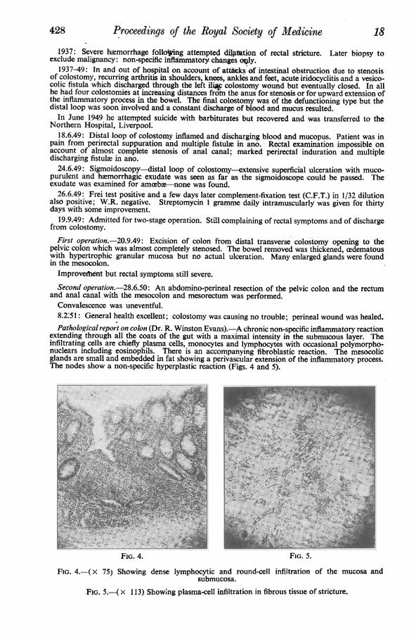
extending through all the coats of the gut with a maximal intensity in the submucous layer. Theinfiltrating cells are chiefly plasma cells, monocytes and lymphocytes with occasional polymorpho-nuclears including eosinophils. There is an accompanying fibroblastic reaction. The mesocolicglands are small and embedded in fat showing a perivascular extension of the inflammatory process.The nodes show a non-specific hyperplastic reaction (Figs. 4 and 5).
~~~~~~~~~~~~~~~~~~~~~~~~~~~~~~~~~~~~~~~~~
FiG. 4. FIG. 5.
FIG. 4.-(x 75) Showing dense lymphocytic and round-cell infiltration of the mucosa andsubmucosa.
FIG. 5.-( x 113) Showing plasma-cell infiltration in fibrous tissue of stricture.

19 Section of Proctology 429
Points of interest.-(l) The problem of diagnosis-dysentery, carcinoma. (2) The severe generalconstitutional effects: arthritis and iridocyclitis. (3) The upward extension of the disease to theright half of the transverse colon. (4) Failure of four colostomies to relieve rectal symptoms.(5) Vesico-colic fistula.
DISCUSSIONIt is believed that the stricture in the male is caused by direct inoculation of the anal
mucosa by pederasty. In the female the primary focus is in the vagina or posterior com-missure and extension occurs either by the medium of the perirectal lymphatics [1, 6, 12]which drain the primary focus, or by direct spread to the adjacent anal mucous membranefrom the primary focus [13]. In the 2 cases described exposure to venereal disease or toirregular intercourse was denied-the male patient was, no doubt, infected in India, butthe female patient had the distinction of acquiring her infection in this country.
Although the stricture usually begins at 2-6 in. from the anus and extends upwardsfor perhaps 2-3 in., as in Case I, occasionally the anal canal is completely obliterated andexceptionally the process extends to involve the whole rectum or even higher, sometimesreaching the cecum. The changes may be quite irregular, stretches of normal bowel beinginterposed between lengths of affected bowel. Complications which are of particularinterest to the surgeon are perforation of the bowel above the level of the stricture, whichis not very uncommon, and the occasional development of carcinoma in association withthe stricture of which several examples have been recorded [14, 15].
Clinicalfeatures and clinical diagnosis.-Although the rate of progress varies considerablyfrom case to case the examples described are typical of the relentless progress to seriouscomplications which occurs with a well-established stricture. To-day attention is beingdirected to the early "pre-stricture" stage of the disease in which treatment with chemo-therapy is so often successful [16]. Grace [13] has found that proctitis without stricturerepresents the earliest objective sign of anorectal lymphogranuloma, and that, untreated,the majority of such cases proceed to the development of a stricture while the proctitis isstill active. The earliest subjective symptoms are the passage of blood and pus per rectum,often without pain, but rectal stricture may develop with no premonitory symptom, andfor this reason Garth [17] recommends routine rectal examination at intervals in femalesknown to have been infected with L.G.V. to ensure early recognition.
Information is gradually accumulating about the constitutional symptoms which mayaccompany the rectal lesion. In the early stage hypersensitivity of the skin, pyrexia and jointpains are common; later, loss of weight, anaemia, arthritis, dermatitis and hyperproteinaemiafrequently develop. The rise in serum protein is largely due to an increase in the globulinfraction and is of some diagnostic significance [18]-normal levels were found in the 2 casesdescribed.Bensaude and Lambling [1] and more recently Gabriel [19] have emphasized that L.G.V.is the chief and possibly the only cause of diffuse inflammatory stricture of the rectum.Patients suffering from resistant unexplained proctitis should be given the benefit of aFrei test. It will be remembered that syphilis, gonorrhoea, chancroid and T.B. often coexistbut do not cause stricture and it is doubtful if dysentery of itself ever causes stricture.Carcinoma may cause difficulty in diagnosis in one of two ways-it may arise in associationwith a fibrous structure or it may assume a fibrous form which closely simulates aninflammatory stricture [20].Many other conditions, well described by Norbury [21], give rise to rectal stricture,most of which have their own special characteristics. In this group are included congenitalstricture, stricture following operation for piles or fistula, or following pile injection,stricture caused by the introduction of too strong chemicals, post-irradiation fibrosis, non-specific pelvic inflammation, actinomycosis, tubercle, bilharzia, ulcerative colitis and perhapsdysentery. Should the diagnosis of these conditions present any difficulty L.G.V. can beexcluded by the use of the Frei and C.F. tests.Diagnostic tests.-Before referring to the routine diagnostic tests 2 laboratory procedureswill be mentioned:(a) Biopsy-although there are no pathognomonic changes in fibrous stricture due toL.G.V. biopsy may be very valuable in excluding carcinoma.(b) Isolation of the virus-this is a difficult procedure and rarely necessary when thesimpler diagnostic tests are available.The original Frei antigen prepared from buboes, and its successor the Mouse Brain antigen,have been largely replaced by the modern Yolk Sac antigen together with a special controlpreparation, known commercially as Lygranum skin test and Lygranum skin test control [22].

430 Proceedings of the Royal Society of Medicine 20
Following infection with L.G.V. the Frei test usually remains positive for the remainderof the patient's life even though the actual infection may have resolved, and it thereforedoes not indicate whether the disease is active or quiescent. This difficulty has been over-come by the introduction of a complement-fixation test. A positive C.F.T. of 1/32dilution or over may be accepted as evidence of an active lesion [23], and repeatedestimations will indicate increasing or decreasing activity. Both the modern Frei test andthe C.F.T. are highly specific [3]. It is true that the virus of L.G.V. is closely allied tothat of psittacosis and that positive Frei and positive C.F. tests may be obtained in psittacosisusing L.G.V. reagents, but in actual practice confusion is unlikely to occur-in a patientsuspected of L.G.V. a positive Frei test with a positive C.F.T. of 1/16 dilution or overis good evidence of active infection with this disease [23].
Treatment.-In the early stage of proctitis resolution may be expected to take place withsulphonamide therapy in about 75% of cases [3]; whilst aureomycin and terramycinpromise to be even more effective [30, 31]. If these measures fail or if the patient is seenfor the first time with an established fibrous stricture, surgery alone can prevent progressivenarrowing and septic complications.The historical aspects of surgical treatment are well reviewed by Wright [15]. Dilatation
by bougie may have a limited application in selected cases; internal and external proctotomyand restricted excisions of the Whitehead type are generally condemned. Probably wideexcision with anastomosis would be advised to-day in place of the plastic operationdescribed by Lockhart-Mummery and Lloyd-Davies [24, 26] at a time when the risks ofoperation were greater. Bensaude and Lambling [1] advocated medical treatment, i.e.diathermic dilatation and antiseptics, and restricted surgery to colostomy and evacuationof abscesses. In so doing they voiced the general opinion of the time, 1936, although somesurgeons, notably Hartmann [25], had already shown that radical surgery has much tooffer-even a functional anus in selected cases-for a condition which resists all palliativemeasures.As in the 2 cases described colostomy, temporary or permanent, is by no means always
free from troubles:(1) Hernia, antegrade or retrograde prolapse, stenosis and fistula still occur in spite of the
many techniques designed to prevent them.(2) In spite of a defunctioning type of colostomy and the use of chemotherapy perirectal
discharge and local sepsis frequently persist [5, 27].(3) Very occasionally the infection extends upwards to involve the distal loop of the
colostomy [28].(4) Even if the proctitis should resolve and the stricture respond to dilatation by bougie
closure of the colostomy is as a rule followed by a recurrence of the condition [29].Recognizing these difficulties, and because so many patients with fibrous stricture of
the rectum belong to the lower age-group, many surgeons to-day advise a radical excisionconducted in stages:
(a) Preliminary exploration to assess the extent of the disease in the bowel, followedby a defunctioning tolostomy at a higher level.
(b) Complete excision of all the diseased tissue, less radical than for carcinoma but toinclude both the bowel and the involved perirectal tissues and glands. This is followed insuitable cases by restoration of continuity by anastomosis of the colon and the rectal stump.
(c) If continuity has been restored, closure of the colostomy.The second operation must be related in timing and in extent to the patient's general and
local condition and for the poor-risk patient Patterson's mucosal stripping operation mayprovide relief [29]. When the anal canal and sphincters are extensively involved in fibrosis,abdomino-perineal or perineal excision with permanent colostomy will be advised. Whenthe site of the stricture is above the anal canal some method of restoration of the continuityof the tract after resection usually proves possible, using an abdominal or perineal approachand either suture or a pull-through technique. In the intermediate cases in which the analcanal is more or less involved, a Pauchet type of operation may be selected, but it is doubtfulif a continent anus will be obtained.Bacon [5] has published a series of 24 cases treated by a modified two-stage Lockhart-
Mummery technique in which there was no mortality and in which the end-results werevery encouraging. Breidenbach [12] reported 18 thre-stage operations of excision by a
perineal approach with suture to testore c?rntinuity; theire was no mortality, the sphincterfunction was good and there was no protruding mucosa to cause moisture and discomfort.

21 Section of Proctology 431
Wright [15] has recorded his experience of the Pauchet technique and although he was lessfortunate, for he had 3 deaths in a series of 26 patients, he regarded the operation and itslate results as satisfactory. He concluded that early diagnosis followed by an excision opera-tion is the best treatment of fibrous stricture of the rectum.
In conclusion.-Two examples are described of fibrous stricture of the rectum: a rarecondition in this country. Progress has been achieved in the past fifteen years in threedirections by: (1) Improvements in diagnostic methods. (2) The recognition of the earlierstages of the disease and of the general constitutional effects. (3) Advances in surgicaltreatment.
It is recognized that the radical operation for fibrous stricture is frequently beset withdifficulties far in excess of those commonly experienced in operations for carcinoma ofthe rectum [15]. In spite of these difficulties encouraging results have been obtained and itseems likely that radical surgery has come to stay. Resection with a permanent colostomybrings relief and may be necessary, but with a high stricture treated at a reasonably earlystage the prospects of resection with restoration of continuity leaving a normally functioninganus are good.
My thanks are due to my colleagues Dr. R. Winston Evans, for the pathological reportsand photomicrographs, and Mr. W. R. Hunter, who kindly transferred the second caseto my care.
REFERENCES1 BENSAUDE, R., and LAMBLING, A. (1936) Proc. R. Soc. Med., 29, 1441.2 FREI, W. (1938) J. Amer. med. Ass., 1lb, 1653.3 GRACE, A. W. (1941) Bull. N.Y. Acad. Med., 17, 627.4 COSTELLO, M. J., and D'AVANZO, C. S. (1948) Arch. Derm. Syph. Chicago, 57, 112.5 BACON, H. E. (1941) Sth. med. J., La Grange, 34, 31.6 WRIGHT, L. T., FREEMAN, W. A., and BOLDEN, J. V. (1946) Arch. Surg., 53, 499.7 Ross, A. 0. F. (1951) Personal communication.8 MORGAN NAUNTON, C. (1937) Practitioner, 89, 421.9 BATCHELOR, R. C. L. (1937) Edinb. med. J., 44, 129.10 LEWIS, R. I. (1941) Brit. J. Radiol., 14, 298.11 PRINGLE, J. S. M. (1940) Irish J. med. Sci., 6th series, 169, 21.12 BREIDENBACH, L., and SLAIrrERY, L. R. (1948) Ann. Surg., 128, 1079.13 GRACE, A. W. (1943) J. Amer. med. Ass., 122, 74.14 LICCIONE, W. T. (1936) Amer. J. Surg., 31, 551.15 WRIGHT, L. T., BERG, B. N., BOLDEN, J. V., and FREEMAN, W. A. (1946) Surg. Gynec. Obstet.,
82, 449.16 GRAY, L. A., and BARNES, M. L. (1940) Amer. J. Surg., 48, 277.17 GARTH, W. (1939) N.Y. St. J. Med., 39, 1432.18 GUTMAN, A. B. (1939) N.Y. St. J. Med., 39, 1420.19 GABRIEL, W. B. (1948) The Principles and Practice of Rectal Surgery. 4th Ed., London: 288.20 TURNER, G. GREY (1936) Proc. R. Soc. Med., 29, 1459.21 NORBURY, L. E. C. (1933) Lancet (ii), 1418.22 RAKE, G., MCKEE, C. M., SHAFFER, M. F. (1940) Proc. Soc. exp. Biol., N.Y., 43, 332.23 BEDSON, S. T., BARDWELL, L. P., KING, E. J., and BISHoP, L. W. S. (1949) J. Clin. Path., 2,
241.24 LOCKHART-MUMMERY, J. P., and LLOYD-DAVIES, 0. V. (1935-36) Brit. J. Surg., 23, 19.25 HARTMANN, H. (1922) Lancet (i), 307.26 LOCKHART-MUMMERY, J. P. Diseases of the Rectum and Colon. 2nd Ed., Baltimore: 189.27 WOODS, F. M., and HANLON, C. R. (1944) Ann. Surg., 120, 598.28 BARBER, W. H., and MURPHY, W. B. (1941) Ann. Surg., 113, 30.29 PATTERSON, R. H. (1941) Ann. Surg., 114, 847.30 PRIGOT, A., WRIGHT, L. T., LOGAN, M. A., de LUCA, F. R. (1949) N.Y. St. J. Med., 49, 1911.31 ANNOTATION (1950) Lancet, (i) 1081.
A film on IIeo-anal Anastomosis was shown by Sir HENEAGE OGiLVIE and Mr. ROWANWEBB. The operation filmed was performed by Mr. John Devine and Mr. Rowan Webbin Melbourn=, Australia.