fighting cancer with transition metal complexes:from … · fighting cancer with transition metal...
TRANSCRIPT

Fighting Cancer with Transition Metal Complexes: FromNaked DNA to Protein and Chromatin Targeting StrategiesGiulia Palermo,[a] Alessandra Magistrato,[b] Tina Riedel,[a] Thibaud von Erlach,[a]
Curt A. Davey,[c] Paul J. Dyson,[a] and Ursula Rothlisberger*[a]
ChemMedChem 2016, 11, 1199 – 1210 Ó 2016 The Authors. Published by Wiley-VCH Verlag GmbH & Co. KGaA, Weinheim1199
ReviewsDOI: 10.1002/cmdc.201500478
1. Introduction
Certain transition metals compounds have been demonstratedto effectively act as anticancer agents.[1] In particular, the inor-
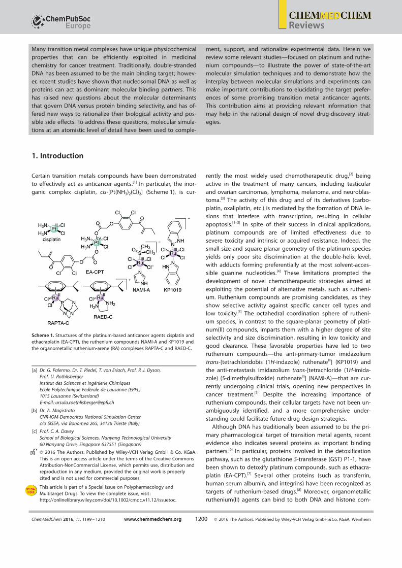
ganic complex cisplatin, cis-[Pt(NH3)2(Cl)2] (Scheme 1), is cur-
rently the most widely used chemotherapeutic drug,[2] beingactive in the treatment of many cancers, including testicular
and ovarian carcinomas, lymphoma, melanoma, and neuroblas-toma.[3] The activity of this drug and of its derivatives (carbo-
platin, oxaliplatin, etc.) is mediated by the formation of DNA le-sions that interfere with transcription, resulting in cellular
apoptosis.[1–3] In spite of their success in clinical applications,
platinum compounds are of limited effectiveness due tosevere toxicity and intrinsic or acquired resistance. Indeed, the
small size and square planar geometry of the platinum speciesyields only poor site discrimination at the double-helix level,
with adducts forming preferentially at the most solvent-acces-sible guanine nucleotides.[4] These limitations prompted the
development of novel chemotherapeutic strategies aimed at
exploiting the potential of alternative metals, such as rutheni-um. Ruthenium compounds are promising candidates, as they
show selective activity against specific cancer cell types andlow toxicity.[5] The octahedral coordination sphere of rutheni-
um species, in contrast to the square-planar geometry of plati-num(II) compounds, imparts them with a higher degree of siteselectivity and size discrimination, resulting in low toxicity and
good clearance. These favorable properties have led to tworuthenium compounds—the anti-primary-tumor imidazoliumtrans-[tetrachloridobis (1H-indazole) ruthenateIII] (KP1019) andthe anti-metastasis imidazolium trans-[tetrachloride (1H-imida-
zole) (S-dimethylsulfoxide) ruthenateIII] (NAMI-A)—that are cur-rently undergoing clinical trials, opening new perspectives in
cancer treatment.[5] Despite the increasing importance ofruthenium compounds, their cellular targets have not been un-ambiguously identified, and a more comprehensive under-
standing could facilitate future drug design strategies.Although DNA has traditionally been assumed to be the pri-
mary pharmacological target of transition metal agents, recentevidence also indicates several proteins as important binding
partners.[6] In particular, proteins involved in the detoxification
pathway, such as the glutathione S-transferase (GST) P1-1, havebeen shown to detoxify platinum compounds, such as ethacra-
platin (EA-CPT).[7] Several other proteins (such as transferrin,human serum albumin, and integrins) have been recognized as
targets of ruthenium-based drugs.[8] Moreover, organometallicruthenium(II) agents can bind to both DNA and histone com-
Many transition metal complexes have unique physicochemicalproperties that can be efficiently exploited in medicinal
chemistry for cancer treatment. Traditionally, double-strandedDNA has been assumed to be the main binding target; howev-
er, recent studies have shown that nucleosomal DNA as well asproteins can act as dominant molecular binding partners. Thishas raised new questions about the molecular determinants
that govern DNA versus protein binding selectivity, and has of-fered new ways to rationalize their biological activity and pos-
sible side effects. To address these questions, molecular simula-tions at an atomistic level of detail have been used to comple-
ment, support, and rationalize experimental data. Herein wereview some relevant studies—focused on platinum and ruthe-
nium compounds—to illustrate the power of state-of-the-artmolecular simulation techniques and to demonstrate how the
interplay between molecular simulations and experiments canmake important contributions to elucidating the target prefer-
ences of some promising transition metal anticancer agents.
This contribution aims at providing relevant information thatmay help in the rational design of novel drug-discovery strat-
egies.
Scheme 1. Structures of the platinum-based anticancer agents cisplatin andethacraplatin (EA-CPT), the ruthenium compounds NAMI-A and KP1019 andthe organometallic ruthenium-arene (RA) complexes RAPTA-C and RAED-C.
[a] Dr. G. Palermo, Dr. T. Riedel, T. von Erlach, Prof. P. J. Dyson,Prof. U. RothlisbergerInstitut des Sciences et Ing¦nierie ChimiquesEcole Polytechnique F¦d¦rale de Lausanne (EPFL)1015 Lausanne (Switzerland)E-mail : [email protected]
[b] Dr. A. MagistratoCNR-IOM-Democritos National Simulation Centerc/o SISSA, via Bonomea 265, 34136 Trieste (Italy)
[c] Prof. C. A. DaveySchool of Biological Sciences, Nanyang Technological University60 Nanyang Drive, Singapore 637551 (Singapore)
Ó 2016 The Authors. Published by Wiley-VCH Verlag GmbH & Co. KGaA.This is an open access article under the terms of the Creative CommonsAttribution-NonCommercial License, which permits use, distribution andreproduction in any medium, provided the original work is properlycited and is not used for commercial purposes.
This article is part of a Special Issue on Polypharmacology andMultitarget Drugs. To view the complete issue, visit :http://onlinelibrary.wiley.com/doi/10.1002/cmdc.v11.12/issuetoc.
ChemMedChem 2016, 11, 1199 – 1210 www.chemmedchem.org Ó 2016 The Authors. Published by Wiley-VCH Verlag GmbH & Co. KGaA, Weinheim1200
Reviews

ponents of nucleosome core particles (NCP), which are the fun-damental unit of chromatin, composed of chromosomal DNA
wrapped around a histone protein core.[9] NCPs have the fun-damental role of compacting DNA in eukaryotic cells, where
the majority of DNA is present in a packed conformation,rather than a naked form, such as in free oligonucleotides or
non-histone protein–DNA complexes. In contrast, experimentalin vitro studies and molecular simulations usually consider
naked DNA to rationalize binding modes and the pharmaco-logical action of these types of compounds.
However, the structure of nucleosomal DNA is markedly dif-ferent from that of free double-stranded DNA (dsDNA).[10]
Indeed, the flexibility of naked DNA easily allows drug-inducedstructural adaptations,[11] whereas nucleosomal DNA is highlyrigid, peculiarly bent and restrained by the histone compo-
nents. In addition, the possibility of drug binding at the levelof the histone proteins has been shown to directly interferewith the binding of chromatin transcription factors that modu-late gene expression in cancer cells,[9a, b, 12] and possible epige-
netic routes to anticancer therapies have thus emerged aspromising alternatives.
In recent years, simulations at the molecular and electronic
structure levels have been used to complement experimental
Dr. Giulia Palermo is a postdoctoral sci-
entist at the EPFL in the research
group of Prof. Rothlisberger, where
she is working on the characterization
of nucleosome dynamics and on chro-
matin drug design by applying classi-
cal and hybrid QM/MM methods. This
research project involves close collabo-
ration with the research groups of
Prof. Dyson (bioinorganic chemistry)
and Prof. Davey (X-ray crystallogra-
phy).
Dr. Alessandra Magistrato leads the
Laboratory of Computational Biochem-
istry at the Democritos National Simu-
lation Center c/o International School
of Advanced Studies (SISSA) in Trieste,
Italy. Her main expertise is in computa-
tional bioinorganic chemistry. She is in-
vestigating the mechanisms of drug–
DNA interactions and the role of bio-
logically relevant metal ions in non-
coding RNAs and enzymes.
Dr. Tina Riedel obtained her PhD in
bioorganic chemistry at the University
of Zurich in 2011 on the design of syn-
thetic vaccines in Prof. John A. Robin-
son’s group. For her postdoctoral work
she joined Prof. Dyson’s research
group at EPFL, where she focuses on
the preclinical development of rutheni-
um-based anticancer drugs and drug
combinations against solid tumors and
tumor microenvironments.
Thibaud von Erlach is a PhD student in
Prof. Rothlisberger’s research group,
where he is studying the mechanism
of action of novel ruthenium- and
osmium-based anticancer agents.
Prof. Curtis A. Davey is an associate
professor in the School of Biological
Sciences at Nanyang Technological
University (Singapore), where he leads
the Laboratory of Genomic Structural
Biology. His expertise in nucleosome
structure and dynamics is dedicated to
understanding epigenetic factors in
genomic regulation and the develop-
ment of metal-based chromatin-target-
ing anticancer drugs.
Prof. Paul J. Dyson is the director of
the Institute of Chemical Sciences and
Engineering at the EPFL and he also
heads the Laboratory of Organometal-
lic and Medicinal Chemistry. His re-
search interests include the develop-
ment of novel metal-based anticancer
agents. In 2015, his contribution to
this field was recognized with the Bio-
inorganic Chemistry Award of the
Royal Society of Chemistry.
Prof. Ursula Rothlisberger is full Profes-
sor of Computational Chemistry and
Biochemistry at the EPFL. Her research
interests are in the development and
application of density-functional-based
mixed QM/MM methods. Her contribu-
tions to the field of theoretical chemis-
try have been awarded with the Ru-
zicka Medal and the 2005 Dirac Medal
of the World Organization of Theoreti-
cally Oriented Chemists (WATOC). In
2015, she was elected to the Interna-
tional Academy of Quantum Molecular
Sciences (IAQMS).
ChemMedChem 2016, 11, 1199 – 1210 www.chemmedchem.org Ó 2016 The Authors. Published by Wiley-VCH Verlag GmbH & Co. KGaA, Weinheim1201
Reviews

studies and provide valuable additional information. Besidesidentifying the binding location and binding mode of metallo-
drugs to dsDNA and proteins, they have also helped in ration-alizing target selectivity between protein versus DNA, and
assess the differences in the interaction with naked versuspacked DNA.[9a, 13] High-resolution structures of nucleosome–
drug adducts have recently become available,[4, 9a, 10, 14] thuspaving the way for realistic molecular dynamics (MD) simula-tions based on classical force fields and on quantum mechan-
ics (QM). In particular, QM methods are necessary for a properdescription of the electronic structure and energetics of transi-
tion metal compounds, the intricate electronic properties ofwhich are often not adequately described at the force-fieldlevel. However, the large size of protein–DNA targets makesa description at the full QM level intractable. A hybrid quan-
tum mechanics/molecular mechanics (QM/MM) approach ele-gantly overcomes these limitations by treating the transitionmetal agent and its direct amino acid/nucleic acid ligands at
the QM level, while the remaining part of the system, includingthe rest of the target biomolecule in explicit solution, is de-
scribed with a classical force field.[15] QM/MM simulations com-bined with ab initio MD were able to accurately predict the
structural and energetic features of covalent target modifica-
tions.[16] Furthermore, ab initio QM/MM MD can be employedfor generating accurate in situ force fields for target-bound
transition metal compounds, which can be employed to runlong-time-scale classical MD. By using a “force-matching ap-
proach”, the classical potentials are derived on the fly fromQM/MM data, thus taking into account the changes in the
electronic structure properties induced by the protein/nucleic
acid environment as well as temperature effects.[17]
Herein we review several computational and experimental
studies, selected from our work, aimed at characterizing the in-teraction of some promising transition metal anticancer agents
with their biological targets. In particular, we report how classi-cal and QM/MM MD simulations have contributed to decipher
the binding and reaction mechanisms of covalently bound
transition metal compounds to proteins and DNA. We alsoshow how the interplay between experiments and computa-tions has helped in clarifying and rationalizing protein versusDNA selectivity, thus addressing relevant biological questions
at the molecular level. Finally, this article aims at providing anoverview of recent computational and experimental studies,
which have contributed to elucidating the targeting character-istics of transition metal anticancer agents. This informationshould facilitate novel drug-discovery strategies and ultimately
lead to more selective anticancer agents.
2. Computational Methods
The computational work that we review here is based mainly
on classical and hybrid QM/MM MD simulations. We introducethe basic concepts of these methods below, and the interested
reader is referred to the review articles and books for a moreextensive description of these computational methods.[15b–d, 18]
2.1. Force-field-based MD
Force-field (FF)-based molecular simulations use the solutionof the classical equations of motion for a set of particles to de-
scribe the time evolution of a system at finite temperature.[18a]
The output is a trajectory that represents the molecular system
as a function of time. This trajectory provides insight into thedynamic and thermodynamic properties of the system underinvestigation. In classical MD, the potential energy of the
system is determined by an empirical FF that is parameterizedto reproduce experimental or ab initio data. The FF is defined
as the sum of different contributions, and is usually composedof bonded terms, describing bond, angle bending, and torsion-al degrees of freedom and non-bonded terms, accounting forvan der Waals and electrostatic forces. A generic form of the
potential energy in commonly used FF for biomolecular sys-tems is given in Equation (1):
V ¼Xbonds
K RðR¢ReqÞ2 þXangles
K qðq¢qeqÞ2þ
Xdihedrals
V n
2½1þ cosðn�¢gÞ¤ þ
Xi<j
�Aij
R12ij
¢ Bij
R6ij
þ qiqje2
4pe0Rij
� ð1Þ
To date, the most commonly used FFs in the simulations ofbiological systems are OPLS,[19] AMBER,[20] GROMOS,[21] and
CHARMM.[22] These FFs have been proven to excellently per-form in simulations of proteins and peptides. However, severe
DNA distortions and unbalanced a/g transitions were found insimulations performed with AMBER parm99 FF.[20a,b] To over-
come this problem, a refinement of this FF has been devel-
oped by the group of Orozco.[20c,d] In the novel parmbsc0 FFs,a correct representation of the a/g transitions is provided and
stable trajectories on the multi-microsecond scale can be gen-erated.
Classical MD simulations can be coupled to methods ena-bling the enhancement of phase space sampling (i.e. , en-
hanced sampling techniques), such as metadynamics,[23] accel-
erated MD,[24] among others, and free-energy methods such asfree-energy perturbation, umbrella sampling, adaptive biasingforce, and thermodynamic integration.[18b, 25] These methodsallow the study of biophysical processes that occur on the mi-
crosecond-to-millisecond (or longer) time scale, which cannotbe directly sampled in the typical time scale of (all-atom) MD
simulations (i.e. , hundreds of nanoseconds to microseconds).By providing an accurate description of the associated free-energy landscape, these methods have been shown to be par-
ticularly efficient for the study of complex conformationalchanges of folded and unfolded structures,[26] as well as for
ligand binding.[27]
2.1. QM/MM MD
FFs can be combined with QM methods in the so-called QM/
MM approach. Originally proposed in 1976 by Warshel andLevitt for studying the enzymatic reaction in lysozyme,[28] many
different QM/MM implementations have been proposed overthe past few decades, with widespread applications in biology
ChemMedChem 2016, 11, 1199 – 1210 www.chemmedchem.org Ó 2016 The Authors. Published by Wiley-VCH Verlag GmbH & Co. KGaA, Weinheim1202
Reviews

and materials science.[15b–d, 29] Popular hybrid QM/MM schemesare the ONIOM[30] method included in the Gaussian suite of
programs,[31] or the fully Hamiltonian coupling approaches in-cluded in the CPMD[32] and CP2K codes.[33] In QM/MM studies
of enzymatic catalysis and inhibition, the region of interest ofthe model system (the enzyme’s active site and/or the ligand
binding pocket) is treated at a higher level of accuracy (QM
level), while the remainder of the system is treated at the MMlevel of theory (Figure 1). In the general form of a hybrid QM/
MM scheme, the total Hamiltonian (H) of the system contains
the Hamiltonians for the quantum (HQM) and classical (HMM)systems and the interaction between the QM and MM regions
(HQM=MM):
H ¼ HQM þHMM þHQM=MM ð2Þ
where the QM Hamiltonian (HQM) can be based on differentquantum chemical electronic structure methods, spanningfrom semiempirical to ab initio Hartree–Fock or density func-tional theory (DFT). In every QM/MM implementation, particu-
lar care must be taken to achieve a rigorous treatment of thecoupling between the QM and MM regions, as described by
the interaction Hamiltonian HQM=MM. This is especially true forthe description of covalent bonds between the QM and MMregions and the treatment of the electrostatic term. To cope
with the split of covalent bonds between the QM and MM re-gions, either linking hydrogen atoms or specially parameter-
ized pseudo-atoms are introduced, thus saturating the valenceof the terminal QM atoms. Concerning the electrostatic interac-
tions, the simplest way to electrostatically couple the QM and
MM regions uses a mechanical embedding scheme, in whichthe electrostatic interactions between the two regions are
treated at the MM level. More rigorous is the electrostatic em-bedding scheme, in which the electrostatic field of the classical
environment polarizes the QM electronic charge density andthe interaction between MM point charges and QM electron
density is incorporated in HQM. In a third polarized embeddingscheme, the polarization effects of the QM region on the MM
part are also considered self-consistently in the framework ofa polarizable FF. The remaining bonding and van der Waals in-
teractions between QM and MM regions are treated classically.Since their first appearance,[28] QM/MM approaches have
been successfully applied to a growing number of drug-design-related studies and to elucidate enzymatic mecha-nisms.[15b, c, 16a, 27b, 34] Moreover, continuous developments in the
field have enabled studies of the reaction mechanisms of bio-logical systems of increasing size (up to 200 000 atoms) andcomplexity.[35] Notably, the 2013 Nobel Prize in Chemistry wasgiven in recognition of the seminal contributions of Karplus,
Levitt, and Warshel in developing multiscale models for com-plex chemical systems. The QM/MM scheme was specifically
mentioned in honoring the groundbreaking work of the Nobel
laureates in the field of molecular simulations. The QM/MMmethod, in combination with first-principles (Car–Parrinello)
MD, is widely employed for the study of anticancer drug–target interactions.[11, 15] Herein we review applications of the
fully Hamiltonian QM/MM extension to Car–Parrinello MD de-veloped by Rothlisberger and co-workers.[36] In this approach,
the electrostatic effects of the classical environment are taken
into account via an electrostatic embedding scheme in theform of an additional contribution to the external potential
acting on the QM system. The Pauli repulsion between theelectrons and the classical point charges is mimicked through
the use of a screened Coulomb potential in order to avoidover-polarization of the electron density near positively
charged classical point charges (i.e. , the so-called electronic
spill-out effect). This QM/MM approach is implemented in theCar–Parrinello code (CPMD)[32] based on DFT in combination
with the classical AMBER[20] and GROMOS[37] FFs using particlemesh Ewald summation to treat long-range electrostatic inter-
actions.The QM (Car–Parrinello)/MM method can be used in combi-
nation with the “force-matching approach” for generating reli-
able in situ force fields for nonstandard residues, such asamino/nucleic acids bound to transition metal complexes. Inthe method developed by Maurer et al. ,[17] the QM region ischosen in such a way to include all components of the system
for which no parameters are available. Finite-temperature QM(Car–Parrinello)/MM MD simulations are used to generate a tra-
jectory of reference configurations. Next, the nuclear forcesacting on the atoms of the QM subsystem are extracted fromthe obtained trajectory and stored. A set of optimal atomic
point charges that reproduces the electrostatic potential andfield in the surrounding of the QM region is determined,
taking into account all trajectory configurations. The storedforces serve as targets for the subsequent parameter-fitting
scheme. Indeed, the force-field parameters are determined insuch a way as to optimally reproduce the electrostatic proper-ties and the nuclear forces of the QM subsystem. The opti-
mized FF parameters obtained can be used to perform molec-ular simulations with the accuracy of a QM/MM treatment at
the computational cost of classical MD.
Figure 1. Representative QM/MM partitioning of a biological system, shownfor RAPTA-C covalently bound to two guanine bases in double-strandedDNA (dsDNA). The QM atoms (i.e. , RAPTA-C and the coordinated guanines)are in ball-and-stick representation. The remaining part of the system, in-cluding dsDNA (shown as ribbons), water molecules (shown as sticks) andcounter-ions (not shown) are treated at the classical (MM) level. The box onthe right highlights the QM region, showing the electronic density (shownwith an isovalue of 0.01 au).
ChemMedChem 2016, 11, 1199 – 1210 www.chemmedchem.org Ó 2016 The Authors. Published by Wiley-VCH Verlag GmbH & Co. KGaA, Weinheim1203
Reviews

3. Platinum Compounds and Protein Binding
3.1. Molecular basis for overcoming Pt-based drugresistance
The action of cisplatin and related platinum compounds is
greatly limited by the occurrence of drug resistance, which hasbeen associated, among other factors such as the recognition
of platinated DNA by repair proteins,[38] to an overexpressionof the p-class glutathione S-transferase (GST P1-1) enzyme incancer cells.[3] GST P1-1 is a detoxification enzyme within the
mercapturic acid pathway that catalyzes the conjugation ofxenobiotics to glutathione, thus leading to the elimination of
toxic compounds. Recently, a novel PtIV compound, termedethacraplatin (EA-CPT, Scheme 1), has been reported to exert
anticancer activity and simultaneously inhibit GST P1-1, thus
overcoming the associated drug resistance. EA-CPT has theability to be reduced intracellularly to release a cytotoxic PtII
moiety and two ethacrynate (EA) molecules, which are directlyresponsible for GST P1-1 inhibition.
To understand the nature of the drug–protein interaction,a detailed experimental and theoretical study has been con-
ducted.[7] X-ray crystallography captured the configuration of
PtII after its release from the EA-CPT molecule and the bindingof the EA fragments (Figure 2 a). In the crystal structure, PtII is
coordinated by two cysteine residues (C101/C101’) and bya chloride ion at the protein dimer interface, while the releasedEA fragments locate within two accessible hydrophobic pock-ets. QM/MM simulations have been used to establish the
nature of the remaining exogenous ligand coordinated to thePtII center, which could not be discerned from the electrondensity map in the X-ray structure. As a result, a fourth PtII
ligand—most likely a hydroxy group—is required for ensuringthe stability of the active site during QM/MM MD. Subsequent-
ly, molecular simulations have been used to investigate howthe binding between the intact EA-CPT and GST P1-1 takes
place. Classical MD simulations were carried out in combina-tion with metadynamics,[23] which allowed a dynamic docking
of EA-CPT from the bulk to the target active site, efficiently ex-ploring different binding poses and constructing the associat-
ed free-energy profile. These simulations enabled discrimina-tion among various binding routes, revealing that EA-CPT pref-
erentially approaches the cysteine residues at the proteindimer interface (Figure 2 b).
This binding mode is in agreement with previous structural
studies of the EA ligand in complex with GST P1-1.[39] Overall,the work suggested that EA-CPT first migrates to the GSTP1-1 dimer interface, where it is subsequently reduced andcleaved, permitting the diffusion of both a cytotoxic PtII spe-
cies and of the EA fragments, which locate within the hydro-phobic enzyme cavities. According to this mechanism, the PtIV
ion of EA-CPT is reduced upon binding at the dimer interface
to PtII, the two ethacrynate ligands are released and inhibitGST P1-1, thus allowing the PtII species to exert its cytotoxic
action without being inactivated by GST-mediated resistance.
4. Ruthenium Compounds
4.1. Active species of RuIII-based drugs
Among Ru-based anticancer drugs, promising candidates areNAMI-A and KP1019 (Scheme 1).[5] Both compounds are octa-
hedral, bearing four chlorides and differing in the axial ligand.
NAMI-A has been evaluated in several in vivo models and hasbeen shown to prevent the development and growth of pul-
monary metastases in all the solid tumors on which it hasbeen tested in vivo, including Lewis lung carcinoma,[40] MCa
mammary carcinoma,[41] TS/A mammary adenocarcinoma,[42]
and human tumors in mice.[43] KP1019, on the other hand, sig-
nificantly decreases tumor growth in several in vivo models, in-
cluding chemoresistant tumors, such as colorectal cancer. BothNAMI-A and KP1019 have already completed phase I and II
clinical trials.[5] Despite their structural similarity, NAMI-A andKP1019, as well as the imidazole analogue of the latter(KP418), have completely different biological effects, principallydue to their different targeting abilities. NAMI-A weekly binds
to DNA,[44] whereas it strongly binds to several proteins (integ-rins, transferrin, human serum albumin, human carbonic anhy-
drase, and lysozyme).[8] In contrast, KP418 reacts with DNA, in-hibiting DNA synthesis.[45] Because NAMI-A can prevent meta-stasis in part by the inhibition of angiogenesis in several exper-
imental models, it is believed that its mechanism of actionmight include modulation of various protein targets, such as
proteases, protein kinases, and integrins, all known to play keyroles in cell motility and invasion.[8] However, it is not fully
clear which biological targets are responsible for its activity.
This lack of knowledge has hampered a full mechanistic under-standing of the mode of action of NAMI-A. Like cisplatin, Ru
compounds are administered as prodrugs and predominantlyremain in their less reactive chloride form at high chloride con-
centration, such as in blood plasma. At low chloride concentra-tion, as in the cellular environment, they rapidly undergo aqua-
Figure 2. a) Crystal structure of GST P1-1, including the ethacrynic acid (EA)moieties and the PtII ion.[7] GST P1-1 is represented as a molecular surface,highlighting the two protein dimers with gray and pink ribbons. The EAmoiety (violet) is shown in space-filling representation, while the Pt ion isshown as a green sphere. C101/C101’ are also shown as sticks. b) Bindingmode of the intact EA-CPT approaching the GST P1-1 dimer interface fromclassical and QM/MM simulations.
ChemMedChem 2016, 11, 1199 – 1210 www.chemmedchem.org Ó 2016 The Authors. Published by Wiley-VCH Verlag GmbH & Co. KGaA, Weinheim1204
Reviews

tion, converting them into biologically active metabolites(Scheme 2).
The ligand exchange reaction activates the Ru agent, as theaqua ligands can rapidly be replaced by the electron-donor li-
gands of biomolecules (i.e. , proteins and DNA). Thus, under-
standing the exchange kinetics in solution is of paramount im-portance to identify the active metabolites and the ease by
which they are formed.Theoretical studies have focused on the study of the kinetics
of ligand–water exchange of NAMI-A and KP418, such as ra-tionalizing the differences between the most abundant metab-
olites formed in solution. An extensive study based on DFT-
B3LYP calculations in implicit solvent has been performed onNAMI-A and KP418 complexes considering both RuII and RuIII
oxidation states.[46] The two compounds exhibit remarkably dif-ferent redox potentials (Em, versus normal hydrogen electrode
NHE), which are 0.235 and ¢0.275 V for NAMI-A and KP418, re-spectively (Table 1). Because of its Em, NAMI-A can be easily re-
duced in vivo by biological reductants, while this process may
be more difficult for KP418.Considering that the reduction of NAMI-A rapidly occurs in
vivo, as mediated by biological reductants, it is likely that themost abundant species in solution would be the RuII-monoa-
qua or cis- or trans-RuII-diaqua metabolites (Scheme 2). In con-trast, in the absence of reductants, the RuIII compound would
immediately dissociate DMSO and retain the RuIII oxidationstate as observed in X-ray studies (Figure 3).[8g] The X-ray struc-
ture of the adduct between NAMI-A in its RuIII form and car-bonic anhydrase indicates that NAMI-A behaves like a multi-
stage drug, progressively releasing all ligands, and with the Rucenter binding to the final protein target residues (Figure 3).
Thus, depending on the bioavailability of reductants, a mixture
of cis- and trans-RuII-diaqua NAMI-A metabolites may be active,or the DMSO ligand might be immediately lost, activating RuIII
for the protein target.The reduction of KP104 is more difficult and if it occurs, it
has no effect on the hydrolytic properties of the molecule.[46]
In this case the most active species appears to be the cis-RuII-
diaqua isomer (Scheme 2). The reduction of KP418 most likelyoccurs after the second hydrolysis step, because the Em valuesincrease as hydrolysis proceeds (Table 1). Thus, for KP104, the
most active metabolite likely maintains the chemical differen-ces of the parent compound, as the imidazole ligands are still
present in the cis-diaquo-RuIII metabolite (Scheme 2). Taken to-gether, these different kinetic properties appear to affect the
biodistribution of the two drugs and in turn their binding pref-
erences to biological targets.
4.2. RuII-arene (RA) compounds and DNA targeting
RuII-arene (RA) compounds have emerged as promising alter-natives to platinum compounds.[47] The prototypes are [RuII(h6-
Scheme 2. Structures of the most active metabolites of a), b) NAMI-A andc) KP418. The cis- and trans-RuII-diaqua metabolites of NAMI-A are shownafter the biological reduction of RuIII into RuII has occurred.
Table 1. Calculated and experimental redox potentials of NAMI-A andKP418 metabolites.
Em [V][a] NAMI-A KP418Calcd.[b] Exptl. Calcd.[b] Exptl.
R 0.17 0.235 ¢0.18 ¢0.275PICl 0.34 0.337 ¢0.01PIDMSO(Im) ¢0.13 ¢0.13PII2Cl cis 0.67 0.12PII2Cl trans 0.57 0.07PIICl,DMSO(Im) ¢0.17 ¢0.17PIII3Cl 1.02 0.53PIII2Cl,DMSO(Im) 0.09 0.09
[a] Redox potential values were calculated all along the reaction pathwayfor hydrolysis of NAMI-A; products from different steps of the reactionare indicated as PIa,b,c, in which I refers to the hydrolysis step, and a,b,crefer to ligand exchanges during hydrolysis. [b] Em was calculated at theDFT/B3LYP level using the 6-31G-(d,p) and Lanl2DZ basis sets on N, S, C,O, H, Cl, and Ru atoms; the conductor-like polarizable continuum model(CPCM) was used.
Figure 3. Crystal structure of NAMI-A bound to human carbonic anhydra-se.[8g] The drug remains in the RuIII oxidation state, but looses all its ligands,transferring the imidazole ligand (Im) onto the active site ZnII ion. The Rucenter (purple sphere) is coordinated by H64 and by the N62 backbone, aswell as to three water molecules (red spheres). Key enzyme residues(brown), and the imidazole moiety of NAMI-A (green) are shown as sticks.
ChemMedChem 2016, 11, 1199 – 1210 www.chemmedchem.org Ó 2016 The Authors. Published by Wiley-VCH Verlag GmbH & Co. KGaA, Weinheim1205
Reviews

arene)Cl(ethylenediamine)] (RAED)[48] and [RuII(h6-are-ne)Cl2(1,3,5-triaza-7-phosphoadamantane)] (RAPTA)[49]
(Scheme 1). These “piano stool” complexes are characterizedby a p-bonded arene ligand as the “seat of the stool”, with
a monodentate phosphine 1,3,5-triaza-7-phosphadamantane(PTA) or a chelating ethylenediamine (ED), and chloride ligands
occupying the remaining coordination sites. The arene ligandprovides a hydrophobic surface that fosters a high degree of
selectivity toward biomolecular targets.[50] As for RuIII-based
drugs, the ligand exchange kinetics can be exploited to modu-late the rate of reaction of RA compounds with biomoleculartargets, by substituting other (labile) ligands in place of thechlorides.[50, 51] The versatility of RA compounds prompted the
development of related compounds, leading to a vast numberof compounds that have been evaluated for cytotoxicity in
cancer cells. Building on the knowledge of the main molecular
target of cisplatin—i.e. , dsDNA—it has been suggested thatRA compounds target guanine bases at the most readily acces-
sible electron donor atom (N7).[52] However, an early (rigid)docking study for an organometallic complex, [Cp2Mo]2 + , indi-
cated that transition metal-arene compounds are too bulky tobind to the DNA major groove in a manner analogous to that
of cisplatin.[53]
To investigate the binding processes of the monofunctionalRAED-C and of the bifunctional RAPTA-C compounds to
dsDNA, classical and ab initio QM/MM MD simulations havebeen performed.[13b] These simulations considered as a drug
target a model sequence of dsDNA containing two guaninebases in the central part of a 12-mer dsDNA, as previously
used in studies of cisplatin binding.[54] The initial approach of
RAED-C to dsDNA has been investigated by placing the [Ru(h6-p-cymene)(ED)]2 + moiety at a ~20 æ distance from the target
guanine (i.e. , G6, Figure 4 a), considering different starting con-ditions facing both the minor and major DNA grooves. Uncon-
strained classical MD showed that RAED-C is easily accommo-dated within the major groove of the dsDNA on a ~15 ns time
scale, exhibiting strong selectivity for GC-rich sequences. In
these classical MD studies, the RuII ion samples configurationdistances as close as 4 æ from one of the DNA binding atoms
(i.e. , N7@G6) reaching almost binding distances. Moreover, theamino group of the ED moiety forms a characteristic hydrogenbond with O6@G6, in agreement with previous studies.[55] MDsimulations show that the high flexibility of dsDNA decreases
the steric hindrance of the bulky p-cymene group, allowingRAED to fit easily within the major groove, allowing covalent
binding at G6. Starting from these configurations, the forma-tion of the coordination bond between RAED-C and N7@G6
has been studied by QM/MM MD in combination with the
thermodynamic integration approach, which allows calculatingthe free energy of a process along a selected reaction coordi-
nate.[18b] By using this approach, a dissociative mechanism ofwater/N7@G6 exchange characterized by a free-energy barrier
of ~11 kcal mol¢1 for the formation of the covalent RAED-C–DNA complex was observed. For the dissociation of RAED-C
from DNA, a free-energy barrier of ~21 kcal mol¢1 was calculat-
ed. The binding of RAPTA-C to dsDNA has been suggested tooccur in a similar way. Indeed, by calculating the electrostatic
potential (ESP) of the [Ru(h6-p-cymene)(PTA)]2 + moiety and ofthe target dsDNA, it was shown that the binding at the major
groove level should be driven mainly by electrostatic interac-tions (Figure 4 b). As indicated by these calculations, the highly
negative ESP of the dsDNA major groove allows the formation
of a suitable binding cavity for the positively charged RA com-pounds.
To gain insight into the long-time-scale DNA distortions in-duced by the covalent binding of RA compounds to dsDNA
oligonucleotides, classical MD simulations of the RA com-pounds-DNA adducts were carried out, by employing a QM/
MM MD tuned (force-matched) FF. During nanosecond-long
classical MD simulations, RAED-C and RAPTA-C induce differentstructural distortions to dsDNA. Namely, RAED-C binding
causes a large increase in the rise between T5 and G6 bases,such that the typical Watson–Crick base pairing between T5
and A20 is broken (Figure 4 c). This results in a substantialopening of the major groove and partial DNA unwinding, in
agreement with previous experiments, suggesting the forma-
tion of a local single-stranded DNA.[56] On the other hand, thebinding of RAPTA-C introduces a global overall bending of the
Figure 4. a) Binding of RAED-C within the major groove of a dsDNA dodecamer, obtained from nanosecond-time-scale classical MD simulations. The graph re-ports the time evolution of the distance between the Ru and the N7 atom of G16 (black line) and of the target guanine G6 (dashed line), highlighting thehigh selectivity of RAED-C for binding to guanine residues. b) The electrostatic properties of RAPTA-C and of the target dsDNA are shown. The electrostaticpotential (ESP) was calculated and mapped onto the solvent-accessible surfaces of both the target dsDNA and of the [Ru(h6-p-cymene)(PTA)]2 + moiety [red =
negative (¢10 kTe¢1) ; blue = positive (+ 10 kTe¢1)] . c) Long-time-scale distortion of a 12-mer dsDNA upon covalent binding of RA compounds. The RAED-Cadduct formation induces a large rise between T5 and G6 bases, thus leading to breaking of the typical Watson–Crick base pairing. RAPTA-C introducesa global bending toward the major groove of dsDNA.
ChemMedChem 2016, 11, 1199 – 1210 www.chemmedchem.org Ó 2016 The Authors. Published by Wiley-VCH Verlag GmbH & Co. KGaA, Weinheim1206
Reviews

DNA (~408) toward the major groove. This structural distortionis similar to the DNA bent configuration induced by the widely
used anticancer drug cisplatin, which is believed to play a cru-cial role in its mechanism of anticancer activity.[57]
Overall, molecular simulations have shown that even bulkyRA compounds can bind to DNA due to the high flexibility of
the double strand, which allows rapid accommodation of thecompounds within its wide major groove. However, upon co-valent binding at the target guanine, RAED-C and RAPTA-C
induce different local and global perturbations to the nakeddsDNA oligonucleotide structure.
4.3. RA compounds and nucleosome targeting
It was recently demonstrated that RA compounds bind directlyto NCPs,[9a, b] which are the fundamental unit of chromatin and
are composed of chromosomal DNA of 145–147 base pairs(bp), wrapped around an octamer of four core histone proteins
(H3, H4, H2A and H2B, Figure 5 a). The packaging of thegenome into nucleosomes raises the possibility to form either
potential adducts at different DNA sites, or to form protein ad-
ducts at exposed sites of the histone core.[58] The latter are par-ticularly interesting, because histone binding may directly in-
fluence gene expression, opening new avenues for possibleepigenetic cancer therapies.[9b, 12]
At the nucleosome level, the binding of RA compounds isvery different from that of cisplatin and other cytotoxic plati-
num-based agents, which bind to many different DNA siteswithin the NCP.[4, 14, 59] By using quantitative bioanalytical meth-
ods, it has been shown that the chromatin-bound adducts incancer cells treated with RAPTA-C are primarily associated withthe protein components, while RAED preferentially targets the
DNA components of chromatin.[9a] X-ray crystallography re-vealed three well-defined histone binding sites for RAPTA-C(Figure 5 a), whereas RAED-C forms adducts preferentially atthe DNA sites with only one additional binding site at the his-tone level. RAED-C binds at guanine sites of the nucleosomalDNA, in analogy with its binding mode to naked DNA,[56, 60]
however, with a vastly different site selectivity, as most of the
reactive sites on naked DNA are not accessible in the NCP dueto the histone packaging. Indeed, the only accessible sites for
adduct formation are at the termini and at locations 1.2 and2.5 double-helical turns away from the NCP center [also called
superhelix location (SHL)�1.5 and SHL�2.5] . Remarkably,both RAPTA-C and RAED-C preferentially bind to glutamate
sites of the histone components, while displaying very differ-
ent histone (RAPTA-C) versus DNA (RAED-C) specificities.To understand the molecular basis of this site selectivity,
QM/MM simulations of the adduct formation of RAPTA-C andRAED-C were performed at selected histone and DNA sites
(Figure 5 b). Whereas the two compounds present a similarfree-energy barrier (~20 kcal mol¢1) to bind to the histone
sites, the barrier for adduct formation at the DNA sites is two-
fold higher for RAPTA-C (~30 kcal mol¢1) than for RAED-C (~15 kcal mol¢1). This difference in free energy at the DNA level is
due to the steric constraints of the nucleosomal double helix,which hampers the accommodation of the larger PTA ligand
during adduct formation. As a consequence, the activationfree-energy barrier for RAPTA-C binding to DNA considerably
increases while adduct stability is significantly decreased so
that the binding of RAPTA-C to DNA is both kinetically andthermodynamically unfavorable (Figure 5 b). Instead, the steric
hindrance of the PTA ligand favors the accommodation ofRAPTA-C at the histone sites via shape and hydrophobic com-
plementarity. This evidence points to the steric difference be-tween the PTA and ED ligands as the main factor underlying
histone versus DNA site preference. This also suggests that themuch higher cytotoxicity of RAED-C may be related to itslesion-forming proclivity, whereas the propensity of RAPTA-C
to form protein adducts may be at the origin of its distinctlydifferent therapeutic action.[9a]
4.4. RAPTA compounds and chromatin compaction
The binding of RAPTA-C occurs at the so-called NCP acidicpatch, which is a prominently negatively charged region of the
histone components, located at the H2A/H2B interface(Figure 6) and composed by eight negatively charged residues
(E56, E61, E64, D90, E91, E92, E102, E110).[9] Among these resi-dues, E61 and E64 coordinate the RuII center of RAPTA-C in
Figure 5. a) Nucleosomal adducts of RAED-C (left panel) and RAPTA-C (rightpanel).[9a] Histone proteins are shown in blue (H3), green (H4), yellow (H2A),and red (H2B) ribbons, while the two 145-nucleotide DNA strands are shownas cyan and violet sticks. RAED-C and RAPTA-C are shown in space-fillingrepresentation. RAED-C preferentially associates to the nucleosomal DNA(Sites SHL ¢1.5/ + 1.5), while also binding at the histone components (Sites2 and 4/4’). RAPTA-C selectively forms adducts at the protein histones(Sites 1–3). b) Free-energy profiles for adduct formation by RAED-C andRAPTA-C at histone glutamate (Site 2, left panel) or DNA guanine (Sites SHL¢1.5/ + 1.5, right panel), as obtained from QM/MM simulations via the ther-modynamic integration approach (i.e. , by integrating the constraint forcealong the selected reaction coordinate, which is reported on the x-axes ofthe graphs). Selected snapshots of the nucleosomal adducts are also report-ed. RA compounds and the NCP reactive residues (DNA guanine and histoneglutamate), which were treated at the DFT(BLYP)/QM level, are shown inCPK representation. Adapted from Ref. [9a] .
ChemMedChem 2016, 11, 1199 – 1210 www.chemmedchem.org Ó 2016 The Authors. Published by Wiley-VCH Verlag GmbH & Co. KGaA, Weinheim1207
Reviews

Site 2, while E102 in conjunction with H106 coordinates
RAPTA-C in Site 3.The NCP acidic patch is a characteristic hot-spot for the bind-
ing of chromatin factors, which are enzymes that alter thedegree of chromatin compaction by specific interactions at the
NCP level. Chromatin factors are therefore crucially involved inregulating histone post-translational modifications and directly
control gene expression.[12] The crystal structures of chromatinfactors bound to the NCP are characterized by a common argi-
nine interaction motif that binds the NCP acidic patch. RAPTA-C substitutes the arginine interaction motif of chromatin fac-
tors, by the positively charged RuII ion engaging ligand coordi-nation with the glutamate residues of the NCP acidic patch.
This suggests that RAPTA-C interferes directly with the modula-tion mechanisms of chromatin compaction and with the his-tone post-translational modifications.
Figure 6 b–d shows the superpositions of the crystal com-plex of the NCP and RAPTA-C with the structures of the RCC1
(PDB ID: 3MVD),[61] Sir3 (PDB ID: 3TU4),[62] and PRC1 (PDB ID:4R8P)[63] chromatin factors bound to the NCP. A net overlap of
RAPTA-C with the arginine interaction motif of these chromatinfactors is observed, indicating that RAPTA-C binding directly in-
terferes with chromatin-factor-mediated regulation. In detail,
the b-propeller protein RCC1 (regulator of chromatin conden-sation, Figure 6 b) is a guanine exchange factor for the Ran
GTPase protein, which is critical in regulating important eu-karyotic cellular functions, such as nuclear transport and mito-
sis.[61] RCC1 binds to the histone core of the NCP using R223,which interacts with the E61, D90, and E92 residues of the NCP
acidic patch. Analogously to RCC1, the bacterial Sir3 (silent in-
formation regulator, Figure 6 c) protein, which establishes tran-scriptionally repressive chromatin states, binds to the NCP
acidic patch by using Arg29 as an anchor.[62] The arginine inter-action motif is also observed in the PRC1 (polycomb repressive
complex, Figure 6 d) protein, which ubiquitylates the nucleoso-mal histone H2A K119 residue, acting as a transcriptional re-
pressor with a crucial role in several human cancers.[63] PRC1
extends R98 into the NCP acidic cavity, interacting with theE61, D90, and E92 residues.
The crystallographic studies indicate the direct interferenceof RAPTA-C with the mechanisms of chromatin compaction
and histone post-translational modifications. Thus, the bindingof RAPTA compounds at the histone level impacts the epige-
netic mechanisms at the molecular level and exerts a direct
action on gene expression.
5. Conclusions
Herein we review several informative studies that have critical-ly contributed to elucidating key aspects of the targeting char-
acteristics of promising organometallic anticancer agents.These studies provide insight into the main factors that maybe responsible for modulating the binding preferences ofthese compounds toward DNA and protein targets.
We show that subtle changes of the ligand spheres may
strongly affect the redox potential of the drugs with directimpact on the nature of the most likely metabolite species
available, and consequently on the biodistribution and biologi-cal activity of the compounds. We also report how the ligandexchange mechanism, which can be modulated by changing
the number of chloride ligands that are exchanged by waterduring the formation of the active drugs, affects the binding
mechanism at the target level. We showcase the remarkablydifferent characteristics of drug–DNA interactions for naked
Figure 6. a) Electrostatic properties of the histone protein core, highlightingthe NCP acidic patch, a negatively charged region located at the groove in-terface of the H2A and H2B histones. The ESP was calculated and mappedonto the protein solvent-accessible surfaces [red = negative (¢5 kTe¢1) ;blue = positive (+ 5 kTe¢1)] of apo NCP (PDB ID: 1AOI).[9c] A close-up view ofthe NCP acidic patch is shown at right, showing eight negatively chargedresidues (E56, E61, E64, D90, E91, E92, E102, E110) as sticks. Asterisks indicatethe E61, E64, and E102 residues that are engaged in ligand coordinationwith RAPTA-C in the adduct X-ray structure.[9b] b)–d) Superpositions of thecrystal adduct of the NCP with RAPTA-C[9b] with the structures of theb) RCC1 (PDB ID: 3MVD),[61] c) Sir3 (PDB ID: 3TU43TU4),[62] and d) PRC1 (PDBID: 4R8P)[63] chromatin factors bound to the NCP. RAPTA-C is shown to over-lap with the arginine interaction motif of chromatin factors, directly interfer-ing with their molecular mechanism of nucleosome binding. For eachsystem, a close-up view of the arginine interaction motif is provided, high-lighting the interaction of a conserved arginine with the NCP E61, D90, andE92 residues. Chromatin factors (blue) are shown as ribbons. RAPTA-C isshown in space-filling representation, as well as the conserved arginine resi-due of chromatin factors. For clarity, RAPTA-C bound at Site 1 is omitted.
ChemMedChem 2016, 11, 1199 – 1210 www.chemmedchem.org Ó 2016 The Authors. Published by Wiley-VCH Verlag GmbH & Co. KGaA, Weinheim1208
Reviews

and packed DNA. Whereas the former is a highly adaptableand flexible target, the latter acts as a sterically highly selective
construct. Steric effects due to bulky ligands also appear as theessential molecular discriminators for histone versus DNA bind-
ing. This selectivity can markedly affect the antitumor/antime-tastatic properties of Ru-based drugs.
As a further remark, we show that drug binding at the levelof histone proteins directly interferes with the molecular mech-
anisms of chromatin compaction and gene expression in
cancer cells. This is a crucial point, considering that the selec-tive targeting of the histone components can interfere with
the epigenetic mechanisms at the molecular level, thus pavingthe way for novel drug-discovery strategies. Overall this review
shows how the synergism between experiments and molecularsimulations may contribute to an in-depth comprehension ofthe mechanism of action of metal-based drugs, elucidating
mechanistic details at an atomistic level, which are often inac-cessible to experiments and molecular simulations taken singu-
larly.
Acknowledgements
A.M. thanks Dr. A. V. Vargiu and Prof. P. Ruggerone (University ofCagliari), Prof. P. Carloni (German Research School for Simulation
Sciences, Forschungszentrum Jìlich) and Dr. A. Robertazzi (FreieUniversit�t Berlin) for having contributed to the work presented
in this review. P.J.D. and U.R. gratefully acknowledge fundingfrom the Swiss National Science Foundation. C.A.D. thanks the
Singapore Ministry of Education, Academic Research Fund Tier 3
Program (grant MOE2012-T3-1-001).
Keywords: antitumor agents · drug design · medicinal
chemistry · metal complexes · molecular dynamics
[1] a) G. Sava, A. Bergamo, P. J. Dyson, Dalton Trans. 2011, 40, 9069 – 9075;b) C. G. Hartinger, P. J. Dyson, Chem. Soc. Rev. 2009, 38, 391 – 401;c) C. G. Hartinger, N. Metzler-Nolte, P. J. Dyson, G. Sava, Organometallics2012, 31, 5677 – 5685.
[2] Y. W. Jung, S. J. Lippard, Chem. Rev. 2007, 107, 1387 – 1407.[3] L. Kelland, Nat. Rev. Cancer 2007, 7, 573 – 584.[4] B. Wu, G. E. Davey, A. A. Nazarov, P. J. Dyson, C. A. Davey, Nucleic Acids
Res. 2011, 39, 8200 – 8212.[5] W. H. Ang, A. Casini, G. Sava, P. J. Dyson, J. Organomet. Chem. 2011, 696,
989 – 998.[6] a) P. Heffeter, K. Bock, B. Atil, M. A. R. Hoda, W. Korner, C. Bartel, U. Jung-
wirth, B. K. Keppler, M. Micksche, W. Berger, G. Koellensperger, J. Biol.Inorg. Chem. 2010, 15, 737 – 748; b) D. A. Wolters, M. Stefanopoulou, P. J.Dyson, M. Groessl, Metallomics 2012, 4, 1185 – 1196.
[7] L. J. Parker, L. C. Italiano, C. J. Morton, N. C. Hancock, D. B. Ascher, J. B.Aitken, H. H. Harris, P. Campomanes, U. Rothlisberger, A. De Luca, M. LoBello, W. H. Ang, P. J. Dyson, M. W. Parker, Chem. Eur. J. 2011, 17, 7806 –7816.
[8] a) L. Messori, P. Orioli, D. Vullo, E. Alessio, E. Iengo, Eur. J. Biochem. FEBS2000, 267, 1206 – 1213; b) A. Bergamo, L. Messori, F. Piccioli, M. Coc-chietto, G. Sava, Invest. New Drugs 2003, 21, 401 – 411; c) F. Frausin, V.Scarcia, M. Cocchietto, A. Furlani, B. Serli, E. Alessio, G. Sava, J. Pharma-col. Exp. Ther. 2005, 313, 227 – 233; d) M. Ravera, S. Baracco, C. Cassino,D. Colangelo, G. Bagni, G. Sava, D. Osella, J. Inorg. Biochem. 2004, 98,984 – 990; e) G. Sava, F. Frausin, M. Cocchietto, F. Vita, E. Podda, P. Spes-sotto, A. Furlani, V. Scarcia, G. Zabucchi, Eur. J. Cancer 2004, 40, 1383 –1396; f) S. Kapitza, M. Pongratz, M. A. Jakupec, P. Heffeter, W. Berger, L.
Lackinger, B. K. Keppler, B. Marian, J. Cancer Res. Clin. Oncol. 2005, 131,101 – 110; g) A. Casini, C. Temperini, C. Gabbiani, C. T. Supuran, L. Mes-sori, ChemMedChem 2010, 5, 1989 – 1994.
[9] a) Z. Adhireksan, G. E. Davey, P. Campomanes, M. Groessl, C. M. Clavel,
H. Yu, A. A. Nazarov, C. H. Yeo, W. H. Ang, P. Droge, U. Rothlisberger, P. J.Dyson, C. A. Davey, Nat. Commun. 2014, 5, 3462; b) B. Wu, M. S. Ong, M.Groessl, Z. Adhireksan, C. G. Hartinger, P. J. Dyson, C. A. Davey, Chem.
Eur. J. 2011, 17, 3562 – 3566; c) K. Luger, A. W. M�der, R. K. Richmond,D. F. Sargent, T. J. Richmond, Nature 1997, 389, 251 – 260.
[10] a) T. J. Richmond, C. A. Davey, Nature 2003, 423, 145 – 150; b) C. A.Davey, T. J. Richmond, Proc. Natl. Acad. Sci. USA 2002, 99, 11169 – 11174.
[11] A. V. Vargiu, A. Magistrato, ChemMedChem 2014, 9, 1966 – 1981.[12] R. K. McGinty, S. Tan, Chem. Rev. 2015, 115, 2255 – 2273.[13] a) C. Gossens, A. Dorcier, P. J. Dyson, U. Rothlisberger, Organometallics
2007, 26, 3969 – 3975; b) C. Gossens, I. Tavernelli, U. Rothlisberger, J.
Am. Chem. Soc. 2008, 130, 10921 – 10928; c) C. Gossens, I. Tavernelli, U.Rothlisberger, J. Phys. Chem. A 2009, 113, 11888 – 11897.
[14] E. Y. Chua, G. E. Davey, C. F. Chin, P. Droge, W. H. Ang, C. A. Davey, Nucle-ic Acids Res. 2015, 43, 5284 – 5296.
[15] a) K. Spiegel, A. Magistrato, Org. Biomol. Chem. 2006, 4, 2507 – 2517;b) P. Carloni, U. Rothlisberger, M. Parrinello, Acc. Chem. Res. 2002, 35,455 – 464; c) E. Brunk, U. Rothlisberger, Chem. Rev. 2015, 115, 6217 –6263; d) E. Brunk, N. Ashari, P. Athri, P. Campomanes, F. F. de Carvalho,
B. F. E. Curchod, P. Diamantis, M. Doemer, J. Garrec, A. Laktionov, M. Mic-ciarelli, M. Neri, G. Palermo, T. J. Penfold, S. Vanni, I. Tavernelli, U. Rothlis-berger, Chimia 2011, 65, 667 – 671.
[16] a) G. Palermo, P. Campomanes, A. Cavalli, U. Rothlisberger, M. De Vivo, J.
Phys. Chem. B 2015, 119, 789 – 801; b) G. Palermo, A. Cavalli, M. L. Klein,M. Alfonso-Prieto, M. Dal Peraro, M. De Vivo, Acc. Chem. Res. 2015, 48,220 – 228; c) G. Palermo, D. Branduardi, M. Masetti, A. Lodola, M. Mor, D.Piomelli, A. Cavalli, M. De Vivo, J. Med. Chem. 2011, 54, 6612 – 6623;
d) G. Palermo, U. Rothlisberger, A. Cavalli, M. De Vivo, Eur. J. Med. Chem.2015, 91, 15 – 26; e) G. Palermo, P. Campomanes, M. Neri, D. Piomelli, A.Cavalli, U. Rothlisberger, M. De Vivo, J. Chem. Theory Comput. 2013, 9,1202 – 1213.
[17] a) P. Maurer, A. Laio, H. W. Hugosson, M. C. Colombo, U. Rothlisberger, J.Chem. Theory Comput. 2007, 3, 628 – 639; b) M. Doemer, P. Maurer, P.Campomanes, I. Tavernelli, U. Rothlisberger, J. Chem. Theory Comput.2014, 10, 412 – 422.
[18] a) D. Frenkel, B. Smit, Understanding Molecular Simulation, 2nd ed. , Aca-demic Press, San Diego, 2002 ; b) R. Baron, J. A. McCammon, Annu. Rev.Phys. Chem. 2013, 64, 151 – 175.
[19] W. L. Jorgensen, D. S. Maxwell, J. Tirado-Rives, J. Am. Chem. Soc. 1996,
118, 11225 – 11236.[20] a) W. D. Cornell, P. Cieplak, C. I. Baily, I. R. Gould, K. M. Merz, D. C. Fergu-
son, T. Fox, J. W. Caldwell, P. A. Kollman, J. Am. Chem. Soc. 1995, 117,5179 – 5197; b) Y. Duan, C. Wu, S. Chowdhury, M. C. Lee, G. Xiong, W.
Zhang, R. Yang, P. Cieplak, R. Luo, T. Lee, J. Caldwell, J. Wang, P. Kollman,J. Comput. Chem. 2003, 24, 1999 – 2012; c) A. P¦rez, I. March�n, D.Svozil, J. Sponer, T. E. Cheatham III, C. A. Laughton, M. Orozco, Biophys.J. 2007, 92, 3817 – 3829; d) A. P¦rez, F. J. Luque, M. Orozco, Acc. Chem.
Res. 2012, 45, 196 – 205.[21] M. Christen, P. H. Hunenberger, D. Bakowies, R. Baron, R. Burgi, D. P.
Geerke, T. N. Heinz, M. A. Kastenholz, V. Krautler, C. Oostenbrink, C.Peter, D. Trzesniak, W. F. van Gunsteren, J. Comput. Chem. 2005, 26,
1719 – 1751.[22] a) A. D. MacKerell, D. J. Bashford, M. Bellott, R. L. Dunbrack, D. J. J. Evan-
seck, J. M. Field, S. Fischer, J. Gao, H. Guo, S. Ha, D. Joseph-McCarthy, L.
Kuchnir, K. Kuczera, K. T. F. Lau, C. Mattos, S. Michnick, T. Ngo, D. T.Nguyen, B. Prodhom, E. W. Reiher, B. I. Roux, M. Schlenkrich, J. C. Smith,R. Stote, J. Straub, M. Watanabe, J. Wiorkiewicz-Kuczera, D. Yin, M. Kar-plus, J. Phys. Chem. B 1998, 102, 3586 – 3616; b) J. Wang, R. M. Wolf,
J. W. Caldwell, P. A. Kollman, D. A. Case, J. Comput. Chem. 2004, 25,1157 – 1174; c) K. Vanommeslaeghe, E. Hatcher, C. Acharya, S. Kundu, S.Zhong, J. Shim, E. Darian, O. Guvench, P. Lopes, I. Vorobyov, A. D. Mack-erell, Jr. , J. Comput. Chem. 2010, 31, 671 – 690.
[23] a) A. Laio, M. Parrinello, Proc. Natl. Acad. Sci. USA 2002, 99, 12562 –12566; b) B. Ensing, M. De Vivo, Z. W. Liu, P. Moore, M. L. Klein, Acc.Chem. Res. 2006, 39, 73 – 81.
ChemMedChem 2016, 11, 1199 – 1210 www.chemmedchem.org Ó 2016 The Authors. Published by Wiley-VCH Verlag GmbH & Co. KGaA, Weinheim1209
Reviews

[24] a) D. Hamelberg, J. Mongan, J. A. McCammon, J. Chem. Phys. 2004, 120,11919 – 11929; b) Y. L. Miao, V. A. Feher, J. A. McCammon, J. Chem.Theory Comput. 2015, 11, 3584 – 3595.
[25] a) W. L. Jorgensen, Acc. Chem. Res. 2009, 42, 724 – 733; b) W. L. Jorgen-sen, L. L. Thomas, J. Chem. Theory Comput. 2008, 4, 869 – 876.
[26] a) Y. L. Miao, S. E. Nichols, P. M. Gasper, V. T. Metzger, J. A. McCammon,Proc. Natl. Acad. Sci. USA 2013, 110, 10982 – 10987; b) J. Wereszczynski,J. A. McCammon, Proc. Natl. Acad. Sci. USA 2012, 109, 7759 – 7764.
[27] a) I. Bisha, A. Laio, A. Magistrato, A. Giorgetti, J. Sgrignani, J. Chem.Theory Comput. 2013, 9, 1240 – 1246; b) “Computational Chemistry forDrug Discovery”: G. Palermo, M. De Vivo in Encyclopedia of Nanotech-nology, 2nd ed. Springer, Heidelberg, 2015, 1 – 15.
[28] A. Warshel, M. Levitt, J. Mol. Biol. 1976, 103, 227 – 249.[29] a) R. A. Friesner, V. Guallar, Annu. Rev. Phys. Chem. 2005, 56, 389 – 427;
b) H. M. Senn, W. Thiel, Angew. Chem. Int. Ed. 2009, 48, 1198 – 1229;Angew. Chem. 2009, 121, 1220 – 1254; c) J. L. Gao, Acc. Chem. Res. 1996,29, 298 – 305.
[30] T. Vreven, K. S. Byun, I. Komaromi, S. Dapprich, J. A. Montgomery, K. Mo-rokuma, M. J. Frisch, J. Chem. Theory Comput. 2006, 2, 815 – 826.
[31] Gaussian 09: M. J. Frisch, G. W. Trucks, H. B. Schlegel, G. E. Scuseria,M. A. Robb, J. R. Cheeseman, G. Scalmani, V. Barone, B. Mennucci, G. A.Petersson, H. Nakatsuji, M. Caricato, X. Li, H. P. Hratchian, A. F. Izmaylov,J. Bloino, G. Zheng, J. L. Sonnenberg, M. Hada, M. Ehara, K. Toyota, R.Fukuda, J. Hasegawa, M. Ishida, T. Nakajima, Y. Honda, O. Kitao, H.Nakai, T. Vreven, J. A. Montgomery, Jr. , J. E. Peralta, F. Ogliaro, M. Bear-park, J. J. Heyd, E. Brothers, K. N. Kudin, V. N. Staroverov, R. Kobayashi, J.Normand, K. Raghavachari, A. Rendell, J. C. Burant, S. S. Iyengar, J.Tomasi, M. Cossi, N. Rega, J. M. Millam, M. Klene, J. E. Knox, J. B. Cross, V.Bakken, C. Adamo, J. Jaramillo, R. Gomperts, R. E. Stratmann, O. Yazyev,A. J. Austin, R. Cammi, C. Pomelli, J. W. Ochterski, R. L. Martin, K. Moro-kuma, V. G. Zakrzewski, G. A. Voth, P. Salvador, J. J. Dannenberg, S. Dap-prich, A. D. Daniels, ©. Farkas, J. B. Foresman, J. V. Ortiz, J. Cioslowski,D. J. Fox, Gaussian Inc. , Wallingford, CT (USA), 2009.
[32] CPMD, www.cpmd.org, Copyright IBM Corp. 1990 – 2008, Copyright MPIfìr Festkçrperforschung Stuttgart 1997 – 2001.
[33] T. Laino, F. Mohamed, A. Laio, M. Parrinello, J. Chem. Theory Comput.2005, 1, 1176 – 1184.
[34] a) X. Biarn¦s, A. ArdÀvol, J. Iglesias-Fern�ndez, A. Planas, C. Rovira, J.Am. Chem. Soc. 2011, 133, 20301 – 20309; b) P. Campomanes, M. Neri,B. A. C. Horta, U. F. Rohrig, S. Vanni, I. Tavernelli, U. Rothlisberger, J. Am.Chem. Soc. 2014, 136, 3842 – 3851; c) M. De Vivo, M. Dal Peraro, M. L.Klein, J. Am. Chem. Soc. 2008, 130, 10955 – 10962.
[35] G. Palermo, M. Stenta, A. Cavalli, M. Dal Peraro, M. De Vivo, J. Chem.Theory Comput. 2013, 9, 857 – 862.
[36] A. Laio, J. VandeVondele, U. Rothlisberger, J. Chem. Phys. 2002, 116,6941 – 6947.
[37] W. R. P. Scott, P. H. Hunenberger, I. G. Tironi, A. E. Mark, S. R. Billeter, J.Fennen, A. E. Torda, T. Huber, P. Kruger, W. F. van Gunsteren, J. Phys.Chem. A 1999, 103, 3596 – 3607.
[38] K. Spiegel, A. Magistrato, P. Carloni, J. Reedijk, M. L. Klein, J. Phys. Chem.B 2007, 111, 11873 – 11876.
[39] A. J. Oakley, J. Rossjohn, M. LoBello, A. M. Caccuri, G. Federici, M. W.Parker, Biochemistry 1997, 36, 576 – 585.
[40] G. Sava, I. Capozzi, K. Clerici, G. Gagliardi, E. Alessio, G. Mestroni, Clin.Exp. Metastasis 1998, 16, 371 – 379.
[41] G. Sava, A. Bergamo, S. Zorzet, B. Gava, C. Casarsa, M. Cocchietto, A.Furlani, V. Scarcia, B. Serli, E. Iengo, E. Alessio, G. Mestroni, Eur. J. Cancer2002, 38, 427 – 435.
[42] A. Bergamo, R. Gagliardi, V. Scarcia, A. Furlani, E. Alessio, G. Mestroni, G.Sava, J. Pharmacol. Exp. Ther. 1999, 289, 559 – 564.
[43] G. Sava, S. Zorzet, C. Turrin, F. Vita, M. R. Soranzo, G. Zabucchi, M. Coc-chietto, A. Bergamo, S. DiGiovine, G. Pezzoni, L. Sartor, S. Garbisa, Clin.Cancer Res. 2003, 9, 1898 – 1905.
[44] V. Brabec, O. Novakova, Drug Resist. Updates 2006, 9, 111 – 122.[45] E. Holler, W. Schaller, B. Keppler, Arzneim.-Forsch. 1991, 41, 1065 – 1068.[46] A. V. Vargiu, A. Robertazzi, A. Magistrato, P. Ruggerone, P. Carloni, J.
Phys. Chem. B 2008, 112, 4401 – 4409.[47] G. Sìss-Fink, Dalton Trans. 2010, 39, 1673 – 1688.[48] R. E. Morris, R. E. Aird, P. D. Murdoch, H. M. Chen, J. Cummings, N. D.
Hughes, S. Parsons, A. Parkin, G. Boyd, D. I. Jodrell, P. J. Sadler, J. Med.Chem. 2001, 44, 3616 – 3621.
[49] A. Dorcier, P. J. Dyson, C. Gossens, U. Rothlisberger, R. Scopelliti, I. Taver-nelli, Organometallics 2005, 24, 2114 – 2123.
[50] A. A. Nazarov, C. G. Hartinger, P. J. Dyson, J. Organomet. Chem. 2014,751, 251 – 260.
[51] M. Stebler-Roethlisberger, W. Hummel, P. A. Pittet, H. B. Burgi, A. Ludi,A. E. Merbach, Inorg. Chem. 1988, 27, 1358 – 1363.
[52] a) P. Nowak-Sliwinska, J. R. van Beijnum, A. Casini, A. A. Nazarov, G. Wag-nieres, H. van den Bergh, P. J. Dyson, A. W. Griffioen, J. Med. Chem. 2011,54, 3895 – 3902; b) C. Scolaro, A. Bergamo, L. Brescacin, R. Delfino, M.Cocchietto, G. Laurenczy, T. J. Geldbach, G. Sava, P. J. Dyson, J. Med.Chem. 2005, 48, 4161 – 4171.
[53] H. M. Chen, J. A. Parkinson, R. E. Morris, P. J. Sadler, J. Am. Chem. Soc.2003, 125, 173 – 186.
[54] L. Y. Kuo, M. G. Kanatzidis, M. Sabat, A. L. Tipton, T. J. Marks, J. Am.Chem. Soc. 1991, 113, 9027 – 9045.
[55] P. M. Takahara, A. C. Rosenzweig, C. A. Frederick, S. J. Lippard, Nature1995, 377, 649 – 652.
[56] C. Gossens, I. Tavernelli, U. Rothlisberger, J. Chem. Theory Comput. 2007,3, 1212 – 1222.
[57] O. Novakova, H. M. Chen, O. Vrana, A. Rodger, P. J. Sadler, V. Brabec, Bio-chemistry 2003, 42, 11544 – 11554.
[58] E. R. Jamieson, S. J. Lippard, Chem. Rev. 1999, 99, 2467 – 2498.[59] G. E. Davey, C. A. Davey, Chem. Biol. Drug Des. 2008, 72, 165 – 170.[60] B. Wu, P. Droge, C. A. Davey, Nat. Chem. Biol. 2008, 4, 110 – 112.[61] R. D. Makde, J. R. England, H. Yennawar, S. Tan, Nature 2010, 467, 562 –
566.[62] K. J. Armache, J. D. Garlick, D. Canzio, G. J. Narlikar, R. E. Kingston, Sci-
ence 2011, 334, 977 – 982.[63] R. K. McGinty, R. C. Henrici, S. Tan, Nature 2014, 514, 591 – 596.
Received: October 14, 2015
Published online on December 4, 2015
ChemMedChem 2016, 11, 1199 – 1210 www.chemmedchem.org Ó 2016 The Authors. Published by Wiley-VCH Verlag GmbH & Co. KGaA, Weinheim1210
Reviews