figure 1: multimodal analysis of the human hippocampus in ... · pdf fileco-funded by the...
TRANSCRIPT

Co-funded by the European Union
SP2 D2.7.1 FINAL PU = Public 23-May-2017 Page 1 / 76
Figure 1: Multimodal analysis of the human hippocampus in a joint collaboration of CEA and FZJ, a post mortem human hippocampus was imaged with structural and diffusion
MRI using an ultra-high field and strong gradient scanner (Task 2.2.4). The hippocampus was segmented based on the T2-weighted MRI measurements by a neuroanatomy expert.
The same tissue sample was cryo-sectioned afterwards and subjected to 3D-Polarized Light Imaging to

Co-funded by the European Union
SP2 D2.7.1 FINAL PU = Public 23-May-2017 Page 2 / 76
reveal the fibre architecture also at microscopic scales. This example illustrates how different “windows to the brain” are being exploited to bridge the different spatial scales of the multi-level organisation of
the human brain

Co-funded by the European Union
SP2 D2.7.1 FINAL PU = Public 23-May-2017 Page 3 / 76
Project Number: 720270 Project Title: Human Brain Project SGA1
Document Title: D2.7.1 SP2 Human Brain Organisation – Results for SGA1 Period 1_v4
Document Filename: SP2 D2.7.1 FINAL
Deliverable Number: SGA1 D2.7.1
Deliverable Type: Report
Work Package(s): WPs 2.1, 2.2, 2.3, 2.4, 2.5, 2.6, 2.7
Dissemination Level: PU (= public)
Planned Delivery Date: SGA1 M12 / 31 March 2017
Actual Delivery Date: SGA1 M14 / 23 May 2017
Authors: Katrin AMUNTS, JUELICH (P20), UDUS (P24); Francesco PAVONE, LENS (P40); Jean-Francois MANGIN, CEA (P11); Sabine BRADLER, JUELICH (P20)
Compiling Editors: Sabine BRADLER, JUELICH (P20)
Contributors:
Thomas Bourgeron, IP (P34); Sven Cichon, UNIBAS (P121); Huib MANSELDER, VU (P113); Karl ZILLES, JUELICH (P20); Cyril POUPON, CEA (P11); Markus AXER, JUELICH (P20); Joachim LÜBKE, JUELICH (P20); Simon EICKHOFF, UDUS (P24), JUELICH (P20); Bertrand THIRION, INRIA (P33); Rainer GOEBEL, UM (P117); Wim Vanduffel, KUL (P111); Pieter Roelfsema, KNAW (P91), Rui Costa, FCHAMP (P23); Dirk FELDMEYER, JUELICH (P20); Jean-Philippe LACHAUX, UCBL (P108); John ASHBURNER, UCL (P82), Timo DICKSCHEID, JUELICH (P20)
SciTechCoord Review: EPFL (P1): Jeff MULLER, Martin TELEFONT, Marie-Elisabeth COLIN
UHEI (P47): Martina SCHMALHOLZ, Sabine SCHNEIDER
Editorial Review: EPFL (P1): Guy WILLIS, Colin McKINNON
Abstract: This Deliverable describes the results of SP2, CDP3 and CDP4 in the first period (M01-M12) of SGA1.
Keywords: Human Brain Organisation, human, monkey, rodent, cytoarchitectonics, receptor autoradiography, PLI, DTI, connectivity, MRT, iEEG, genomics, Atlas, maps

Co-funded by the European Union
SP2 D2.7.1 FINAL PU = Public 23-May-2017 Page 4 / 76
Table of Contents 1. SP Leader’s Overview......................................................................................... 7
1.1 Key Personnel ............................................................................................... 7 1.2 Progress ...................................................................................................... 7 1.3 Deviations ................................................................................................... 7 1.4 Impact of work done to date............................................................................. 7 1.5 Priorities for the remainder of the phase ............................................................. 8
2. Work Package 2.1 Human Neurogenomics ............................................................... 9 2.1 Key Personnel ............................................................................................... 9 2.2 WP Leader’s Overview .................................................................................... 9 2.3 Priorities for the remainder of the phase ............................................................. 9 2.4 Milestones .................................................................................................. 11 2.5 Task 2.1.1 Imaging Genomics of the Human Brain .................................................. 12 2.6 Task 2.1.2 Genotype-phenotype analysis of carriers of synaptic mutations ................... 13
3. Work Package 2.2 Morphology and Architecture of the Human Brain: A Multi-Level and Multi-Modal Approach ............................................................................................... 15
3.1 Key Personnel .............................................................................................. 15 3.2 WP Leader’s Overview ................................................................................... 15 3.3 Priorities for the remainder of the phase ............................................................ 15 3.4 Milestones .................................................................................................. 17 3.5 Task 2.2.1 Cytoarchitectonic Segregation of the Human Cerebral Cortex ..................... 18 3.6 Task 2.2.2 Cell Types, Synapses, and their Quantitative Characterisation in the Human Brain 19 3.7 Task 2.2.3 Transmitter Receptors in Cortical and Subcortical Regions and Layers of the Human Brain 22 3.8 Task 2.2.4 Connectivity within and between Cortical Areas and their Microstructure ....... 24 3.9 Task 2.2.5 Quantitative Characterisation of Fibre Tracts ......................................... 26 3.10 Task 2.2.6 Morphological and functional connectivity of human cortical microcircuits ..... 27
4. Work Package 2.3 Function and Variability ............................................................ 29 4.1 Key Personnel .............................................................................................. 29 4.2 WP Leader’s Overview ................................................................................... 29 4.3 Priorities for the remainder of the phase ............................................................ 29 4.4 Milestones .................................................................................................. 30 4.5 Task 2.3.1 Atlasing and Cognitive Meta-Analysis .................................................... 31 4.6 Task 2.3.2 Multi-modal regional mapping ............................................................ 31
5. Work Package 2.4 Comparative Computational Architecture of Multi-Modal Processing Streams (Systems Physiology) ............................................................................. 33
5.1 Key Personnel .............................................................................................. 33 5.2 WP Leader’s Overview ................................................................................... 33 5.3 Priorities for the remainder of the phase ............................................................ 33 5.4 Milestones .................................................................................................. 34 5.5 Task 2.4.1 Multi-scale Processing in Space, Time and Frequency ................................ 35 5.6 Task 2.4.2 The Role of Attention and Perception and Learning .................................. 35 5.7 Task 2.4.3 Revealing Columnar-Level Feature Codes in VWFA and FFA ......................... 36 5.8 Task 2.4.4 Development of an Empirically-Derived Brain Atlas on Sensorimotor Integration 37
6. Work Package 2.5 Integrative Maps & Models .......................................................... 40 6.1 Key Personnel .............................................................................................. 40 6.2 WP Leader’s Overview ................................................................................... 40 6.3 Priorities for the remainder of the phase ............................................................ 40 6.4 Milestones .................................................................................................. 41 6.5 Task 2.5.1 Matching Microscopic and In Vivo Parcellations ....................................... 42

Co-funded by the European Union
SP2 D2.7.1 FINAL PU = Public 23-May-2017 Page 5 / 76
6.6 Task 2.5.2 Co-Design Big Data Analytics: Integrating Connectivity Across Scales ............. 43 6.7 Task 2.5.3 Human Intracranial Electrophysiology Data and Tools ................................ 45
7. Work Package 2.6 Co-Design/Methods and Big Data Analytics ...................................... 47 7.1 Key Personnel .............................................................................................. 47 7.2 WP Leader’s Overview ................................................................................... 47 7.3 Priorities for the remainder of the phase ............................................................ 47 7.4 Milestones .................................................................................................. 48 7.5 Task 2.6.1 Learning and Applying Shape and Multi-modal Appearance Models ................ 49 7.6 Task 2.6.2 Multimodal Alignment with Macro- and Microanatomical Patterns ................. 50 7.7 Task 2.6.3 Mutual Tissue/Fibre Models for Simulation of dMRI and 3D-PLI Measurements .. 51 7.8 Task 2.6.4 Big Data Methods for Extracting Quantitative Data in High-Resolution Imagery . 52 7.9 Task 2.6.5 Co-design of the HBP Atlas-Based Big Data Analytics ................................. 53
8. Work Package 2.7 Coordination and Management .................................................... 58 8.1 Key Personnel .............................................................................................. 58 8.2 WP Leader’s Overview ................................................................................... 58 8.3 Priorities for the remainder of the phase ............................................................ 58 8.4 Milestones .................................................................................................. 59 8.5 Task 2.7.1 Ethic and Innovation ........................................................................ 60 8.6 Task 2.7.2 Scientific Coordination and Management ............................................... 60
9. Co-Design Project 3 Multi-Level Human Brain Atlas .................................................. 61 9.1 Key Personnel .............................................................................................. 61 9.2 CDP Leader’s Overview .................................................................................. 61 9.3 Use Case Progress ......................................................................................... 61 9.4 Priorities for the remainder of the phase ............................................................ 63
10. Co-Design Project 4 “Visuo-Motor Integration” ....................................................... 64 10.1 Key Personnel .............................................................................................. 64 10.2 CDP Leader’s Overview .................................................................................. 64 10.3 Use Case Progress ......................................................................................... 66 10.4 Priorities for the remainder of the phase ............................................................ 67
11. Publications .................................................................................................... 68 12. Dissemination .................................................................................................. 75 13. Education ....................................................................................................... 75 14. Ethics ............................................................................................................ 76 15. Innovation ...................................................................................................... 76 16. Open Research Data .......................................................................................... 76
List of Tables Table 1: Milestones for WP2.1 Human Neurogenomics ........................................... 11
Table 2: Milestones for WP2.2 Morphology and Architecture of the Human Brain: A multi-level and multi-modal approach ............................................................... 17
Table 3: Milestones for WP2.3 Function and Variability ......................................... 30
Table 4: Milestones for WP2.4 Comparative Computational of Multi-modal Processing Streams (Systems Physiology) ............................................................................. 34
Table 5: Milestones for WP2.5 Integrative Maps & Models ....................................... 41
Table 6: Milestones for WP2.6 Co-design/Methods and Big Data Analytics ................... 48
Table 7: Milestones for WP2.7 Coordination and Management ................................. 59

Co-funded by the European Union
SP2 D2.7.1 FINAL PU = Public 23-May-2017 Page 6 / 76
List of Figures Figure 1: Multimodal analysis of the human hippocampus in a joint collaboration of CEA and
FZJ, a post mortem human hippocampus was imaged with structural and diffusion MRI using an ultra-high field and strong gradient scanner (Task 2.2.4). ....................... 1
Figure 2: Mapping of the lateral orbitofrontal cortex in a coronal histological sections of a human brain using image analysis and an observer-independent approach. Several new areas, Fo4-9, has been detected, which exceeds the number of areas of the classical map of Brodmann by a factor of at least 3. .................................................. 18
Figure 3: Assessment of development of different tissue compartments from fusiform faces and places as obtained by in vivo MRI in children and adults, and underlying cytoarchitectonic maps of areas FG2 and FD3. Tissue development is correlated with a specific increase in recognizing faces (Gomez et al., Science, 2017). ................... 19
Figure 4: Molecular characterisation of human brain tissue stained with different antibodies: parvalbumin (PV), glial fibrillary acidic protein (GFAP), neurofilament and NeuN. Scale bar = 50 µm ....................................................................................... 21
Figure 5: Myelin autofluorescence enhancement – The preparation protocol allows imaging of myelinated fibres (red circles) with high resolution without exogenous labelling with both confocal and two-photon fluorescence microscopies. Image scale bar = 50 µm .. 22
Figure 6: Connectivity profiles in the poscentral gyrus .......................................... 42
Figure 7: Maps from the Archi database and JUELICH ............................................ 43
Figure 8: Mockup of the 3D atlas viewer interface, together with a visual workflow for the JUGEX tool. ....................................................................................... 56
Figure 9: Projecting 3D fibre from a subject morphologically compliant with the big brain [CEA] ............................................................................................... 57
Figure 10: Schematic overview of links between different SPs within CDP4. ................. 64
Figure 11: A. Illustration of the VGG16 encoder and a randomly initialised decoder. B. Natural image stimulus in the verification data set (not seen by the network during training). C. Empirical salience as reflected by the density of human eye fixations. D. Network prediction of salience. ................................................................ 65
Figure 12: A. Activity profiles of left SG neurons in response to constant input applied to the left LLBN (dashed lines reflect zero activity). B. Activity profiles of right SG neurons in response to constant input applied to the left LLBN (dashed lines as in A). C. Oblique saccades produced by the model in response to different proportions of input to its horizontal and vertical component. D. Tuning curve of the left EBN exhibiting a cardioid-like shape. ......................................................................................... 66

Co-funded by the European Union
SP2 D2.7.1 FINAL PU = Public 23-May-2017 Page 7 / 76
1. SP Leader’s Overview
1.1 Key Personnel Subproject Leader: Katrin AMUNTS (JUELICH, UDUS)
Subproject Deputy Leader: Jean-Francois MANGIN (CEA)
Subproject Deputy Leader: Francesco PAVONE (LENS)
Subproject Manager: Sabine BRADLER (JUELICH)
1.2 Progress What went particularly well?
The 2nd Technical Review in June has been successful for SP2, and confirmed the setting and strategy of the Subproject. Moreover, the preparation of the SGA2 phase is progressing well, taking into account specific recommendations of the Review. SP2 will have a new SP structure, in particular to better meet the need/requests of the Platforms, and to more clearly reflect the objectives of the Subproject in its WPs.
The collection of intracranial electrode recordings in the context of epilepsy surgery has caught the attention of Giacomo RIZZOLATTI (University of Parma, Italy), a key figure in Neurosciences, who will join SP2. A Partnering Project is being prepared as the result. Other external partners have contacted SP2, to become affiliated through FLAG-ERA calls.
The progress in all Work Packages and Tasks is very good, without any delays or deviations.
Co-Organisation of the EITN workshop on consciousness on the 9-10th March 2017 with SP3, SP6 and CDP3 members. Intensive coordination process with SP5 and SP7, to align the activities towards human brain atlas and big data analytics, including physical and virtual conferences.
1.3 Deviations What didn’t go according to plan?
SP2 has to further strengthen collaboration with the modelling and simulation SPs. The SGA2 plans for SP2 are going in this direction. There will be Work Packages, which generate data of the hippocampus, visuo-motor areas and the temporal lobe, which will be used for modelling and simulation.
SP2 has to address further educational issues, such as a student workshop. For this purpose, we sent recommendations to the education Team for their SGA2 plans, such as a human brain atlas teaching course.
SP2 has to put more effort on outreach activities to gain more visibility in the scientific community. As one outreach activity and community building event SP2 plans to organise a “Human Brain Atlas Workshop” in SGA2 in cooperation with SP5. Furthermore, SP2 will co-organise and support the Brain-inspired Computing International Workshop 2017 in Cetraro, Italy, and actively contributes to global brain initiatives (e.g., resulting in a joint publication in Neuron, 2015, and presentation at the WHO in Geneva).
All milestones in M12 are achieved.
No changes to the DoA work plan will need to be made.
1.4 Impact of work done to date SP2 worked on the generation of data and the development of tools, which will contribute to the Human Brain Atlas (e.g. T2.2.1), modelling and simulation (e.g. T2.2.6). Specific Tasks

Co-funded by the European Union
SP2 D2.7.1 FINAL PU = Public 23-May-2017 Page 8 / 76
are working on bridging the gap between species (i.e. mouse, monkey and human) helping to understand the results obtained by applying different techniques to the same regions of the brain (T2.2.2). The development of the Project Lifecycle Application (PLA) improved the dataflow and strengthened collaboration across the entire HBP and appeared to be very useful for SGA2 planning. The collaboration between SP2 and SP4 on connectivity matrices is growing, through links with G. DECO and V. JIRSA. A joint postdoc located in EITN is now planned to strengthen the collaboration. Research in SP2 is pioneering Use Cases towards big data analytics, thus contributing to Fenix. In SGA2, planning of the phase was based on 10 Use Cases. Researchers of SP2 published in a number of top-journals during the M12, including Science, Nature, Am. J. Psych., Neuron and others.
1.5 Priorities for the remainder of the phase Short comment from SP leader.
We are currently putting a lot of efforts in SGA2 planning and focus on activities to make human brain data available to a broader research community. This includes not only comprehensive annotation and documentation of data, but also the development of new atlas tools and integration of already developed tools into the Neuroinformatics Platforms in co-design with SP5. Such activities will potentially address a very large community of users in the field of neuroimaging. To meet the EC request of openness of the project, we are planning a joint Open Call on single cell genomics together with SP1. Furthermore, we are strengthening our collaboration with SP7 in terms of big data analytics, targeting high-resolution post-mortem data and high-throughput neuroimaging data from large cohorts. Software tools for the analysis of functional data and meta-analytic approaches (Simon EICKHOFF) will be made available through SP5 to the wider science community. In addition, we are planning to set up collaboration towards drug innovation (Jean-Pierre CHANGEUX, Paolo CARLONI) by molecular analysis of neurotransmitters.

Co-funded by the European Union
SP2 D2.7.1 FINAL PU = Public 23-May-2017 Page 9 / 76
2. Work Package 2.1 Human Neurogenomics
2.1 Key Personnel Work Package Leader: Thomas Bourgeron (IP)
2.2 WP Leader’s Overview • What went particularly well?
The WP is progressing very well. It is focused on the identification of genetic variants affecting brain structure/function and increasing the risk of neurologic or neuropsychiatric conditions. We have two complementary approaches that investigate both the roles of common and rare genetic variants.
Regarding the common variants, the laboratory of Sven CICHON (UNIBAS) in collaboration with researchers at JUELICH has collected genetic, epigenetic, MRI, and neuropsychological data on 1000 (healthy) individuals from the general population and have performed two imaging genetics studies on this sample, one focusing on the global impact of SNPs associated with Late-onset Alzheimer’s Disease (LOAD) on cortical atrophy, and one focusing on the identification of common genetic variants influencing grey matter volume in brain regions that have previously been shown to show grey matter loss across different psychiatric disorders.
Regarding the rare variants, the laboratory of Thomas BOURGERON is currently investigating two cohorts coming from France (French Autism Exome Project) and from the Faroe Islands (Autism in the Faroe Islands). To date, more than 700 individuals have undergone whole exome sequencing (WES) or whole genome sequencing (WGS). Using this resource, several rare, de novo mutations affecting known genes for ASD were identified (for example: SHANK3, SCN2A, GRIN2B or MECP2). The brain anatomy and function of the patients carrying these mutations is currently being investigated. Our preliminary results show that in a group of 35 patients with SHANK3 deletions, ten had corpus callosum (CC) abnormalities (thin or agenesis of the CC).
• What didn’t go according to plan?
A problem for both Tasks was the short-term (and retrospective) recruitment of personnel. In the Bourgeron lab, Thomas ROLLAND, the initial candidate, was successful in obtaining a permanent position from the CNRS in France to stay in the laboratory. This is very good news for the Project, but we had to identify a new candidate to recruit for the Project in order to boost the analyses. Several candidates were interviewed and a selection will be made in May.
• Impact of work done
The aim of this WP is the identification of genetic variants influencing the structure and/or function of the human brain as well as behaviour. We are basically establishing genotype-phenotype relationships. The identification of such variants influencing brain phenotypes at different levels (structural/functional/behavioural) is an important step towards dissecting the underlying biological processes at the molecular level. Besides this, it is expected that studies in cohorts with particular brain diseases (such as the investigation of autism spectrum disorders) will also help to improve diagnostic tools and the development of knowledge-based treatments in the future.
2.3 Priorities for the remainder of the phase The project is going particularly well with the identification of common genetic variation influencing cortical volume in several brain regions that have an impact in neurodegenerative/neuropsychiatric disorders, and of mutations in genes for ASD. For the

Co-funded by the European Union
SP2 D2.7.1 FINAL PU = Public 23-May-2017 Page 10 / 76
next phase of the Project, imaging genetics analyses will be performed for additional structural brain phenotypes. Identified variants will be followed-up in additional datasets and further bioinformatics/biostatistical analyses will be performed to shed more light on the biological processes influenced by these variants. For the ASD mutations, we will use this resource and focus our analysis on the genotype-phenotype relationships. We will especially investigate the subset of patients with both genetic and structural MRI data (> 250 patients). We will also use two new tools that we developed in the laboratory. Gravity (http://gravity.pasteur.fr/) allows rapid visualisation and analysis of all the exonic variants (including copy-number variants) stored in a database by mapping them on a protein-protein interaction (PPI) network. BrainBox (http://brainbox.pasteur.fr/) allows you to visualise, segment and annotate collaboratively any brain MRI dataset available online. We will also recruit the post-doctoral fellow /engineer in April/May 2017.

Co-funded by the European Union
SP2 D2.7.1 FINAL PU = Public 23-May-2017 Page 11 / 76
2.4 Milestones Table 1: Milestones for WP2.1 Human Neurogenomics
MS No. Milestone Name Lead Partner Task(s) involved Expected
Month Achieved
Month Comments
----- ----- ---- ---- ---- ----- No Milestones in M12

Co-funded by the European Union
SP2 D2.7.1 FINAL PU = Public 23-May-2017 Page 12 / 76
2.5 Task 2.1.1 Imaging Genomics of the Human Brain 2.5.1 Key Personnel Task Leader: Sven CICHON (USB, UNIBAS)
2.5.2 SGA1 DoA Goals We have collected genetic, epigenetic, MRI, and neuropsychological data on large cohorts of individuals from the general population, as well as from patients with diverse psychiatric conditions and their relatives. In conjunction with WPs 2.2, 2.3, 2.4, and 2.5, we will identify genetic and epigenetic factors contributing to inter-individual variation in morphology, architecture and cognitive performance. The identified genetic variants can subsequently be tested by the simulation methods in SP6. This Task will provide important insights into individual genes and basic biological processes involved in these phenotypes and their variation. A different aspect will be the investigation of the heritability of brain phenotypes captured by frequent genetic variations. Further, we will search to partition this heritability into different biological pathways.
2.5.3 Component Progress 2.5.3.1 SP2 – Genetic and epigenetic factors contributing to inter-individual variation
on morphology, architecture and cognitive performance
Component description from PLA: We will collect genetic, epigenetic, MRI, and neuropsychological data on large cohorts of individuals from the general population as well as from patients with diverse psychiatric conditions and their relatives. We will identify genetic and epigenetic factors contributing to inter-individual variation on morphology, architecture and cognitive performance.
CDP to which Component contributes (if relevant): CDP3 Multi-Level Human Brain Atlas. Information on genetic factors that influence structural and functional properties in particular brain regions can be incorporated into the Multi-Level Human Brain Atlas.
Progress on Component: We have collected genetic, epigenetic, MRI, and neuropsychological data on approximately 1000 individuals from the general population. In SGA1 so far, we aimed to test the cumulative influence of genetic variation that had recently been found to be associated with Late-onset Alzheimer’s Disease (LOAD) in large genome-wide association studies (GWAS), with cortical atrophy. We selected the 20 strongest associated SNPs and mapped these SNPs to genes and then to seven biological pathways. Cumulative genetic risk scores were computed for SNPs within these pathways and scores were correlated with cortical atrophy. We obtained evidence that patterns of cortical atrophy in older adults from the general population are significantly influenced by pathway-specific genetic risk for LOAD. The genetically defined anatomical subtypes may be useful to distinguish between cortical atrophy in LOAD and normal aging. Publications of the results are underway.
In a second study, we aimed to identify common genetic factors involved in grey matter loss of brain regions observed across different psychiatric disorders. The underlying ROIs come from Eickhoff and colleagues who recently performed a voxel-based morphometry meta-analysis that investigated large patient-control samples of major psychiatric disorders (schizophrenia, bipolar disorder, depression, addiction, obsessive-compulsive disorder, and anxiety). The authors identified that patterns of grey matter loss converged across the six diagnostic groups in the bilateral insula and the dorsal anterior cingulate cortex (Goodkind*, Eickhoff*, et al. 2015, JAMA Psychiatry). The three regions formed a common neurobiological substrate that also showed evidence of functional connectivity during tasks and at resting. In the present study, we aimed to identify genetic factors (SNPs) that influence the substrate formation. We thus conducted a GWAS of the substrate volume in several thousand individuals from four population cohorts and identified a genome-wide significant locus. The finding was supported by replication in an independent cohort. In the next part of the study, we aim to search for an overlap between our data and SNPs that contribute to the risk for

Co-funded by the European Union
SP2 D2.7.1 FINAL PU = Public 23-May-2017 Page 13 / 76
schizophrenia, bipolar disorder, and depression, representing the largest samples of Goodkind, Eickhoff, et al. For this purpose, we will apply latest methods (sign test, polygenic risk scoring, LD score regression) on the largest GWAS of the three disorders available to date (Ripke et al. 2014, Nature; Mühleisen*, Leber*, Schulze*, et al. 2014, Nature Communications; Hyde et al. 2016, Nature Genetics).
Datasets are not yet published and therefore not yet accessible or reusable.
Quality Control:
• Upstream Component name (from PLA): For the work done so far, no upstream Component in SGA1 was needed. Further studies in SGA1 will include results from upstream Components.
• Downstream Component name (from PLA): Genetic variants/risk scores influencing patterns of cortical atrophy will be delivered to the Human Brain Atlas through CDP3. It will be explored how these data can be integrated into the Atlas.
2.6 Task 2.1.2 Genotype-phenotype analysis of carriers of synaptic mutations
2.6.1 Key Personnel Task Leader: Thomas BOURGERON (IP)
2.6.2 SGA1 DoA Goals The objective of this Task is to identify mutations affecting genes involved in neuropsychiatric disorders such as autism spectrum disorders (ASD). We analysed the genetic profile of 2815 individuals (1085 patients with ASD + 1089 relatives + 641 controls) with single nucleotide polymorphisms (SNPs) data as well as 699 individuals (115 patients with ASD + 354 relatives + 230 controls) with Whole Exome Sequencing (WES) or Whole Genome Sequencing (WGS) data. Using the SNP data, we called the copy-number variants (CNV) using two algorithms QuantiSNP and PennCNV. Using the WES/WGS data, we first called all the single nucleotide variants (SNVs) using GATK and FreeBayes. We then used Lumpy, erds and delly to detect the CNVs. The visualisation of the CNVs is performed using SnipPeep and SVPV. For variants affecting coding regions, we use GRAVITY (http://gravity.pasteur.fr/), a new tool developed in our laboratory that allows a rapid analysis of all the exonic variants by mapping them on a protein-protein network. The set of variants will be then analysed in order to identify genotype-phenotype correlations at the clinical level and at the brain anatomy/function for the patients with MRI/EEG data available.
2.6.3 Component Progress 2.6.3.1 SP2 – Genetic variants and individual genomic profiles
Short description of Component from PLA: We will identify individuals carrying deleterious variants in synaptic genes, provide a genomic profile of the individuals including whole genome sequencing and a phenotypic profile including clinical, MRI and high-resolution EEG data.
Progress on Component: We identified new mutations in ASD-risk genes (for example SEMA5A, ANK3, SCN2A, GRIN2B, SHANK3, CNTN6, PANX3, LPHN3, NRXN2, SYNGAP1, CNTNAP2 GRID2, GRIA3, KCNQ3, SOX5 and TRPC5). We also identified new compelling candidate genes for ASD (for example ACTL6B and RIMS4). ACTL6B belongs to the chromatin remodelling brain-specific BAF (bBAF) complex and is required for post-mitotic neural development and dendritic outgrowth. During neural development, ACTL6B participates to the switch from a stem/progenitor to a post-mitotic chromatin remodelling mechanism. This switch occurs as neurons exit the cell cycle and become committed to their adult state. RIMS4 (Regulating

Co-funded by the European Union
SP2 D2.7.1 FINAL PU = Public 23-May-2017 Page 14 / 76
Synaptic Membrane Exocytosis 4) is a key player of synaptic membrane exocytosis. Interestingly, RIMS1 mutations were previously associated with ASD, but with very little clinical information on the patients. In our case, the individual carrying a de novo stop mutation of RIMS4 is diagnosed with Asperger syndrome (Full Scale IQ=114; Performance IQ=108; Verbal IQ=116). In summary, our results suggest that the genetic causes of ASD in the Faroe Islands are not different from other European populations. Interestingly, inbreeding seems to be only a small risk for ASD. A paper reporting both cohorts should be submitted in the end of 2017. At the clinical level, we also recently investigated 85 patients with different 22q13 rearrangements (78 deletions and 7 duplications that include the gene coding the synaptic scaffolding protein SHANK3). We first explored their clinical features and provide evidence for frequent corpus callosum abnormalities. We then mapped candidate genomic regions at the 22q13 locus associated with risk of clinical features, and suggest a second locus associated with absence of speech. Finally, in some cases, we identified additional rearrangements at loci associated with ASD, potentially modulating the severity of the syndrome. We also report the first SHANK3 deletion transmitted to five affected daughters by a mother without intellectual disability nor ASD, suggesting that some individuals could compensate for such mutations. A paper reporting this study is currently under review. Thomas Bourgeron is also leading the genetics work package of the EU-AIMS, the largest European project on the research on ASD. The SNP/CNV of more than 1000 individuals (500 patients and 500 relatives and controls) with in depth phenotyping (for example EEG and MRI) will be available in 2017. This unique resource will be used to increase our statistical power and to detect new genotype-phenotype relationships.

Co-funded by the European Union
SP2 D2.7.1 FINAL PU = Public 23-May-2017 Page 15 / 76
3. Work Package 2.2 Morphology and Architecture of the Human Brain: A Multi-Level and Multi-Modal Approach
3.1 Key Personnel Work Package Leader: Francesco Pavone (LENS)
3.2 WP Leader’s Overview • What went particularly well?
From the cytoarchitectonic perspective, we obtained new probabilistic maps of the human brain in several areas, and mapped some of them onto the BigBrain with 20-fold higher spatial resolution. First neuronal spatial distributions have been obtained with two-photon microscopy. Biophysical, electrophysiological and morphological properties of human cortical neurons have been characterised.
Concerning spatial resolution of receptors, we obtained 15 distribution patterns in several regions of the cortex, and compared them with same data on other mammalian species.
Connectivity has been studied at different scales: at the macroscale, advanced methods for MRI image acquisition, analysis and registration have been set up and validated. At the meso- to microscale, polarized light (PLI) imaging has been performed on two entire human hippocampi. At the microscale, a protocol to render myelin autofluorescent has been set up, and applied onto PLI-imaged samples, opening the way towards a correlative approach fusing MRI, PLI and two-photon microscopy. At the ultrastructural level, quantitative characterisation of fibre bundles using electron microscopy has been set up.
• What didn’t go according to plan?
We are experiencing some staining problems in cleared specimens with a fraction of tested antibodies. We are now exploring a much larger cohort of antibodies and different sample clearing methods that preserve much better antigen reactivity.
Cytoarchitectonic mapping revealed a more detailed parcellation than expected, increasing mapping time.
• Impact of work done
Almost all of the work done will populate the Multi-Level Human Brain Atlas developed in CDP3 with qualitative and quantitative datasets. This reference atlas will provide a unique concentration of multi-scale and multi-level information about the human brain.
Biophysical, electrophysiological and morphological properties of neurons have been used to build first ever data-drive biophysical models of human neurons.
Technological advancements obtained in the WP – from MRI data analysis to optical imaging – will have a significant impact for the neuroimaging and neurophotonics communities outside HBP, eventually providing neuroscientists with ever better technical tools.
3.3 Priorities for the remainder of the phase The immunostaining issues encountered need to be overcome to allow optical mapping of a larger number of cell types. Also, the potential impact of longer mapping times due to smaller parcellations needs to be defined and possible workarounds to speed up acquisition should be investigated.
Acquisition of all the cytoarchitectonic datasets needs to be completed, and data have to be properly integrated into the Multi-Level Human Brain Atlas. All partners should devote significant effort also to data integration and to spatial registration, working in tight connection with WP 2.6.

Co-funded by the European Union
SP2 D2.7.1 FINAL PU = Public 23-May-2017 Page 16 / 76
Correlation of multiple scales in connectivity analysis needs to be further continued, allowing quantitative comparison of MRI, PLI and optical microscopy methods. MRI analysis methods for connectivity inference will be further developed and validated.

Co-funded by the European Union
SP2 D2.7.1 FINAL PU = Public 23-May-2017 Page 17 / 76
3.4 Milestones Table 2: Milestones for WP2.2 Morphology and Architecture of the Human Brain: A multi-level and multi-modal approach
MS No. Milestone Name Lead Partner Task(s) involved Expected
Month Achieved
Month Comments
MS2.2.1 Mean densities of 15 different receptors in human anterior, mid- and posterior cingulate cortex
JUELICH T2.2.3 12 12 Excel-Table of the data (mean densities based on six to nine different human hemispheres) delivered to CDP3

Co-funded by the European Union
SP2 D2.7.1 FINAL PU = Public 23-May-2017 Page 18 / 76
3.5 Task 2.2.1 Cytoarchitectonic Segregation of the Human Cerebral Cortex
3.5.1 Key Personnel Task Leader: Katrin Amunts (UDUS, JUELICH)
3.5.2 SGA1 DoA Goals This Task aims to provide cytoarchitectonic probabilistic map, based on quantitative estimates of cytoarchitectonic organisation with respect to cortical layers. The objectives in SGA1 are 90% coverage probabilistic maps, 50% coverage BigBrain, verified with microscopic detail and community driven whole brain parcellations.
3.5.3 Component Progress 3.5.3.1 SP2 – 90% coverage probabilistic cytoarchitectonic maps
Component description from PLA: 90% coverage cytoarchitectonic probabilistic map based on quantitative estimates of cytoarchitectonic organization with respect to cortical layers (preferential sites of intra- or inter-hemispheric connections, input and output layers, local circuitry).
CDP to which Component contributes: CDP3 – Multi-Level Human Brain Atlas, Use Case: CDP3-P4 and CDP3-P3
Progress on Component: Cytoarchitectonic mapping projects have been started during the first 12 months of SGA1 in the following areas: Frontal opercular areas, orbitofrontal, lateral-prefrontal, anterior insular. Several areas are “in progress” including higher auditory, dorsolateral-prefrontal, temporal, cingulate, allocortical, but also subcortical nuclei (bed nucleus, amygdala, ruber, subthalamus). Mapping has been finished and manuscript drafts or drafts of doctoral theses have been prepared/submitted for several areas including SMA/preSMA, posterior parietal, temporo-insular cortex, Globus pallidus, dorsal Striatum. In addition, a series of areas in the BigBrain has been mapped including striate and extrastriate visual areas, somatosensory areas, subthalamic nucleus and other nuclei and areas.
Figure 2: Mapping of the lateral orbitofrontal cortex in a coronal histological sections of a human brain using image analysis and an observer-independent approach. Several
new areas, Fo4-9, has been detected, which exceeds the number of areas of the classical map of Brodmann by a factor of at least 3.
Cytoarchitectonic maps have been verified against functional and connectivity measures, and results have been published in leading international journals. In particular, these maps

Co-funded by the European Union
SP2 D2.7.1 FINAL PU = Public 23-May-2017 Page 19 / 76
served as a basis to elucidate the mechanisms of areal specialisation in higher visual areas involved in face and place recognition during ontogeny and development, which was published in Science (2017).
Figure 3: Assessment of development of different tissue compartments from fusiform
faces and places as obtained by in vivo MRI in children and adults, and underlying cytoarchitectonic maps of areas FG2 and FD3. Tissue development is correlated with a
specific increase in recognizing faces (Gomez et al., Science, 2017).
Maps of the frontopolar region of the human brain have demonstrated a differential involvement in brains of patients with major depression, which also resulted in a publication in a top journal (Am. J. Psychi., 2016). The hippocampal maps contributed to an international initiative, targeting at a comprehensive, uniform parcellation of this region. Several of the analysed regions show a more detailed parcellation than expected, which increases the mapping time. A release (milestone) is planned for M24. Work was done by UDUS.
Datasets “in-progress” are not yet published and therefore not yet accessible or reusable.
Quality Control:
• Upstream Component: No upstream Component in SGA1 is needed for the work and results of this Task/Component.
• Downstream Component: Finalised and published probabilistic maps will be delivered to and integrated in the Human Brain Atlas through CDP3. CDP3 is working on tools to integrate this data into the Atlas.
3.6 Task 2.2.2 Cell Types, Synapses, and their Quantitative Characterisation in the Human Brain
3.6.1 Key Personnel Task Leader: Francesco PAVONE (LENS, CNR)
Other Researcher: Huib MANSVELDER (VU)

Co-funded by the European Union
SP2 D2.7.1 FINAL PU = Public 23-May-2017 Page 20 / 76
3.6.2 SGA1 DoA Goals Multilevel maps of quantitative cell characterisation, morphologies and fibre distributions in selected regions of the human brain using various microscopy techniques as two-photon fluorescence microscope and light-sheet microscope.
3.6.3 Component Progress 3.6.3.1 SP2 – Maps of different human neural circuits
Component description from PLA: Maps of different neuronal circuits of mm-size selected regions of human brain will be obtained using serial two photon microscopy. Immunostaining and clearing techniques will be developed starting from the protocols optimised in T1.3.3 of SP1.
CDP to which Component contributes (if relevant): None.
Progress on Component: Maps of NeuN stained neurons of healthy cortex were performed using two-photon fluorescence microscope. We found some difficulties on images analysis with the Terastitching tool due to the presence of multi-coloured stacks (NeuN and DAPI). This issue will be addressed in the future months by T2.6.4.
The work was done by LENS and CNR-INO.
Dataset: 1 Map of neuronal distribution of NeuN stained cells in human healthy cortex obtained with two-photon fluorescence microscope. To be further analysed and uploaded on CINECA in next months.
Quality Control:
• Upstream Component name: Optimisation of Clarity for whole brain imaging from T1.3.3 (Sacconi Task Leader): finished Component with good result
• Upstream Component name: Cell counts, cell and vascular segmentation for selected areas in human from T2.6.4 (Pavone task leader): intermediate release
• Downstream Component name: Mapping of cellular structures onto molecular architecture for T2.2.3 (Zilles task leader): intermediate release
3.6.3.2 SP2 – Multilevel maps of quantitative cell distributions and morphologies
Component description from PLA: Multilevel maps of quantitative cell distributions and morphologies in selected regions of human brain using serial two photon and confocal light sheet microscopy. Immunocytochemical staining methods for marker of different interneuron types in the human neocortex will be optimized in order to obtain physiology, morphology and molecular type data.
CDP to which Component contributes (if relevant): none
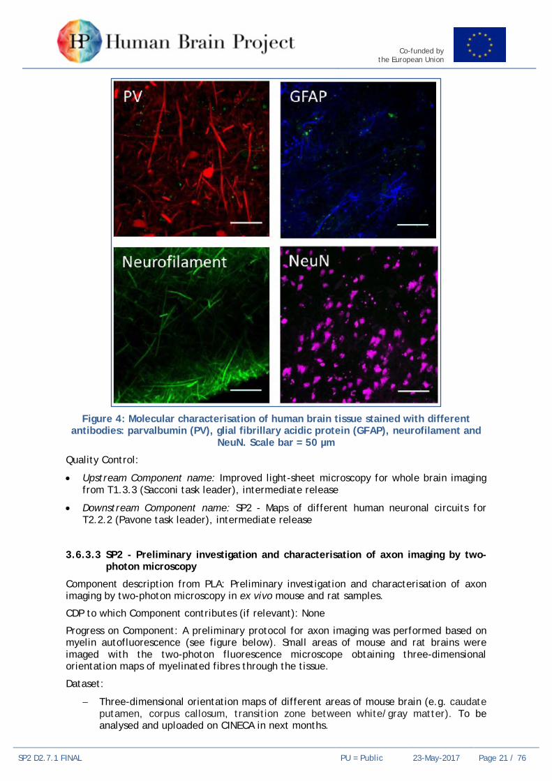
Progress on Component: Different antibodies were tested by LENS and CNR-INO to neurons and interneurons discrimination (e.g. NeuN, PV, VIP, Somatostatin, MAP2, GAD67, Neurofilament) but only some of them have worked well (NeuN, PV and neurofilament, see picture below). In future antibodies from different company will be tested to overcome this issue. First trials of tissue imaging with light sheet microscopy was performed. We found some difficulties on sample mounting that will be addressed in the future months.
At VU, antibodies were tested on human slices to discriminate interneuron subtypes. PV, CR, CB and GAD67 worked well on human neurons.
Co-funded by the European Union
SP2 D2.7.1 FINAL PU = Public 23-May-2017 Page 21 / 76
Figure 4: Molecular characterisation of human brain tissue stained with different
antibodies: parvalbumin (PV), glial fibrillary acidic protein (GFAP), neurofilament and NeuN. Scale bar = 50 µm
Quality Control:
• Upstream Component name: Improved light-sheet microscopy for whole brain imaging from T1.3.3 (Sacconi task leader), intermediate release
• Downstream Component name: SP2 - Maps of different human neuronal circuits for T2.2.2 (Pavone task leader), intermediate release
3.6.3.3 SP2 - Preliminary investigation and characterisation of axon imaging by two-photon microscopy
Component description from PLA: Preliminary investigation and characterisation of axon imaging by two-photon microscopy in ex vivo mouse and rat samples.
CDP to which Component contributes (if relevant): None
Progress on Component: A preliminary protocol for axon imaging was performed based on myelin autofluorescence (see figure below). Small areas of mouse and rat brains were imaged with the two-photon fluorescence microscope obtaining three-dimensional orientation maps of myelinated fibres through the tissue.
Dataset:
− Three-dimensional orientation maps of different areas of mouse brain (e.g. caudate putamen, corpus callosum, transition zone between white/gray matter). To be analysed and uploaded on CINECA in next months.

Co-funded by the European Union
SP2 D2.7.1 FINAL PU = Public 23-May-2017 Page 22 / 76
− Three-dimensional orientation maps of different areas of rat brain (e.g. anterior commisure, transition zone between corpus callosum/cingulum, optic tract, and part of the caudate putamen). To be analysed and uploaded on CINECA in next months.
Figure 5: Myelin autofluorescence enhancement – The preparation protocol allows imaging of myelinated fibres (red circles) with high resolution without exogenous
labelling with both confocal and two-photon fluorescence microscopies. Image scale bar = 50 µm
Quality Control:
• Upstream Component: none
• Downstream Component: component of SGA2 T2.3.4 Integration of high-resolution connectivity across scales and modalities
3.7 Task 2.2.3 Transmitter Receptors in Cortical and Subcortical Regions and Layers of the Human Brain
3.7.1 Key Personnel Task Leader: Karl ZILLES (JUELICH)
Other Researchers: Francesco PAVONE (LENS, CNR), Irene COSTANTINI (LENS), Leonardo SACCONI (CNR)
3.7.2 SGA1 DoA Goals The goal of this Task is to characterise the expression patterns of multiple receptors for the classical neurotransmitters glutamate, GABA, acetylcholine, serotonin, noradrenaline, dopamine, and adenosine in selected human brain regions. The data, i.e. the mean receptor densities in cytoarchitectonically identified cortical regions, resulting from this analysis will be integrated into the HBP atlas for selected areas of the initial Big Brain parcellation.
3.7.3 Component Progress 3.7.3.1 SP2 – Quantification of multiple receptor distributions for selected areas
Component description from PLA: Quantification of the densities (fmol/mg protein) of up to 19 different receptor binding sites for glutamate, GABA, acetylcholine, serotonin, noradrenaline, dopamine, and adenosine in primary sensory and motor cortical areas, in hippocampus, cingulate and entorhinal cortex, as well as in the basal ganglia, selected thalamic and brainstem nuclei using receptor autoradiography. We will provide a table of the data (mean densities based on six to nine different human hemispheres), color-coded autoradiographs, and laminar profile curves for each of the above listed brain structures.

Co-funded by the European Union
SP2 D2.7.1 FINAL PU = Public 23-May-2017 Page 23 / 76
CDP to which Component contributes (if relevant): CDP3, Multi-Level Human Brain Atlas, CDP3-P4
Progress on Component: only JUELICH contributed to this component
We analysed the distribution patterns of 15 different receptors in the primary visual, somatosensory and auditory areas of the human brain to determine whether differences in their regional expression may characterise principal subdivisions of the cortex into primary sensory, motor, and hierarchically higher sensory or multimodal areas. Additionally, we compared these results with data obtained from non-human primate, scandentia, rodent, afrotherian, and marsupial brains to determine whether transmitter receptors enable the identification of primary sensory areas throughout various mammalian species. All three primary sensory areas can be clearly delineated from surrounding cortical areas in the human brain by their conspicuously higher muscarinic M2, GABAergic GABAA, noradrenergic 2 and the serotonergic 5-HT2 receptor densities. The interspecies comparison revealed that the M2 receptor, and to a lesser degree also the 2 and 5-HT2 receptors can be considered as an evolutionary conserved molecular marker of areas V1, S1 and A1 in all examined species, with the notable exception of the marsupial brain. These data demonstrate, that receptor studies, by identifying primary sensory areas, may provide a powerful tool for the analysis of cortical evolution.
Milestone 2.2.1 achieved. Data provided to CDP3.
Milestone name: mean densities of 15 different receptors in human anterior mid and posterior cingulate cortex
Dataset (primary sensory areas): http://dx.doi.org/10.1016/B978-0-12-804042-3.00043-9, Comparative Analysis of Receptor Types That Identify Primary Cortical Sensory Areas, accessible yes, reusable yes.
Quality Control:
• Upstream component: To date there have been no releases which were essential for the progress of this component
• Downstream component: SP2 - Integration of papaya prototype with JuBrain atlas and receptor measurements into NIP backend + Task 2.6.5 (SGA1) + provided Excel tables with data necessary to create “receptor fingerprints” (polar coordinate plot showing the mean [+ SD; dashed line] densities [in fmol/mg protein] of 16 receptors [names specified on each axis] in a given area. Data based on the analysis of 8 human hemispheres), and the corresponding “receptor profiles” (Plots depicting laminar-specific changes in receptor density from the pial surface to the border between layer IV and the white matter. X axis codes for cortical depth, Y axis for receptor density in fmol/mg protein) as well as 16 colour coded autoradiographs, each showing the laminar distribution of a single receptor in a specific cortical area of a single brain + data provided has been successfully integrated into the Jubrain Atlas via the papaya prototype.
3.7.3.2 SP2 – Mapping of cellular structures onto molecular architecture
Component description from PLA: Mapping cellular structures onto molecular architecture using serial two photons microscopy in combination of clearing and immunostaining techniques. We will provide images of spatial distribution of different types of receptors together with different kind of cells, ultra-resolution imaging of receptors and sub-cellular structures.
CDP to which Component contributes (if relevant): None.
Progress on Component: Different antibodies against various receptors were tested (e.g. M2R, GluR, GABAxR) but they have not worked well. There wasn’t significant contrast between the signal and noise to allow imaging. In future, others antibodies from different companies will be tested to overcome this issue and some enhancing fluorescence protocol will be tested. Work was done by CNR-INO.

Co-funded by the European Union
SP2 D2.7.1 FINAL PU = Public 23-May-2017 Page 24 / 76
Dataset: none.
Links: none.
Quality Control:
• Upstream Component: Optimisation of Clarity for whole brain imaging, T1.3.3 (Sacconi task leader), finished component with good result
• Downstream Component: Multilevel maps of quantitative cell distributions and morphologies, T2.2.2 (Pavone leader), we couldn’t yet provide the protocol for receptors staining
3.8 Task 2.2.4 Connectivity within and between Cortical Areas and their Microstructure
3.8.1 Key Personnel Task Leader: Cyril POUPON (CEA)
Other Researcher: Markus AXER (JUELICH), Jean-François MANGIN (CEA), Pamela GUEVARA (Univ of Concepcion, Chile), Nicole SCHUBERT (JUELICH)
PhD students: Nicole LABRA (CEA), Justine BEAUJOIN (CEA), Kevin GINSBURGER (CEA)
3.8.2 SGA1 DoA Goals The objectives of Task 2.2.4 are four-fold:
1) produce an atlas of U-fibre bundles,
2) deliver a massive MRI database including anatomical/quantitative/diffusion MRI scans to map the white matter microstructure including the axon diameter and density, the microvasculature volume fraction, the myelin water fraction; profiles will be provided along the white matter bundles included in the bundle atlas designed in the frame of the Ramp-Up and SGA1 phases,
3) provide a dual high resolution PLI / dMRI dataset of the same post mortem hippocampus specimen and design methods to align them as well as to perform comparison of the structural connectivity obtained across the microscopic and mesoscopic scales,
4) provide information about the cortical layer reached by each bundle and describe the architecture of fibres entering gyri and connecting to cortical areas.
3.8.3 Component Progress 3.8.3.1 SP2 – 3D-PLI data for selected human anatomical structures
Description of Component from PLA: Registration and curation of 3D-PLI data for selected anatomical structures into the atlas. The data will be registered to the Big Brain template.
CDP to which Component contributes (if relevant): CDP3 Multi-Level Human Brain Atlas, CDP3-P4 Enrichment of the Human Brain Atlas with qualitative and quantitative datasets
Progress on Component: Two post-mortem human hippocampi from two subjects have been cryo-sectioned (60 µm section thickness). One of those hippocampi has been scanned with high resolution dMRI at Neurospin (cf. T2.5.2 for details) beforehand. 3D-PLI scanning has been started (and is still continuing) for both samples. Zeineh et al. (2016) provided a microstructural delineation and fibre tract identification (e.g., endofolial pathway, Schaffer collaterals, performant path system) at section level for one of the samples. Recent work focused on setting up a pipeline required for 3D reconstruction of the 3D-PLI scanned and analysed images (Ali et al., 2017). Datasets are not yet fully published and therefore not yet accessible, but should be delivered as soon as they get published Those reconstructions will finally be aligned to the Big Brain template. Work was done by JUELICH.

Co-funded by the European Union
SP2 D2.7.1 FINAL PU = Public 23-May-2017 Page 25 / 76
Links: see publications.
Quality Control:
• Upstream Component: There is currently no component, which appears to be critical for the successful achievement of the task.
• Downstream Component: Finalised and published 3D fibre orientation maps will be delivered to and integrated in the Human Brain Atlas through CDP3.
3.8.3.2 SP2 – Map of human fibre bundles and their microstructure
Description of Component from PLA: The data comes from Task 2.2.4. This Task will provide an atlas of white matter fibre bundle, aggregating 40 deep white matter structures and over 100 U-fibre bundles. Inter-individual variability in the MNI space will be represented as probabilistic atlases. 3D representations of the bundles will be used to visualise connectivity in the atlas interface. For each bundle, the microstructural organisation of axons will be provided as series computed along the bundle. Diffusion-based measurements like FA or MD will be of interest for Medical Informatics. Axon diameters and axon densities will be of interest for modelling brain connectivity using generative approaches mimicking development.
CDP to which Component contributes (if relevant): CDP3 Multi-Level Human Brain Atlas, CDP3-P4 Enrichment of the Human Brain Atlas with qualitative and quantitative
Progress on Component: The map of human long fibre bundles provided in the Ramp-Up Phase is now completed with a map of 100 short U-fibres elaborated by CEA through a collaboration with the University of Concepcion in Chile, elaborated from the ARCHI database as planned. Associated publication: Guevara et al., 2017. After a first release of a bundle atlas including 40 long and 100 short white matter bundles, the preparation of a second richer release is actually ongoing, relying on a subset of the HCP database making use of a refined alignment across subjects. This work is performed jointly with Task 5.3.2 aiming at integrating HCP dataset in the NIP. A dedicated tool was developed in the frame of SGA1 to align fields of diffusion-based probability displacement functions (PDFs) or orientation distribution functions inferred from the multiple-shell dMRI HCP dataset using a non-linear approach. This novel registration method was proven to significantly improve the registration of dMRI datasets with respect to scalar methods, thus allowing sharper probabilistic maps for long and short white matter bundles as well as yielding the emergence of additional short white matter bundles to be delivered in the second release of the bundle atlas. The methods were accepted for communication in the next OHBM conference (Ginsburger et al, 2017) and recently submitted to the MICCAI 2017 conference (Ginsburger et al, 2017). The acquisition of the massive BICKET (Brain Imaging of the Cytoarchitecture using a Key Emerging Technology) MRI dataset is started (CEA). The imaging protocol is finalised including 10 sessions of MRI scans per subject with the inclusion of 20 subjects, for a total of 400h of acquisitions scheduled in the period March 2017-March 2018. The protocol includes a massive set of anatomical, relaxometry, diffusion MRI scans plus an additional 10 minutes of resting-state fMRI at the beginning of each imaging session. A new decoding software tool to infer the microstructural features is under development, and part of it has already been accepted that will be presented during the next ISMRM conference (Ginsburger et al, 2017). The high resolution dMRI acquisition of the human hippocampus specimen is also listed as a component of Task T2.5.2 and the progress on this Component achieved by CEA and JUELICH was therefore detailed in this Task. The last topic focuses on the study of the architecture of fibres when they enter a gyrus and then connect to cortical areas; this topic is a hot topic in the field of connectomics as the poor resolution of in vivo dMRI data (~1mm) does not allow to accurately map the sharp turns of fibres entering the cortex, and does not allow to map the fibre architecture within the cortical layer; to this aim, a dedicated dMRI acquisition with mesoscopic resolution was started on a post mortem human hemisphere at 11.7T that will give access to 300 micrometer dMRI data (CEA); one block of the hemisphere localised in the visuo-spatial area will be scanned with PLI at a microscopic

Co-funded by the European Union
SP2 D2.7.1 FINAL PU = Public 23-May-2017 Page 26 / 76
scale (JUELICH), which will allow comparison between scales (in vivo millimetre / mesoscopic / microscopic). In parallel of the dMRI acquisition, the development of a novel anatomically-informed global tractography approach was started involving the use of adequate anatomical priors stemming from the cortical ribbon in order to drive the creation of fibres specifically when they enter gyri, thus allowing them to follow sharp turns before connecting to the cortical areas. The actual tool was first designed to run on a clinical dataset and will benefit from a code redesign in the frame of SGA2 to allow its use on a high-performance computing facility in order to allow massive global tractography at multiple scales. This preliminary work was accepted as an oral presentation in the frame of the ISMRM conference (Teillac et al, 2017) and recently submitted to the MICCAI conference (Teillac et al, 2017).
Datasets are not yet fully published and therefore not yet accessible but should be delivered as soon as they get published.
Quality Control:
• Upstream Component name (from PLA): “In vivo fibre tract scans and diffusion-based data on major and U-shaped tracts were” and “Probabilistic atlas of long white matter bundles” components are provided by the team in charge of this Task (e.g. CEA and Univ Chili); the other upstream Components (functional maps, architectonic maps, etc.) are not critical for the achievement of the Task and they will be easily exploited through intersection with the bundle atlas after integration in the HBP atlas standard spaces .
• Downstream Component: Finalised and published probabilistic maps of long and short white matter bundles as well as their quantitative microstructural features will be delivered to and integrated in the Human Brain Atlas through CDP3.
3.9 Task 2.2.5 Quantitative Characterisation of Fibre Tracts 3.9.1 Key Personnel Task Leader: Joachim LÜBKE (JUELICH)
3.9.2 SGA1 DoA Goals
The major aim of this study is to quantitatively described axons in various fibre tracts of the human brain. This will be achieved using high-end, fine scale electron microscopy.
3.9.3 Component Progress
3.9.3.1 SP2 – 3D models of fibre tracts
Description of Component from PLA: Quantitative analysis of various fibre tracts in the human CNS, including those in different neocortical regions, the hippocampal formation and other important fibre tracts, such as corpus callosum, their trajectory and dimension at the electron microscopic level. Finally, fibre tracts are 3D-reconstracted.
CDP to which Component contributes (if relevant): None
Progress on Component: The main problem working with human tissue is its availability and quality for such fine-scale investigations. Post mortem brain cannot be used for such quantitative investigations due to bad preservation of the ultrastructure. The last year was spent to collect human brain tissue from various sources that have the quality needed for the investigations. We have started with the quantitative analysis and 3D-reconstruction of fibre bundles in the corpus callosum at the subcellular level
For each dataset: Datasets are not accessible to the public. We plan to submit an abstract to the Annual Neuroscience meeting this year.
Links: not applicable
Quality Control:

Co-funded by the European Union
SP2 D2.7.1 FINAL PU = Public 23-May-2017 Page 27 / 76
• Task responsibility by PI.
• Adjusting a protocol for SEM-FIB electron microscopy that would allow higher throughput and areas of interest in the samples.
3.10 Task 2.2.6 Morphological and functional connectivity of human cortical microcircuits
3.10.1 Key Personnel Task Leader: Huib MANSVELDER (VU)
3.10.2 SGA1 DoA Goals The aim of this Task is to map the structure and function of microcircuits of pyramidal and interneurons in human neocortex. In this Task, strategic data that is lacking from the literature will be generated and we will bring together all other efforts and data that are generated by labs worldwide. In addition, to reconstruct functional cortical circuits, this Task will closely collaborate, interact and share data with the modelling and informatics platforms.
The objectives for SGA1 are to:
• Map subcellular function of different neuron types, linking morphological and functional properties of dendrites.
• Provide morphology of synaptic input profile and synaptic connection strength for connected pairs of cortical pyramidal neurons, linking morphological and functional properties of axons.
3.10.3 Component Progress 3.10.3.1 SP2 – Morphological data of human neocortical pyramidal neurons
Description of Component from PLA: Map subcellular function of different neuron types.
CDP to which Component contributes (if relevant): CDP3 – Multi-Level Human Brain Atlas, CDP3-P4
Progress on Component: Datasets on biophysical properties of membranes from human cortical neurons that were shared with partner Idan SEGEV (HUJI) as indicated in the previous reporting period for input for the first ever data-driven biophysical models of human neurons, have now been published.
Data sets of morphologies and electrophysiology-codes of the very same pyramidal neurons from layers 2 and 3 of human temporal cortex were transferred to partner Segev (WP6) for modelling and simulation in the previous period and were supplemented with new data sets of similar type. The manuscript on this combined data-modelling work is now being prepared for submission for publication.
Contribution of each Partner in Task to the above: All work was done by partner Mansvelder, VU Amsterdam
For each dataset: linked to publication http://dx.doi.org/10.7554/eLife.16553.
Quality Control:
• All data were shared with team Segev and his tasks in SP4 and SP6, quality control see publication.
3.10.3.2 SP2 – Morphological cortical connectivity profiles
Description of Component from PLA: Morphological connectivity profiles and synaptic connection strength for connected pairs of cortical pyramidal neurons.

Co-funded by the European Union
SP2 D2.7.1 FINAL PU = Public 23-May-2017 Page 28 / 76
CDP to which Component contributes (if relevant): CDP3 – Multi-Level Human Brain Atlas, CDP3-P4
Progress on Component: Data on connectivity profiles and synaptic connection strength for connected pairs of cortical pyramidal neurons have been transferred to the team of Idan SEGEV (HUJI).
Contribution of each Partner in Task to the above: All work was done by partner Mansvelder, VU Amsterdam
For each dataset: N/A

Co-funded by the European Union
SP2 D2.7.1 FINAL PU = Public 23-May-2017 Page 29 / 76
4. Work Package 2.3 Function and Variability
4.1 Key Personnel Work Package Leader: Simon Eickhoff (JUELICH)
4.2 WP Leader’s Overview The work planned in WP2.3 has been performed according to plan. In particular, we have continued the acquisition of the IBC data from the Ramp-Up Phase without any problems. A public data release has been performed (http://neurovault.org/collections/2138/). In addition, we have published the first tri-modal (resting-state connectivity, task co-activations and diffusion tractography) parcellation at the start of 2017, providing a new map of the left dorsal premotor cortex (Genon et al., 2017). Based on the infrastructure set up in the aforementioned project, we have continued over the last month to refine the pipeline into a common framework for multimodal brain parcellation. In particular, we have established an infrastructure for using the same methods and computational framework across all three modalities and added structural covariance as another feature of brain organisation. Initial work using this framework is currently being performed on the hippocampus as a region of particular interest in the HBP. The initial results, which also address the dependency of brain parcellations on the investigated sample of subjects as well as methodological choices in the data processing indicate several important new aspects. First, the influence of different (equally plausible) processing options is moderate but still evident. Second, the congruency across datasets is higher than across modalities for any one dataset. Third, in line with previous more anecdotal literature, we observed a functional segregation of the human hippocampus in the anterior-posterior rather than in the medial-lateral direction as evident in cytoarchitecture. Providing an important conceptual framework for this kind of multi-modal investigations, we have recently published a concept paper (Eickhoff et al., 2017) outlining challenges and approaches for the multi-modal mapping of the human brain.
4.3 Priorities for the remainder of the phase For the remainder of SGA1 we will continue the acquisition of the IBC data and provide additional analyses of this unique dataset. The work on the hippocampus parcellation should largely be finished within the rest of the phase, providing the first quantifiable results on the relationship between different in vivo brain parcellation approaches as well as novel insights into the functional compartmentalisation of the human hippocampus. In close integration with SP5 and CDP3, we are currently setting up a pipeline for multi-modal brain parcellation that can run on the JURECA HPC environment, which will dramatically speed up the process and allow more detailed investigations of additional regions. Through a second student representing an in-kind contribution to the project, we are now starting to perform a second study in the same manner as performed for the hippocampus also for the basal ganglia.

Co-funded by the European Union
SP2 D2.7.1 FINAL PU = Public 23-May-2017 Page 30 / 76
4.4 Milestones Table 3: Milestones for WP2.3 Function and Variability
MS No. Milestone Name Lead Partner Task(s) involved Expected
Month Achieved
Month Comments
MS2.3.1 Framework for multi-modal mapping established for one initial region of interest
JUELICH T2.3.2 12 12 Achieved. We have used the hippocampus as the initial region of interest and established the pipeline for its multi-modal parcellation.

Co-funded by the European Union
SP2 D2.7.1 FINAL PU = Public 23-May-2017 Page 31 / 76
4.5 Task 2.3.1 Atlasing and Cognitive Meta-Analysis 4.5.1 Key Personnel Task Leader: Bertrand THIRION (INRIA)
Other Researcher: Jean-Francois MANGIN (CEA)
Other researcher: Ana Luisa Grilo PINHO
Engineer: Isabelle COURCOL
4.5.2 SGA1 DoA Goals
This Task aims to produce a set of brain maps that provide a complete description of brain anatomical organisation, connectivity and functional organisation. A specific aim is to create a high-resolution atlas of cognitive function, that will provide the first objective basis to compare cognitive components to their neural substrate and thus revisit the ontology of cognitive processes from a data-driven perspective. At the same time, it will make up the first cognitive atlas of the human brain. The individual data analysis allows a highly resolved picture (1.5mm isotropic). In the framework of SGA1, we are committed to deliver 50 such brain maps in each of the 12 subjects of the “Individual brain Charting cohort”.
4.5.3 Component Progress 4.5.3.1 SP2 – full human brain activity maps (volumes)
Description of Component from PLA: We provide fifty maps of brain activity spanning various cognitive contrasts at 1.5mm resolution, recoded in 12 subjects. The images are sampled in the volume.
Progress on Component: Data acquisition has been very active, with about 80 acquisitions performed since the beginning of SGA1. The acquired data comprise in particular a language protocol from NeuroSpin, a video watching experiments (3 full acquisitions), a retinotopy experiment (one full acquisition), high resolution diffusion and high-resolution T1 and T2 images. In parallel, we are preparing future acquisitions for the remainder of SGA1: mental time travel protocol, resting-state, a reward protocol, possibly a parietal protocol (saccades and computation), and another movie watching protocol. We also consider performing quantitative T1 and T2 experiments. All the acquired data are pre-processed based on a unique pipeline that relies on state-of-the-art techniques. The data have been pushed to the Jureca server for availability with HBP. A first release of the resulting brain maps has taken place in the Neurovault interface (http://neurovault.org/collections/2138/). We are currently preparing a journal publication to describe our procedures and intermediate results. MRI acquisitions were done at CEA. Remaining work was done by INRIA.
4.6 Task 2.3.2 Multi-modal regional mapping 4.6.1 Key Personnel Task Leader: Simon EICKHOFF (JUELICH)
4.6.2 SGA1 DoA Goals This Task will employ multi-modal in vivo mapping of regional modules in large, population based samples to investigate key questions on regional brain organisation. In doing so, we will provide a new, validated model of how brain modules may be defined from multi-modal in vivo data, which can then be applied to provide robust, valid parcellations of the human brain.

Co-funded by the European Union
SP2 D2.7.1 FINAL PU = Public 23-May-2017 Page 32 / 76
4.6.3 Component Progress 4.6.3.1 SP2 – Selected multimodal regional maps with cognitive features
Description of Component (from PLA): We will employ multi-modal in vivo mapping of regional modules in large, population-based samples to investigate the key questions on regional brain organisation. In doing so, we will provide a new, validated model of how brain modules may be defined from multi-modal in vivo data, which can then be applied to provide robust, valid parcellations of the human brain.
CDP to which Component contributes (if relevant): CDP3 – Multi-level Human Brain Atlas, Use Cases: CDP3-P4 and CDP3-P6.
Progress on Component: Progress as planned. We worked on the hippocampus as the first use-case due to the focus on this structure within SP2 in SGA2. Initial parcellations using several different modalities have been computed, these are currently being evaluated against each other.
Dataset is not yet accessible.
Quality Control:
• Upstream component: Software components of Task 5.4.4: Implementation of atlas-based analysis pipelines and Pre-processing pipeline for raw neuroimaging data
• Downstream component: Until now, we did not generate and deliver data to another component because we just implemented the pipeline.

Co-funded by the European Union
SP2 D2.7.1 FINAL PU = Public 23-May-2017 Page 33 / 76
5. Work Package 2.4 Comparative Computational Architecture of Multi-Modal Processing Streams (Systems Physiology)
5.1 Key Personnel Work Package Leader: Rainer GOEBEL (UM)
5.2 WP Leader’s Overview Work in WP2.4 progressed well in most Tasks. As a continuation of the work started in the Ramp-Up Phase, we have further provided important multi-level multi-species data that will serve as relevant constraints for the creation of models and brain simulations, especially for the question how bottom-up and top-down effects are integrated in layers of early visual cortex. The “mesoscopic” focus on cortical columns and cortical layers is critically needed to simulate the human brain and to bridge animal and human data. With respect to CDP4 (Task 2.4.4), we made more progress than expected not only specifying a state-of-the-art cognitive architecture for visuo-motor integration tasks but also completing the implementation of a first neural network model that ran successfully as a closed-loop prototype on the Neurorobotics Platform. While guided by SP2, this work was only possible through close collaboration with SP4, SP7 and SP10 resulting in co-development and improvements of IT platforms, especially the capabilities of the NEST simulation environment. The first prototype model incorporates also a deep learning autoencoder predicting saliency distributions for eye movements that match human performance.
While the development of cognitive architectures and experimental non-human primate (NHP) and human measurements and analyses progressed well, we experienced delays for running the planned human texture segregation / attention studies. In order to separate functional activity across cortical layers as good as possible in the human brain, this study was scheduled for 9.4 Tesla fMRI but unexpected technical issues lead to delays. Since the technical issues seem to be solved, we will start with these measurements in April/May in Maastricht; in case of further technical issues, the study will be executed on the 7 Tesla fMRI scanner that has been shown to be good enough for roughly separating cortical layers and cortical columns (e.g. De Martino et al., 2015).
5.3 Priorities for the remainder of the phase For the remainder of the SGA1 phase, we will continue acquiring strategically relevant multi-level multi-species data for atlas building and for the creation of computational (neural network) models. In the context of CDP4, we will work on completing the visuo-motor computational architecture continuing to co-developing the IT platforms to make them better suited for use by (cognitive) neuroscientists.

Co-funded by the European Union
SP2 D2.7.1 FINAL PU = Public 23-May-2017 Page 34 / 76
5.4 Milestones Table 4: Milestones for WP2.4 Comparative Computational of Multi-modal Processing Streams (Systems Physiology)
MS No. Milestone Name Lead Partner Task(s) involved Expected
Month Achieved
Month Comments
---- ---- ---- ---- ---- ---- No Milestone in M12

Co-funded by the European Union
SP2 D2.7.1 FINAL PU = Public 23-May-2017 Page 35 / 76
5.5 Task 2.4.1 Multi-scale Processing in Space, Time and Frequency 5.5.1 Key Personnel Task Leader: Wim VANDUFFEL (KUL)
Other Researcher: Pieter ROELFSEMA (KNAW)
5.5.2 SGA1 DoA Goals It is not well understood how neurons in lower and higher areas that represent the visual (auditory) world at different spatial (spectral) and temporal scales interact with each other. By recording activity in the non-human primate and mouse with electrophysiology in the different cortical layers and with fMRI while changing activity with perturbation methods, such as optogenetics, we will increase our understanding of the interactions between brain regions at different levels of the cortical hierarchy and at different scales. In addition, we will compare brain activity in non-human primates to data obtained in mice within the same perceptual task.
5.5.3 Component Progress 5.5.3.1 SP2 – Computational architecture of the functional organization and visual and
auditory processing streams and human and macaque monkey
Description of Component from PLA: Definition of the computational architecture of the multi-scale, multi-level functional organisation in visual and auditory processing streams of the human and macaque monkey brain. High resolution human and monkey fMRI data will be generated.
CDP to which Component contributes (if relevant): CDP4 “Visuo-Motor Integration”
Progress on Component: Top-down and feedforward inputs into cortical areas arrive in different cortical layers. In the primary visual cortex (area V1) feedforward input from the LGN arrives in layer 4, and feedback from higher visual areas (such as V2) arrives in layer 1, 2 and 5. An important aim has been to compare the activity in the different cortical layers, using recordings from the different layers of macaque monkeys and human high-field fMRI. The high field fMRI results in humans are currently being measured and the results will be compared to published results from monkey laminar recordings in area V1 in a texture-segregration task (Self et al., Current Biology, 2013). We have now also recorded the neuronal correlates of attention and working memory in the different layers of V1 (van Kerkoerle et al., Nature Communications, 2017). We found that top-down influences mainly impact on activity in the superficial and deep layers and that these effects are weaker in layer 4. Furthermore, we found that the top-down effects are associated with extra synaptic inputs into layers 1, 2 and 5, precisely matching the known terminals of feedback connections into V1. We have submitted a review about the comparison of human and monkey laminar data (Self et al., accepted NeuroImage).
In addition, we have had a unique opportunity to record spiking activity in area V3 of a human patient, who was implanted with electrodes as part of a treatment for epilepsy. We were able to replicate our previous findings on the activity profiles of texture segregation and attention shifts, hence confirming that the macaque monkey is an excellent model for visual processing in the human visual cortex (Self et al., PLoS Biology, 2016).
5.6 Task 2.4.2 The Role of Attention and Perception and Learning 5.6.1 Key Personnel Task Leader: Rainer GOEBEL (UM)
Other Researcher: Wim VANDUFFEL (KUL), Rui COSTA (FCHAMP)

Co-funded by the European Union
SP2 D2.7.1 FINAL PU = Public 23-May-2017 Page 36 / 76
5.6.2 SGA1 DoA Goals This Task aims to deepen our understanding of attentional selection. Attentional top-down signals are thought to originate in fronto-parietal brain areas and modulate activity via feedback connections in sensory cortex. Much of the evidence supporting this theory, however, comes from either correlation-based type of experiments, or from a surprisingly limited number of causal-oriented experiments, whereby activity in one brain region (e.g. in frontal cortex) is perturbed, while measuring changes in behaviour or activity in target areas. However, most methods to (reversibly) inactivate a region have a rather poor spatial and/or temporal resolution.
5.6.3 Component Progress 5.6.3.1 SP2 – Selective attention in perception and learning in humans and monkeys
Description of Component from PLA: We will use a top-down and bottom-up cued selective attention task and perform a comparative study targeting occipito-parietal and frontal cortex of NHP and human patients using intracranial electrophysiology (human/monkey), high-resolution fMRI (human/monkey) and focal perturbations (monkey) based on inactivating optogenetics and novel pathway and direction-selective inactivation methods.
CDP to which Component contributes (if relevant): CDP4 “Visuo-Motor Integration”
Progress on Component: The monkey data have been collected and are being analysed. We first focused on the involvement of parietal areas (combined electrophysiology, fMRI, and optogenetic inactivation). We are currently preparing the data for publication (KUL).
Experiments with human fMRI at 7 Tesla (UM) are currently being designed using the same paradigm as used for the NHP experiments (KUL). The experiments are scheduled for execution in Q3/Q4 of 2017.
5.7 Task 2.4.3 Revealing Columnar-Level Feature Codes in VWFA and FFA
5.7.1 Key Personnel
Task Leader: Rainer GOEBEL (UM)
Other Researchers: Dirk FELDMEYER (JUELICH)
5.7.2 SGA1 DoA Goals
Sub-millimetre ultra-high field fMRI provides the unique possibility to unravel the columnar-level features coded in brain areas that have no exact homologue in other species, such as areas involved in reading or perception of human faces. We will use 7 and 9.4 Tesla fMRI to unravel representational sub-units in higher-level visual areas, including the visual word form area (VWFA) and several face areas (OFA, FFA1 (pFus) and FFA2 (mFus)). This will be performed by opposite but converging experimental approaches (high-resolution fMRI in humans, cellular level in rodents). The aim is to unravel how different types of sensory input affect the structural and functional organisation of individual neurons and neuronal networks
5.7.3 Component Progress 5.7.3.1 SP2 – Properties of excitatory neurons GABAergic interneurons and the neuronal
networks in sensory cortical areas in rodents
Description of Component from PLA: The columnar organisation of sensory areas is a feature that is already expressed at the cellular level, i.e. on the scale of a few μm, leading to properties like retino- (as well as tono- and somatotopic) coding, which cannot be resolved by sub-millimetre fMRI. Using a correlated electrophysiological/morphological approach, we plan to investigate the properties of excitatory neurons and GABAergic interneurons and the

Co-funded by the European Union
SP2 D2.7.1 FINAL PU = Public 23-May-2017 Page 37 / 76
neuronal networks formed by them in different sensory cortical areas (primary visual, auditory and somatosensory cortex) in brains of rodents.
Progress on Component: The structural, electrophysiological and synaptic data on interneurons in the rodent somatosensory cortex have been collected and analysed in depth. Two publications are under review and a third one is in preparation. We are currently in the process of gathering morphological data on interneuron types in layer 6 of the prefrontal cortex. Work was done by JUELICH.
5.7.3.2 SP2 – Ultra-high field fMRI of sub-units in higher-level visual areas and face areas in human and monkey
Description of Component from PLA: We will use 7 and 9.4 Tesla fMRI to unravel representational sub-units in higher-level visual areas, including the visual word form area (VWFA) and several face areas (OFA, FFA1 (pFus) and FFA2 (mFus)). This will be performed by opposite but converging experimental approaches using: a) rich stimulus sets presented at different positions and sizes on the screen containing words and faces, respectively, and b) fragments of letter strings and face parts (e.g., eyes, nose and mouth). An extended population receptive field (pRF) estimation approach and several competing encoding neural network models will be used to analyse the data guiding the assignment of the most likely stimulus features to voxels within the targeted specialised brain areas. The same sub-millimetre fMRI approach will be also performed in monkeys to elucidate species-specific processing of letter string-derived shape features as well as faces and face parts from the same (monkey) and different (human) species.
CDP to which Component contributes (if relevant): CDP4 “Visuo-Motor Integration”
Progress on Component: We (UM) have designed the stimuli to probe “faciotopy” in the human face areas in collaboration with Kriegeskorte (Cambridge, SP3 T3.1.1 PLP) and first pilot data has been acquired at 7 Tesla. The main measurements (10 or more subjects) are scheduled to start in April. In collaboration with Carmine Gnolo (Maastricht), we furthermore developed a generative model creating arrangements of oriented face features (simple bar-like features for mouth, nose, eyes) to investigate the neural mechanisms calculating a holistic measure of “faceness”.
As an extension of a previous study (Emmerling et al., 2016), we investigate the neural correlates of letter perception and letter imagery using sub-millimetre 7 Tesla data and advanced pRF modelling. The analysis results from the first 3 subjects indicate that the reconstruction of letter shapes is not only possible for seen but also for purely imagined letters using activity in early visual cortex (V1-V3). For imagined letter shapes, the activity in early visual cortex is very weak likely resulting from top-down (imagery-driven) activation (see also Task 2.4.2 and SP3.1). This hypothesis will be tested by selectively reconstructing visual content from upper (supra-granular), middle and lower (infra-granular) layer compartments. Since feedback connections into V1 spare the middle layers, it is expected that visual content can be better reconstructed from upper (and eventually lower) layer compartments while reconstruction of seen letter shapes should work best for middle layers.
KUL partner: High resolution fMRI data of the face-patch system in monkeys have been collected. The data are being analysed. In a next step, we will use novel statistical methods to link the monkey with the human data.
5.8 Task 2.4.4 Development of an Empirically-Derived Brain Atlas on Sensorimotor Integration
5.8.1 Key Personnel Task Leader: Rainer GOEBEL (UM)

Co-funded by the European Union
SP2 D2.7.1 FINAL PU = Public 23-May-2017 Page 38 / 76
5.8.2 SGA1 DoA Goals This Task contributes to CDP4 developing a neurobiologically realistic model of sensorimotor integration performing advanced object recognition and spatial localisation tasks. The visuo-motor integration tasks selected for SGA1 are goal-directed eye movements including saccades and smooth pursuit executed in specific (top-down modulating) contexts.
5.8.3 Component Progress 5.8.3.1 SP2 – Brain atlas of sensorimotor integration
Description of Component from PLA: Development of a neurobiologically realistic model of sensorimotor integration performing advanced object recognition and spatial localisation tasks. Strategic 7T fMRI data will be acquired and analysed to provide specific information on neural networks underlying selected sensorimotor integration tasks (oculomotor tasks in SGA1).
CDP to which Component contributes (if relevant): CDP4 “Visuo-Motor Integration”.
Progress on Component: Based on multi-level and multi-species brain data we specified a cognitive architecture to assign and model computational functions of more than 20 human brain areas subserving visuo-motor integration tasks. In SGA1, we focus on goal-directed eye movements including saccades and smooth pursuit. The brain areas of the cognitive architecture calculate salient regions from the visual input in parietal cortex, where it is integrated and transformed from sensory reference frames (e.g., in eye- and head-centred coordinates) to motor-relevant reference frames (e.g., body-centred coordinates). This information is fed forward to premotor cortex and frontal eye fields (FEF) and integrated with information from prefrontal cortex about action goals and contexts (e.g. instructions such as “look at the red object”) before final motor output is sent to primary motor cortex, relayed via the corticospinal tracts, and modulated by the basal ganglia. The cognitive architecture is currently implemented in NEST (see 19.3.2) using non-spiking point neurons. The architecture and assumed functions in specific model brain areas is constrained by existing monkey and human electrophysiological and neuroimaging data. We have also designed a 7 Tesla study to perform a visuo-motor test battery (including saccade – anti-saccade task, visual search, natural viewing) at an unprecedented spatial functional resolution of 0.8 mm iso-voxel. The design of the 7 Tesla study has been completed and its execution is scheduled for June/July. The data will be used to validate the developed cognitive architecture and to fine-tune architectural decisions.
5.8.3.2 Network architecture of visuo-motor integration (model)
Description of Component from PLA: Python implementation of visuo-motor network architecture using dynamic activation functions. The network architecture contains visual input layer, computation of saliency map, selection of targets and generation of saccades. The architecture forms a closed-loop interacting with sub-cortical structures.
CDP to which Component contributes (if relevant): CDP 4 “Visuo-Motor Integration”.
Progress on Component: We have started the implementation of the developed cognitive architecture as a non-spiking neural network model in Python (PyNEST). The overall network will model the computational functions of more than 20 human brain areas subserving visuo-motor integration tasks. We have already completed two important sub-functions, a model of the saccade generation (needed to link to the Neurorobotics Platform) and the calculation of salient regions as target for eye movements. The output model is implemented as a rate neuron model of the saccade generator (SG) in the reticular formation (RF) a proposed by Gancarz & Grossberg (1998) and a publication is in preparation (Senden et al. 2017). For the saliency calculation, we have developed a deep convolutional autoencoder network able to automatically learn a mapping from natural images to topological saliency distributions. The encoding network employs the pretrained VGG16 model (Simonyan & Zisserman, 2014) for feature extraction whereas the decoding network was randomly initialized. The entire

Co-funded by the European Union
SP2 D2.7.1 FINAL PU = Public 23-May-2017 Page 39 / 76
forward stream of the autoencoder involves convolution, max-pooling, dilated convolution, concatenation, and upsampling. After training, the network shows good generalisation as it performs well with previously unseen data sets. Specifically, the predicted salience distribution given previously unseen natural images shows good correspondence to empirical salience distributions as reflected by human fixation patterns. This network forms the front-end of the larger architecture of visuo-motor integration, providing salience distributions as input to a target selection process.

Co-funded by the European Union
SP2 D2.7.1 FINAL PU = Public 23-May-2017 Page 40 / 76
6. Work Package 2.5 Integrative Maps & Models
6.1 Key Personnel Work Package Leader: Jean-Francois MANGIN (CEA)
6.2 WP Leader’s Overview This integrative WP is at the crossroad of several tasks initiated during the RUP. Hence, after several years of work, these tasks are now delivering high quality and curated results allowing seamless integration. Therefore, our targets relative to the link between post-mortem databases of JUELICH and in vivo database of Neurospin (Archi database in the NIP) were reached faster than expected. Hence, we are already experimenting with the new dataset to be integrated in the NIP, namely the outstanding Human Connectome Project dataset, for which things were more challenging during the last year, because some of the Oxford’s toolbox required to perform the analysis were still missing a GPU speed-up component. The advent of the last public release in March 2017 is overcoming most of our difficulties and will allow us to proceed forward rapidly. The connectivity matrices generated by this WP (joint work of T2.5.1 & T2.5.2) have just been delivered for a component of SP4 (T4.4.1) and will be at the core of a service to the community provided through the NIP portal. The number of matrices delivered will increase rapidly with the integration of the HCP dataset and its massive processing using SP7 facility (joint work with T5.3.2 & T5.4.4). Relative to the joint acquisitions of PLI and diffusion data, we had some unexpected difficulties with crystals forming in the histological slices before PLI acquisition. The PLI acquisitions can be exploited but they are not optimal. A second experiment with a mouse brain convinced us that the problem was probably related to the high number of experiments performed with the MRI to tune the diffusion acquisition. Hence, we have triggered the acquisition of a new hippocampus specimen on the 11,7T magnet, which shall overcome the problem. The growth of our database of multimodal data including ECOG acquisitions of epileptic patients is raising a lot of interest in the neuroscience community. We are gaining momentum with the arrival in SGA2 of an opinion leader in the field, Giacomo Rizzolatti. We are on the verge to federate the community, which shall happen during SGA2.
6.3 Priorities for the remainder of the phase For the second year of SGA1, the main priorities are:
T2.5.1: Illustrate how the sulcus-based controlled alignment of post mortem and in vivo data create new opportunity to match the areas of multimodal parcellations, in order to pave the way to the use cases planned in SGA2 relative to the HBP’s atlas usage.
T2.5.2: Finalise at least one joint PLI / diffusion MRI acquisition; perform global tractography from the dMRI dataset; assess the cortical layer reached by some of the tracked bundles in the PLI 2D slices for feasibility.
2T.5.3: Finalise the software HiBop that will be used during SGA2 as a standalone component connected with the NIP repository to attract and federate the ECOG community.

Co-funded by the European Union
SP2 D2.7.1 FINAL PU = Public 23-May-2017 Page 41 / 76
6.4 Milestones Table 5: Milestones for WP2.5 Integrative Maps & Models
MS No. Milestone Name Lead Partner Task(s) involved Expected
Month Achieved
Month Comments
MS2.5.1 Sulcus-based alignment of cytoarchitectonic and diffusion-based parcellations
CEA T2.5.1 12 12 Achieved (see task 2.5.1)

Co-funded by the European Union
SP2 D2.7.1 FINAL PU = Public 23-May-2017 Page 42 / 76
6.5 Task 2.5.1 Matching Microscopic and In Vivo Parcellations 6.5.1 Key Personnel Task Leader: Jean-Francois MANGIN (CEA)
Research fellow: D. RIVIÈRE (CEA), F. POUPON (CEA)
Postdoc: J. LEBENBERG (CEA), S. LEFRANC (CEA)
6.5.2 SGA1 DoA Goals The aim in this Task is to provide complementary views of a unique brain architecture. This is a complex process because each brain parcellation provides only a partial view of the complete multilevel segregation and we do not have access to all parcellations in the same individual. Hence, the complementary parcellations stemming from microscopic features on one side and in vivo imaging on the other side have to be aggregated, which is the goal of this Task. Bridging information issued from dedicated SP2 research programs are used to guide the process. In SGA1, the link between some architectural features and the cortical folding pattern is exploited to match Cyto-architectonics versus fMRI-based MRI-connectivity based parcellation, using sulcus-based alignment methods from Task 2.6.2.
6.5.3 Component Progress 6.5.3.1 SP2 – New human brain parcellations based on microscopic post mortem and in
vivo data
Description of Component from PLA: New macroscopic parcellations based on in vivo diffusion data, and alignment of these parcellations with post mortem parcellations (cytoarchitectonics) and the fMRI-based parcellations.
CDP to which Component contributes (if relevant): This Component contributes to the CDP3 (the HBP Human Brain Atlas) through 4 use cases (P3, P4, P6, P8). Numerous atlas usage relies on parcellations and their integration.
Progress on Component: To complement post mortem architectonics parcellations, and task-fMRI-based parcellations, this Task has finalised work initiated during the RUP to generate dMRI-connectivity-based parcellations from the 79 in vivo datasets of the Archi database included in the NIP [Lefranc et al. (2016) Groupwise connectivity-based parcellation of the whole human cortical surface using watershed-driven dimension reduction. Med Image Anal 30:11-29 and Fig.]. Population-based and individual parcellations have been computed in order to support a variety of research programs aiming at comparing and matching these new parcellations with the other ones. The processing pipelines are now in the process of being tuned to the large HCP dataset currently in process of integration into the NIP through Task 5.3.2.
Figure 6: Connectivity profiles in the poscentral gyrus

Co-funded by the European Union
SP2 D2.7.1 FINAL PU = Public 23-May-2017 Page 43 / 76
During the last 12 months, we have also finalised the sulcus-based controlled alignment between the 79 subjects of the Archi database (integrated in the NIP) and the post-mortem individual cytoarchitectonic maps of JUELICH (including the BigBrain). This was a challenge because of the unusual nature of histological images. It was performed thanks to the software developed in the context of Task 2.6.2. We have now generated and quality checked all the sulcus-based transformations between each individual native spaces and the set of standard spaces taken into account by the CDP3 for the HBP atlas. All in vivo and post mortem individual parcellations can now be put in any standard space available in CDP3 for comparisons (MNI, Collin and BigBrain), which corresponds to the Milestone MS2.5.1 (see Figure 6)
Figure 7: Maps from the Archi database and JUELICH
Quality Control:
• All the key upstream components have been received and used for MS 2.5.1: the alignment toolbox of T2.6.2 (SGA1), the cytoarchitectonic maps of T2.2.2 (RUP) and T2.2.1 (SGA1), the functional maps of T2.1.1 (RUP), the diffusion data of T2.2.2 (RUP). The SGA1 Components are upgraded regularly.
• Downstream: Interactions with T2.6.1 are part of the development of the toolbox delivered by T2.6.2; the tuning of the parcellation pipelines for HPC is done in collaboration with T2.6.5; The connectivity-based parcellations have been delivered to T2.5.2 for the computation of connectivity matrices.
6.6 Task 2.5.2 Co-Design Big Data Analytics: Integrating Connectivity Across Scales
6.6.1 Key Personnel Task Leader: Markus AXER (JUELICH)
Other researchers: Jean-François MANGIN (CEA), Denis RIVIÈRE (CEA), Cyril POUPON (CEA)
Post-docs: Sandrine LEFRANC (CEA)
PhD students: Justine BEAUJOIN (CEA), Kevin GINSBURGER (CEA), Daniel SCHMITZ (JUELICH)
6.6.2 SGA1 DoA Goals The goals of this Task are:

Co-funded by the European Union
SP2 D2.7.1 FINAL PU = Public 23-May-2017 Page 44 / 76
• Delivery of pairwise structural connectivity matrices for different brain parcellation systems with a focus on the HBP’s atlas nodes
• Delivery of multi-modal (dMRI and 3D-PLI) hippocampus datasets aligned across scales
6.6.3 Component Progress 6.6.3.1 SP2 – Human connectivity matrix from atlas parcels
Description of Component from PLA: This dataset is produced by Task 2.5.2. This Task will compute pairwise structural connectivity information for connectome nodes defined from post mortem, diffusion (T 2.5.1) or functional data. Hence this Task will provide the connectivity matrix used to serve the connectome-oriented requests to the HBP atlas. Connectivity of the nodes will be inferred from diffusion MRI tractograms but also from in vivo functional acquisitions (fMRI, MEG, EEG, depth electrodes, etc.). We will provide average connectivity and a large set of individual connectivity matrices to allow study of inter-individual variability. A typical application planned in SP4 is the simulation of resting state networks from the connectome matrix for modelling epileptic seizures.
CDP to which Component: CDP3-P4 and CDP3-P8
Progress on Component: Three sets of structural connectivity matrices have already been computed for each of the 79 subjects of the Archi database, using two of the most standard public parcellations (AAL, freesurfer) and a more advanced connectivity-based parcellation adapted to each individual (Constellation toolbox, developed in the context of HBP, Lefranc et al., Med. Image Anal., 2016). These datasets have been sent to partners of SP4 in the context of CDP3, who will provide complementary fMRI-based connectivity matrices (T4.4.1 Gustavo DECO, Gorka ZAMORA-LÓPEZ). Additional parcellations will be used following the same process. Larger and refined matrices will be generated using the HCP dataset integrated in the NIP through T5.3.2. Work was done by CEA.
Quality Control:
• Upstream Component: There is currently no Component which appears to be critical for the successful achievement of the Task.
• Downstream Component: Finalised and published 3D fibre orientation maps will be delivered to and integrated in the Human Brain Atlas through CDP3.
6.6.3.2 SP2 – Crossing scales between dMRI and 3D-PLI
Description of Component from PLA: Two datasets of a post mortem human hippocampus will be acquired with high-resolution dMRI and 3D-PLI at complementary spatial scales. The 3D-volumes will be aligned; the obtained fibre distributions/densities in different regions of the hippocampus will be compared to each other.
CDP to which Component contributes (if relevant): CDP3-P4 Enrichment of the Human Brain Atlas with qualitative and quantitative datasets
Progress on Component: The high resolution dMRI acquisition of the human hippocampus specimen provided by JUELICH is completed (CEA) and provides a unique dataset of dMRI data acquired at 11.7T with a resolution of 300 micrometres over the entire specimen; a successful reconstruction of the polysynaptic circuit has been reached as well as the segmentation of the laminar structure of the cornus Ammonis using diffusion MR microscopy; the specimen was sent back to INM1 for 3D-PLI acquisition, aiming to perform a joint 3D-PLI/dMRI analysis in the coming months. Results were disseminated in conferences (Beaujoin et al, 2016, 2017) and recently submitted to Brain Structure and Function. In addition to the non-linear registration approach developed with the Heidelberg partner, the alignment tool developed in the frame of Task 2.2.4 that exploits the 3D information provided by the orientation distribution functions (ODFs) computed from both 3D-PLI and dMRI datasets will be used to accurately register them and therefore serve as a cornerstone to navigate across the scales offered by the two modalities (Ginsburger et al, 2016). Hippocampus cryo-

Co-funded by the European Union
SP2 D2.7.1 FINAL PU = Public 23-May-2017 Page 45 / 76
sectioning has been finished and early quality checks at the histological section level revealed unexpected crystal formations across the entire tissue. Since crystals exhibit birefringence, they become detectable in 3D-PLI measurements. From a second experiment with a mouse brain there is evidence that the crystal growth might have been related to the high number of experiments performed with the MRI to tune the diffusion acquisition. Hence, we have triggered the acquisition of a new hippocampus specimen on the 11,7T magnet, which shall overcome the problem. In addition, 3D-PLI scanning has been started (and is still continuing) for the current joint hippocampus sample. 90 sections (from about 500 sections; 60 µm section thickness) have been digitised and transferred into fibre orientation maps (FOMs) for both resolutions 64 µm and 1.3 µm. 3D reconstruction of all blockface images has been finished; volume registration of the T2 MRI dataset into the blockface volume is currently being explored. This step will help to identify the location of each 3D-PLI section within the diffusion MRI volume. Further, work focuses on section-wise 3D reconstruction utilising the registration pipeline described in T2.2.4 (Ali et al., 2017). The JUELICH group has also implemented an algorithm to generate fibre ODFs from high-resolution 3D-PLI fibre orientation vectors (Paper submitted). This will make MRI-based ODFs alignment and comparison with 3D-PLI-based ODFs possible.
Datasets are not yet published in a journal paper and therefore not yet accessible but will be made usable as soon as they get published.
Quality Control:
• Upstream Component: There is currently no Component which appears to be critical for the successful achievement of the Task.
• Downstream Component: 3D fibre orientation maps will be delivered to and integrated in the Human Brain Atlas through CDP3. Due to the (unexpected) crystal formation in the tissue, a second hippocampus will be started to be scanned already within SGA1 (but work will be continued in SGA2).
6.7 Task 2.5.3 Human Intracranial Electrophysiology Data and Tools 6.7.1 Key Personnel Task Leader: Jean-Philippe LACHAUX (UCBL)
6.7.2 SGA1 DoA Goals This Task will provide intracranial EEG recordings with their exact localisation relative to the HBP atlas features, including the connectome nodes. All subjects are epileptic patients with a documented file description precisely their special context. During SGA1, the dataset of outstanding EEG recordings will be enlarged and complemented with brain activation maps, together with the scripts based on open-source software (HiBoP), in order to ensure the reproducibility of the results.
6.7.3 Component Progress 6.7.3.1 SP2 – Human iEEG recordings
Description of Component from PLA: This data is produced in Task 2.5.3. It will provide intracranial EEG recording with their exact localisation relative to the HBP atlas features, including the connectomes nodes. As a result of the Ramp-Up Phase, electrophysiological data from thirty epileptic patients have been delivered at the beginning of 2016. Human atlas data will be hosted at the HPAC infrastructure data repositories, in the context of CDP3. As a result, users can retrieve electrophysiological data from all sites by spatial queries to one of the accepted template spaces, either based on a radius search to a given set of 3D coordinates, or directly by the selection of cortical areas. During SGA1, this dataset of outstanding EEG recording will be enlarged and complemented with brain activation maps (raw, pre-processed data and analysis results), together with the scripts used to process the

Co-funded by the European Union
SP2 D2.7.1 FINAL PU = Public 23-May-2017 Page 46 / 76
data. The provided data are highly relevant for clinically-oriented users, modellers and theoreticians, as well as methodologists interested in testing novel signal processing algorithms (for instance to measure functional connectivity).
CDP to which Component contributes (if relevant): CDP3 - Multi-Level Human Brain Atlas, Use Case P4
Progress on Component: iEEG data acquisition in patients is a steady, stationary process, which takes manpower to complete. In the last 12 months, we kept recording from new patients and gathered new data from previous patients (pre-HBP), to reach 15 new patients. In addition, we have been developing a new task of interest for modellers and clinicians alike, which is designed to trigger activity in the hippocampus (learning series of words). We also advanced our analysis procedure for simultaneous SEEG/fMRI data to identify correlations between high-frequency [50-150 Hz] iEEG signal component and BOLD signal (improved analysis of previous dataset). Link to publication by Saignavongs et al. 2017 (http://dx.doi.org/10.1142/S0129065717500010). iEEG data will be uploaded to the server at the end of SGA1.
6.7.3.2 SP2 – Human intracranial electrophysiology tools
Description of Component from PLA: Human Intracranial Electrophysiology Tools. The tool is a software (“HiBoP”) to load and visualise large amounts of multimodal functional brain data, with a strong emphasis on intracranial EEG from large (>30) groups of patients, but with the possibility to juxtapose fMRI data. iEEG are shown on 3D models of the participants’ brain dynamically, with millisecond time resolution.
CDP to which Component contributes (if relevant): CDP 3 – Multi-Level Human Brain Atlas, Use Case P4
Progress on Component: The HiBoP software delivered at the end of the first period was a beta version with a strong need for major improvements. In the last 12 months, two engineers paid on HBP budget worked literally side by side with iEEG users (neuroscientists), who used HiBoP on a daily basis for analysis of functional localisers data and report any bug they could find, which were rapidly corrected to stabilise the software. In addition, they added several important functionalities for HiBoP, such as the ability to group, label, exclude, visualise recording sites onto individual patients’ anatomy; immediate access to high-frequency activity maps at the single trial level on individual site, correlation between response amplitude across two sites for functional connectivity. In 12 months, HiBoP has switched from a demo to a fully functional program actually used by researchers and students in the lab. Still, many new functionalities need to be developed and tested (systematic correlation between BOLD and iEEG, anatomical connectivity, easy connection with database etc.). We plan to deliver at the end of SGA1 a windows version, a Linux version and a Mac OS version.

Co-funded by the European Union
SP2 D2.7.1 FINAL PU = Public 23-May-2017 Page 47 / 76
7. Work Package 2.6 Co-Design/Methods and Big Data Analytics
7.1 Key Personnel Work Package Leader: John ASHBURNER (UCL)
7.2 WP Leader’s Overview • Much of the data required by the HBP is image-based and to be shared among the project
(and to external researchers) via the human brain atlas of CDP3. This WP mostly concerns development of image processing algorithms required by CDP3. Overall, progress has been reasonably good, and most Tasks were able to employ people to do the required work. Unlike many ICT projects, Tasks in WP2.6 tend to require some experimenting to determine which strategies work, prior to implementing the final code. Many strategies have been explored, and the Tasks have now settled on the final strategies that they intend to adopt. Some tasks have now implemented prototypes and have them running on the JUELICH supercomputers for parallel computation.
• As a result of the brief employment period, the delays in finance and the requirement of having specialist skills that are in demand by the private sector, there have been some delays in employing personnel.
• It is too early to assess impact, particularly as work only began in earnest relatively recently.
7.3 Priorities for the remainder of the phase As most of the exploratory work has now been done, the efforts will now be on implementing software to meet the requirements of CDP3. In addition to coding, this will require extensive testing using a wide variety of data. Many of the Tasks involve image registration, which is an application area that is impossible to make completely robust. Many further incremental modifications will be needed to increase robustness and accuracy, particularly for certain more challenging datasets. Further synergy between Tasks is to be expected later on in SGA1.
Many of the Tasks have required new algorithms to be developed, which will be written up for the scientific literature. User documentation will also be needed.

Co-funded by the European Union
SP2 D2.7.1 FINAL PU = Public 23-May-2017 Page 48 / 76
7.4 Milestones Table 6: Milestones for WP2.6 Co-design/Methods and Big Data Analytics
MS No. Milestone Name Lead Partner Task(s) involved Expected
Month Achieved
Month Comments
---- ---- ---- ---- ---- ---- No Milestones in M12

Co-funded by the European Union
SP2 D2.7.1 FINAL PU = Public 23-May-2017 Page 49 / 76
7.5 Task 2.6.1 Learning and Applying Shape and Multi-modal Appearance Models
7.5.1 Key Personnel Task Leader: John ASHBURNER (UCL)
7.5.2 SGA1 DoA Goals The goal is to produce a basic image registration framework that uses an empirically derived model of anatomical variability to achieve more precise and robust whole-brain image alignment. The first step is to develop a framework for computing the main modes of variability from a large sample of brain MRI. The second step is to use these derived modes of variability to better model the prior covariance of shape variability of brain images, in order to achieve more accurate and robust image alignment.
7.5.3 Component Progress 7.5.3.1 SP2 – Shape and appearance models for human brain variability
Description of Component from PLA: Develop a coherent unsupervised model for directly learning a dictionary of principal geodesics that best encode geometric variability, and implement a parallel version of the learning algorithm that may be executed on several CPUs. The ultimate goal is to implement an algorithm for using a learned model to infer optimal alignment with new image data, and thereby allow to project and superimpose atlas parcellation onto new MR scans (beyond SGA1).
CDP to which Component: This Component is an essential tool for multimodal integration of the Human Brain Atlas targeted by CDP3 – Multi-Level Human Brain Atlas, P5. It is especially relevant for data that are of a modality or quality that is unsuitable for applying the results of Task 2.6.2.
Progress on Component: Development of this work is in conjunction with a Task in SP8, T8.3.11 Brain morphological features. Most of the preliminary work has focused on finessing a principal geodesic analysis (PGA) algorithm using 2D images, with the intention of making a fully 3D implementation more straightforward for postdocs employed in future. Recent work on the 2D PGA framework has focussed on ensuring that it converges, that the integrations of the diffeomorphisms are stable and that it gives accurate estimates of the main modes of anatomical variability. Several versions of probabilistic formulations have been investigated, and a final approach for SGA1 has been settled on. So far, decisions about strategy have been based on how well the approach can encode the shape variability of a large dataset of small 2D images (MNIST), which is widely used by the neural network community. Using relatively small training sets (fewer than about 1000 per category), the model is able to capture shape variability more accurately than state-of-the-art deep learning methods. A simple 3D implementation of PGA has also been implemented and tested using IXI data. Delays in financing mean that no postdocs are yet employed, so all development so far is done by the PI (UCL).
The work has made use of the following datasets:
MNIST: https://en.wikipedia.org/wiki/MNIST_database.
IXI: http://brain-development.org/ixi-dataset/
All software packages are under development and therefore not yet accessible.
Quality Control:
• Upstream Component: There is currently no component that appears to be critical for the successful achievement of the Task.

Co-funded by the European Union
SP2 D2.7.1 FINAL PU = Public 23-May-2017 Page 50 / 76
• Downstream Component: The results will be delivered to and integrated in the Human Brain Atlas through CDP3.
7.6 Task 2.6.2 Multimodal Alignment with Macro- and Microanatomical Patterns
7.6.1 Key Personnel Task Leader: Timo DICKSCHEID (JUELICH)
Other Researcher: Jean-Francois MANGIN (CEA)
7.6.2 SGA1 DoA Goals The Task goal is to develop robust spatial alignment methods for human brain data across scales. The planned achievements for SGA1 are 1) a refined computer algorithm for automatic detection of cortical sulci from T1-weighted MRI scans and their use for 3D to 3D nonlinear image registration, as well as 2) an assessment of the suitability of a range of tissue microstructures for alignment of small-field-of-view microscopy data with whole brain atlas data. The latter requires implementation of a range of microstructure detectors, as well as a matching procedure.
7.6.3 Component Progress 7.6.3.1 SP2 – Improved version of sulcus-based cross modality alignment toolbox
Description of Component from PLA: Building upon previous work in the context of the DISCO software, a further improvement of alignment will be implemented based on cortical sulci that have been previously identified using a pattern recognition approach.
CDP to which Component contributes (if relevant): This Component is an essential tool for multimodal integration of the Human Brain Atlas targeted by CDP3 – Multi-Level Human Brain Atlas, P5.
Progress on Component: The API between DISCO and the complementary DARTEL software (SPM) has been improved. Reverse engineering has been performed to allow seamless composition of the spatial transformations yielded by the two complementary approaches. The resulting sulcus-based alignment procedure has been used in T2.5.1 to perform controlled alignment of all the post-mortem and in vivo data dedicated to the NIP into the different standard spaces available in the HBP atlas.
The complete software is integrated as a component toolbox into the mature BrainVISA framework (http://brainvisa.info) and is now ready to be converted as a plugin to build a cloud-based service in SGA2. Work was done by CEA.
Quality Control:
• Upstream Components: The key upstream Component used for validation during joint work with T2.5.1, namely the cytoarchitectonic maps of T2.2.2 (RUP) & T2.2.1, have been received and are upgraded regularly.
• Downstream Components: The automatic sulcus recognition software providing the landmarks used by the toolbox has been applied to the 79 brains of the Archi database of the NIP. Recognition errors have been manually corrected for each brain, and the resulting database is on the verge of being transferred to T2.6.1 as an input for methodological development.

Co-funded by the European Union
SP2 D2.7.1 FINAL PU = Public 23-May-2017 Page 51 / 76
7.6.3.2 SP2 – Classifier for microstructural landmarks that can be used for cross-scale alignment of PLI data
Description of Component: A machine-learning based classifier that robustly detects microstructural landmarks in PLI data, that can be found at different scales. The focus is to align data from a polarization microscope (which delivers data at the micrometre scale) with that of a one-shot device (which delivers data in the order of 60 micron).
CDP to which Component contributes (if relevant): This Component contributes to CDP3 P5 (Alignment tools for incoming data).
Progress on Component: The work in this Task has initially focused on cell-stained histological data instead of PLI. We developed methods for the detection and matching of features representing vessel-like structures and bright blob-like structures that often represent Virchow-Robin spaces. Our initial approach detected vessel-like structures as Haar-like features coupled with a variant of AdaBoost to learn a classification function (Viola et al. 2001), and bright blobs by morphological filtering of the response of a Maximally stable extremal regions detector (MSER; Matas et al. 2004). To obtain a computationally more efficient algorithm, we then implemented a unified approach for both based on a Support Vector Machine (SVM) that works in the space of principal modes of 2D histograms of image patches, counting for each patch the number of pixels over different grayscale ranges with respect to their distance to the centre pixel. Algorithms have been installed on the JUELICH supercomputers for parallel computation. The downstream use for cross-scale PLI alignment has not yet been provided, but we are already in contact with the PLI group to evaluate the algorithm as a next step.
7.6.3.3 SP2 – Robust matching procedure for incomplete sets of microstructures detected at different scales
Description of Component: A matching procedure based on appearance and spatial constraints for cross-scale microstructures, which exploits a coarse pre-alignment as well as the planarity of scanned histological sections to overcome the inherent difficulty of matching tissue microstructures with poor distinctiveness.
CDP to which Component contributes (if relevant): This Component contributes to CDP3 P5 (Alignment tools for incoming data).
Progress on Component: For the microstructural features detected with the above component, a matching algorithm has been implemented that uses 1) similarity of the features used in the SVM classifier and 2) patterns of spatial arrangement of the features in the 2D section plane. We first evaluated several states of the art approaches for spatially supported matching (2D-homography driven RANSAC, Graph Transform Matching by Aguilar et al., IVC 2009) that did not show acceptable performance due to the sparseness of the features and high amount of redundant responses. The new approach evaluates the spatial configuration of feature triplets, and exploits congruency of the spanned triangles. It has been successfully applied to use the vessel- and blob-like features for registration of regions in consecutive sections at 1 micrometre resolution Algorithms have been installed on the JUELICH supercomputers for parallel computation.
The downstream use for cross-scale PLI alignment has not yet been provided, but we are already in contact with the PLI group to evaluate the algorithm as a next step.
7.7 Task 2.6.3 Mutual Tissue/Fibre Models for Simulation of dMRI and 3D-PLI Measurements
7.7.1 Key Personnel Task Leader: Markus AXER (JUELICH)
Other Researcher: Cyril POUPON (CEA)

Co-funded by the European Union
SP2 D2.7.1 FINAL PU = Public 23-May-2017 Page 52 / 76
PhD student: Felix MATUSCHKE (JUELICH), Miriam MENZEL (JUELICH)
7.7.2 SGA1 DoA Goals This Task is twofold: (1) Create a library of real case membrane geometries (tubes and simplified cell shapes with different arrangements) and intrinsic birefringence; and (2) finalize the 3D-PLI simulator and the diffusion simulator.
7.7.3 Component Progress 7.7.3.1 SP2 – Simulated tissue/fibre models
Description of Component from PLA: Simulated tissue/fibre models will be designed in a joint research program as basis for simulated 3D-PLI and dMRI measurements and analyses. We will generate a dictionary of synthetic datasets providing a broad spectrum of modelled, realistic fibre arrangements.
CDP to which Component contributes (if relevant): None.
Progress on Component: An extension of the dMRI simulator provided along with the Connectomist software (CEA) has been started to make it usable on any high-performance computing facility; this extension mainly involves a refactoring of the code to allow distribution of data as well as the distribution of the Monte-Carlo simulations across the nodes of the HPC facility, each node already benefiting from a multithreaded process over the cores of the node. The development of a novel software library to design tissue models has also been started, first aiming at building complex geometries of fibres tuned with respect to a limited set of parameters like the mean orientation of the fibres, the angular dispersion of the fibres, the maximum tortuosity of fibres, the distribution of axon diameters, the axon volume fraction. The objective is here to write a library that will be used to create artificial tissue mimicking white matter with a common distributed file format that the 3D-PLI and dMRI simulators will be able to read, in order to perform 3D-PLI and dMRI simulations and better model and understand the origins of the signal, to ultimately develop novel dictionary decoding approaches. Two abstracts in direct relation to this work were accepted for communication in the ISMRM and OHBM conferences (Estournet et al, 2017) and a journal paper is in preparation. For 3D-PLI, the development of two complementary simulation approaches has been initiated to model the interaction of polarized light with brain tissue together with its measurement in a polarimetric setup. The first type of simulation focuses solely on the effect of tissue birefringence and is based on the Jones calculus, an efficient matrix formalism to describe macroscopic effects of linear optical components exerted to a light beam. The related software tool is referred to as simPLI. The second simulation technology is based on computational electrodynamics to describe the propagation of a polarized light wave through brain tissue by means of Maxwell’s equations (Menzel et al., 2016). While simPLI has been optimized to perform efficiently on the HPC facility JURECA at the Supercomputing Centre Jülich, the electrodynamics based simulation utilizes the JUQUEEN supercomputer architecture.
All software packages are under development and therefore not yet accessible.
Quality Control:
• Upstream Component: There is currently no Component which appears to be critical for the successful achievement of the Task.
• Downstream Component: The results will be delivered to and integrated in the Human Brain Atlas through CDP3.
7.8 Task 2.6.4 Big Data Methods for Extracting Quantitative Data in High-Resolution Imagery

Co-funded by the European Union
SP2 D2.7.1 FINAL PU = Public 23-May-2017 Page 53 / 76
7.8.1 Key Personnel Task Leader: Francesco PAVONE (LENS)
Task contributors: Ludovico SILVESTRI (LENS), Giacomo MAZZAMUTO (LENS)
7.8.2 SGA1 DoA Goals High-resolution 3D microscopic imagery of brain tissues includes a wealth of valuable data that needs to be processed in an automatic fashion to produce reliable quantitative information. The goal of this Task is to implement efficient automatic methods for handling high-resolution microscopic imagery. Ideally, specific software tools should be able to perform different levels of processing including: stitching of overlapping 3D tiles, cell counting, cell segmentation, vascular segmentation.
7.8.3 Component Progress 7.8.3.1 SP2 – Cell counts, cell and vascular segmentation for selected areas in human
Description of Component from PLA: Registration and curation of cell counts and segmentation for selected cytoarchitectonic areas into the atlas. The cell counts and segmentation should be accessible by queries to the ontologies, and visual selections of areas in the JuBrain parcellations. In SGA1, they are presented as an overlay in the 3D viewer, depicting precomputed plots.
CDP to which Component contributes (if relevant): CDP3, use case CDP3-P4 Enrichment of the Human Brain Atlas with qualitative and quantitative datasets
Progress on Component: We are focusing on the development of a dedicated software tool to stitch high-resolution 3D microscopic tiles. In effect, we found that existing open source software solutions do not perform and scale well given the extent of our datasets (several TB of data). The stitching tool that we are developing leverages the computing power of modern GPUs and is therefore set to be more efficient than existing solutions. The software is being developed in Python and makes use of state-of-the-art computing libraries such as TensorFlow, an open source software library for numerical computation using data flow graphs developed by Google. In the next period, stitched images produced with the software tool described above will be fed to cell segmentation and counting algorithms. In particular, to achieve high throughput in the analysis pipeline, we will port and improve existing algorithms for brain cell finding to a GPU architecture.
Quality Control:
• Upstream Component:
SP2 - 3D-PLI data for selected human anatomical structures (Axer): we have received provisional data to help component design.
Data analytics workflows for the human brain atlas (Dickscheid): we have not received any prototype. According to PLA they will be released at M24.
• Downstream Components:
SP2 - Mapping of cellular structures onto molecular architecture (Pavone): we received help to co-design the Component. Nothing final has been provided, as the release is on M24.
SP2 - Multilevel maps of quantitative cell distributions and morphologies (Pavone): we received help to co-design the Component. Nothing final has been provided, as the release is on M24.
SP2 - Maps of different human neuronal circuits (Pavone): we received help to co-design the Component. Nothing final has been provided, as the release is on M24.
7.9 Task 2.6.5 Co-design of the HBP Atlas-Based Big Data Analytics

Co-funded by the European Union
SP2 D2.7.1 FINAL PU = Public 23-May-2017 Page 54 / 76
7.9.1 Key Personnel Task Leader: Timo DICKSCHEID (JUELICH)
Other Researcher: Markus AXER (JUELICH)
Other Researcher: Jean-Francois MANGIN (CEA)
Other Researcher: Rainer GOEBEL (UM)
7.9.2 SGA1 DoA Goals Task 2.6.5 is dedicated to the integration of tools and datasets into the Neuroinformatics Platform, and the development of analysis workflows for large image datasets and simulations for the HPAC Platform. The Task hereby ensures a tight integration of SP2 with the Platforms. The goal is to work on different, strategically important levels for co-designing the Platforms:
• On the level of data management, especially targeting the transfer of human data from labs through HPC centres to the NIP,
• on the level of datasets, especially contributing to provision of SP2 datasets to task 5.3.1,
• on the level of data-specific tool development for integration with the web-based human atlas in conjunction with 5.4.3, and
• on the level of image processing and data analytics workflows to be operated on the HPAC Platform.
7.9.3 Component Progress 7.9.3.1 SP2 – Co-design of workflow for exposing high-resolution data from HPC centres
to the HBP portal with metadata integration
Description of Component from PLA: Co-design of a workflow for exposing high-resolution data from remote sites (e.g. JUELICH) to the HBP portal (SP5), including an agreement about data types, image service (BBIC, DVID, or similar technology), and permission management. Based on the report, a developer has a clear plan how to implement a particular scenario where large datasets from an HBP partner need to be registered and visualised in the NIP, including data transfer, conversion, curation.
CDP to which Component contributes (if relevant): This Component contributes directly to CDP3 products P2 (3D interactive big data viewer), P5 (Alignment tools for incoming data), and P3 (Initial set of linked template datasets with labelled parcellations).
Progress on Component: In order to identify the missing Components for such an end-to-end workflow, we analysed the complete pipeline of data travelling from a HBP partner lab that is not directly connected to a HPC centre, down to visualisation of a final dataset in a web-based viewer of the Neuroinformatics Platform. At the start of the chain, we started to work as early adopters of the FENIX platform to realise an update of a larger neuroimaging dataset from INRIA (the IBC dataset, B. Thirion). We provided an HPAC account to INRIA, and migrated the RUP release of the data from JUELICH’s JADE server to the GPFS system underyling FENIX. We are currently discussing proper permission management and file system organisation with members of SP7. At the end of the chain, we took part in evaluation and discussion of proper HBP image services to be deployed by SP5. Several members of our team in JUELICH attended the cross-SP workshop on HBP Image Service and Viewer Architecture in Düsseldorf on Feb 15th, 2017. The results had implications for the work on Papaya prototype integration (see below component).
Partner UM is establishing pipelines to provide sub-millimetre human 7 and 9.4T functional MRI datasets (see WP2.4) for integration in the atlas. The datasets contain volumetric data at a resolution of 0.8mm iso-voxel sampled using an equi-volume layer stratification model (Kemper et al., NeuroImage, 2017) from visual, auditory and multisensory stimulation

Co-funded by the European Union
SP2 D2.7.1 FINAL PU = Public 23-May-2017 Page 55 / 76
experiments. We also tested and compared different volumetric and cortical alignment schemes to integrate post-mortem and in vivo functional atlases in collaboration with JUELICH and Stanford (Rosenke et al., NeuroImage, 2017).”
In order to render cross-scale alignment and comparison of different datasets targeting the brain’s fibre architecture possible, two methods were implemented. The first method is based on feature detection and matching (Ali, Rohr, Axer, Amunts, Eils, Wörz. Registration of ultra-high resolution 3D PLI data of human brain sections to their corresponding high-resolution counterpart, IEEE International Symposium on Biomedical Imaging (2017)). It provides a robust way to identify subsets of 3D-PLI based brain tissue images scanned at 1,3 resolution in whole brain section images scanned at 64 µm resolution. Second, an algorithm was developed that provides a statistical representation of fibre orientation vectors (orientation distribution function, ODF) gained from 3D-PLI (Axer, Strohmer, Gräßel, Bücker, Dohmen, Reckfort, Zilles, Amunts. Estimating fiber orientation distribution functions in 3D-Polarized Light Imaging. Frontiers in Neuroanatomy 10 (2016)). Such ODF is directly comparable to diffusion MRI results and supports cross-modality alignment. To demonstrate a viable way for cross-modality alignment of histological datasets highlighting different structural features, a study on three rat brains has been carried out (Schubert, Axer, Schober, Huynh, Huysegoms, Palomero-Gallagher, Bjaalie, Leergaard, Kirlangic, Amunts, Zilles. 3D reconstructed cyto-, muscarinic M2 receptor, and fiber architecture of the rat brain registered to the Waxholm space atlas. Frontiers in Neuroanatomy 10 (2016)).
7.9.3.2 SP2 – Integration of Papaya prototype with JuBrain atlas and receptor measurements into NIP backend
Description of Component from PLA: The existing papaya viewer prototype which allows to interactively retrieve receptor data per brain region from a 3D view of the JuBrain atlas will be tightly integrated to use the NIP backend service for querying the data. The current prototype only uses local datasets and structured text files, shipped within the Collaboratory container.
CDP to which Component contributes (if relevant): This Component contributes directly to CDP3 product P3 (Initial set of linked template datasets with labelled parcellations) and P6.
Progress on Component: For this purpose, a new employee has been hired in JUELICH (Dr. Haimasree BHATTACHARYA), who started in M10 due to the delayed recruitment process. She investigated the software code of the Papaya viewer with custom HBP extensions, studied documentation of the SP5 APIs, and joined several cross-SP meetings around the human atlas. In discussions with WP5.4, it turned out that Papaya will most likely not exceed the prototype stage, since all HBP-specific features can be directly integrated into the new multi-resolution 3D atlas viewer developed in T5.4.3. Since the NIP backend services are not yet ready providing receptor distributions to the atlas viewer, we chose to implement a web-compatible implementation of the JUGEX tool (JUELICH), which performs statistical analysis of Allen Gene expressions between two brain regions selected from the JUBRAIN cytoarchitectonic atlas. As a next step, this implementation will be connected with the 3D atlas viewer so that users can interactively select areas and assess gene expressions. This will provide a key Use Case for the integration of external data repositories, and is coordinated with Task 5.3.2 (CEA). We plan to integrate a prototype of the JUGEX tool with the new 3D atlas viewer around M20, and currently work with T5.4.3 on an appropriate interface design (see Figure 7). Integration with the SP5 APIs (Image Services, Knowledge Graph) is expected to be delayed, but still to happen in SGA1.
Link to the 3 D Atlas viewer in the software catalogue: https://collab.humanbrainproject.eu/#/collab/19/nav/2108?state=software,atlas-viewer-neuroglancer Link to the conversion scripts for neuroglancer in the software catalogue:

Co-funded by the European Union
SP2 D2.7.1 FINAL PU = Public 23-May-2017 Page 56 / 76
https://collab.humanbrainproject.eu/#/collab/19/nav/2108?state=software,neuroglancer-conversion-scripts Link to the Web JUGEX in the software catalogue: https://collab.humanbrainproject.eu/#/collab/19/nav/2108?state=software,Web%20JuGex Link to PyTiff in the software catalogue: https://collab.humanbrainproject.eu/#/collab/19/nav/2108?state=software,pytiff
Figure 8: Mockup of the 3D atlas viewer interface, together with a visual workflow for
the JUGEX tool.
7.9.3.3 SP2 – Workflow for automatic prediction of annotations in high-resolution histological sections across consecutive slides
Description of Component from PLA: Implementation of a novel workflow for predicting missing annotations in high-resolution images of histological series. The workflow will be based on a deep neural network architecture which is suitable for image segmentation tasks, and use a sparse set of manual annotations in some slices of the same stack as its training data to predict the same annotations in other sections. The network will therefore specialise for the case of one specific texture class in one image stack from the same subject. This way we expect to improve automatisation in cytoarchitectonic mapping, and be able to provide dense mappings for regions in whole-brain histological datasets.
CDP to which Component contributes (if relevant): This work contributed directly to CDP3-P4 (Initial qualitative and quantitative datasets).
Progress on Component: We have filled a part-time position (Christian SCHIFFER) for implementing prediction of missing annotations of brain regions in stacks of high-resolution images. We started to perform transfer learning from on a convolutional neural network architecture that has been previously trained for texture-based segmentation of cortical areas (Spitzer et al.; ISBI 2017, in press). The network is fine-tuned for distinguishing one particular cortical region, described by a sparse set of expert annotations in histological sections, from other cortical areas in the same subject (i.e. same image stack). The work is directly supervised by Hannah SPITZER (JUELICH) who developed the original convnet.
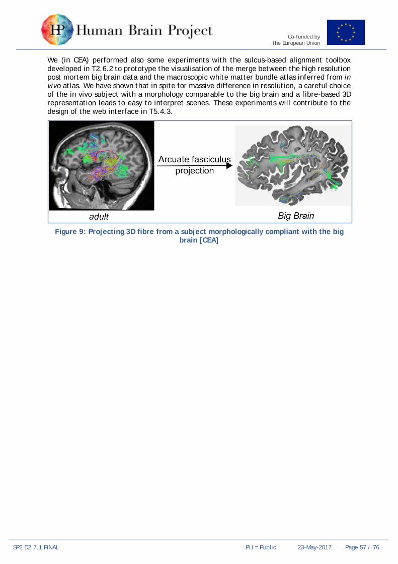
Co-funded by the European Union
SP2 D2.7.1 FINAL PU = Public 23-May-2017 Page 57 / 76
We (in CEA) performed also some experiments with the sulcus-based alignment toolbox developed in T2.6.2 to prototype the visualisation of the merge between the high resolution post mortem big brain data and the macroscopic white matter bundle atlas inferred from in vivo atlas. We have shown that in spite for massive difference in resolution, a careful choice of the in vivo subject with a morphology comparable to the big brain and a fibre-based 3D representation leads to easy to interpret scenes. These experiments will contribute to the design of the web interface in T5.4.3.
Figure 9: Projecting 3D fibre from a subject morphologically compliant with the big
brain [CEA]

Co-funded by the European Union
SP2 D2.7.1 FINAL PU = Public 23-May-2017 Page 58 / 76
8. Work Package 2.7 Coordination and Management
8.1 Key Personnel Work Package Leader: Katrin AMUNTS (JUELICH)
8.2 WP Leader’s Overview • What went particularly well? The review in June went very well for SP2 as well as the
still ongoing planning of the SGA2 phase.
• What didn’t go according to plan? The delay of SGA1 signature resulted in some administrational problems. Partners were not able to hire people or finance existing staff, which resulted in a delay of starting research in these areas.
8.3 Priorities for the remainder of the phase We will finish the SGA2 proposal in the next weeks, prepare the Open Call mentioned in the SP Leader’s report, prepare the review in June and the Summit in October, and increase outreach activities.

Co-funded by the European Union
SP2 D2.7.1 FINAL PU = Public 23-May-2017 Page 59 / 76
8.4 Milestones Table 7: Milestones for WP2.7 Coordination and Management
MS No. Milestone Name Lead Partner Task(s) involved Expected
Month Achieved
Month Comments
---- ---- ---- ---- ---- ---- No Milestones in M12

Co-funded by the European Union
SP2 D2.7.1 FINAL PU = Public 23-May-2017 Page 60 of 76
8.5 Task 2.7.1 Ethic and Innovation 8.5.1 Key Personnel Task Leader: Wim VANDUFFEL (KUL)
8.5.2 SGA1 DoA Goals Task 2.7.1 coordinates with the External Relations Team on issues related to innovation, coordinates with the Ethics Rapporteur and with SP12 on issues to ethics, and provides support to Partners related to innovation and ethics.
8.5.3 Component Progress 8.5.3.1 SP2 – Ethics and Innovation
Description of Component from PLA: Coordination of scientific activities within SP2 and their interaction with other SPs, the entire HBP, and the larger community.
CDP to which Component contributes (if relevant): None
Progress on Component: SP2 participated in EM/Rapporteur/EAB – Trilateral Meeting (02.03.2017) and will be present in the Bristol meeting (28./29.03.2017)
We made sure that the ethical protocols from the Amsterdam and Leuven groups are handled with confidentiality.
8.6 Task 2.7.2 Scientific Coordination and Management 8.6.1 Key Personnel Task Leader: Katrin AMUNTS (JUELICH, UDUS)
8.6.2 SGA1 DoA Goals Task 2.7.2 is responsible for resource allocation and use, quality control, planning of the next phase (SGA2), coordination of internal reviews (KPI, Milestones, Deliverables), performance and risk management, reporting, organisation of meetings, community building and documentation.
8.6.3 Component Progress 8.6.3.1 SP2 – Scientific coordination and management
Description of Component from PLA: This Task is responsible for resource allocation and use, quality control, planning SGA2, coordination of Reviews, performance and risk management, reporting, organisation of meetings, community building and documentation.
CDP to which Component contributes (if relevant): None.
Progress on Component: We coordinated and organised SP2’s contribution to the Open Day, Summit and the STOA event in Brussels (29./30.11.2016), prepared the RUP review (21.-23.06.2015), organised three physical meetings in Florence, Leuven and Frankfurt, eight meetings of JUELICH/UDUS/UNIBAS partners, coordinated SGA2 planning, presented SGA2 plans in the Malaga meeting (19.-22.02.2017), prepared a semester report and Deliverable in M06, and contributed to the Science and Technology Coordinators and Community Coordinators groups.
Katrin Amunts chairs the SIB and attends DIR meetings as Scientific Research Director. She is also member of the gender committee, and of the team coordinating international collaboration. Vera Kleber coordinated SIB activities.

Co-funded by the European Union
SP2 D2.7.1 FINAL PU = Public 23-May-2017 Page 61 of 76
9. Co-Design Project 3 Multi-Level Human Brain Atlas
9.1 Key Personnel CDP Science Leader: Katrin AMUNTS (JUELICH)
CDP Implementation Leader: Timo DICKSCHEID (JUELICH)
9.2 CDP Leader’s Overview A significant effect of the CDP3 is that we established a HBP-community around the human brain atlas that covers a range of SPs, and initiated many fruitful discussions. SP2 was a major driver for developing a concept of a European Research Infrastructure, from a neuroscience perspective. This concept has been published in Neuron (2016). These discussions resulted in significant scientific input, and had a strong impact onto both the reorganisation of SP5 (DPIT) and the work plan proposal for SGA2. We organised three physical meetings: One parallel session for the human brain atlas at each of the summits in Madrid and Florence, as well as a full-day workshop in Düsseldorf on Jan 30, 2017. It turned out that Product 7 (Realignment of knowledge graph for intersubject variability) is a particularly difficult topic. To cover all aspects of human variability in the NIP, a larger set of additional attributes would be required in the KnowledgeGraph that is very difficult to underpin with appropriate datasets. We therefore decided during the meeting in Florence, to postpone this aspect to a later phase of the HBP. A specifically fruitful work is product P8 “Modelling and model validation using human quantitative data”, which has led to close links between SP2 (esp. Mangin), SP5 (esp. Dickscheid) and SP4 (esp. Deco, Gorka).
9.3 Use Case Progress 9.3.1 CDP3-P1 2D interactive atlas viewer with annotation capabilities Use Case Leader: Timo Dickscheid (JUELICH)
The 2D atlas viewer is covered by the multiresolution viewer delivered by EPFL at the end of the RUP (after proposing the CDP3), as well as the work currently undertaken by Task 5.4.2. A possible continuation of the EPFL version is currently negotiated between the BBP staff and UIO. Annotation capabilities for the 2D atlas viewer have been postponed due to the delayed start of SP5, and in favour of a focus on the most important functionality.
Contributing Tasks: 5.4.2
9.3.2 CDP3-P2 3D interactive big data viewer The big data viewer is established in Task 5.4.3, “Development of 3D High-Volumetric Interactive Atlas Viewer”. Due to the delayed start of SP5, 5.4.3 is still in the recruitment process, but with joint efforts from Tasks 2.6.5 and 5.3.3, a test installation of the neuroglancer project (https://github.com/google/neuroglancer) has been deployed on a webserver in JUELICH (https://www.jubrain.fz-juelich.de/apps/neuroglancer/BigBrainRelease.2015) that points to a Version of the Big Brain dataset that we specifically converted to a format that is suitable for web-based streaming, and made available via http. This test installation has been used to present the neuroglancer to prospective users in the CDP3 group and collect feedback about necessary adoptions.
Link to the 3 D Atlas viewer in the software catalogue:
https://collab.humanbrainproject.eu/#/collab/19/nav/2108?state=software,atlas-viewer-neuroglancer
Contributing Tasks: 5.4.3, 2.6.5

Co-funded by the European Union
SP2 D2.7.1 FINAL PU = Public 23-May-2017 Page 62 of 76
9.3.3 CDP3-P3 Provision and maintenance of template datasets with labelled parcellations in the Human Brain Atlas
The provision of template datasets is covered by data delivery from SP2 in the Ramp-Up Phase, and task 5.3.1 for curation of human atlas data. The initially targeted template datasets are
1. the JuBrain cytoarchitectonic atlas with MNI Colin27 MRI,
2. the infant atlas provided by G. Dehaehne-Lambertz,
3. an fMRI-based parcellation based on many subjects,
4. a DTI-based white matter parcellation into bundles,
5. the Big Brain high-resolution template.
Due to the delayed start of SP5, 5.3.1 is still in the recruitment process, so the integration of these template datasets is not yet completed. However, initial curated versions of 1, 2, 5 as well as a pre-release of 4 (CEA) are already registered in the NIP and integrated into EPFL’s multi-resolution 2D atlas viewer. All of them require further integration work to be performed by WP5.1 and T5.3.1. CDP3 identified the integration of spatial transformations between the human macroscale template spaces as an urgent open issue, and proposes a new WP5.3 task on this topic in SGA2. The goal is to actually make transitions between the template spaces interactively accessible, allowing to transfer parcellations and data across these spaces effortlessly. Transformations should be composable and invertable.
Contributing Tasks: 5.3.1, 2.2.1, 2.5.1, 2.6.5
9.3.4 CDP3-P4 Enrichment of the Human Brain Atlas with qualitative and quantitative datasets
Use Case Leader: Timo DICKSCHEID (JUELICH)
Similar to P3, a set of qualitative and quantitative datasets are delivered from SP2 in the Ramp-Up Phase, and Task 5.3.1 needs to work on their integration. We decided to focus on the integration of area-specific receptor data, and some iEEEG measurements that are actionally spatially anchored to the atlas via MNI coordinates. We started discussions with “5.4.4 – Spatial Search Application” about possibilities to retrieve such spatially anchored data through visual retrieval from within the atlas viewers. We will work towards that goal, but an extended atlas viewer release with such interactive capabilities will most likely be available only in SGA2.
Contributing Tasks: 5.3.1, 2.2.1, 2.2.3, 2.2.4, 2.2.6, 2.3.2, 2.5.1, 2.5.2, 2.5.3, 2.6.4, 2.6.5
9.3.5 CDP3-P5 Interactive spatial alignment tools for human brain data Use Case Leader: Timo DICKSCHEID (JUELICH)
The conceptual work within this product has led to an updated work plan for SGA1 Tasks 5.3.4 “Cross-scale Interactive Spatial Alignment Tools for Partial Volumes” and 5.1.2 “Integrating 2D Atlas Viewers and Manual Spatial Registration Tools”. We have a significantly better understanding of the co-developments necessary between those two Tasks, and started already to share a common codebase. We are confident to provide manual 3D registration of high-resolution partial volumes to reference atlases towards the end of SGA1.
Contributing Tasks: 5.4.3, 5.1.2, 2.6.5, 2.6.1, 2.6.2
9.3.6 CDP3-P6 Interactive region-based analysis of different modalities Use Case Leader: Timo DICKSCHEID (JUELICH)

Co-funded by the European Union
SP2 D2.7.1 FINAL PU = Public 23-May-2017 Page 63 of 76
The work for this product is mainly carried out by Task 2.6.5. We have started to implement a javascript-based tool for querying gene expressions from the Allen human brain atlas, spatially filtered by cytoarchitectonic brain regions from the JuBRain atlas. This code will soon be integrated into the HBP customised version of the Papaya viewer, side by side with functionality for querying region-specific receptor distributions. As an initial useful tool, we will provide interactive differential analysis of gene expressions corresponding to two different brain regions.
Contributing Tasks: 2.6.5, 2.5.1, 2.6.1, 2.6.2, 2.3.2
9.3.7 CDP3-P7 Revision of human brain ontologies to reflect intersubject variability
Use Case Leader: Timo DICKSCHEID (JUELICH)
This product has been discussed during all physical meetings of CDP3, with the result that it is very difficult to take practical action on update human brain ontologies to cover all factors of variability. The main reasons are a lack of data that covers all aspects of variability, and the limited resources in the SGA1 work plan of SP5. A decision was made to postpone this product to a later stage of the HBP. However, a request has been made to SP12 (Changeux, Evers) to produce an initial table of required semantic attributes that would be needed in the KnowledgeGraph. The idea is to iterate on such a list of attributes together with the corresponding Tasks in SP5, and continue to elaborate on human variability.
9.3.8 CDP3-P8 Modelling and model validation using human quantitative data This product has led to a very fruitful collaboration between SP2, SP5 and SP4. In particular, SP2 (CEA) is already providing connectivity matrices from different sources of connectivity data, grouped by different cortical parcellations to SP4 who will use them for constraining their resting-state models. While this is currently done in direct communication between the partners, the plan is to provide such functionality directly from within the human atlas through the NIP in the future. The idea is to have a user select a cortical parcellation as well as a connectivity dataset, and provide a module which generates the corresponding connectivity matrix. An initial version will rely on precomputed connectivity data, customized to different parcellations available in the atlas.
Contributing Tasks: 2.5.1, 2.5.2
9.4 Priorities for the remainder of the phase The priority in CDP3 will be to curate the essential datasets, and push development efforts for the 3D atlas viewer as well as initial interactive tools (P6) to make multimodal data integration practically visible for end users. Particular efforts will be made that tools that have been developed by PIs of SP2 are being made available through the collab.

Co-funded by the European Union
SP2 D2.7.1 FINAL PU = Public 23-May-2017 Page 64 of 76
10. Co-Design Project 4 “Visuo-Motor Integration”
10.1 Key Personnel CDP Science Leader: Rainer GOEBEL (Maastricht University)
CDP Implementation Leader: Sonja GRÜN (JUELICH)
10.2 CDP Leader’s Overview Work within CDP4 occurs in close collaboration between SP2, SP3, SP4, SP7, and SP10 (for a graphic overview see figure 4.2.1). Specifically, SP2 is responsible for the development of a cognitive architecture of visuo-motor integration based on empirical data (P1: Neural sensorimotor integration model). Within SP3 the relevance of empirical and model eye-movements is evaluated for object recognition as they determine which information is integrated across saccades. SP2 and SP4 together perform the essential task of translating the cognitive architecture into large-scale networks of dynamic mean field and population models thus increasing its biological plausibility (P2: Large-scale models on sensorimotor integration). Additionally, SP4 provides tools necessary for the validation of simulated against empirical data. CDP4 further relies on as well as drives the continued extension of the NEST platform including the development of methods for simulating non-spiking neurons and (changes in) their interactions (P3: Optimized generic architectures for sensory-guided neuromotor control). Finally, CDP4 aims to integrate the cognitive architecture into a closed-loop embodied system (P4: Neurorobotic closed-loop engine embodying sensory-guided neuromotor control) and hence benefits from and contributes to the Neurorobotics Platform developed in SP10.
In the first year of SGA1 we made more progress than expected; we not only specified a state-of-the-art cognitive architecture for visuo-motor integration tasks but also completed the implementation of a first neural network model that ran successfully as a closed-loop prototype on the Neurorobotics Platform (video available). While guided by SP2, this work was only possible through close collaboration with SP4, SP7 and SP10 resulting in co-development and improvements of IT platforms, especially the capabilities of the NEST simulation environment.
Figure 10: Schematic overview of links between different SPs within CDP4.
Based on multi-level, multi-species brain data obtained within SP2, we established a cognitive architecture of visuo-motor integration. The cognitive architecture specifies a

Co-funded by the European Union
SP2 D2.7.1 FINAL PU = Public 23-May-2017 Page 65 of 76
range of interdependent computational functions and identifies several cortical and sub-cortical brain regions subserving these functions in the human. In SGA1, we focus on goal-directed saccadic and smooth-pursuit eye movements. Functionally, the cognitive architecture begins by extracting a salience distribution from visual input revealing regions of distinctive and potentially relevant features. Based on these salience distributions, a competitive decision-making process selects a target for an eye-movement taking the recent history of eye movements into account. Finally, a saccade is initiated to displace the eyes from current fixation towards the selected target. Structurally, saliency computation is performed by visually responsive neurons in the lateral intraparietal cortex (LIP), the frontal eye fields (FEF), and the superficial layers of the superior colliculus (SCs). Target selection is carried out by movement neurons in the LIP and FEF as well as the intermediate layers of the superior colliculus (SCi). Finally, saccades are generated by a network of neurons in the reticular formation (RF) of the brain stem.
The cognitive architecture is currently implemented as a network of rate neurons in NEST (SP7) using a Python interface for legibility (PyNEST). We have already implemented two of the previously described sub-functions. This includes salience calculation (front-end link to Neurorobotics Platform; SP10) and saccade generation (back-end link to Neurorobotics Platform). For salience calculation, we have developed a deep convolutional autoencoder network (Fig. 4.2.2 A) able to automatically learn a mapping from natural images to topological saliency distributions. The encoding network employs the pretrained VGG16 model (Simonyan & Zisserman, 2014) for feature extraction whereas the decoding network was randomly initialised and trained on recorded human saliency data. The entire forward stream of the autoencoder involves convolution, max-pooling, dilated convolution, concatenation, and upsampling. After training, the network shows good generalisation as it performs well with previously unseen data sets. Specifically, the predicted salience distribution (Fig. 4.2.2 D) given previously unseen natural images (Fig. 4.2.2 B), shows good correspondence to empirical salience distributions (Fig. 4.2.2 C) as reflected by human fixation patterns.
Figure 11: A. Illustration of the VGG16 encoder and a randomly initialised decoder. B.
Natural image stimulus in the verification data set (not seen by the network during

Co-funded by the European Union
SP2 D2.7.1 FINAL PU = Public 23-May-2017 Page 66 of 76
training). C. Empirical salience as reflected by the density of human eye fixations. D. Network prediction of salience.
For saccade generation, we implemented a rate neuron model of the reticular formation proposed by Gancarz & Grossberg (1998). The SG consists of a horizontal and a vertical component each with two long-lead burst neurons (LLBNs), excitatory burst neurons (EBNs), inhibitory burst neurons (IBNs), and tonic neurons (TNs). Within each component, the two directions (left-right, down-up) interact antagonistically. Additionally, both components share a single omnipause neuron (OPN) which tonically inhibits each EBN as long as no saccade is being initiated. Based on network interactions among these RF neurons, the model reproduces typical neuronal activation profiles (Fig. 4.2.3 A) observed during saccade generation, realistic saccade trajectories (Fig 4.2.3 B), and cell tuning properties (Fig. 4.2.3 C).
Figure 12: A. Activity profiles of left SG neurons in response to constant input applied to
the left LLBN (dashed lines reflect zero activity). B. Activity profiles of right SG neurons in response to constant input applied to the left LLBN (dashed lines as in A). C. Oblique saccades produced by the model in response to different proportions of input to its
horizontal and vertical component. D. Tuning curve of the left EBN exhibiting a cardioid-like shape.
In parallel to these efforts, current work in SP4 investigates how population models behave collectively when forming a large network of interacting brain areas. Establishing the prerequisites and conditions under which large-scale brain networks exhibit stable behaviour will allow for a translation of the simplified cognitive architecture employing rate neurons to more biologically realistic brain systems. However, this translation will likely not occur within SGA1.
10.3 Use Case Progress For the Use Case “Comparative analysis of experimental and simulated data” we compared saliency/eye movement predictions of the model with a large empirical human data set

Co-funded by the European Union
SP2 D2.7.1 FINAL PU = Public 23-May-2017 Page 67 of 76
revealing a high degree of consistency. We currently also prepare quantitative comparisons between model generated activity states in modules of the network with measured activity amplitudes in involved parietal, frontal and subcortical human brain areas.
10.4 Priorities for the remainder of the phase In the remainder of SGA1, we aim to implement the complete architecture of the eye movement model continuing usage and co-development of the relevant IT platforms to make them better suited for use by (cognitive) neuroscientists in the future. The resulting product and platform co-development should form a solid basis to substantially extent the model in SGA2 to enable more general visuo-motor tasks including goal-directed reaching hand movements and simulation of related patient data such as altered eye movement patterns in spatial neglect and autism.

Co-funded by the European Union
SP2 D2.7.1 FINAL PU = Public 23-May-2017 Page 68 of 76
11. Publications Articles in Journals
Amunts K, Ebell C, Muller J, Telefont M, Knoll A, Lippert T (2016) The human brain project: creating a European research infrastructure to decode the human brain. Neuron 92:574-581.
Axer M, Strohmer S, Gräßel D, Bücker O, Dohmen M, Reckfort J, Zilles K, Amunts K (2016) Estimating fiber orientation distribution functions in 3D-Polarized Light Imaging. Frontiers in Neuroanatomy 10:1-12.
Balan P, Gerits A, Mantini D, Vanduffel W (2017) Selective TMS-induced modulation of functional connectivity correlates with changes in behavior. NeuroImage 149:361-378.
Balsters JH, Mantini D, Apps MA, Eickhoff SB, Wenderoth N (2016) Connectivity-based parcellation increases network detection sensitivity in resting state fMRI: An investigation into the cingulate cortex in autism. NeuroImage Clinical 11:494-507.
Bastin J, Deman P, David O, Gueguen M, Benis D, Minotti L, Lachaux JP, Kahane P (2016) Direct recordings from human anterior insula reveal its leading role within the error-monitoring network. Cerebral Cortex pii: bhv352.
Bzdok D, Varoquaux G, Grisel O, Eickenberg M, Poupon C, Thirion B (2016) Formal Models of the Network Co-occurrence Underlying Mental Operations. PLoS Computational Biology 12: e1004994 .
Bzdok D, Varoquaux G, Thirion B (2016) Neuroimaging Research: From Null-Hypothesis Falsification to Out-of-sample Generalization. Educational and Psychological Measurement, SAGE Publications.
Cieslik EC, Seidler I, Laird AR, Fox PT, Eickhoff SB (2016) Different involvement of subregions within dorsal premotor and medial frontal cortex for pro- and antisaccades. Neuroscience and Biobehavioral Reviews 68:256-269.
Corlier J, Rimsky-Robert D, Valderrama M, Lehongre K, Adam C, Clémenceau S, Lachaux JP, Navarro V (2016) Self-induced intracerebral gamma oscillations in the human cortex. Brain 109:3084-3091.
Cortese S, Castellanos FX, Eickhoff CR, D'Acunto G, Masi G, Fox PT, Laird AR, Eickhoff SB (2016) Functional Decoding and Meta-Analytic Connectivity Modeling in Adult Attention-Deficit/Hyperactivity Disorder. Biological Psychiatry 80:896-904.
Degenhardt F, Heinemann B, Strohmaier J, Pfohl MA, Giegling I, Hofmann A, Ludwig KU, Witt SH, Ludwig M, Forstner AJ, Albus M, Schwab SG, Borrmann-Hassenbach M, Lennertz L, Wagner M, Hoffmann P, Rujescu D, Maier W, Cichon S, Rietschel M, Nöthen MM (2016) Identification of rare variants in KCTD13 at the schizophrenia risk locus 16p11.2. Psychiatr Genet. 26:293-296.
Eickenberg M, Gramfort A, Varoquaux G, Thirion B (2017) Seeing it all: Convolutional network layers map the function of the human visual system. NeuroImage 152:184-194.
Eickhoff SB, Nichols TE, Laird AR, Hoffstaedter F, Amunts K, Fox PT, Bzdok D, Eickhoff CR (2016) Behavior, sensitivity, and power of activation likelihood estimation characterized by massive empirical simulation. Neuroimage 137: 70-85.
Eickhoff SB, Laird AR, Fox PM, Lancaster JL, Fox PT (2016) Implementation errors in the GingerALE Software: Description and recommendations. Human Brain Mapping 38:7-11.
Eickhoff SB, Constable R, Thomas Yeo BT (2017) Topographic organization of the cerebral cortex and brain cartography. NeuroImage pii: S1053-8119(17)30122-2.

Co-funded by the European Union
SP2 D2.7.1 FINAL PU = Public 23-May-2017 Page 69 of 76
Eyal G, Verhoog MB, Testa-Silva G, Deitcher Y, Lodder JC, Benavides-Piccione R, Morales J, DeFelipe J, de Kock CP, Mansvelder HD, Segev I (2016) Unique membrane properties and enhanced signal processing in human neocortical neurons. Elife pii: e16553.
Fan L, Li H, Zhuo J, Zhang Y, Wang J, Chen L, Yang Z, Chu C, Xie S, Laird AR, Fox PT, Eickhoff SB, Yu C, Jiang T (2016) The Human Brainnetome Atlas: A New Brain Atlas Based on Connectional Architecture. Cerebral Cortex 26:3508-3526.
Genon S, Li H, Fan L, Muller VI, Cieslik EC, Hoffstaedter F, Reid AT, Langner R, Grefkes C, Fox PT, Moebus S, Caspers S, Amunts K, Jiang T, Eickhoff SB (2016) The Right Dorsal Premotor Mosaic: Organization, Functions, and Connectivity. Cerebral Cortex pii: bhw065.
Genon S, Reid A, Li H, Fan L, Müller VI, Cieslik EC, Hoffstaedter F, Langner R, Grefkes C, Laird AR, Fox PT, Jiang T, Amunts K, Eickhoff SB (2017) The heterogeneity of the left dorsal premotor cortex evidenced by multimodal connectivity-based parcellation and functional characterization. NeuroImage pii: S1053-8119(17)30145-3.
Gomez J, Barnett MA, Natu V, Mezer A, Palomero-Gallagher N, Weiner KS, Amunts K, Zilles K, Grill-Spector K (2017) Microstructural proliferation in human cortex is coupled with the development of face processing. Science 355:68-71.
Guevara M, Román C, Houenou J, Duclap D, Poupon C, Mangin JF, Guevara P (2017) Reproducibility of superficial white matter tracts using diffusion-weighted imaging tractography. Neuroimage 147:703-725.
Hinney A, Kesselmeier M, Jall S, Volckmar AL, Föcker M, Antel J; GCAN; WTCCC3, Heid IM, Winkler TW; GIANT, Grant SF; EGG, Guo Y, Bergen AW, Kaye W, Berrettini W, Hakonarson H; Price Foundation Collaborative Group; Children’s Hospital of Philadelphia/Price Foundation, Herpertz-Dahlmann B, de Zwaan M, Herzog W, Ehrlich S, Zipfel S, Egberts KM, Adan R, Brandys M, van Elburg A, Boraska Perica V, Franklin CS, Tschöp MH, Zeggini E, Bulik CM, Collier D, Scherag A, Müller TD, Hebebrand J (2016) Evidence for three genetic loci involved in both anorexia nervosa risk and variation of body mass index. Molecular Psychiatry 22:192-201.
Jeurissen D, Self MW, Roelfsema PR (2016) Serial grouping of 2D-image regions with object-based attention in humans. eLife 5: e14320.
Jockwitz C, Caspers S, Lux S, Eickhoff SB, Jütten K, Lenzen S, Moebus S, Pundt N, Reid A, Hoffstaedter F, Jöckel KH, Erbel R, Cichon S, Nöthen MM, Shah NJ, Zilles K, Amunts K (2017)
Influence of age and cognitive performance on resting-state brain networks of older adults in a population-based cohort. Cortex 89: 28-44.
Jockwitz C, Caspers S, Lux S, Jütten K, Schleicher A, Eickhoff SB, Amunts K, Zilles K (2016) Age- and function-related regional changes in cortical folding of the default mode network in older adults. Brain Struct Funct 222: 83-99.
Kedo O, Zilles K, Amunts K (2016) Advances in cytoarchitectonic mapping of the human amygdala and the hippocampus. International Journal of Clinical & Medical Imaging 2:3.
Kerestes R, Chase HW, Phillips ML, Ladouceur CD, Eickhoff SB (2017) Multimodal evaluation of the amygdala’s functional connectivity. NeuroImage 148:219-229.
Kogler L, Muller VI, Seidel EM, Boubela R, Kalcher K, Moser E, Habel U, Gur RC, Eickhoff SB, Derntl B (2016) Sex differences in the functional connectivity of the amygdalae in association with cortisol. Neuroimage 134:410-423.
Lefranc S, Roca P, Perrot M, Poupon C, Le Bihan D, Mangin JF, Rivière D (2016) Groupwise connectivity-based parcellation of the whole human cortical surface using watershed-driven dimension reduction. Medical Image Analysis 30:11-29.
Lührs A, Bücker O, Axer M (2016) Towards large-scale fiber orientation models of the brain - automation and parallelization of a seeded region growing segmentation of high-resolution

Co-funded by the European Union
SP2 D2.7.1 FINAL PU = Public 23-May-2017 Page 70 of 76
brain section images. In: Brain-Inspired Computing, K. Amunts, L. Grandinetti, T. Lippert, and N. Petkov, eds. (Springer International Publishing).
Mangin JF, Lebenberg J, Lefranc N, Labra N, Auzias G, Labit M, Guevara M, Mohlberg H, Roca P, Guevara P, Leroy F, Dehaene-Lambertz G, Cachia A, Dickscheid T, Coulon O, Poupon C, Rivière D, Amunts K, Sun ZY (2016) Spatial normalization of brain images and beyond. Medical Image Analysis 33:127-133.
Menzel M, Axer M, De Raedt H, Michielsen K (2016) Finite-Difference Time-Domain Simulation for Three-dimensional Polarized Light Imaging. Book chapter In: Brain-Inspired Computing, K. Amunts, L. Grandinetti, T. Lippert, and N. Petkov, eds. (Springer International Publishing).
Müller V, Cieslik E, Serbanescu I, Laird A, Fox P, Eickhoff S (2016) Altered Brain Activity in Unipolar Depression Revisited: Meta-analyses of Neuroimaging Studies. JAMA Psychiatry 74:47-55.
Nelissen K, Fiave P, Vanduffel W (2017) Decoding grasping movements from the parieto-frontal reaching circuit in the non-human primate. Cerebral Cortex. doi: http://dx.doi.org/ 10.1093/cercor/bhx037.
Nichols TE, Das S, Eickhoff SB, Evans AC, Glatard T, Hanke M, Kriegeskorte N, Milham MP, Poldrack RA, Poline JB, Proal E, Thirion B, Van Essen DC, White T, Yeo BT (2017) Best practices in data analysis and sharing in neuroimaging using MRI. Nature Neuroscience 20 doi: 10.1038/nn.4500.
Nostro AD, Muller VI, Reid AT, Eickhoff, SB (2016) Correlations Between Personality and Brain Structure: A Crucial Role of Gender. Cerebral Cortex DOI: 10.1093/cercor/bhw191.
Poeppl TB, Langguth B, Rupprecht R, Laird AR, Eickhoff SB (2016) A neural circuit encoding sexual preference in humans. Neuroscience and Biobehavioral Reviews 68: 530-536.
Poeppl TB, Muller VI, Hoffstaedter F, Bzdok D, Laird AR, Fox PT, Langguth B, Rupprecht R, Sorg C, Riedl V, Goya-Maldonado R, Gruber O, Eickhoff SB (2016) Imbalance in subregional connectivity of the right temporoparietal junction in major depression. Human Brain Mapping 37:2931-2942.
Poeppl TB, Langguth B, Rupprecht R, Safron A, Bzdok D, Laird AR, Eickhoff SB (2016) The neural basis of sex differences in sexual behaviour: A quantitative meta-analysis. Front Neuroendocrinol. 43:28-43.
Poort J, Self MW, van Vugt B, Malkki H, Roelfsema PR (2016) Texture segregation causes early figure enhancement and later ground suppression in areas V1 and V4 of visual cortex. Cerebral Cortex 26:3964–3976.
Quaglio G, Corbetta M, Karapiperis T, Amunts K, Koroshetz W, Yamamori T, Draghia-Akli R (2017) Understanding the brain through large, multidisciplinary research initiatives. Lancet Neurol 16:283-284.
Premereur E, Taubert J, Janssen P, Vogels R, Vanduffel W (2016) Effective Connectivity Reveals Largely Independent Parallel Networks of Face and Body Patches. Current Biology 26:1-11.
Roelfsema PR, de Lange FP (2016) Early visual cortex as a multi-scale cognitive blackboard. Annu. Rev. Vision Sci. 2:131-151.
Saignavongs M, Ciumas C, Petton M, Bouet R, Boulogne S, Rheims S, Carmichael DW, Lachaux JP, Ryvlin P (2016) Neural Activity Elicited by a Cognitive Task can be Detected in Single-Trials with Simultaneous Intracerebral EEG-fMRI Recordings. International Journal of Neural Systems 27: 1750001.

Co-funded by the European Union
SP2 D2.7.1 FINAL PU = Public 23-May-2017 Page 71 of 76
Schilbach L, Derntl B, Aleman A, Caspers S, Clos M, Diederen KM, Gruber O, Kogler L, Liemburg EJ, Sommer IE, Muller VI, Cieslik EC, Eickhoff SB (2016) Differential Patterns of Dysconnectivity in Mirror Neuron and Mentalizing Networks in Schizophrenia. Schizophrenia Bulletin 42:1135-1148.
Schubert N, Axer M, Schober M, Huynh AM, Huysegoms M, Palomero-Gallagher N, Bjaalie JG, Leergaard TB, Kirlangic ME, Amunts K, Zilles K (2016) 3D Reconstructed cyto- muscarinic M2 receptor, and fiber archtiecture of the rat brain registered to the Waxholm Space Atlas. Frontiers in Neuroanatomy 10:1-13.
Silvestri L, Costantini I, Sacconi L, Pavone FS. (2016) Clearing of fixed tissue: a review from a microscopist's perspective. J Biomed Opt. 21:081205.
Varikuti DP, Hoffstaedter F, Genon S, Schwender H, Reid AT, Eickhoff SB (2016) Resting-state test-retest reliability of a priori defined canonical networks over different preprocessing steps. Brain Structure and Function 222:1447.
Verhoog MB, Obermayer J, Kortleven CA, Wilbers R, Wester J, Baayen JC, De Kock CP, Meredith RM, Mansvelder HD (2016) Layer-specific cholinergic control of human and mouse cortical synaptic plasticity. Nature Communications 7:12826.
Weiner KS, Barnett MA, Lorenz S, Caspers J, Stigliani A, Amunts K, Zilles K, Fischl B, Grill-Spector K (2016) The cytoarchitecture of domain-specific regions in human high-level visual cortex. Cereb Cortex 27:146-161.
Zayats T, Jacobsen KK, Kleppe R, Jacob CP, Kittel-Schneider S, Ribasés M, Ramos-Quiroga JA, Richarte V, Casas M, Mota NR, Grevet EH, Klein M, Corominas J, Bralten J, Galesloot T, Vasquez AA, Herms S, Forstner AJ, Larsson H, Breen G, Asherson P, Gross-Lesch S, Lesch KP, Cichon S, Gabrielsen MB, Holmen OL, Bau CH, Buitelaar J, Kiemeney L, Faraone SV, Cormand B, Franke B, Reif A, Haavik J, Johansson S (2016) Exome chip analyses in adult attention deficit hyperactivity disorder. Transl Psychiatry 6:e923.
Zilles K, Palomer-Gallagher N (2017) Comparative Analysis of receptor types that identify primary cortical sensory areas. Book chapter In: Kaas, Evolution of nervous systems 2e. vol 2, pp. 225-245. Oxfort: Elsevier.
Zilles K, Palomero-Gallagher N, Gräßel D, Schlömer P, Cremer M, Woods R, Amunts K, Axer M (2016) High-resolution fiber and fiber tract imaging using polarized light microscopy in the human, monkey, rat and mouse brain. Book chapter In: Rockland K.S. (Ed.), Axons and Brain Architecture, pp. 369-389. Elsevier Academic Press, San Diego.
Publications in conference proceedings
Adibpour P, Dehaene-Lambertz G, Dubois J (2016) Anatomo-functional correlates of visual interhemispheric communication throughout infancy. Proc. 22nd Annual Meeting of the Organization for Human Brain Mapping (OHBM), Geneva.
Acciai L, Costantini I, Pavone FS, Conti V, Guerrini R, Soda P, Iannello G (2016) Towards automated neuron tracing via global and local 3D images analysis. Proc. International Symposium on Biomedical Imaging, Prague.
Allegra Mascaro AL, Costantini I, Margoni E, Iannello G, Bria A, Sacconi L, Pavone FS (2016) Label-free NIR reflectance imaging as a complimentary tool for two-photon fluorescence microscopy: multimodal investigation of stroke. Proc. SPIE 9712, Multiphoton Microscopy in the Biomedical Sciences XVI.
Ali S, Rohr K, Axer M, Gräßel D, Schlömer P, Amunts K, Eils R, Wörz S (2017) Spline-based multimodal image registration of 3D PLI data of the human brain. Proc. of Bildverarbeitung für die Medizin (BVM).

Co-funded by the European Union
SP2 D2.7.1 FINAL PU = Public 23-May-2017 Page 72 of 76
Ali S, Rohr K, Axer M, Gräßel D, Schlömer P, Amunts K, Eils R, Wörz S (2017) Elastic registration of high-resolution 3D PLI data of the human brain. Proc. IEEE International Symposium on Biomedical Imaging (ISBI) 2017, Melbourne, Australia.
Ali S, Rohr K, Axer M, Amunts K, Eils R, Wörz S (2017) Registration of ultra-high resolution 3D PLI data of human brain sections to their corresponding high resolution counterpart. Proc. IEEE International Symposium on Biomedical Imaging (ISBI) 2017, Melbourne, Australia.
Beaujoin J, Boumezbeur F, Bernard J, Axer M, Mangin J-F, Poupon C (2016) Post-mortem mapping of the inner connectivity of the human hippocampus using diffusion MRI at 11.7T. Proc. 22nd annual meeting of the Organization for Human Brain Mapping (OHBM), Geneva, Switzerland.
Beaujoin J, Destrieux C, Bernard J, Poupon F, Mangin, J-F, Poupon C (2017) Mapping of the human brainstem connectivity and microstructure using diffusion MRI at 11.7T. Proc. 23nd annual meeting of the OHBM Vancouver 2017.
Beaujoin J, Destrieux C, Bernard J, Poupon F, Mangin J-F, Poupon C (2017) Characterization of the brainstem connectivity and its microstructure using diffusion MR microscopy at ultra-high field (UHF) with strong gradients. Proc. ISMRM Hawai 2017, USA.
Bludau S, Dickscheid T, Iannilli F, Amunts K (2016) 3D-reconstruction of cell distributions in the human subthalamic nucleus at 1 micron resolution. Proc. 22nd annual meeting of the Organization for Human Brain Mapping (OHBM), Geneva, Switzerland.
Cieslik E, Müller V, Janssen L, Eickhoff S (2016) Working memory in schizophrenia: a neuroimaging meta-analytic and VBM study. Proc. 22nd Annual Meeting of the Organization for Human Brain Mapping (OHBM), Geneva, Switzerland.
Costantini I, Allegra Mascaro AL, Margoni E, Iannello G, Bria GA, Sacconi L, Pavone FS (2016) Combination of two-photon fluorescence microscopy and label-free near-infrared reflectance: a new complementary approach for brain imaging. In: Biomedical Optics 2016, OSA Technical Digest (online) (Optical Society of America, 2016), paper JW3A.23.
Dadi K, Alexandre Abraham A, Mehdi Rahim M, Thirion B, Varoquaux G (2016) Comparing functional connectivity based predictive models across datasets. In: 2016 International Workshop on Pattern Recognition in Neuroimaging (PRNI).
Di Giovanna AP, Silvestri L, Müllenbroich MC, Allegra Mascaro AL, Sacconi L, Pavone FS (2016) High-Resolution Reconstruction of Whole Mouse Brain Vasculature with Light-Sheet Microscopy. In: Biomedical Optics 2016, OSA Technical Digest (online) (Optical Society of America, 2016), paper BTu4D.4.
Dohmatob E, Mensch A, Varoquaux G, Thirion B (2016) Learning brain regions via large-scale online structured sparse dictionary-learning. In: Neural Information Processing Systems (NIPS), Dec 2016, Barcelona, Spain.
Estournet D, Beaujoin J, Poupon F, Teillac A, Mangin J-F, Poupon C (2017) Axon diameter mapping using diffusion MRI Monte-Carlo simulations and fingerprint approaches. Proc. 23nd annual meeting of the OHBM Vancouver 2017.
Estournet D, Beaujoin J, Poupon F, Teillac A, Mangin J-F, Poupon C (2017) Axon diameter mapping using diffusion MR microscopy embedded in a Monte-Carlo based fingerprint approach. Proc. ISMRM Hawaii 2017, 22.-27. April 2017.
Genon S, Li H, Fan L, Müller V, Cieslik E, Hoffstaedter F, Reid A, Langner R, Grefkes C, Fox P, Moebus S, Caspers S, Amunts K, Jiang T, Eickhoff S (2016) Multimodal evidence of a rostro-caudal and ventro-dorsal organization within the right dorsal promotor cortex. Proc. 22nd Annual Meeting of the Organization for Human Brain Mapping (OHBM), Geneva, Switzerland.

Co-funded by the European Union
SP2 D2.7.1 FINAL PU = Public 23-May-2017 Page 73 of 76
Ginsburger K, Poupon F, Teillac A, Mangin J-F, Poupon C (2017) Non-rigid Diffeomorphic Registration of dMRI Data using Mean Apparent Propagator Images. Proc. 23nd annual meeting of the OHBM Vancouver 2017.
Ginsburger K, Mangin J-F, Poupon C (2017) Analytical solution for restricted diffusion in multilayered cylinders using the extended Multiple Correlation Function approach. Proc. ISMRM Hawaii 2017, 22.-27. April 2017.
Gräßel D, Palomero-Gallagher N, Axer M, Amunts K, Zeineh M, Zilles K (2016) Spotlight Imaging to explore high-resolution fiber orientation maps of the rat and human brain. Proc. 22nd Annual Meeting of the Organization for Human Brain Mapping (OHBM), Geneva, Switzerland.
Hänel C, Demiralp A, Axer M, Gräßel D, Henschel B, Kuhlen T (2017) Interactive level-of-detail visualization of 3D-Polarized Light Imaging data using spherical harmonics. Submitted to the Eurographics Conference on Visualization (EuroVis) 2017, Barcelona, Spain.
Hoffstaedter F, Reid A, Grefkes C, zu Eulenberg P, Eickhoff S (2016) Functional hierarchy in the optokinetic nystagmus network reveals functional specific sub-networks. Proc. 22nd Annual Meeting of the Organization for Human Brain Mapping (OHBM), Geneva, Switzerland.
Hoyos-Idrobo A, Varoquaux G, Thirion B (2016) Fast brain decoding with random sampling and random projections. In: PRNI 2016: the 6th International Workshop on Pattern Recognition in Neuroimaging, Jun 2016, Trento, Italy.
Langner R, Latz A, Cieslik E, Hoffstaedter F, Pundt N, Moebus S, Caspers S, Amunts K, Eickhoff S (2016) Is age-related decline in cognition action control mediated by functional connectivity changes? Proc. 22nd Annual Meeting of the Organization for Human Brain Mapping (OHBM), Geneva, Switzerland.
Latz A, Hoffstaedter F, Cieslik E, Caspers S, Moebus S, Pundt N, Amunts K, Eickhoff S, Langner R (2016) Neural correlates of age-related changes in cognitive action control. Proc. 22nd Annual Meeting of the Organization for Human Brain Mapping (OHBM), Geneva, Switzerland.
Lebenberg J, Labit M, Auzias G, Kabdebon C, Leroy F, Hertz-Pannier L, Poupon C, Dehaene-Lambertz G, Mangin J-F, Dubois J (2016) Sulcus-based alignment of infant brains to study cortical maturation with a AAL-like atlas. Proc. 22nd Annual Meeting of the Organization for Human Brain Mapping (OHBM), Geneva, Switzerland.
Mensch A, Varoquaux G, Thirion B (2016) Compressed Online Dictionary Learning for Fast fMRI Decomposition. In: International Symposium on Biomedical Imaging (ISBI), Prague, Czech Republic.
Müller V, Cieslik E, Serbanescu I, Eickhoff S (2016) ALE meta-analysis on altered brain activity in major depression revised: A problem of reproducibility. Proc. 22nd Annual Meeting of the Organization for Human Brain Mapping (OHBM), Geneva, Switzerland.
Palomero-Gallagher N, Schleicher A, Amunts K, Zilles K (2016) Molecular correlates of resting state networks. Proc. 22nd Annual Meeting of the Organization for Human Brain Mapping (OHBM), Geneva, Switzerland.
Pinho AL, Varoquaux G, Thirion B (2017) Mapping Cognitive concepts to brain activity with high-resolution data and a cognitive atlas. Proc. 23nd Annual Meeting of the Organization for Human Brain Mapping (OHBM).
Reid A, Bzdok D, Langner R, Fox P, Laird A, Amunts K, Eickhoff S, Eickhoff C (2016) Multimodal connectivity mapping of the left anterior and posterior lateral prefrontal cortex. Proc. 22nd Annual Meeting of the Organization for Human Brain Mapping (OHBM), Geneva, Switzerland.

Co-funded by the European Union
SP2 D2.7.1 FINAL PU = Public 23-May-2017 Page 74 of 76
Schober M, Axer M, Huysegoms M, Schubert N, Amunts K, Dickscheid T (2016) Morphing Image Masks for Stacked Histological Sections Using Laplace’s Equation. Proc. of Bildverarbeitung für die Medizin (BVM).
Schubert N, Axer M, Schober M, Huynh A, Huysegoms M, Palomero-Gallagher N, Bjaalie J, Leergard JG, Amunts K, Zilles K (2016) Cyto-, receptor and fiber architecture of the rat brain registered to the Waxholm Space. Proc. 22nd Annual Meeting of the Organization for Human Brain Mapping (OHBM), Geneva, Switzerland.
Sigl B, Caspers S, Mohlberg H, Cieslik E, Eickhoff S, Amunts K (2016) The human dorsal premotor cortex – cytoarchitecture, maps and function. Proc. 22nd Annual Meeting of the Organization for Human Brain Mapping (OHBM), Geneva, Switzerland.
Silvestri L, Rudinskiy N, Paciscopi M, Müllenbroich MC, Costantini I, Sacconi L, Frasconi P, Hyman BT, Pavone FS (2016) Mapping whole-brain activity with cellular resolution by light-sheet microscopy and high-throughput image analysis. Proc. SPIE 9690, Clinical and Translational Neurophotonics; Neural Imaging and Sensing; Optogenetics and Optical Manipulation, 969012.
Spitzer H, Stibane D, Caspers S, Zilles K, Amunts K, Dickscheid T (2016) Feasibility of deep learning for automatic parcellation of cortical regions in histological sections. Proc. 22nd Annual Meeting of the Organization for Human Brain Mapping (OHBM), Geneva, Switzerland.
Spitzer H, Amunts K, Harmeling S, Dickscheid T (2017) Parcellation of visual cortex on high-resolution histological brain sections using convolutional neural networks. Proc. IEEE International Symposium on Biomedical Imaging, Melbourne.
Teillac A, Poupon F, Mangin J-F, Poupon C (2017). A novel anatomy-based constrained global tractography. Procs of the 25th ISMRM conference, Hawaii, USA.
Varikuti D, Hoffstaedter F, Genon S, Schwender H, Reid A, Eickhoff S (2016) Resting-state test-retest reliability over different preprocessing steps. Proc. 22nd Annual Meeting of the Organization for Human Brain Mapping (OHBM), Geneva, Switzerland.
Varoquaux G, Kowalski M, Thirion B (2016) Social-sparsity brain decoders: faster spatial sparsity. In: Pattern Recognition in NeuroImaging, Jun 2016, Trento, Italy.
Others (poster, oral presentation on conference without publication in proceeding)
Beaujoin J, Boumezbeur F, Bernard J, Axer M, Mangin J-F, Poupon C (2016) Oral presentation: Post-mortem inference of the inner connectivity of the human hippocampus using ultra-high field diffusion MRI at 11.7T. 25th annual meeting of the International Societe for Magnetic Resonance in Medicine (ISMRM), 7-13 May 2016, Singapore.
Beaujoin J, Boumezbeur F, Bernard J, Axer M, Mangin J-F, Poupon C (2016) Poster: Microstructure of the human brain hippocampus revealed by diffusion MR microscopy at 11.7T. 33rd annual meeting of the European Society for Magnetic Resonance in Medicine, 29 September-1 October 2016, Vienna, Austria.
Costantini I, Silvestri L, Menzel M, Axer M, Amunts K, Pavone FS (2016) Poster: Integrated dual approach for 3D reconstruction of myelinated fibers orientation: combination of polarized light imaging and two-photon fluorescence microscopy. SfN, San Diego, 12-16 November 2016.
Costantini I, Menzel M, Silvestri L, Schubert N, Axer M, Amunts K, Pavone FS (2017) Poster: Correlative label-free two-photon fluorescence microscopy and polarized light imaging for 3D reconstruction of myelinated fibers orientation. SPIE /BiOS 2017, 29 January, 2017, San Francisco, USA.
Mullenbroich MC, Silvestri L, Turrini L, Di Giovanna AP, Alterini T, Gheisari A, Ricci P, Sacconi L, Vanzi F, Pavone FS (2017) Oral presentation: Bessel-beam light-sheet microscopy for high-

Co-funded by the European Union
SP2 D2.7.1 FINAL PU = Public 23-May-2017 Page 75 of 76
fidelity functional and structural whole-brain imaging. SPIE BiOS Photonics West, 30 January, 2017, San Francisco, USA.
12. Dissemination 5 April 2016: SPIE’s Photonic Europe event in Brussels – talk by Katrin Amunts about “Human Brain Atlas – neuroscientific and computational challenges”.
20-21 April 2016: Katrin Amunts and Thomas Lippert organized SMHB (Supercomputing and Modeling of the Human Brain) General Assembly in the Forschungszentrum Jülich, Germany.
1 June 2016: Katrin Amunts was invited to give a talk “Human brain CCF” at the NIH BRAIN Initiative webinar on Interoperable Cell Type Brain Atlases.
26-30 June 2016: 22nd annual meeting of the Organization for Human Brain Mapping (OHBM) in Geneva, Switzerland – SP2 partners contributes by educational courses, oral presentations and posters (see publications) to this conference.
30 June 2016: Katrin Amunts gave a talk “OHBM meets WHO: European Human Brain Project” at WHO in Geneva, Switzerland.
18. July 2016: Katrin Amunts was interviewed with Leonid Schneider, Science Journalist “For better science” about the aims of HBP.
2 August 2016: Multi-Council Working Group (MCWG) Meeting (WebEx meeting), Brain Research through advancing innovative neurotechnologies (BRAIN) – Talk by Katrin Amunts about “Human Brain Project”.
19 September 2016: Coordinating Global Brain Projects at Rockefeller University and Columbia University in New York – Talk by Katrin Amunts about “The Human Brain Project”.
12 October 2016: Open Day at the HBP Summit – presentation of research in SP2 to the public.
29-30 November 2016: Participation of SP2 and the entire HBP on the “STOA Event: Understanding the Human Brain: A new Era of Big Neuroscience” in the European Parliament in Brussels - presentation of main achievements to Members of the Parliament.
28 November 2016: F.S. Pavone gave an oral presentation “News and opportunities from the Human Brain Project, Understanding and fighting dementia: an Italy-UK Symposium” at a Symposium in London, UK.
12-14 December 2016: Timo Dickscheid, Francesco Pavone and Rainer Goebel attend the US Brain Initiative Meeting in Washington to strengthen collaboration between the Initiatives.
02 February 2017: Katrin Amunts, Timo Dickscheid and other researchers from JUELICH have been interviewed by IEEE Spectrum regarding HBP, Human Brain Atlas and Neuroinformatics Platform. The article will follow.
13. Education Katrin Amunts was chair of the Young Researchers Event on 12 April 2016 in Budapest.
3rd HBP Education School – Future neuroscience – The multiscale brain from genes to behaviour, 1 December 2016, Obergurgl University Centre, Austria: Miriam Menzel (JUELICH) was part of the organisation committee. Francesco Pavone (LENS) and Simon Eickhoff (Danilo Bzdok) were docents at this event.
Katrin Amunts and Karl Zilles (JUELICH) gave talk in the Educational Course “Anatomy and its impact on structural and functional imaging” during the 21st Annual Meeting of the Organization for Human Brain Mapping, Geneva, 26 June 2016.

Co-funded by the European Union
SP2 D2.7.1 FINAL PU = Public 23-May-2017 Page 76 of 76
14. Ethics SP2 participated in EM / Rapporteurs / EAB - Trilateral Meeting. We discussed about uploading sensitive documents (Tresorit) and the security of these documents. The EM will review submissions without necessarily uploading in Tresorit. EC reviewers will be put into contact immediately with the PIs upon request. Ethical protocols do not have to be translated in English, only summaries need to be provided. The EM will flag missing information during its internal review.
There are SOPs being established for incidental findings. Each institution has its own procedures but we hope to streamline it in the future via the SOPs. These SOPs will be reviewed in Month 12 by the DIR. Reply: Firstly, approval for scientific research is only approved by local authorities if there is rationale for it. Secondly, the PLA provides an overview of research activities and how they feed into each other - so this covers the Q of unnecessary research duplication.
A one-pager has been adapted in terms of data protection, data anonymisation and incidental findings.
SP2 will be present at the Ethics meeting in Bristol (March 28, 29).
15. Innovation No patents, copyright, other IPRs, contacts with industry, etc. this period.
16. Open Research Data Can be found in the description of each component.