filamentation of asparagine synthetase in saccharomyces
TRANSCRIPT

RESEARCH ARTICLE
Filamentation of asparagine synthetase
in Saccharomyces cerevisiae
Shanshan Zhang1,2,3, Kang Ding1,2,4, Qing-Ji Shen5, Suwen Zhao1,4, Ji-Long LiuID1,5*
1 School of Life Science and Technology, ShanghaiTech University, Shanghai, China, 2 University of
Chinese Academy of Sciences, Beijing, China, 3 Shanghai Institute of Biochemistry and Cell biology,
Chinese Academy of Sciences, Shanghai, China, 4 iHuman Institute, ShanghaiTech University, Shanghai,
China, 5 MRC Functional Genomics Unit, Department of Physiology, Anatomy and Genetics, University of
Oxford, Oxford, United Kingdom
* [email protected], [email protected]
Abstract
Asparagine synthetase (ASNS) and CTP synthase (CTPS) are two metabolic enzymes cru-
cial for glutamine homeostasis. A genome-wide screening in Saccharomyces cerevisiae
reveal that both ASNS and CTPS form filamentous structures termed cytoophidia. Although
CTPS cytoophidia were well documented in recent years, the filamentation of ASNS is less
studied. Using the budding yeast as a model system, here we confirm that two ASNS pro-
teins, Asn1 and Asn2, are capable of forming cytoophidia in diauxic and stationary phases.
We find that glucose deprivation induces ASNS filament formation. Although ASNS and
CTPS form distinct cytoophidia with different lengths, both structures locate adjacently to
each other in most cells. Moreover, we demonstrate that the Asn1 cytoophidia colocalize
with the Asn2 cytoophidia, while Asn2 filament assembly is largely dependent on Asn1. In
addition, we are able to alter Asn1 filamentation by mutagenizing key sites on the dimer inter-
face. Finally, we show that ASN1D330V promotes filamentation. The ASN1D330V mutation
impedes cell growth in an ASN2 knockout background, while growing normally in an ASN2
wild-type background. Together, this study reveals a connection between ASNS and CTPS
cytoophidia and the differential filament-forming capability between two ASNS paralogs.
Author summary
Asparagine synthetase (ASNS) is an essential enzyme for biosynthesis of asparagine. We
have recently shown that ASNS, similar to CTP synthase (CTPS), can assemble into
snake-shaped structures termed cytoophidia. In this study, we reveal that the ASNS cytoo-
phidium stays close with the CTPS cytoophidium in most cells. Two ASNS proteins, Asn1
and Asn2, localize in the same structure. The Asn1 protein is important for the formation
of the Asn2 filaments. Mutant cells with branching Asn1 cytoophidia grow slower than
wild-type cells. Our findings provide a better understanding of the ASNS cytoophidium as
well as its relationship with the CTPS cytoophidium.
PLOS Genetics | https://doi.org/10.1371/journal.pgen.1007737 October 26, 2018 1 / 19
a1111111111
a1111111111
a1111111111
a1111111111
a1111111111
OPEN ACCESS
Citation: Zhang S, Ding K, Shen Q-J, Zhao S, Liu J-
L (2018) Filamentation of asparagine synthetase
in Saccharomyces cerevisiae. PLoS Genet 14(10):
e1007737. https://doi.org/10.1371/journal.
pgen.1007737
Editor: Gregory P. Copenhaver, The University of
North Carolina at Chapel Hill, UNITED STATES
Received: June 30, 2018
Accepted: October 3, 2018
Published: October 26, 2018
Copyright: © 2018 Zhang et al. This is an open
access article distributed under the terms of the
Creative Commons Attribution License, which
permits unrestricted use, distribution, and
reproduction in any medium, provided the original
author and source are credited.
Data Availability Statement: All relevant data are
within the paper and its Supporting Information
files.
Funding: This work was supported by
ShanghaiTech University (SZ and JLL) and the UK
Medical Research Council (Grant #
MC_UU_12021/3 and MC_U137788471 to JLL).
The funders had no role in study design, data
collection and analysis, decision to publish, or
preparation of the manuscript.
Competing interests: The authors have declared
that no competing interests exist.

Introduction
Intracellular compartmentation is crucial for the function of a cell. In 2010, three studies
reported that the metabolic enzyme CTP synthase (CTPS), forms filamentous compartments,
termed cytoophidia, in fruit flies, bacteria and budding yeast cells [1–3]. Subsequent studies
revealed that the CTPS cytoophidium also exists in fission yeast, human and Arabidopsis cells
[4–7]. CTPS can form cytoophidia not only in the cytoplasm but also in the nucleus of eukary-
otic cells [8–10].
A genome-wide screening identified that at least 23 proteins, including CTPS and aspara-
gine synthetase (ASNS), can form filaments in budding yeast [9]. Both CTPS and ASNS are
glutamine-utilizing enzymes. While CTPS converts the nucleotide UTP into CTP, the enzyme
ASNS catalyzes the conversion of L-aspartate into L-asparagine. Both enzymes have a signifi-
cant impact on glutamine homeostasis [11, 12].
In Saccharomyces cerevisiae, there are two ASNS genes, ASN1 and ASN2, and two CTPSgenes, URA7 and URA8 [13, 14]. Genetic studies have demonstrated that asparagine auxotro-
phy in yeast results from a combination of ASN1 and ASN2 mutations, while neither ASN1nor ASN2 mutation can individually lead to total auxotrophy [15]. Double ASN1 and ASN2mutants have no effect on cell cycle progression in S. cerevisiae, while ASNS mutation lead to
G1 phase arrest in hamster [13, 16]. ASNS knockdown significantly deregulated the expression
of CDK4, CDK6 and Cyclin D1 and suppressed the growth of melanoma cells and epidermoid
carcinoma cells [17].
To better understand filamentation of metabolic enzymes, here we use ASNS and CTPS
in S. cerevisiae as examples. We are particularly interested in the following important ques-
tions: 1) Is ASNS filamentation sensitive to glucose deprivation? 2) Is ASNS present in
the same cytoophidium as CTPS? 3) Does the Asn1 cytoophidium behave identically to the
Asn2 cytoophidium? 4) What are the consequences when the ASNS filamentation is
disturbed?
Results
ASNS can form cytoophidia in S. cerevisiaeASNS has been discovered to form cytoophidia in the cytoplasm and nucleus in vivo [9]. Using
GFP-tagged strains, we confirmed that both Asn1 and Asn2 could form cytoophidia in the
cytoplasm and nucleus (Fig 1A and 1B). Furthermore, the GFP tagged Asn1 or Asn2 cells were
cultured in different growth phases to test the cytoophidium abundance. Previous studies
defined the different growth phases of budding yeast [18, 19]. We collected a population of
cells cultured for 6 hours, 24 hours and 7 days. These three durations represent the exponen-
tial, diauxic and stationary phases, respectively (Fig 1A–1C). In the exponential phase, neither
Asn1 nor Asn2 formed detectable cytoophidia under light microscopy. In contrast, we could
detect Asn1 and Asn2 cytoophidia in the diauxic phase. The percentage of cells containing
cytoophidia was 35.97% (Asn1) and 18.84% (Asn2), respectively. In the stationary phase,
80.69% of cells showed Asn1 cytoophidia and 90.82% cells contained Asn2 cytoophidia. After
transitioning from the exponential phase to the stationary phase, it was clear that the abun-
dances of both Asn1 and Asn2 cytoophidia were greatly increased (Fig 1C). The alignment of
the Asn1 and Asn2 proteins shows 88% identity (Fig 1D). These results imply that the forma-
tion of cytoophidia correlates with the growth phases and the cytoophidium responds to the
change of culture condition. These data confirm our previous study in which we screened
4159 GFP-tagged proteins in budding yeast [9].
Filamentation of asparagine synthetase
PLOS Genetics | https://doi.org/10.1371/journal.pgen.1007737 October 26, 2018 2 / 19

Glucose deprivation induces ASNS cytoophidium formation
CTPS has been found to form filaments due to the deprivation of carbon source [3]. Among
five filament-forming enzymes (Gcd2-GFP, Glt1-GFP, Psa1-GFP, Sui2-GFP and Ura7-GFP),
only Ura7 reacted to carbon deprivation. To test whether carbon deprivation regulation is a
CTP synthase-exclusive process or a more general process, we conducted the glucose depriva-
tion experiment in Asn1-GFP and Asn2-GFP cells.
Results indicate that ASNS based filaments show similar behavior to CTP synthase-based
filaments under carbon source starvation. Distilled water treatment induced filament forma-
tion in both Asn1-GFP and Asn2-GFP strains and YP media induced filaments in the
Asn1-GFP strain (Fig 2A and 2B). The add-in of glucose showed a severe inhibition effect to
those starvation-induced filaments in both strains. Asn1 and Asn2 filament positive cells per-
centages were merely around 10%, far less than the percentage of Asn1 and Asn2 filaments
when the strains reach the stationary phase (Fig 2A). At the diauxic shift, when cells begin to
utilize other carbon sources rather than glucose, the add-in of glucose pulls cells back to glu-
cose metabolism. Results showed that the add-in of glucose severely reduced filaments in both
Asn1-GFP and Asn2-GFP strains (Fig 2C).
Fig 1. Asn1 and Asn2 form filaments in S. cerevisiae. (A) Confocal images of yeast cells showing the filamentation capacity of Asn1 and Asn2 in exponential, diauxic
and stationary phase. Scale bar, 5 μm. Asn1 and Asn2 are marked by GFP. Cell wall is stained by Calcofluor White stain. (B) In stationary phase, both Asn1 and Asn2
form cytoophidia in the cytoplasm and nucleus. Scale bar, 2 μm. The nuclei are labelled with DAPI. White arrows indicate nuclear cytoophidia and red arrows indicate
cytoplasmic cytoophidia. (C) Quantification of the cells containing visible cytoophidia in the exponential, diauxic and stationary phase. �P<0.05 and ���P<0.0001.
Error bars show SEM. (D) The amino acid sequence alignment of Asn1 and Asn2.
https://doi.org/10.1371/journal.pgen.1007737.g001
Filamentation of asparagine synthetase
PLOS Genetics | https://doi.org/10.1371/journal.pgen.1007737 October 26, 2018 3 / 19

Fig 2. Filament formation regulated by carbon nutrient starvation. (A, B) deprivation of glucose in media induces Asn1 and Asn2 cytoophidia in the exponential
phase. (C) The addition of glucose at the diauxic shift causes Asn1 and Asn2 cytoophidia to disassemble. Error bars show 95% confidence interval. Scale bars, 10 μm.
YP stands for yeast extract peptone medium without dextrose.
https://doi.org/10.1371/journal.pgen.1007737.g002
Filamentation of asparagine synthetase
PLOS Genetics | https://doi.org/10.1371/journal.pgen.1007737 October 26, 2018 4 / 19

Spatial association of ASNS cytoophidia and CTPS cytoophidia
ASNS is responsible for the biosynthesis of aspargine, while CTPS catalyzes the rate-limiting
reaction for the de novo synthesis of the pyrimidine nucleotide CTP. In budding yeast, both
ASNS and CTPS use glutamine as the substrate and both enzymes have the ability to form
cytoophidia. To study the relationship between CTPS and ASNS, we co-expressed GFP-tagged
Ura7 (CTPS) and mCherry-tagged Asn1 (ASNS) in the same strain. We classified the spatial
relationship of the Ura7 cytoophidium and the Asn1 cytoophidium into the following four cat-
egories. 1) Adjacent (The Ura7 cytoophidium localizes adjacently to the Asn1 cytoophidium,
either head-to-head or side-by-side). 2) Separated (The Ura7 cytoophidium and the Asn1
cytoophidium localize separately with each other). 3) Ura7 only (Only Ura7-GFP is detect-
able). 4) Asn1 only (Only Asn1-mCherry is detectable) (Fig 3). Among 791 fluorescence posi-
tive cells growing at the stationary phase, 84% fell into the “Adjacent” category, while only 9%
cells showed separated distributions of Asn1 and Ura7 (Fig 3C). Moreover, very few cells
showed only one type of cytoophidia (5% Ura7 only and 2% Asn1 only, respectively).
CTPS and ASNS can form cytoophidia both in the cytoplasm and in the nucleus. Nuclear
cytoophidia are much smaller and more difficult to be detected than cytoplasmic cytoophidia.
The data in Fig 3 were mostly focusing on the cytoplasmic cytoophidia. To determine the rela-
tionship of nuclear cytoophidia between Ura7 and Asn1, we captured many cells under high
resolution. In most cases, one nucleus contains one Ura7 positive cytoophidium and one Asn1
positive cytoophidium. The Asn1 nuclear cytoophidium seemed always localizing adjacently
to the Ura7 nuclear cytoophidium, no matter whether the Asn1 cytoplasmic cytoophidium is
adjacent to or separated from the Ura7 cytoplasmic cytoophidium in the same cell (Fig 4). The
intimate spatial relationship both in the cytoplasm and in the nucleus suggest a functional
coordination between ASNS and CTPS cytoophidia. Further studies are required for under-
standing the coordinated filamentation between these two metabolic enzymes.
Asn1 cytoophidia colocalize with Asn2 cytoophidia
Disrupting either ASN1 or ASN2 alone has no effect on growth rate but simultaneous disrup-
tion of ASN1 and ASN2 leads to an asparagine auxotroph mutant [15]. Since both Asn1 and
Asn2 formed cytoophidia and they had high identity, we were curious about the subcellular
localization of Asn1 and Asn2 cytoophidia. To this end, we tagged Asn1 with GFP and tagged
Asn2 with mCherry to obtain a double-labelling strain. Confocal images of double-labelling
cells showed that Asn1 cytoophidia colocalize with Asn2 cytoophidia both in the cytoplasm
and in the nucleus (Fig 5A). Super-resolution images that were obtained under stimulated
emission depletion (STED) microscopy confirmed the colocalization of Asn1 and Asn2 cytoo-
phidia (Fig 5B).
Asn1 is influential for the filamentation of Asn2
Next, we wanted to know whether filamentation of Asn1 and Asn2 is independent with each
other. Our strategy was to knockout one ASNS and then tag the other ASNS protein with GFP.
We generated the following four strains: 1) Asn1-GFP (A1G), 2) Asn2-GFP (A2G), 3)
Asn1-GFP Asn2-knockout (A1G A2KO), and 4) Asn2-GFP Asn1-knockout (A2G A1KO). We
checked the morphology and measured the abundance of the ASNS cytoophidia. ASNS cytoo-
phidia were detectable in most A1G, A2G and A1G A2KO cells, but only very few A2G A1KO
cells showed ASNS cytoophidia (Fig 6A). Whilst only 9.85% of the A2G A1KO cells showed
the Asn2 cytoophidium, more than 80% of the A2G cells contain the Asn2 cytoophidium
(Fig 6B). These results make us believe that Asn1 has a great impact on the filamentation of
Asn2. In the A1G and A1G A2 KO cells, the abundances of ASNS cytoophidia reached 87.12%
Filamentation of asparagine synthetase
PLOS Genetics | https://doi.org/10.1371/journal.pgen.1007737 October 26, 2018 5 / 19
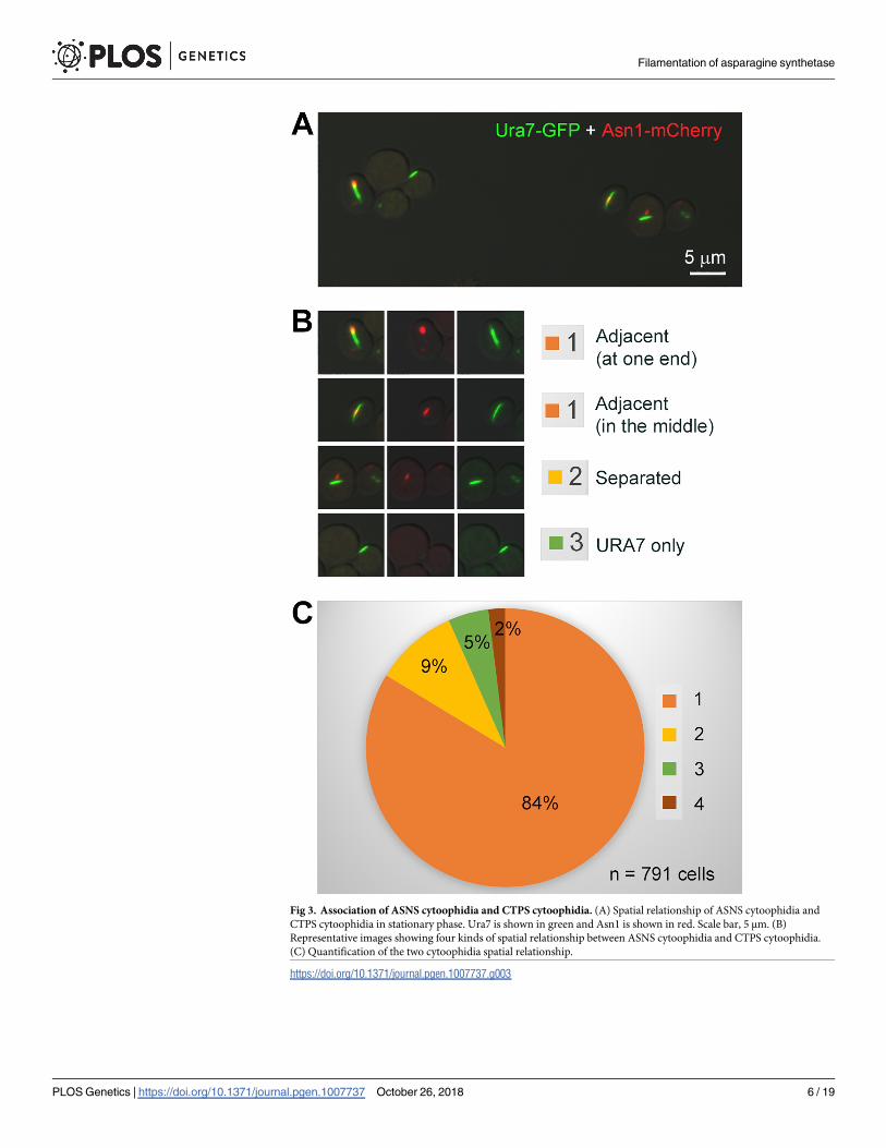
Fig 3. Association of ASNS cytoophidia and CTPS cytoophidia. (A) Spatial relationship of ASNS cytoophidia and
CTPS cytoophidia in stationary phase. Ura7 is shown in green and Asn1 is shown in red. Scale bar, 5 μm. (B)
Representative images showing four kinds of spatial relationship between ASNS cytoophidia and CTPS cytoophidia.
(C) Quantification of the two cytoophidia spatial relationship.
https://doi.org/10.1371/journal.pgen.1007737.g003
Filamentation of asparagine synthetase
PLOS Genetics | https://doi.org/10.1371/journal.pgen.1007737 October 26, 2018 6 / 19

and 92.01%, respectively (Fig 6B). In addition, we compared the ASNS protein level and
growth rate among these four strains. ASNS protein levels showed no significant difference
among all four strains (Fig 6C). To study whether the ASNS knockout has an effect on cell
growth, we did spot assay experiments to test the cell growth rate. Our results showed that nei-
ther ASN1 knockout nor ASN2 knockout alone affected cell growth rate (Fig 6D).
Fig 4. Magnified images of ASNS cytoophidia and CTPS cytoophidia in cytoplasm and nucleus. (A) ASNS cytoophidia and CTPS cytoophidia in nucleus
have similar association pattern as in cytoplasm. Scale bar, 2 μm. (B-D) High magnification of yeast cells show that the spatial relationship of ASNS
cytoophidia and CTPS cytoophidia in nucleus. Scale bar, 2 μm.
https://doi.org/10.1371/journal.pgen.1007737.g004
Filamentation of asparagine synthetase
PLOS Genetics | https://doi.org/10.1371/journal.pgen.1007737 October 26, 2018 7 / 19

Dimerization interface of Asn1
The biosynthesis of asparagine from aspartic acid occurs in three steps: activation of aspartate,
glutamine hydrolysis, and synthesis of a beta-aspartyl-AMP intermediate and its subsequent
reaction with ammonia [20]. A study of E. coli ASNS B (ASNB) showed that ASNS consists of
two distinct domains: an N-terminal domain that mediates the hydrolysis of glutamine to glu-
tamate and a C-terminal domain that is involved in the activation of aspartate by ATP [20].
The dimeric interface of E. coli ASNB is formed by both the N- and C-terminal region of each
monomer, which includes an α-helix-turn-β-sheet structure (A13 –S40) of the N-terminal and
an α-helix-turn-α-helix structure (V301 –M339) of the C-terminal domain, with both ligand-
binding sites being distant from the dimer interface. Several salt-bridge pairs enhance the
binding between two dimers, such as R17:D306, R25:D310, R28:D30, E48:R334 in ASNB. In
addition, R25 and R334 could potentially form a cation-π interaction with Y313 and W34
respectively.
The homology model of Asn1 shared the same dimer interface and conserved salt-bridge
interactions with the structure of E. coli ASNB, including K25:D330, R28:D330, E48:R354, as
in Asn1 (Fig 7). K25 and R354 also formed cation-π interactions with Y333 and W34, respec-
tively. We chose D330V, R354E and E48K for the following research (Fig 7B and 7C), aiming
to disrupt or decrease the dimerization thus filament formation.
Disrupting dimerization of Asn1 affects its filamentation
We utilized a site-directed mutagenesis strategy to target the specific site on Asn1 dimerization
interface. To avoid the complementary effect of Asn1 and Asn2, we knocked out Asn2 in
Fig 5. Asn1 cytoophidium colocalizes with Asn2 cytoophidium. Confocal images (A) and STED images (B) of yeast cells
displayingthe colocalization of Asn1 cytoophidium and Asn2 cytoophidium. Scale bar, 5 μm. ASN1 is tagged with GFP and ASN2
is tagged with mCherry. Double tagged yeast cells are collcected in stationary phase.
https://doi.org/10.1371/journal.pgen.1007737.g005
Filamentation of asparagine synthetase
PLOS Genetics | https://doi.org/10.1371/journal.pgen.1007737 October 26, 2018 8 / 19

order to study the Asn1 cytoophidium only. We first generated three GFP tagged Asn1 mutant
strains (D330V, R354E and E48K). Then we knocked out ASN2 in each of these three ASN1mutant strains. Eventually we obtained the following eight strains: ASN1WT-GFP ASN2 WT,
ASN1D330V-GFP ASN2 WT, ASN1R354E-GFP ASN2 WT, ASN1E48K-GFP ASN2 WT,
ASN1WT-GFP ASN2 KO, ASN1D330V-GFP ASN2 KO, ASN1R354E-GFP ASN2 KO and
ASN1E48K-GFP ASN2 KO.
Similar to ASN1WT, both ASN1E48K and ASN1R354E showed one large cytoplasmic cytoophi-
dium and one small nuclear cytoophidium in ASN2 knockout background (Fig 8A, 8B and 8C).
Fig 6. Asn2 cytoophidium is dependent on Asn1. (A) Confocal images of cytoophidium in A1G, A2G, A1G A2KO, A2G A1KO cells. Scale bar, 2 μm. (B)
Quantification of cytoophidium abundance in A1G, A2G, A1G A2KO, A2G A1KO cells showing that Asn1 is crucial for Asn2 cytoophidium formation. ���P<0.0001.
n.s. = not significant. Error bars show SEM. (C) Western blot analysis of ASNS protein level and quantification of the relative ASNS abundance in A1G, A2G, A1G
A2KO, A2G A1KO cells. n.s. = not significant. Error bars show SEM. (D) Spot assay of A1G, A2G, A1G A2KO and A2G A1KO cells. Eight microliters of four 10-fold
serial dilution of each yeast culture was spotted.
https://doi.org/10.1371/journal.pgen.1007737.g006
Filamentation of asparagine synthetase
PLOS Genetics | https://doi.org/10.1371/journal.pgen.1007737 October 26, 2018 9 / 19

A typical cytoplasmic cytoophidium is straight without any branches or slightly curved. How-
ever, the Asn1 cytoophidium in ASN1D330V-GFP ASN2 KO cells displayed irregular morphol-
ogy with a variety of shapes, many of which contain multiple branches (Fig 8D–8H). In ASN2
wild type background, ASN1WT, ASN1R354E and ASN1E48K present filament structure (Fig 8I–
8K). Whereas the morphology change of ASN1D330V cytoophidium in ASN1D330V-GFP ASN2
WT cells is less dramatic (Fig 8L).
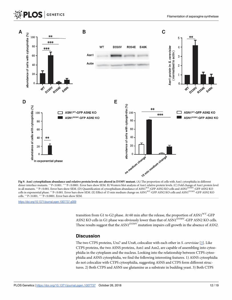
Furthermore, we checked the percentage of cells containing cytoophidia and the Asn1 pro-
tein level in the stationary phase. In the ASN1WT-GFP ASN2 KO strain, the percentage of cells
with cytoophidia reached 22.72%. When we cultured mutant strains in the same medium,
60.91% of ASN1D330V-GFP ASN2 KO cells showed obvious Asn1 cytoophidia in the stationary
phase, while only 3.65% and 8.81% of ASN1R354E-GFP ASN2 KO cells and ASN1E48K-GFP
ASN2 KO cells, respectively, contained Asn1 cytoophidia (Fig 9A). The protein level of Asn1
in ASN1D330V-GFP ASN2 KO cells was upregulated significantly comparing with the WT con-
trol and the other two mutants (R354E and E48K) (Fig 9B and 9C).
ASN1D330V promotes filamentation
In ASN1-GFP cells, we have already shown that there are no visible Asn1 cytoophidia in the
exponential phase (Fig 1B). To investigate whether the Asn1D330V had a similar behavior to
Asn1WT, we collected ASN1WT-GFP ASN2 KO cells and ASN1D330V-GFP ASN2 KO cells after
a 6-hour culture. In the ASN1WT-GFP ASN2 KO cells, the Asn1 cytoophidium was still unde-
tectable in the exponential phase. To our surprise, 21.58% of ASN1D330V-GFP ASN2 KO cells
showed visible Asn1 cytoophidia (Fig 9D). These results suggest that D330V promotes the
assembly of Asn1 cytoophidia.
To determine if the assembly of ASNS cytoophidia is sensitive to nutrient availability, we
cultured both ASN1WT-GFP ASN2 KO cells and ASN1D330V-GFP ASN2 KO cells both for 10
Fig 7. Homology model of Asn1 and its dimer. (A) Homology model of Asn1 monomer using E.coil ASNB as reference. N-terminal domain
containing the glutamine binding site and C-terminal domain containing the AMP binding site are shown in cyan and yellow, respectively. Glutamine
is shown in green and AMP in magenta. (B) Homology model of Asn1 dimer. N-N’ presents Asn1 dimer interface. N’ is shown as hydrophobicity
surface view. Blue represents the hydrophilic surface area and orange represents the hydrophobic surface area. (C) Asn1 dimer interface. Key residues
in protein are labelled and mutation sites may have a strong effect on dimerization of Asn1 are highlighted with pink (D330, R354, E48).
https://doi.org/10.1371/journal.pgen.1007737.g007
Filamentation of asparagine synthetase
PLOS Genetics | https://doi.org/10.1371/journal.pgen.1007737 October 26, 2018 10 / 19

days and then switched them to fresh media for 15 minutes. The percentage of cells containing
cytoophidia dropped from 23.76% to 2.63% in the ASN1WT-GFP ASN2 KO strain, while
decreasing from 82.71% to 18.21% in the ASN1D330V-GFP ASN2 KO strain (Fig 9E). In con-
clusion, both Asn1WT and Asn1D330V cytoophidia are sensitive to culture conditions.
The ASN1D330V cells grow slowly in the absence of ASN2We used spot assays to compare the growth rates of various strains. In a normal ASN2 back-
ground, the growth rate of ASN1D330V-GFP cells was similar to that of ASN1WT-GFP and of
the other two mutants, ASN1R354E-GFP and ASN1E48K-GFP (Fig 10A). Both ASN1R354E-GFP
and ASN1E48K-GFP cells grew normally in an ASN2 KO background. However, the growth of
ASN1D330V-GFP cells slowed down when ASN2 was knockout (Fig 10B).
Next, we used the alpha mating factor to arrest the ASN1WT-GFP ASN2 KO cells and
ASN1D330V-GFP ASN2 KO cells in G1 phase, and subsequently released those cells at the same
time. We collected those groups at 0 min, 30 min, 60 min and 90 min after alpha factor release
and analyzed the cell cycle by FACS (Fig 10C). At 30 min after release, there were two distinct
peaks formed by ASN1WT-GFP ASN2 KO cells and more than a half of cells successfully pro-
gressed from the G1 to G2 phase. However, ASN1D330V-GFP ASN2 KO cells delayed the
Fig 8. Cytoophidia in Asn1 mutants. (A-D) Morphology of Asn1WT, Asn1E48K, Asn1R354E and Asn1D330V cytoophidium in ASN2 knockout background. (E-H)
Varied morphology of Asn1D330V cytoophidium in ASN1D330V-GFP ASN2 KO cells. (I-L) Morphology of Asn1WT, Asn1E48K, Asn1R354E and Asn1D330V cytoophidium
in ASN2 wild type background. Scale bar, 2 μm.
https://doi.org/10.1371/journal.pgen.1007737.g008
Filamentation of asparagine synthetase
PLOS Genetics | https://doi.org/10.1371/journal.pgen.1007737 October 26, 2018 11 / 19
transition from G1 to G2 phase. At 60 min after the release, the proportion of ASN1WT-GFP
ASN2 KO cells in G1 phase was obviously lower than that of ASN1D330V-GFP ASN2 KO cells.
These results suggest that the ASN1D330V mutation impairs cell growth in the absence of ASN2.
Discussion
The two CTPS proteins, Ura7 and Ura8, colocalize with each other in S. cerevisiae [3]. Like
CTPS proteins, the two ASNS proteins, Asn1 and Asn2, are capable of assembling into cytoo-
phidia in the cytoplasm and the nucleus. Looking into the relationship between CTPS cytoo-
phidia and ASNS cytoophidia, we find the following interesting features. 1) ASNS cytoophidia
do not colocalize with CTPS cytoophidia, suggesting ASNS and CTPS form different struc-
tures. 2) Both CTPS and ASNS use glutamine as a substrate in budding yeast. 3) Both CTPS
Fig 9. Asn1 cytoophidium abundance and relative protein levels are altered in D330V mutant. (A) The proportion of cells with Asn1 cytoophidia in different
dimer interface mutants. ��P<0.001. ���P<0.0001. Error bars show SEM. B) Western blot analysis of Asn1 relative protein levels. (C) Fold change of Asn1 protein level
in all mutants. ��P<0.001. Error bars show SEM. (D) Quantification of cytoophidium abundance of ASN1WT-GFP ASN2 KO cells and ASN1D330V-GFP ASN2 KO
cells in exponential phase. ��P<0.001. Error bars show SEM. (E) Effect of 15 min medium change on ASN1WT-GFP ASN2 KO cells and ASN1D330V-GFP ASN2 KO
cells. ��P<0.001. ���P<0.0001. Error bars show SEM.
https://doi.org/10.1371/journal.pgen.1007737.g009
Filamentation of asparagine synthetase
PLOS Genetics | https://doi.org/10.1371/journal.pgen.1007737 October 26, 2018 12 / 19

and ASNS form cytoophidia not only in the cytoplasm, but also in the nucleus. 4) The number
of ASNS cytoophida, similar to that of CTPS cytoophidia, is one in the cytoplasm and one in
the nucleus in many cells. 5) In most budding yeast cells that we analyzed, the ASNS cytoophi-
dia localize adjacently to the CTPS cytoophidia. These two types of structures can touch with
each other head-to-head or side-by-side. 6) The close spatial relationship between ASNS and
CTPS does not limited to the cytoplasm. In the nucleus, we often observe that the ASNS fila-
ment sits next to the CTPS filament. 7) The length of the ASNS cytoplasmic cytoophidium is
shorter than that of the CTPS cytoplasmic cytoophidium. 8) The length of the ASNS nuclear
cytoophidium is shorter than that of the CTPS nuclear cytoophidium. 9) Nuclear cytoophidia
are much smaller than cytoplasmic cytoophidia, whether the composition is CTPS or ASNS.
10) Both CTPS and ASNS cytoophidia are very sensitive to nutrient changes and growth
phases. Further studies are required to understand the dynamic interaction and functional
connection, if any, between ASNS and CTPS cytoophidia. Previous studies have shown that
CTPS cytoophidia have interfilament interaction with inosine monophosphate dehydrogenase
Fig 10. ASN1D330V-GFP ASN2 KO cells show lower growth rate and slower cell cycle progression. (A) Spot assay of mutants before
ASN2 knockout (ASN1WT-GFP ASN2 WT, ASN1D330V-GFP ASN2 WT, ASN1R354E-GFP ASN2 WT and ASN1E48K-GFP ASN2 WT). (B)
Spot assay of mutants after ASN2 knockout (ASN1WT-GFP ASN2 KO, ASN1D330V-GFP ASN2 KO, ASN1R354E-GFP ASN2 KO and
ASN1E48K-GFP ASN2 KO). (C) FACS analysis of ASN1WT-GFP ASN2 KO cells and ASN1D330V-GFP ASN2 KO cells.
https://doi.org/10.1371/journal.pgen.1007737.g010
Filamentation of asparagine synthetase
PLOS Genetics | https://doi.org/10.1371/journal.pgen.1007737 October 26, 2018 13 / 19
(IMPDH) cytoophidia [21, 22], suggesting that the interaction between two cytoophidia may
be a phenomenon more general than we have appreciated.
Asn1 and Asn2 have identical distribution pattern, raising the possibility that Asn1 and
Asn2 have the same filament-forming capability. To our surprise, our data indicate that Asn1
and Asn2 behave distinctly in the absence of its partner. Removing Asn2 does not prevent
Asn1 to form cytoophidia in most cells. However, Asn2 alone can rarely form filaments in the
absence of Asn1. The discrepancy between Asn1 and Asn2 provides an interesting angle to
study the mechanism of the filamentation of metabolic enzymes in general. It would be inter-
ested to test if differences in transcription, translation, stability or half-life between Asn1 and
Asn2 influence their filamentation outcomes.
Three mutations in the dimerization interface of Asn1 were designed aiming to disrupt the
filament formation. We observe no obviously morphological changes of cytoophidia in
ASN1R354E-GFP ASN2 KO and ASN1E48K-GFP ASN2 KO cells, although these two mutant
strains show less cells containing cytoophidia than the wild-type strain. The third mutation,
ASN1D330V, results in branched cytoophidia, which are hardly seen in the wild-type strain. We
also find that ASN1D330V promotes cytoophidia formation, which reminds us the famous E6V
mutation in beta hemoglobin leads to the polymerization of hemoglobin tetramers. Here the
reason for Asn1 filamentation may be similar. A new protein-protein interface may be induced
by the D330V mutation, either through a stacking or bridging and stacking mechanism [23].
The Asn1 protein level in ASN1D330V-GFP ASN2 KO cells is higher than that in ASN1WT-GFP
ASN2 KO, ASN1R354E-GFP ASN2 KO and ASN1E48K-GFP ASN2 KO cells. However, the
growth rate of ASN1D330V-GFP ASN2 KO cells are much slower than the other three strains.
Currently we are unable to tell whether branched cytoophidia in ASN1D330V-GFP ASN2 KO
cells contribute to high protein level and/or slow grow rate. Ultrastructural analysis of cytoo-
phidia containing wild-type and mutant Asn1 proteins will help us gain insights into the
molecular mechanism of ASNS cytoophidium assembly.
CTPS cytoophidia was first reported by three studies in 2010 [1–3]. Eight years later, the
physiological role of CTPS cytoophidia remains elusive. There were some lines of evidence
support that forming filaments can sequester or promote the enzymatic activity of CTPS in
various organisms [24–28]. Does the ASNS cytoophidium have similar functions of the CTPS
cytoophidium? Since there are quite a few metabolic enzymes showing the filament-forming
capability, we speculate that cytoophidia have more general functions in the cell [21, 22]. The
close proximity between ASNS and CTPS cytoophidia raises the possibility that both enzymes
function coordinately in maintaining metabolic homeostasis in the cytoplasm and nucleus.
In summary, we use budding yeast as a model system to study the filamentation of two glu-
tamine-utilizing enzymes CTPS and ASNS. We have detected the close relationship between
CTPS and ASNS cytoophidia both in the cytoplasm and in the nucleus. Moreover, we have
demonstrated that two ASNS proteins exhibit differential filament-forming capability even
their distribution seems identical in the cytoplasm and nucleus. Finally, we identify a muta-
tion, ASN1D330V, which leads to branched cytoophidia, increased protein level and delayed
growth. Our results provide new opportunities to study how filamentation and compartmenta-
tion impact glutamine metabolism.
Materials and methods
Yeast strains and growth conditions
The yeast strains used in this study are derived from BY4741 [29]. All the S. cerevisiae strains
used are listed in S1 Table. Yeast cells were cultured at 30 ˚C on YPD rich media (1% Bacto
yeast extract, 2% peptone, 2% glucose) or SC medium with appropriate supplements
Filamentation of asparagine synthetase
PLOS Genetics | https://doi.org/10.1371/journal.pgen.1007737 October 26, 2018 14 / 19

depending on strains. In glucose deprivation assay, exponential phase cells (12 hours cultur-
ing) were centrifuged and added into previous media, YP media (1% yeast extract, 2% pep-
tone), distilled water, and 2% glucose solution. After 1 hour 32˚C culturing, cells were fixed
and pictures were taken by confocal microscope. In glucose recovery assay, 2% glucose was
added into culturing system. After 1 hour 32˚C culturing, cells were fixed and pictures were
taken by confocal microscope.
Genomic tagging
Fluorescent protein tagging in the genome was done by transforming yeast strains with a PCR
product that encoded a selective marker gene and 5’ and 3’ 40-bp flanking sequences homolo-
gous to the target gene. pFA6a-mCherry(S65T)-kanMX6 plasmid was updated by pFA6a-GFP
(S65T)-kanMX6 [30]. pFA6a-GFP(S65T)-His3MX6 and pFA6a-mCherry(S65T)-KanMX6
were utilized in GFP tagging and mCherry tagging, respectively. The yeast strain BY4741 and
plasmids were gifts from Jinqiu Zhou (SIBS, Shanghai, China). PCR products harboring the
target gene sequence were used to transform the BY4741 haploid wild type strain.
After transformation and homologous recombination, transformants were selected based
on the selective marker. Correct integration results in a C-terminal in-frame GFP fusion,
whose expression is driven by the endogenous promoter. Primers used in GFP or mCherry
tagging of ASN1 and ASN2 are listed in S2 Table. Sequences in upper case indicate homology
to the genome of S. cerevisiae and those in lower case indicate homology to the PCR cassette
employed.
Gene disruption
The ASN1 and ASN2 disruption cassettes were constructed respectively in pRS306 and
pRS303 plasmids. For each gene disruption, the 5’ untranslated region and 3’ region were sub-
cloned into the corresponding vector, cut using specific restriction enzymes, and then trans-
ferred into yeast to replace the given gene with a selective marker by homologous recombina-
tion [31]. The disruption cassettes were both linearized with EcoR I. Gene disruption or
genomic tagging was confirmed by PCR. Primers used in ASN1 or ASN2 deletion are listed in
S3 Table.
Confocal microscopy and stimulated emission depletion microscopy
Yeast cells were spun down for 1 min at 4000 rpm/min. 100 μl of 4% paraformaldehyde were
added to each tube to fix the cells for 10 min at room temperature, and then the cells were
washed once with PBS.
A few microliters of cell suspension were mixed with agarose gel (1.2% low melting temper-
ature agarose and 100 μM N-propyl gallate in YPD) to avoid movement of the cells during
imaging. Images were acquired under 63x objectives on a Zeiss LSM 710 inverted fluorescence
confocal microscope. For the co-localization experiment, super-resolution stimulated emission
depletion (STED) microscopy was used. The abundance of filaments in cells was quantified by
capturing images in at least four different areas containing a minimum of 100 cells each.
Homology modeling of S. cerevisiae Asn1
The homology model of Asn1 of S. cerevisiae and its dimer were built based on the structure of
E. coli ASNB [20] (PDB: 1CT9), by using the MODELLER software [32]. The sequence identity
between Asn1 and ASNB is 47%. Residues on the dimer interfaces are highly conserved.
Filamentation of asparagine synthetase
PLOS Genetics | https://doi.org/10.1371/journal.pgen.1007737 October 26, 2018 15 / 19

Mutagenesis
This method is applied to alter single nucleotides in DNA that has been previously cloned in a
plasmid. The plasmid is then transferred back into yeast cells to generate a strain that carries
the mutated version of the gene of interest on the plasmid but lacks the corresponding wild
type locus in the genome in order to assess the function of the mutant allele. Insert fragments
(including about 1 kb upstream of the promoter element and 0.5 kb downstream of the 30-
untranslated region of GFP-tagged target gene) were cloned into pRS315. Subsequently, this
vector was transformed into an ASN1 deletion strain. This strain was regarded as the control
strain. The vector was also used as the template for point mutant vectors (E48K, D330V and
R354E). DpnI was used to eliminate the template and then point mutant vectors were trans-
formed into the ASN1 deletion strain. Oligonucleotide primers used for site-directed mutagen-
esis are listed in S4 Table.
Protein abundance of Asn1 or mutant Asn1 (E48K, D330V and R354E)
We used Western blots to analyze the protein level of ASNS. Yeast cells were collected at differ-
ent time points and run on an 8% SDS-PAGE gel before transfer to a PVDF membrane (Bio-
Rad). Following incubation in blocking buffer (TBST containing 5% nonfat dry milk (Bio-
Rad)) for 1 h, the membrane was hybridized in blocking buffer containing primary antibody
overnight at 4 ˚C. The membrane was then washed and incubated with a horseradish peroxi-
dase-conjugated secondary antibody for 1 h at room temperature. Detailed information about
the antibodies used, with concentration and source, can be found in S5 Table. Enhanced ECL
was used for signal detection. The image signal was collected by an Amersham Imager 600
(GE Healthcare Life Science). Data were processed and analyzed by Image J software. All
quantifications were done by normalizing protein levels to actin.
Spot assay of yeast cells
Yeast cells were grown overnight and diluted roughly to an optical density (O.D.) of 1.0. Four
or five ten-fold dilutions were spotted onto YPD agar plates. A multichannel pipette was used
to transfer 10 μl cells to 90 μl culture medium. After mixing well, 8 μl of each dilution were
spotted onto the plates. Plates were incubated at 30 ˚C, examined regularly, and photographed
after 48 hours incubation.
Supporting information
S1 Table. S. cerevisiae strains used in this study. The strain name, number and genotype of
strains used in our study are listed in this table. All strains are haploid and derive from the
wild-type haploid (BY4741). The introduced plasmid carries the wild type, mutational version
of Asn1 and the corresponding wild-type locus in the genome is disrupted.
(TIF)
S2 Table. Primers used for genomic tagging. Genomic tagging with fluorescent protein is
based on the homologous recombination. Sequence homology to pFA6a-GFP(S65T)-
His3MX6 for GFP tagging are indicated small letter and in bold. Sequence homology to
pFA6a-mCherry(S65T)-KanMX6 for mCherry tagging are indicated small letter and under-
lined.
(TIF)
S3 Table. Primers used for gene disruption. Gene disruption is based on the fact that linear
DNA fragments carrying a selectable marker gene with homology regions to an interest gene.
Filamentation of asparagine synthetase
PLOS Genetics | https://doi.org/10.1371/journal.pgen.1007737 October 26, 2018 16 / 19

5’ and 3’ untranslated region of ASN1 are amplify by ASN1 UHA-F, ASN1 UHA-R and ASN1
DHA-F, ASN1 DHA-R primers respectively. ASN1 KO testing F and ASN1 KO testing R
primers are used to confirm the genotype. ASN2 disruption strain is obtained by similar
approach.
(TIF)
S4 Table. Oligonucleotide primers used for site-directed mutagenesis. Upstream of the pro-
moter element and coding sequence of ASN1-GFP with selective marker are amplify by A1-UF
and A1-UR primers. Downstream of the 3’—untranslated region of ASN1 is amplified by
A1-DF and A1-DR primers. For point mutant vectors (E48K, D330V, R354E), corresponding
primers are used. Point mutations are indicated in bold and underlined.
(TIF)
S5 Table. Antibodies used with concentration and source. We analyze the protein level of
ASNS by detecting GFP. In this table, we provide the information of antibody used with con-
centration and source.
(TIF)
Acknowledgments
We would like to thank the Liu group for discussion and Minghong He and Jiacheng Liu and
other members from Professor Jinqiu Zhou’s lab at Shanghai Institute of Biochemistry and
Cell Biology (SIBCB), Chinese Academy of Sciences (CAS) for their help on yeast genetics. We
are grateful for the assistance given by the National Center of Protein Science Shanghai
(NCPSS).
Author Contributions
Conceptualization: Shanshan Zhang, Ji-Long Liu.
Formal analysis: Shanshan Zhang, Kang Ding, Qing-Ji Shen, Suwen Zhao, Ji-Long Liu.
Funding acquisition: Ji-Long Liu.
Investigation: Shanshan Zhang, Qing-Ji Shen, Ji-Long Liu.
Methodology: Shanshan Zhang, Kang Ding, Qing-Ji Shen.
Project administration: Ji-Long Liu.
Resources: Ji-Long Liu.
Supervision: Suwen Zhao, Ji-Long Liu.
Writing – original draft: Shanshan Zhang, Kang Ding, Qing-Ji Shen, Ji-Long Liu.
Writing – review & editing: Suwen Zhao, Ji-Long Liu.
References
1. Ingerson-Mahar M, Briegel A, Werner JN, Jensen GJ, Gitai Z. The metabolic enzyme CTP synthase
forms cytoskeletal filaments. Nat Cell Biol. 2010; 12(8):739–46. Epub 2010/07/20. https://doi.org/10.
1038/ncb2087 PMID: 20639870.
2. Liu J-L. Intracellular compartmentation of CTP synthase in Drosophila. Journal of Genetics and Geno-
mics. 2010; 37(5):281–96. https://doi.org/10.1016/S1673-8527(09)60046-1 PMID: 20513629
3. Noree C, Sato BK, Broyer RM, Wilhelm JE. Identification of novel filament-forming proteins in Saccharo-
myces cerevisiae and Drosophila melanogaster. J Cell Biol. 2010; 190(4):541–51. Epub 2010/08/18.
https://doi.org/10.1083/jcb.201003001 PMID: 20713603.
Filamentation of asparagine synthetase
PLOS Genetics | https://doi.org/10.1371/journal.pgen.1007737 October 26, 2018 17 / 19

4. Carcamo WC, Satoh M, Kasahara H, Terada N, Hamazaki T, Chan JY, et al. Induction of cytoplasmic
rods and rings structures by inhibition of the CTP and GTP synthetic pathway in mammalian cells. PLoS
ONE. 2011; 6(12):e29690. Epub 2012/01/06. https://doi.org/10.1371/journal.pone.0029690 PMID:
22220215.
5. Chen K, Zhang J, Tastan OY, Deussen ZA, Siswick MY, Liu JL. Glutamine analogs promote cytoophi-
dium assembly in human and Drosophila cells. J Genet Genomics. 2011; 38(9):391–402. Epub 2011/
09/21. https://doi.org/10.1016/j.jgg.2011.08.004 PMID: 21930098.
6. Chang CC, Jeng YM, Peng M, Keppeke GD, Sung LY, Liu JL. CTP synthase forms the cytoophidium in
human hepatocellular carcinoma. Exp Cell Res. 2017; 361(2):292–9. https://doi.org/10.1016/j.yexcr.
2017.10.030 PMID: 29097181.
7. Daumann M, Hickl D, Zimmer D, DeTar RA, Kunz HH, Mohlmann T. Characterization of filament-form-
ing CTP synthases from Arabidopsis thaliana. The Plant journal: for cell and molecular biology. 2018.
https://doi.org/10.1111/tpj.14032 PMID: 30030857.
8. Gou KM, Chang CC, Shen QJ, Sung LY, Liu JL. CTP synthase forms cytoophidia in the cytoplasm and
nucleus. Exp Cell Res. 2014; 323(1):242–53. Epub 2014/02/08. https://doi.org/10.1016/j.yexcr.2014.
01.029 PMID: 24503052.
9. Shen QJ, Kassim H, Huang Y, Li H, Zhang J, Li G, et al. Filamentation of Metabolic Enzymes in Saccha-
romyces cerevisiae. J Genet Genomics. 2016; 43(6):393–404. https://doi.org/10.1016/j.jgg.2016.03.
008 PMID: 27312010.
10. Zhang J, Hulme L, Liu JL. Asymmetric inheritance of cytoophidia in Schizosaccharomyces pombe. Biol
Open. 2014; 3(11):1092–7. Epub 2014/11/02. https://doi.org/10.1242/bio.20149613 PMID: 25361577.
11. Lin WC, Chakraborty A, Huang SC, Wang PY, Hsieh YJ, Chien KY, et al. Histidine-Dependent Protein
Methylation Is Required for Compartmentalization of CTP Synthase. Cell Rep. 2018; 24(10):2733–45
e7. https://doi.org/10.1016/j.celrep.2018.08.007 PMID: 30184506.
12. Huang H, Vandekeere S, Kalucka J, Bierhansl L, Zecchin A, Bruning U, et al. Role of glutamine and
interlinked asparagine metabolism in vessel formation. The EMBO journal. 2017; 36(16):2334–52.
https://doi.org/10.15252/embj.201695518 PMID: 28659375.
13. Van-Dinh D, Michèle V, Monique BF, Bertrand DF. Cloning of the ASN1 and ASN2 genes encoding
asparagine synthetases in Saccharomyces cerevisiae: differential regulation by the CCAAT-box-bind-
ing factor. Molecular Microbiology. 1996; 22(4):681–92. https://doi.org/10.1046/j.1365-2958.1996.d01-
1715.x PMID: 8951815
14. Nadkarni AK, McDonough VM, Yang WL, Stukey JE, Ozier-Kalogeropoulos O, Carman GM. Differential
biochemical regulation of the URA7- and URA8-encoded CTP synthetases from Saccharomyces cere-
visiae. J Biol Chem. 1995; 270(42):24982–8. PMID: 7559626.
15. Fernando R, Jean-Marie W. Two Asparagine Synthetases in Saccharomyces cerevisiae. European
Journal of Biochemistry. 1980; 108(2):373–7. https://doi.org/10.1111/j.1432-1033.1980.tb04732.x
PMID: 6105958
16. Greco A, Gong SS, Ittmann M, Basilico C. Organization and expression of the cell cycle gene, ts11, that
encodes asparagine synthetase. Mol Cell Biol. 1989; 9(6):2350–9. PMID: 2569668.
17. Li H, Zhou F, Du W, Dou J, Xu Y, Gao W, et al. Knockdown of asparagine synthetase by RNAi sup-
presses cell growth in human melanoma cells and epidermoid carcinoma cells. Biotechnol Appl Bio-
chem. 2016; 63(3):328–33. https://doi.org/10.1002/bab.1383 PMID: 25858017.
18. Werner-Washburne M, Braun EL, Crawford ME, Peck VM. Stationary phase in Saccharomyces cerevi-
siae. Mol Microbiol. 1996; 19(6):1159–66. PMID: 8730858.
19. Herman PK. Stationary phase in yeast. Current opinion in microbiology. 2002; 5(6):602–7. PMID:
12457705.
20. Larsen TM, Boehlein SK, Schuster SM, Richards NG, Thoden JB, Holden HM, et al. Three-dimensional
structure of Escherichia coli asparagine synthetase B: a short journey from substrate to product. Bio-
chemistry. 1999; 38(49):16146–57. PMID: 10587437.
21. Liu JL. The Cytoophidium and Its Kind: Filamentation and Compartmentation of Metabolic Enzymes.
Annu Rev Cell Dev Biol. 2016; 32:349–72. https://doi.org/10.1146/annurev-cellbio-111315-124907
PMID: 27362644.
22. Chang CC, Keppeke GD, Sung LY, Liu JL. Interfilament interaction between IMPDH and CTPS cytoo-
phidia. The FEBS journal. 2018. https://doi.org/10.1111/febs.14624 PMID: 30085408.
23. McPartland L, Heller DM, Eisenberg DS, Hochschild A, Sawaya MR. Atomic insights into the genesis of
cellular filaments by globular proteins. Nat Struct Mol Biol. 2018; 25(8):705–14. https://doi.org/10.1038/
s41594-018-0096-7 PMID: 30076408.
Filamentation of asparagine synthetase
PLOS Genetics | https://doi.org/10.1371/journal.pgen.1007737 October 26, 2018 18 / 19
24. Strochlic TI, Stavrides KP, Thomas SV, Nicolas E, O’Reilly AM, Peterson JR. Ack kinase regulates CTP
synthase filaments during Drosophila oogenesis. EMBO Rep. 2014; 15(11):1184–91. Epub 2014/09/
17. https://doi.org/10.15252/embr.201438688 PMID: 25223282.
25. Petrovska I, Nuske E, Munder MC, Kulasegaran G, Malinovska L, Kroschwald S, et al. Filament forma-
tion by metabolic enzymes is a specific adaptation to an advanced state of cellular starvation. Elife.
2014. Epub 2014/04/29. https://doi.org/10.7554/eLife.02409 PMID: 24771766.
26. Noree C, Monfort E, Shiau AK, Wilhelm JE. Common regulatory control of CTP synthase enzyme activ-
ity and filament formation. Mol Biol Cell. 2014; 25(15):2282–90. Epub 2014/06/13. https://doi.org/10.
1091/mbc.E14-04-0912 PMID: 24920825.
27. Barry RM, Bitbol AF, Lorestani A, Charles EJ, Habrian CH, Hansen JM, et al. Large-scale filament for-
mation inhibits the activity of CTP synthetase. Elife. 2014; 3:e03638. Epub 2014/07/18. https://doi.org/
10.7554/eLife.03638 PMID: 25030911.
28. Aughey GN, Grice SJ, Shen QJ, Xu Y, Chang CC, Azzam G, et al. Nucleotide synthesis is regulated by
cytoophidium formation during neurodevelopment and adaptive metabolism. Biol Open. 2014; 3(11):
1045–56. Epub 2014/10/19. https://doi.org/10.1242/bio.201410165 PMID: 25326513.
29. Winzeler EA, Shoemaker DD, Astromoff A, Liang H, Anderson K, Andre B, et al. Functional characteri-
zation of the S. cerevisiae genome by gene deletion and parallel analysis. Science. 1999; 285(5429):
901–6. PMID: 10436161.
30. Longtine MS, McKenzie A 3rd, Demarini DJ, Shah NG, Wach A, Brachat A, et al. Additional modules for
versatile and economical PCR-based gene deletion and modification in Saccharomyces cerevisiae.
Yeast. 1998; 14(10):953–61. https://doi.org/10.1002/(SICI)1097-0061(199807)14:10<953::AID-
YEA293>3.0.CO;2-U PMID: 9717241.
31. Rothstein R. Targeting, disruption, replacement, and allele rescue: integrative DNA transformation in
yeast. Methods in enzymology. 1991; 194:281–301. PMID: 2005793.
32. Sali A, Blundell TL. Comparative protein modelling by satisfaction of spatial restraints. Journal of molec-
ular biology. 1993; 234(3):779–815. https://doi.org/10.1006/jmbi.1993.1626 PMID: 8254673.
Filamentation of asparagine synthetase
PLOS Genetics | https://doi.org/10.1371/journal.pgen.1007737 October 26, 2018 19 / 19