file name (title): document number (id): date: .‘@glw
TRANSCRIPT

MARSHALLISLANDSl?ILETRACKINGDOCUMENT
Record Number: /ss
File Name (TITLE):
Document Number (ID):
DATE: .‘@gLw +
Previous Location (FROM): /! /%
AUTHOR:
Addditional Information:
OrMIbox: /o
CyMIbox: 0

. -
, .’
y:
OPERAlION CASTLE - FINAL REPORT PROJCCT 4 1 ,
Study of Response of Human Beings
Accidentally Exposed to
Significant Fallout Radiation
by
E. P. Cronkite, Commander, MC. USN V. P. Bond, M.D., Ph.D. L. E. Browning, Lt. Col., MC, USA W. H. Chapman, Lt., MSC, USN
S. H. Cohn, Ph.D. Ji. A. Conard, Commander, MC. USN C. L. Dunham, M.D.
, :i R. S. Farr, Lt., MC, USN W. S. Hall, Commander, MC, USN Ii. Sharp, Lt. (jg), USN N. R. Shulman, Lt., MC, USN
Naval Mcdicttl Rosearch II Bc!thcnda, Mllry;:,n,,
rtnd
U. S. Naval Radiological Ddcnsc Lub San I:rancisco, California

ABSTRACT
FOIIOWIII~ IIIP d(>tonation of Shol 1 1111 Bikini Atoll OII 1 Mi1r1*h l!)!i4, 28 Amt*ri1*:111s i1nd 2J!l
Marshallc~sc~ WC~I’C c~xl)osc~t lo f;~llouI. OW huntlrt~d fifty -HOVIVI of 111~ M;~rsh;rll~~~ w(*~-t- OII lJ(i~+lk
1\1011. 64 Wl’I’(’ 011 lLm~c’l;lp Aloll, iLId IH W(*IY’ OII thtb nt*Ight111rinK ;~loll of AIIIIIKII~;I~~. Thcs 28
Aij1~~r11~:111s WI~I‘I’ on IConl:c~r1k Atoll. Tht* ~)rt*sc~nc’t~ of s1Knific*;ml l’al1~1uI WI lhc~st* i;Iolls was I~rst
~1~~11~f~1111111~11 I1v ;I ~‘IV~~IIY~III~ dosi11i1~lt~1-, 101~~11~~11 011 Ron~t~rlk, WhC~ll IhIS dtYlc*c~ wt*nt (dr sc*illt~ at
loo IIII’ h1. sl101.lly dh. ths 1lt~I111ral ion. Kmc~rKc~ncy surveys tIt~It~ctc~d radii11 it,11 OII t h(t inhabilcd
~llrllls , x1111 cSv;lc.u;lticjll of 111h;tljil;1nts to the* Navxl StaI1on at Kwi1jaltbi11 was l1ro111l1tly carried out.
‘1’111, tlosc~ (,I r;rcl~;~lic~J IO which lhtl individuals were c!xposcti was ~nl~ulatcd fro111 thtl intensities
IIUII~~ OII tlt(b ISI;III~IS iI11d lhtb tl1~*;1y cbxponc11t of the fallout matrrial. The individuals on Rongelap rc~*c~~vr~l alJ~)J‘oxit~i;ltclly I75 r, those on Ailinginae received approximately 69 r, and the Amthri- cans 01) liongt~rtk rtlcSthivc!d an average calculated dose of 76 r. The Marshallese on Utirik re- c*c+vc!d approxin,ately I4 r. The fallout on Rongelap, Ailinginae, and to a lesser extent on Ronge- rik was distinctly visible. No fallout was observed on Utirik. A significant number of indi- viduals on Rongelap suffered from mild nausea and one or two individuals vomited on the day of the r?xposure. With the exception of nausea in one Ailinginae individual.;there were no other definite gastrointestinal symptoms in the other Marshallese or the Americans. The Mar- shallese on Rongelap and Ailinginae, and the Americans experienced to a varying degree burn-
~ng of thr eyes and itching of the skin from 1 to 3 days. Later, signs of radiation injury included definite c>pilation in the Hongelap and Ailinginae groups, and the development of spotty, super- fit:ial, hyperpigmented skin lesions that desquamated from the center of the lesions outwards. In some cases the skin damage was sufficient to result in raw, weeping lesions. There was no full Ihickncbss necrosis of the skin. The Americans developed only minor skin lesions without
ul(~c~ratic~rJ. Thclrca w(*rtt IIO skin Icsionu in 111~ Utlrik natives. All lesions hc~al~~d ratjidly with IIO lurlhc~r I)rl~;Jktlown of I tw skin noted during I Iw period of obsc*rval ion. Mlc*rotic.ulJic. (‘XUIII -
t1;11 IOII (II ~IIC~~JS~~*S of Iha, l(hsious showc4 ~*II;LIIK~*H uSunlly ;tsso(*i:1lc~d wilh ratii;tl 1011 i11Jury. Fully
~‘loll~c~l ~~~tl~v~tiuals :1111i IIIOSC~ rc*mainIng inHido of buildings or huts w(br(* prott~(‘lc*d IO viLryill(: ~I(~gr~~(!h front tli~vc~lo~~rric~nt of I~~sions. H~!n1;1I~~l11~1~~ rh;ugc++ wt.r11 dthf1111lt> in II1tb Iio11~t~lap,
Ailirlginilc~, :III~ Ih(b A111c~ri1*;11t Kr’c1tips. Lyinphopc~nia appt!arc*d pruml)t ly and was persistent for :I prolo1~cd l1c~r1od of lin1c. Neutropenia occurred in all of the individuals with lnitiiil minimum
valut~s occurring around th1, 1 Ith day followed by an increase in the counts i111d a secondary
minimum around the 4Olh to 45th day. The most consistent hematologic change was the de- I)ression in the platelet ceounts. Platelets were below normal when first counted on the 10th day of post-exposure and progressively decreased attaining a minimum between the 25th and 30th day. Although recovery commenced following this minimum, the platelet counts had not returned to normal by the completion of the initial study on the 76th post-exposure day. The incidence of various respiratory and cutaneous infections was identical in all exposure groups and bore no relationship to the hematologic changes.
Urinary excretion of radioisotopes was studied. Small amounts of radioactive material were found. Estimates of total body burden indicate that there is no long term hazard and that ingestion and inhalation of isotopes did not contribute significantly to the initial radiation exposure.

--.^c__I_ -_.. - .----.-~-.. _ __. _ ._ _.__._
FOREWORD
(a) An ovt!r-itI1 dcscriptiou of each dctwation, includinp yield, height of burst, ground zero
location, linlc! 0r detonation, mnbient attnospheric conditions at detonation t itnc>, c~tc.. for the
cqwration.
0,) I)is~ussicm 0r all lm~jec:t results.
(c) A sunln)ary of c:;~c*h projtbct, including objectives and rrsults.
((1) A ~ort~l)lc~t(~ Ilstin(: ol all reports cowrinK the Mllitnry Efftwts ?‘ost Program.
I)(*ta~Ic~l rtqborts OII tlos~n~c*try and intc~rnal radioact ivr ~‘o~~ti~~~~i~~;ltI~~~~ will ~JV put~l~shed as
~ulq~l~~in~~rils IO this rcq>ort.
Muc.11 of thcb nlc!dic;rI t~~rnlinology in this rcq~ort is explainctd in l)i~rc~nthc~sc~s for the bewflt
of the Inynlm. .
.-

. . . . ..1 - .
PREFACE
()114*1x1 101t (‘AS’I’I ,I5 (lid 1101 ill(*iudt* ;I I~IOIII~V~I~~I~ pro~r:ltir. ‘1’11~~ Ihhrl ic*il~biils III i~ro]t*~t 4. 1
W(‘I’(’ tll.;lWll 11~0111 Vill’lOllS I;tl~or;lloric~s ;Illd Wc’l’c’ (Olilily unl)r~~)~;lrt*ii 101’ ;I IlOl~l ~lrogI’;1IIl wtlcw
I\I(~ JOIIII 1K)i) AM’ Mtvl~c*al I~c~sc~;IJ~~~II TI~;IIII UTIS org;~~~i~.cxi :rllcsr liitb o))t~r;ll LOII II;I~ h~gtui.
i)r. .Iohn C. ilu~hc~r. i)iroclor, i)ivtsion oI Hiology ;rnd Mt*dlt*int>, AKC. ;wd Lt Co1 I,. E. I~IYJWI~IIIR, MC, USA. Surct‘on of the Armed For(.os Sl~cr~al Wttapons Projcc’t. st~lt~ctc~d the proj- ot:t officclr and rcquosted that the selection of technical and professional p,crsonnel bc com- ntt!nc:c!d Immodiatoly. Rear Admiral Clarence Brown, Deputy Surgeon General, Medical De- l~artn~ent. USN, gave immediate and complete support of all naval medical research activities and assigned responsibility to CAPT Van Tipton. MC, USN, and CDR Harry Etter, MC, USN, of the Atomic Defense Division, Bureau of Medicine and Surgery, USN, for the implementation of the project.
Since little detailed information was available about dose and initial symptomatology, the research team was organized to include the following talents for the constitution of the emer-
gency mtldical tcam: internal medicine, hematology, radiation technology and radiobiology. In t I)(, scllcc.tion of pc~rsonnt~l thcx emphasis was placed on past experience in biomedical research III (he, ficbld with alorni(* weapons. In addition, provisions were made for a swond txhelon of sl)(ac’i;rl izc4 Ix~rsonnc~l in c.asc: they were needed. Accordingly, a prevrntivtb mcldicint* unit of 11~~ <:orlltrlarltlt~r in-Ch!c>f, lhc:ific Fleet, was alerted for possiblr bn~tt~riologit~ studic*s; blood I);Jrlk ~J(~I.SOIIII(~~, ;III~ ;~tltl~l IOIGII c.iinicians and nurses wt’rc notific*d in cast! c*ondil ions justifitbd Il~*ir st’rv~t’t*s III IIIc- Kw;ijalc~1ii urea. Rc?;lr Admiral Rarlholomt~w HOK;II~. MC. USN, I’ac’ific. l*‘lt*(~t Mtbd~c.;~l Ofl I(:tbr, I~IIII(*(II;II(~J~ mad13 ally nccdcd mc~dic*al fac~ilil1t~s of thcb l%c*iCic Flcc>t ~rv;ll1;l1Jl~~.
i’c!r:~onitc~l wt’rtb 0bla1ncd within the Continental limits of the U. S. for 111th rt~sr;rr(*h team ilh rOllows:
Naval Medical Hlbscbarch Institute (NMRI) 4 rncdical officers (Is. P. Cronkite, project officer, R. A. Conard, N. R. Shulman. and
R. S. Farr) 2 medical service corps officers (W. H. Chapman and R. Sharp) 6 cnli.sted men (C. R. Sipe, P. K. Schork, C. P. A. Strome, W. C. Clutltrr, R. E. Hansell,
J. S. Hamby) U. S. Naval Radiological Defense Laboratory (NRDL)
1 civilian M.D. (V. P. Bond) 1 medicai service corps officer (L. J. Smith) 4 enlisted men (W. H. Gibbs, J. C. Hcndrie, W. S. Argonza, J. Flanagan)
Divi:;ion of Biology and Medicine, AEC 2 civilian M.D.‘s (C. L. Dunham and G. V. LeRoy)
Armed Forces Special Weapons Project (AFSWP) 1 Army Mtbdical Officer (L. E. Browning)
Preliminary studit,:; had bc!c!r~ made by CDH W. S. Hall, MC, USN, Station Mt,tlic.:tl Offic*c*r and his st:~Tf, :mti tl(!(~orllarninntioll of thr iptlivitlual~ was wall untlcrw;ry whc-11 i’rojt~c’t 4. I pc~rson-

1
‘l’llt~ pr0jtsc.l 0111(.1’1. ~‘0~1llll~~utis ;III of IlIt’ Il~Olt~SSl~~llill ;lllti It*t’ll~llt~;1I ll1c~~IlLlt~!s 0I IlIt- Krc’rlI)
for IIII~IV t~xt~~~ll~~nt it~olivalioir. iirili;ilivt~. ;III~I ~tbIu~rl;~~y lt1t1~ I~~)LII’s 01 ~5x1 I’iI work 111~11 Wt’I’t’ (‘5
Yefltiitl for lhe ilt~t~trl~l~Jli~ll~ll~~~ of the clinic-al anti reutrnrclr thjertivt-s. ;llltI for lilt’ l’ill’itl t’OI
Icc11or1 of thr prcliiniu;~ry d;rla in Ilit, fithId. It IS t~nlpliasizr~d Ilrat lht> work was iI c.otbIJt~t’;itivt~
cncit!avor in which all wt*rc Illutuitlly dq)cndcnl upon tliIt!h olhrsr. Thtl willing rfforls of i~ll (‘on- cernc+d (*onslit uted it rt~lnarkahlc example of team- work and sacrifit*c> of pctrsonal ;1mb11 IU~:S ;IIK~
deslrtbs for Ihe good of Ihe project at large. The authors wish to express their gratitude and indebtedness to Dr. John C. Bughtlr, CAPT
Van Tiljlon, arid CDH Harry Etter; CAPT W. E. Kellum, MC, USN, and CAPT T. L. Willmon, Cotnrnanding alld Extkc.utive Officers, respectively. NMRI; CAPT R. A. Hinners, USN, Dlrctclor,
NRDI.. and CAPT A. 1~. t3chnke. MC, USN, Associate Director, NRDL; all of whoin gavu un- limilt~ti SUiJfJor! and rtduced administrativr procrdures to a bare minimum, thus making it possible for tht! unit to be asscmblrd and underway in a matter of hours.
On arrival at Kwajalein, RADM R. S. Clarke. USN, Commanding Officer, U. S. NiIVXl Sta-
t~on. Kwajaltbin, supI)orltld Projocl 4.1 wilh all lhca facilities at his dial)osal. As ;I rc!sult, ;I labor;rlory and 1.11n1c was t!slal~I ishtxti and opemt ing within 24 hours after arrival of tlltx projtM
l’c’rscmnc~l.
I)rojtx*l pt~rsonut~l also wish lo acknowledge the* outstanding contribulions OI Cal C. S. Maup~n, MC, USA. F’icbld Ct)nluliLrld, AFSWP, CAPT H. H, Haight, MC:, USN, Division of Milt- tat-y Alq)lic*ation, AEC; Cal K. Hou~hltm, MC, USAF, Special Weapons Center; CAPT Donald Dt~mt~nI, MC, USN, CINCPAC Flrt~l; Drs. T. L. ‘Shipman, T. White, and P. Harris of Los Alamos
Scicbntrlic Laboratory; Dr. Gordon Dunning, Division of Biology and Medicine. AEC, and Dr. G. V. L,c>Roy. University of Chicago. During all phases of the early care of the cxl)osed indi- viduals, the foregoing participated as much as their other primary duties would permit. In additicln the authors wish to thank them for the extensive and complete data which they collected in thcb atolls or their home laboratories and kindly furnished to the project personnel.
The continuous help and cooperation of Trust Territory representatives and tht*ir aid in obtaining necessary control data on native Marshallese at Majuro is hereby acknowlt~dgcd. The authors arc partlc*ularly indebted to Mr. John Tobin. His help as an intcrprt’tc’r and 1~1s extrnslvt~ knowlcdgo of Ihe Marshallesc language and habits were tnvaluablt>. I~ic~ulcntant .1. S. Thompson. MC, USN, furnisht>d his records on the) exposed indtviduals dt~~oiit;tniiii;ll(~tI by lh(> radial IOI~ Kroul) of Ilit, VP-29 squadron stat ioncd aI Kwajalein.
‘I’h(~ authors wish c~spct*i;llly lo cbxpr’css their admiration for the exccllcnl job tionc, by 111t~ n~txiit~al pt~rsonnt~l of 11~~ U. S. Naval Dinpt~nuary, Kwnjalcin, in coii;pIt~ling lht* t~xlt~ns~vt~ I:II)o. ratory c~x;+rllitl;ll IONS IllitI w(trt! rt~tluirc*ti to obtain a l)rompt initial tXvaluat ion of lht, hcva’rit y ()I lht! r:ttlial ioii injury.
Tti(s aulh~rs ;kr(’ dt~t~ply gratt!ful 10 Dr. David A. Wood of the Univt!rsiIy of California Ho+ pit;1l, San Franc.isc*o, ;IIKI Dr. Edward L. Alpen and Miss Pat Roan of NRDL, for I~IVillu~~bll~ aid in carrying out 111t~ I~islol~att~olo~ic’al cvaluat ion of skin lesions. The extensivtl conI r 1bu1 ions 0r Mr. H. H. Hcclller in the statistical analyses of the data, and of Mr. C. A. Sondhaus uf (ht.
NRDL In dosage t~alcul:ttitrns are gratefu:.ly acknowledged.
8 ‘- . .
.I .
--v_l___.___ __ ._ _-_a -_- -- --- --~ - _ __ _._____
l_l _----_- -____ -- .-- .~ ._._ - _~__ _--..--. .-. ~- ._.. ~._ -. .~-
. ..‘. . . . . . .
.
‘L., :. ’
CONTENTS
ABSTRACT ........
FOlIEWOl~l~ ........
PREFACE ........
ILI>USTRATlONS .......
TABI>BS .........
CHAPTER I INTHODUCTION. ....
1.1 Obj(~c*livcs ........ 1 .2 Goneral D(bsc:ripLion of the Exposed Groupi
. .
. .
.
.
. .
. .
. .
. . 1 .3 Evacuation and Early Care of the Exposed Groups . I .4 Estlmalion of Whole Body External Dose . . . 1.6 l%tim;tlc of Maximal Skin Dose from the Ground .
CHAPTER 2 CLINICAL OBSERVATIONS AND THERAPY .
2.1 2.2 2.3
2.4
2.5
2.6
2.7 2.8
Int reduction . . . . . . . . Symptoms and Signs Related lo Radiation Injury . Clinical Observation and Therally as Related to Hematological Findings . . . . . . . . . . . The Effect of an Epidemic of Upper Respiratory Infection on an Irradiated Population . . . . . . , . . . Coml)arison with the Ulirik Group of Diseases Seen in the Rongelap and AilinKinac Groups During the Period of Observation . . . . . Chances in Weight as an Indication of Disturbance in the Overall Mtbtabol ism . . . . . . . . . . . . Thct Kffc!c*ls of Pregnancy . . . . , . . . . . I)ts(*ussion on Iht! USC* of Antibiotics in this Groul) and in Radiation injury. . . . . . . . . . . . . .
CHAPTEH 3 SKIN LESIONS, EPILATION, AND NAIL PIGMENTATION . . .
3.1 Introduction-- Early Symptomatology Referable to the Skin . . . 3.2 Skin Lesions . . . . . . . . . . . . 3.3 Epilation . . . . . . . . . . . . . 3.4 Nail Pigmentation . . . . . . . . . . . 3.5 Factors Influencing the Severity of Skin Lesions . . . . . 3.6 C:orrc~l:~tion with Hc~matological Findings , . . . , .
5
l’iK(’
3
5
7
. 10
. 12
. 15
. 15
. 15
. 15
. 16
. 22
. 24
. 24
. 24
. 25
. 26
. 29
. 30
. 31
. 33
. 34
. 34
. 34
. 39
. 40
. 41
. 12

-_---1_ ___-.--- -.- -.-. - --.-..--. _~ _ _
CONTENb (Continkdj
3.7 I)lscwssicm . . . . 3 .H Crmvlusious . . . . .
CHAPTER 4 HEMATOLOGY . . . . .
4.1 4.2 4.3 4.4 4.5 4.6 4.7 4.8 4.!1 4.10 4.1 I 4.12
4.13
Introduction . . . . . . . . Gcncral Methods . . . . . . . Methods of Treating Data, Control Groups. . . Hcnmtolo~i~;~l Findings, Gwwral . . . . Rongc*lap Group . . . . . . . AilinKinac~ Gruup . . . . . . . Utirik Group . . . . . . . I~cmgc~rik Gnmp (Anwricans) . . . . . Moucwytc-s ;~rrtl Eosinophilt~s. All Groups . . . Ht~rmlwrit, All Groulw . . . . . . M~~rl~holo~y of I+rlphvral Hlotd . . . .
.
. .
. ,
. .
. .
. .
. .
. .
. .
. .
. .
. .
. .
. . C:clulp;lrlsc)n of Hc~ru:clologiic’ul Findings iu Childrcu and Adults,
.
Rongc!lap Group . . . , . . . . I)isc:ussicm . . . . . . .
CHAPTER 5 INTERNAL RADIOACTIVE CONTAMINATION . .
!i. 1 Introduction ......... 5.2 Physical Environment Studies ...... 5.3 Animal Studies ....... , .. 5.4 Human Studies .......... 5.5 Discussion. ......... 5.6 Conclusion. .........
CHAPTER 6 RECOMMENDATIONS ......
REFERENCES. ..........
ILLUSTRATIONS
CHAl’TElI 1 INTIIOI~UCTlON
1 . I Tlrc$ I ,iving Area on I(ong(:l;lp Island, Indira1 ing the Light,
O~N:II-~~I)I~ ()I Cin~slruction . . . . . . 1 .2 Histogram of l’roportion of Total Dose Contributed by Varloue
Energy Regions lrom the Fallout Radiation . . . . 1.3 Cumulative Dose as a Funclion of Time After the Detonation . 1.4 Comparative Depth Doses ior Bilater,al Exposure from a Small
Source and Multiple Source 4n Exposure with Cobalt-69 . . 1.5 Comparative Depth Doses in a Phantom Man of Initial Atomic Bomb
Radiation and Radiation from a Field of Fission Products . .
CHAPTER 2 CLINICAL OBSERVATIONS AND THERAPY ,’
2.1 The Blood Count Changes in a Two Year Old Child with Fever . . 2.2 The Blood Count Changes in a Fifty Year Old Man with Fever . .
.
.
I’XKI
.I ‘P .I
. 4:)
. 63
. t;;s
. 63
. (4
. 65 ,
. 79
. HO
. no
. HO
. 81
. 81
. 81
. 82
. 82
. H9
. 89
. 89
. 91
. 92
. 96 _ 96
. 97
. 98
. 17
. 18
. 21
. 21
. 22
. 27
. 28
,
.--._ -.__._

ml__ __-__ _--- --.------ --- --- .~-- -...._. . . ._-._
I LLusT#Ail~ol\zs’ (Cdnt iiied)
(‘HAI’TEIC 3 SKIN LESIONS. EI’ILATION. AND NAIL, I’IGMENTATION
(:olor I’l;rtc*s
3 . 1
3 .2
3 .:i
3.4
3.5
3.6
3.7
3.8 3.9 3.10 3.11 3.12
3.13
3.14
3.15
3.16 3.17 3.18
3.19
3.20
3.21
3.22
3.23
3.24
b:;trly Ilyl"~~IIi~nIc~IIt(~d M;l~ulol);I~)~lli~t. Nt*<‘k L~~I+~oII~ $11 16 Ihys. Cast J!), Age‘ 15, F. . . . . . . . N(s(.k I ~(*SIOIIS ;It 28 I);I~H. Wrt I)~*S~t~~;Itl~iIt loI\. Wl\it(a (‘oIo~ IS C:;tlitttliI\t* I ,ot ion. C;IH(L 78. AKP 37, b’. . . . . . . lb~lCgmcsnl inl: Supc~rllc*i;~l Ncc*k l,t~sions aI 40 Days. Hyl)c~~~)i~ltlc~lltc~d Arcbas III~~ C~~~~l~l~l~~t~~ly I)l~scl~an~;llc~d. CiIHc 24, Age‘ 15, F. . . lit*;~l(*d Nc1c.k I ,c-sions ;It 7’7 1);~~s Sllowinc: Dusky I’I~Illt*lltilt ion (II I3;lc’k of N(bc*k. Casct 39, ARtI 15 F. . . . . . . . Hyl)crlrlglnc,nlc~d Raised I’laques and Bullac on Dorsum of Feet and Toes at 28 Days. One Lesion on Left Foot Shows Deeper Involvement. Feet were Painful at This Time. . . . . . . , Lesions 10 Days Later. Bullae Have Broken, Desquamation is Essentially Complete, and Lesions Have Healed. Feet no Longer Painful . Lesions 6 Days Later Showing Repigmentation Except for Small Scar on Dorsum of Left Foot at Site of Deepest Lesion . . , , Extensive Lesions in B?y, Age 13, Case 26 . . . . _ Extensive Lesions in Boy, Age ;3. Case 26 . . . . . Whol(~ Body View of Lesions in Same Case as Plates 3.8.- 3.9 . . Whole Body Viclw of Lesions in Same Case as Plates 3.8-3.9 . . I)c~squamation of Back a~f Scalp at 28 Days. Epilation Occurred l?arlic~r in I~t!squ:tmatt!d Area. (See Plate 3.14.) Note Persistent Ul(*c~ral IOII of Left Ear. . . . . . . . . . EiKhttbc*n Days Lattlr. Scalp Lesions Have Healed With Rcpigmentatton ()I’ Skin. No Hair Growth Evident in Epilated Areas. Ear Ulceration Pc~rsisls With Little Improvement. . . . . . . . Epilalion in Man at 18 Days. Case 79, Age 41. Scalp Lesions Present in Areas of Epilation. (Same Case as in Plates 3.12 and 3.13.) . . Spotty Epilation in Boy. Age 13, at 28 Days. Case 26. Note Scalp Lesions in Areas of Epilation. (Same Case as in Plates 3.8..-3.11.) . Epilalion in 7 Yr. Old Girl at 28 Days. Case 72. . . . . Pigmented Bands in Semilunar Area of Fingernails at 77 Days . .
(X100, H&E) Epidermis: Extensive Transepidermal Damage With L,ess Involved Zones on Either Side . . . . . . . (X100, H&E) Epidermis: Arcades of Minimal Damage Occur in Relation to Excretory Ducts of Sweat Glands. . . . . . (X400, H&E) (Cast> #26) Transepidermal Damage With Disorganization of th(* Malpighian Layer . I . . . . . . . (X100, H&E) (Case # 63) Transepidermal Damage With Disorganization of the Malpighian Layer . . . . . . . . . (X100, H&E) (Case #75) Loose Lamination of Stratunl Corneunl With Outward Papillary Projections and Resultant “Rugose” Appoaranr(> . (x400, H&E) (Case #75) Same as 3.22. Occasional Pcrinuclear Cytoplasmic Halos in Mid Stratum Granulosum . . . . . (x100, H&E) (Case #39) Narrow Rugose Epidermis With Papillary
Extensions Downward of Stratum Malpighii . . . . .
. 37
’ 49(b)
. 49(h)
. 49(b)
. 51((,) * 51((.) . 53((.1) . 53((.1)
. 55((f)
. 55(d)
. 57((Z)
. 57(e)
. 57(c)
. 57(1&)
. 59(.0
* 59(f)
* 59(./j
. 59(l)
. 61 (,r)
* 61(5_)
. 61C.g)

-_--__- ___ ____- __. _._. _-- ~_--. --._._. - ---. .-. - - - _ . . . .
CHAPTER 4 HEMATOLOGY
4.1
4.2
4 .3
4.4
4.5
4.6 4.7
4.8 4.9
4.10
4.11
Serial Changts in Ncutrophilc Cowls of Rongelap Group for Thaw Lws Ihan 5 Years ;rnd Grwtcr than 5 Ytlars of Age . . . Serial Chan~w 111 Lymph~wylt~ Count of Itongc~lap Group for Thost* LC~SS Ih:ln 5 ile;ir~ a11d Grt~iltt~r Ihan 5 YtsiIrS of Agt’ . . . Cornpar;~t ivtb Serial (:hilngt’s in Ihe Tolal Ltukoc’yie, Nwtrophilt~, and I,yml~hoc~ylt~ CtJ~r~ts III Thostl Gro:itt*r than 5 Yrarn Old, I~tJt~~~t~l;i~~ Croup I , . . . . , . . .
S~~I.I;LI 1’1~11 ~~lt*l <Illiltl~t’s in Thoscl Lt+ss ~II;III 15 Years and Cc&ttt~~
than 15 Yt*;~rs of A~41 III 1ho Rongc~lal~ Grouit . . . . St-rml 7’11l;rI I ,twkoc.ylt*. Nwlrt~~~l~llt~, itlltl IJY Ill~tlt~t~yl t2 COUlll Ill
I~:x~~owtl Amc*rit*ans . . . . . . . . Scrlitl l’i;~lt~lt~t ~~JUII~S in Exlwstld Amt!ri~i~ns . . . . S(brwl Eosinophilc Counls on Those Ltw Ihan and Greater than 5 Years of Age of thr Hongelap Group . . . . . . Serial Hemalocrils for All Exposed Groups . . . . Cumulative Neutrophile Counts for the Rongelap, Ailinginae and Majuro Groups at the Time of Peak Depression , . . Cumulat.ive Lymphocyte Counts for the Rongelap, Ailinginae and Majuro Groups at the Time of PeakDepression . . . Cumulative Platelet Counts for the Rongelap, Ailinginae, Utirik and Ma]uro Groups at the Time of Peak Depression . . .
.
CHAPTER 5 INTERNAL RADIOACTIVE CONTAMINATION
5.1 Well, Rwgelap . . . . . . . . . . _ 5.2 Cistern, Rongelap . . . . . . . . . 5.3 Auloraditqraph of Rongclap Chicken Tibia, 1%~ , 21 Hr Exposure
Aninlal Sat*rifit:ed 45 Days Post-exposure. . . . . 5.4 Autoradiograph of Rtmgelap Pig Tibia, 42 Hr Exposure. Animal
Sacrlficc:d 58 Days Post-exposure . . . . . .
TABLES
CHAPTER 1 INTRODUCTION
1 .l Exposed and Control Unexposed Groups . . . . .
CHAPTER 2 CLINICAL OBSERVATIONS AND THERAPY
2.1 Diseases that Appeared During Observation of the Rongelap and Ailinginae Groups . . . . . . . . .
2.2 Diseases that Appeared During Observation of the Utirik Group 2.3 Weight Changes, Rongelap and Ailinginae Groups . . . 2.4 Blood Counts on Pregnant Individuals, Rongelap and Ailinginae
Groups . . . . . . . . . . . 2.5 Patients Treated With Antibiotics ., . . . . .
/ ,’
-. . . *
s$J, iQ’&$G-+‘” _ .c,.+ -”
_,&f l\Ld 12
,: ;
a ~
, . . . . ., . . . . . . . ..‘ . . . . ~ . . . . . r ; Zf . .I. : !
. . . . . . : 8’ ._ . . . , . . .I , . . -. .*
---_ .- ._. :.-A _ --. .-__
. 67
. m
. ti!)
70
. 71
. 72
. 73
. 74
. 75
. 76
. 77
. 90
. 90
. 91
. 92
. 16
. 29
. 30
. 31
. 32
. 32

-- --___ --__----__---- ---___. .___~ -~.-.~-.-_. ._ .._. _. .__ _
(‘HAI’TKIC :I SKIN l,KSIONS, ~:I’ILATION. ANIJ NAIl, I’lGMEN’l’ATION
3.1 L(tsions in Hon~c~lal) Group . . . . . . . 1i.2 L,c*sions in Ailln~ina(~ and Rongerik Groups . . . . . ? .3 Surfarc, Dostas III r-q) Hvquired lo l’rodure Recognizable
l$itlc:rrrial lnjury . . . . . . . . .
CHAPTEH 4 HEMATOLOGY
4.1 4.2 4.X 4.4 4.5 4.6 4.7 4.8 4.9
4.10
4.11
Hr~nlatologic.ill licsult~, Control Groups . . . . . Ron~l~l:qJ Groul~. M(wi Blood Counts By Day and By Age . . Atlll\~inxc~ GI‘IRII~, M~WI Blood Count i3y Day and By Agtb . . 111 i rik Gr-oul). Mt>;ul 13l0od COUIII 13~ Dity and By Ago . . .
kml:c*rik GIYJU~I, MIWI Hlood Counl fIy Way . . . . lton~:c~l~~~~ Grcn~l), M(WI ~IllJcJd Counl~ ;I[ Tinw of I+;tk Dt*prtwaitm
Ailingiwc* Grcbul), M(sitn Blood ~~~JUl~~S it1 Tit~hc> of I’cuk Ih~)rt~nsltm
Hcltlalo(.rit, All Exposurr Groups . . . . . . ~Olll~JarlSlJll By Agt’ of Mean Neutrophile, Lymphocyte and Platelet Counts 111 Ilw Hcqq:lap Group at the Time of Peak Depression . Charactt:ristics of Available Data on the Hematological Effects of Pentttraling Radiation . . . . . . . . Mean Peripheral Blood Couni Values for Several Control
Populations (x 10’) . . . . . . . . .
CHAPTER 5 INTERNAL RADIOACTIVE CONTAMINATION
5.1 Gross Beta Activity in Urine of Rongelap Group . . . . 5.2 Radiochemical Analysis of .Tissues and Urine of Rongelap Pigs 1 .
_~.. _
“I &I. +9ul
!‘;‘I’.”
. 35
. 40
. 45
. 64
. 05
. 66
. 66
. (xi
. 7H
. 79
. 82
. 82
. 84
. 84
. 93
. 94
. .:
13’-14 r. $:@ j’
‘,, .’
.&;<‘i: ‘: . : , . :, , /’
. . , . . . . . r”./;:* .:. ,p...: :: . . . . . . . .
._. .- __ _ ..__~ - -_.~ -~

CHAI’TEH 1
INTRODUCTION
1.1 OBJECTIVES
Project 4.1 was organized with the following specific objectives: (1) To evaluate the severity of the radiation injury in the human beings exposed to the fall-
out radiation. (2) To provide for all necessary medical care for these individuals. (3) To (*onduct a scientific study of radiation injury in human beings.
1.2 GENERAL DESCRIPTION OF THE EXPOSED GROUPS . . _ _. _ __ _ _ -.--. -_
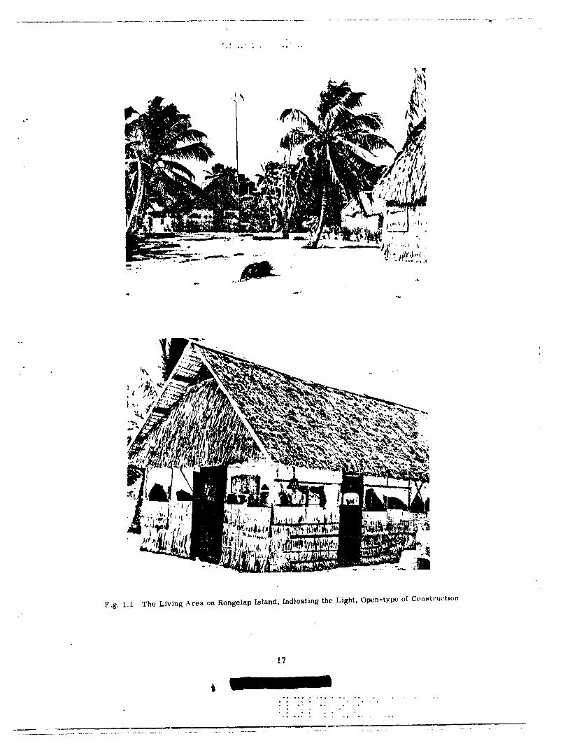
Shol 1 of Operation CASTLE was detonated on 1 March 1954. Followtrr~ the detonation sig- nilic.;lnt amountn of radioactive materials fell on the following popula_ted neil$boring atolls: (1) ltongclal), (2) Ailinginae, (3) Rongerik, and (4) Utirik. Exposure groups arca identified ac- cording lo their geographical location at the time of exposure. The numbers of individuals in- volved, their location, the distance of the atoll on which they were located from the site of the detonation on Bikini, the calculated dose of radiation, the probable time of beginninji of the fall- out and its duration are tabulated in Table 1.1. The Rongelap group received the highest calcu- lated dose. These individuals were living under relatively primitive conditions in lightly con- structed palm houses (Fig. 1.1). The Ailinginae people were a part of the Rongelap group who were on their Ailinginae farms from the time the fallout began to the time of evacuation. Their calculated dose was smaller than that of the other members of their group that had remained on Rongelap. The third and largest group of Mlrshallese, inhabitants of the atoll of Utirik, received the smallest dose of radiation.
The American military personnel exposed on Rongerik were aware of the significance of fallout and promptly put on additional clothing to protect the skin. As far as duties would per- mit, they remained inside of Butler-type buildings. In contrast, most of the native Marshallese remained nut of doors and thus were more heavily contaminated by the matrri;ll falling on the atolls. Some of lhe Marshallese, however, went swimming during the filll(>ut and nWl\y ol the c#hildren waded 111 the water; thus washlnl: a considerable amounl of llrc nmteri:~l from their
skins.
1.3 EVACUATION AND EARLY CARE OF THE EXPOSED GROUPS _. _.
The American military personnel were evacuated to Kwajalein via air in two groups on
2 March. The native Marshallese were evacuated by a combination of air and surface trans-

‘l’~lhll~ I . I E:XI’OSb:I> AND ~‘ON’l’tlOl. IlNKXI’OSb:I) (;ItOlIl’S
Numhvr from Ilikini in (;I’OII~ (Nnul. Miles)
2H I 35
Ii4
I I(
157
I n!i
a:1
270
Ii + 4 hr
H+4hr,
H + 22 hr
Marshallvse,
Control
M:ljuro Group
117
Tlrnl~ of ICvaciutllon
-' ---. I - -~
H + 211.5 hr
(8 rnru)
H ’ :j4 hI
(20 IIlfY~)
Starlcd at
H + 55 hr Completed at
H + 78 hr
:I75 mr/hr
H + 7 days 175
100 mr?‘hr H + 9 duys 40 mr/hr
H + 6 days
69
14
Total Kxposcd - 267
‘IUal Controls - 222
* St%! scctiol\ I .4.:x.
_-...--_.-- ..-. -. -
1.4 ESTIMATION OF WHOLE BODY EXTERNAL DOSE
‘I’hc estimalod v;~lues of external dose given in ‘I8ble 1.1 were based on readings of AN I’DR-30 field instruments. Averages of a l?umber of dose rate measurements on each island :kt ;I given time were used. The readings were t;lken 111 air, ;Lpproximalely 3 feet itbove
ground, severill days iliter the Inhabitints were evacuated. Before this time, adequate surveys with well cnlihrated instrumenls had not been possible.
Several variables which influence the results are indicated below. These will be discussed in greater detail in an addendum report on external dose, which will contain the data, methods .lnd c:IIculations by which the external dose analysis was made.
1.4.1 Energy Distribution of the Fallout Gamma Radiation, $5 Variation With Time, _th$ ___.-..- _- _-. . _ _ __ ._. ___._. Response oi the Meter in__EachE_n_e_rgZle~i~n_and Its Corrf+i_on F+ctp:_.for .the_Tota! -.. -- _.-- Dose Spectrum _-.---__--
Fallout deposited as an effectively infinite plane source resulted in the dose-energy histo- gram shown in Fig. 1.2. Its energy distribution was the result of degradation of the original
16
.
F.g. 1.1 The Living Area on Rongelap Island, Indicating the Light, Open-ty}w of Conntructlon
17
. . . . . ,. . . . . . , . . _ . . . . . ~. . . . . . . . . . . . . . . :. _. . I. ..L . _ ., . . ..1
_- ..- --
_.. ~_ ~~.._...__.__ -.- -- .._ _.~ - ._ -- --- ---~.-~ -.~

----_-- _I-.--..
.-. ___
._ _
__
. .
I .
.
I 1
m-w
.
z cl
3500 lWlO
1 JO
IN
33 U
3d
m
7
_: ” _-
-
_-
A-
: --
c
__
: __
__ -s U
__ -E d
-_
-- 0
-c h
_-
__
.-
-0 0
.

1.4.2 Rate of Decay of the Fallout Mixture
Dec*:Iy r;Iles of fallout samples were measured in the field and in the Iilbor;tt(jry, where :I fairly consislent pattern WBS observed among various locations: and eilnlples. 111 ;Iddition, theoretical c~onuider:1tions b:Iscd on the radiochemical composition of the f:tlloul mixture per- mitted decay rates to be cal(*ulaled for different intervals between tl1e times of inilial exposure and later survey readings. These agreed well with the experimental data, i111d were used both in the dose c*:Il(*ul:Itions during the exposure intervals and in extrapolating the liltel survey
readinKs to earlier Limes.
1.4.3 l’imcL of Arrival of lht, Radioactive Cloud, Durilllon of Lhe FiIllINIl, ;III~ Time cd’ Ev;Icw;~-
lion for E;it:h Cane
Only Ihc 11rne or evacuation is known accurately for all the islands. On Rongerik, however, the time of arrival of the radioactive cloud Wds determined precisely by tl1e continuously re- cSording dose rate monitor at the v :ather station. The fallout became visible iI1 the time the inslrument first indicated lhe presence of a radiation field above background. The material
had the appearance of snow. The times of beginning of fallout on Rongelap and Ailingimae were estimated from similar visual observations, combined with knowledge of the relative distances of these atolls from Bikini and the wind velocities in the ares. Fallout was not ob- served on Utirik, hence the estimate of arrival time there was made on the basis of the Ronge-
rik fallout time, wind, and distance factors. Two extreme possibilities exist relative to the duration of the fallout: the first, that the
tallout occurred entirely within a short time; the second, that it was gradual and extended over :1 period of many hours. The monitoring instrument on Rongerik went off-scale a1 100 mr’ hr, ’ 2 hour after Lhe dose rate began to rise above background. u this rate 0r increase is extra- polated to a point for which subsequent decay would reduce the dose r;lte to the ?;ilues lourtd at later limes, a long fallout is implied. This was taken as one limiting IVast!. ;Ind I.orrespondi11g dosc!s were c*;III:IIlated. tfowcver, Ihe possibility does not seem great that (111s ;tc*~IIaIly OC- csurrcd. Exisling diIt;I are inccbnclusive, but several indications tending lo klvcbr (Iw short Lyme
hyl)c)tl1ttsis art* sun1n1ariaed below.
First: ;I IOII~ r;~iht I)rldKtl)ly would IIO( lw uniformly Iitxtvy thrc)u&out, IIw rirst porl~oii
t)eing Iho most- 1nCt:nsc ;Ind Iht l,nl;In(*e tailing off. The lotal phenomenon IIIIIS tc*IIcis toward the effect of a slIorter falloul. This is supported by monitor data from olher 1Iucle:Ir cvenls.
Second: the estimated durations of fallout, of about 18 hours, wl1ic.h result from the :12~ove
extrapolation for Rongerik and Rongelap, appear too long to have occurred al the distances of these atolls from Bikini, since the wind velocity in the area was high enough for the cloud to pass over the islands in a considerably shorter time.
Third: the accounts of the visibility of the fallout, although conflicting, do not seem to indicate such late cessation.
19
! 1 . . . . . I . . . . I . . . . . . ~.
. 1
. . .,_ .
. .
. . *.
.I.... ,. :_. ,
.I ._

-_I~-__ .--._-. - -_--...-_.-._-_.._ _.... - - .._~._. i ----
Fifth: on Utirik, only ;I short f:~llout time is consistent wtlh the later dose riitt:s observed,
provided the IallouJ began as late as was estimated from wind and distance factors. A OIIP hour dur;rtion* of fallout appears likely. On the other islands the actual fallout time IS known
to have exceeded one hour; however, since the approximate dose discussed above was seen to fit the film dat:~ on ILmgcrik. it W:IS used for the other islands as listed in the calculations in Table 1.1. The hour limit is thus “an effective value.”
If the long fallout case IS also considered, a lower limit for the d&e may also be esti- mated, though the upper limit is taken as most probable. The ranges are then as follows: Rongerik 50 r 104 r; Rongelap 102 r .- 175 r; Ailinginae 53 r - 69 r; and Utirik - 14 r.
The dose value for Rongerik given in Table 1.1 is 75 per cent of the short fallout case value, :Lver:lged for 28.5 and 34 hour exposures. This best expresses the average air dose re- ceived by personnel who spent roughly half their time inside structures where the dose rate
was later found to be roughly half that outdoors. On the other islands no such shielding WdS
present. Figure 1.3, for the Rongelap atoll, illustrates the cumulative dose as a function of time
after the detonaHon. It (*an be seen that the rate of delivery of the dose varied continuously, the major portion being received at the higher dose rate prevatliny in the early portion of the exposure period. 13~ the time that 90 per cent of the dose had been received, for example, the dose rate had fallen lo as than 30 per cent of its initial value. Thus the dose’rate C$ exposure diffyrrd markedly from that usually encountered using x-ray units.
1.4.4 Geometry of the Exposures __ -
A third difference between the type of exposure encountered here and other external ex- posures lay in the geometry of the source. These doses were delivered from a plane source, so that the radiation field did not follow the narrow beam geometry usually employed experi- mentally. In such a diffuse 360” field, the decrease of dose with depth in tissue is less yro- nounced than that resulting from a unilateral or bilateral exposure to an X-ray beam, so that for ;I given energy, the dose at the center of the abdomen is approximately 50 per cent higher than ;I given air dose would imply for the narrow beam case. Figure 1.4 illustrates UI ex-
perirnerrl:ll sinrulatiorj of thr field geometry using ii spherically oriented group of Co” sou rres with ;I t)lr:lnlom placcad at their c-enter, compared with a conventional depth dose curve cjbtained with ;I single sourc*e. It would appear under the circumstances that the midline dose, rather Ihan dos(* measured in ;lir, would be the better parameter in terms predicting biological ef- fects. 01 fhis I)asik, the air dose values stated in Table 1.1 should be multiplied by ;rpproxl- m:~lc~ly 1 .5 III order to c.omp;lre their effects to those of an exposure using ;I narrow beam
gwrwl ry. If this is done, assuming ;I fast fallout of one hour, the following doses in terms of
AII air dose under Iilbor:tLory (*onditions result: Ror@ap 260 r; Ailinginne 100 r; RonKerik 120 r; and Ulirik 21 r.
*While it is obvious that the fallout lasted longer than one hour, calculations of dose are based on an assumed one hour fallout as explained in the text.
20

.- __.- i-m.-p
7 y
-.-
i _
477 EYPOSLIHE,
M~YS~CES~ / &
n
11: RATIO I.%
.
CM MASONITE
DtPlH DO’% OISlRlBUTlON IN CYLINDRICAL PHANTOM, C060FACIL~TY,(NMttI)
-u
. . . . . . . . . I
. . . . ,.:
., .
1. ._ I
., . . . . .
_-- -.. -
___ _ . .~ ~-
~..-~- ---
_ _. ~--.- --
. . . . . . , . . . . . ._. . . . . . . . ; . . . _ _- __

loo0 II. -._---. -- ~_
DFPl ti I)O:>t IN PHANTOM MAN
INIIIAL RAOIATION
--f--J-- HI.!;II)IIAL FIEI I-I
0 IO 20
DEPTH (GM)
30
E’ig. 1.5 Compnralivc Depth Doses in iI Phantom Man of lnltinl Atomic Bomb Radintion ilnd
Itndixtion from ti Field of Fission Products
gamma dose. The midline gamma dose is approximately 60 per cent of the portion of the air gamma dose due to 80 kv radiation or above. This portion in turn is estimated to be 90 per
cent of the gamma dose measured in air by the instrument. Thus the dose at the surface of a phantom exposed to mixed fission product radiation from an external plane source might be expected to be 3,: (0.6)(0.9) or about six times the midline dose, if both occur at 3 feet off the ground. Such a depth dose measurement has in fact been made experimentally at a previous

*F. W. Chambers, Project 2.2b, Residual Gamma Depth Dose Measurements in Unit- Density Material, AFSWP, WT-719, Operation UPSHOT-KNOTHOLE.
23 i *. ..a . . . . . . . . . . . . .
. . .I .:
f . . . .
. . .I .
f . . . . . . . . . . .
____
---
.~_ ._~_.__ .___..~ ~~__ ._. __.. __. ----- -_.- --

-.-_ I___ ___.----.--- -._. - ._._~_._._ _. .-- .
. ..I. . . :.:...-.
. :.. . .
CLINICAL OBSERVATIONS AND THERAPY
.-
It was known ininit~diatc~ly that the t>XplJSed grtJUpS had received a significant anrount of
pt~nt~lrating radiation to the entire, body, extcnsivc contamination of tht, skin, and possible intcr-
nai lloposition of radioactive materials. It was therreIore decided that clinical obscrvnlions
would be as extcntiive and frequent as facilities and personnel permitted in order to rccognizr
and cart. for radiation clfrcts as early as possible. Accordingly complete initial histories and
phy!;ic.al examinations with numerous follow-up examinations were carried out. Surveys of the
sk~:~ W(~I’P conducted at frequent intervals and the detailed skin findings are reported in chaptc>r
3. P~x~t~nsrvc~ hc*n~;~ttJl~~~ic;~l studirs were conducted, the detailed results ol which art* prcscntcd
in c*h:tl)lt*r 4. Ilt:sults of examinaticnls fur urinary excretion of radioisotopes are reported in
I~ll;lj)lt'L. 5. .
III ;1ddlll011 lo I)t!ricItiit* c~xaminutions, routine sick call was held twice &~lly.~‘M~di~:.. (+;I ro
M.I~ ;~v:~~lal)lt! ;I( al I tin,css :IIIC! hosjJit;tl facilitlcs wt’r(’ available at the Kwajalrin N;Iv;I~ D~spc’tl- S.I 1-y.
III vitbw ul’ (ht. witlt~sprt~ad c*onlllc:tlnl: opinions in regard to the va!ut> of various prophylactic
;t11d Il~c~r;tlJt~~tic. IIN*;L.SIII*~~S in i rc~atrnc~nt of radiation effects, it was dccldcd in advant*tB 111iIt Ihctr:tl)y would 1101 IJts Kivctn arl)itr;irily tJut would be instituted as indicated clinically for sj.~c’-
CIVIC* (*ondlllons on ;III ~ntfl.v~tluaI Ibasis. Howevtlr, if sttvere ji~inuluc:ytopc~ili~~ devclqwd (blow
IO(N) ct,lls, (x111) the IJrcll,hyl;tc~tic. ust’ (of antibiotics was to be considcrc~d. Whole blood transfu-
SIOIIC WC’I.(’ likt‘wisch lo I)c used only in t:asc of dt*vclopment of serious anemia.
2.2 SYMPTOMS AND SIGNS RELATED TO RADlATlON INJURY
St)ver;lJ symptoms that developed during the Iirst day or two after exposure probably were
attributable to radiation. Itching and burning of the skin and eyes during this period occurred in over one quartc,r of the Kongelap population, to a lt*sser extent in the Ailinginar and to ;I very
slight t~xt(~llt in the Amcrlt*ans. The skin symtomatoltJh~* might have been due tn part to the
marked alkalinity of thus fallout matcrlal (calcium oxide). About two thirds of the Rougelap group rqmrted naustxl during t.hls early perlod and one tenth of the grocp reported vonliting and diarrhea. Only one’ Ajlinginao individual reported nausea. The people of Utirik and the Americans developed no signs or symptoms that might be related to radiation.
* The symtomatology is based on questionings through an interpreter by several observers.
Despite the repeated interrogations and the inevitable suggestion of the interrogators, the sto-
ries remained remarkably consistent.
24
....... . ............ ............ :. . .............
..........
1 :
.
.....
..........
... ........ ......
._

i

. . . .* ,,* ., and 4 hours later, 104. The two-year old child had itn initial temperature of 101.8 ~c~~I.LY~~ which rose to 103.5 degrees in 4 hours. Roth were given intramuclcul;lr procatne pc~nirrllin when thts sharp rise in lc~nlpcr;lture occurred, and both were without symptoms and ft>vcr the following day. A HVVOIX~ injection of pt*nicillin was civtbn at thin time, and therapy was discon- tinut:tI. In splttb (II the. filet that the II(YII rophilcs rc~rnalnt~d dcljrt!sst>d in both GISCS I~MIK ;lltt>r the Ir!vcbr had pacrtlcad, both tndivtduals rccovercd and had no further tllnc~ss. In F’tgurcl 2. t t hr blood flaunts and platt~lc*t~ of the two-ycitr old patient and the> time of the occurrtBnce of thtj illrre~s with ft:vt:r arc1 illustr;~t(~d. A one-year old boy hnd symptoms of upper resptratory infc>c*tion lot
2 stbv’t!r:ii days bcforc bt~i~ig brought to the cllai~. When he Wits HC’BII, hit+ tcmpcraturc* W:IO 100.11 d(!grcx!s. In addition 10 the ht:;ld colde there was ph:Lryngeal inJection and numt’rous CO~I’N(~ rhonchi (wheczlng) throughoul the chest. A diagnosis of uppcbr respiratory infrction wlth II+ sociat(bd bronchitis W;IH mi\dt? and thr child wu given ;I sinKlc Intrarnuticular 111jt~~~tlo11 of pro- ca:nt: pc~nicSillin. On lhts folIowIng day no rnlee or rhonchi wtbrt* hrard, tht! trmpcrctturc W;IH W”F, :lnd hc rt*maincd ;rYynlplomatIr wlthout furthcbr treatment.
OIK, of th 13 indivjdualb: was iI 5O-yenr old man who reported to the clinic: wtth wt~ahtw~, nervousness, wild ;tlxlorult~;~l p:tin, and shuotiny pain in the upper imlt?rior cht;st, bilutt~r;~l ly. He appeared modcralt*ly ill, his tempcraturc W~H 99.G”F, and the only positive physical finding was moderate tendcrntbes ilr lhc right upper quadran of the abdomen. Since hie granulorytc count rcmilincd low on repeated t:xamination, he WiLS seen at frequent intervals. Within a 10 hour
period the temperature rose to 101.6”F, following which it fell gradually to normal. The ab- dominal tenderness continued for 24 hours and then gradually disappeared during the subsequent two days. A diagnosis of cholecystitis (inflamation of the gall bladder) was.made. No specific therapy was given. In Figure 2.2 his white blood count and platelet counts in relation to the appearance of symptoms are shown.
A female, 38, developed generalized urticaria, fever, and headache, Urticaria and fever subsided within 8 hours without any therapy.
2.3.2 Platelet Counts
All individuals with a platelet count of 100,000 or less were examined daily for evidence of hemorrhage into the skin, mucous membranes, and retinae. Urine was examined’for red cells and albumin, and women were questioned concerning excessive menstruation. There was no evidence of hemorrhage into tissues even though 11 individuals reached platelet levels between 35,000 and 65,000. Two women menstruated when their platelet counts were 150,000 and 130,000 respectively. Both menstruated several extra days and thought that the bleeding was excessive but not sufficient to cause concern.
2.3.3 Hematocrit Changes
Nineteen individuals in the Rongelap group had hematocrits of 35 per cent or below; how- ever, none of these were below 31 per cent. Nine of the 19 were children, aged one to live years who would be expected to have a lower hematocrit than normal adults; four were over 70 years
of age, in which age group a decreased hematocrit is frequently present without obvious cause. Two of the 19 had had menorrhagia (profuse menstruation) prior to the determination, two were
three to four months pregnant and had not received supplementary iron, and two were young women. The low hematocrits that were observed could be explained on the basis of normal physiological variations rather than to the effects of irradiation on hematopolesis (blood for- mation). At no time were whole blood transfusions considered since hematocrits remained within levels consistent with well being and normal activity,
2.4 THE EFFECT OF AN EPIDEMIC OF UPPER RESPIRATORY INFECTION ON AN IRRADiATED POP-tiLATIoN
_.___ _ _ _ __ -. __ -... .
Between the 27th and 42nd post-exposure days an epidemic of upper respiratory diseases (URI) occurred. Fifty-eight per cent of the Rongclap group and 56 per cent of the Aiiinginae group were involved. Seventy per cent of the affected individuals developed symptoms between
26
. . . . , , , , , . . . . . , - . . . . . . . . . . . . * . . . . .* . . . . _ ,. . . *.. . . . . . _ ,.. , . . . . . I1
. “. . .
t . . . . ; ,. -. . . , . . . . . . . . :.‘ -- ..-
----- -~ .-- --

- _
_
1 47 ___
~_‘_ --.
_. ,
_ (
/ I
1 1
I
I I
_. 1
I , / I , I
-.i ----C
/
I
L. ,:,
:.
2 .c ..b
i 7 ,-_
.2 I-
u . . . c
--=+
, I --
\
-/ I 1.
r’
I d Y)
I_
0, aD
r--
UJ
i ,&
, . . . . . . . . . . . . . . . . . .
. . . . . . I
.; :
: :
’ .
.>
. .
*. :~
’ :
C.
.-. *
. .
. .
. .
. .
. _
I_-
-_
--_--
--__
_

T
. .
+
\ 0 I -- -I P
_.~ .- --. --
---
. . . .-.. a/ \
_.. _ __
-.. .--- --a- /
-a
I d
\ .-. ---
\- _.-.. --
0 Q
, 47
r- u)
In *
m
N
- -1
0” 0 In
N
EO
I x s1133
b . _
a \ _-..
_ <
_ _.
_ _
. . __
-..
0
T
-- ____.__.__~
.- --___
__- .-..
~_ ~. - ____ .__ --
_. __..

‘I’ tlllt~ L’. I IJISk:ASI~:S ‘I’lih’l’ AI’I’EAlII~:I) I)Ul(IN(; OIlSI~:I~V:\‘l’lctN
(II*’ ‘1’111: l~~N~;l~:I./\l’ .\NI) r\ll.IN(;IN,\k: (;1~01tI’~
Of the 64 individuals from Rongelap, 27 had no respiratory infectron and of these 13 (48 per c.c!nl) without URI showed a rise In leukocytes; 37 had the infection, and of these, 24 (53 per cent) showed a rise; 7 of these 24 showed a rise three or more days before symptoms ap- peared. Of the 18 from Ailinginae, 8 had no respiratory infection and 3 (37 per cent) of these
showed a rise; 10 had the infection, of these, 3 (33 per cent) showed a rise in count. It is also of interest that not only the irradiated individuals developed the respiratory in-
fection but in addition the medical personnel involved in their care and study also developed equally severe respiratory infections. The respiratory infections consisted of moderatr ma- laise, sore throat with prominent lymphoid follicles, pharyngitis, moderate fever on the first day, and a purulent (pus) nasal and tracheal discharge for 10 days.
2.5 COMPARISON WITH THE UTIRIK GROUP OF DISEASES SEEN IN THE HONGELAP AND AILINGINAE GROUPS DURING TIjE’ PERIOD6-F OB<ERVkTiON __-_ --
The diseases that were seen in the Rongelap and Ailinginae groups during the period of obeervatlon are-listed in Table 2.1. None of these appeared to be related to the cxffrcts of ir- ritdiation, either directly or as a result of the hematologic disturbances. For comparison, the diseases Lhat were seen in the Utirik group during the period of observation are lisled in Table 2.2. The high incidence of gastroenteritis (inflammation of the stomach and intestines) in both groups was probably due to the Marshallese keeping perishable foods unrefrigerated for long periods, and was not seen after this practice was stopped. It would appear that a higher per- centage of the Rongelap-Ailinginae group developed upper respiratory infections compared to the Utirik group. However, all of the Rongelap-Ailinginae group were questioned concerning even mild symptoms of URI, whereas only those of the t tirik group with severe symptoms of
. . ____._.~_ .-.
. . . . . . . ..* -. . ,.. _-, . . . . , . . . . . . ..a - .
__.‘IL__ __._- ---. .- ---_-

1 1 1 I 1 1 1 2
30 1 R
I 1 1 1
1
3
1
URI came to the clinic for treatment. Similarly, the per cent of the Rongelap-Ailinginae group that developed purulent infections appears higher than the Utirik group. However, the diseases are tabulated for the period of observation o! each group, and the Utirik group was observed
for only half as long since they were moved to another island when it was evident thdt further study was unnecessary.
2.6 CHANGES IN WEIGHT AS AN INDICATION OF DISTURBANCE IN THE OVERALL ME- TABOLISM
The body weight of individuals in the Rongelap-Ailinginae group was followed routinely. Since they had an unrestricted diet and all ate well, their change in weight might be taken as an
30
1 1c . . . . . . . . . . . . . . . . . . . . . . . . . . . . . . . . _I . . . .
. - . . . . . . . , . . . .
. . . ‘. . . , . ,-. . .
. . . . . , . . . -. . .
. . . . . . . . . , . . . ,. * . . . . . . .
_- - - ~_.___..__ --. __-. ..- ..___. _-__-_. . . __~_

. .
‘l’;ll,i~~ 2.3 U’KIGII’I CIIANGES. ItONGELAI ANI) hll,lNGINA~: t;l101Il’S
5 3
U.6-- lu.u 0.5 - 10.0
1 3 I !I
Alllnginnc~ Group
7
0
Number ~d~sc~rvcd
Number that g~incd
weight
Average gain (lb)
Sprc:d {)I gain (lb)
Number that lost 6
WC’ ight
Avcragc loss (lb) 2
Sprcxd of loss (lb) 0.5--3
Per cent of jiroup 86
that lost weight .._
9 __?
3
2.7
2-4
6
2
u.5-4.0
67
ment is open to question. Unfortunately, the weight of the individuals from Utirik was not sys- tematically followed and no satisfactory control exists to aid In interpreting the loss in weight of the Roligelap-Ailinglnae group.
2.7 THE EFFECTS ON YREGNAN(=Y
Four women in the Rongelap group were pregnant when brought to Kwajalein. Two were in the first trimester, one in the second trimester, and one in the third trimester. None of these women had abnormal symptoms referable to pregnancy, and as far as can be determined preg- nancy continued in a normal fashion. In the Ailinginae grocp, one woman was in the second trimester. No abnormality was detected. Fetal movement& were unaffected in the individual
in the third trimester. The hematologic changes of the pregnant women are listed in Table 2.4. TWO individuals in the first trimester had marked depression of pla:elets but at no time was
31

------~- --.._- ._.- ___-- ~~- .-_F.-..__ ..___._
, . I .
. . . . . . . . . ,.;.
.
‘I’nl,lt~ “:I 1~1.001) COUNTS ON I’RECNAN’I INIJIVII)~IAI.S ItON(;KI./\l’ ANI) hll.lN(;INhE: GROUP8
Paliiml No.
I
2 3 4
ii
6
7
8 9
JO :I
T:lhlt! 2.5
:i,uuu “.:>uu
:1,1Mt
‘; .uuu
3.200
PATIENTS TREATED WITH ANTIBIOTICS _ _.__
No. of clnys Antibiotic Condition lrented used _ s
.^_. _
Dt*c!p cxtcnwivc! slough of opidcrmls or rt)llt
‘I’cmlh c~xtraction ‘I’cn~th c:xtrM.ion Inrlumod tonsils with high
tornpcralurc and URJ URI and bronchitis with high
LrmJx:i-nture URI, xcverc, with pharyngitis and
high temperature Rapid progressing undermining
impetigo Traumntic gangrene of foot Cystitis Furunclc on buttock Furuncle on forehead
- ..-- -. -. __._. - .-
32
2 PcnicIllin
1 Penicillin 1 Prnlcillin
2 Ponicill in
1 Penicillin
2 Penicillin
2 Penicillin
I Penicillin 5 Gantrasin 2 Penicillin 1 Panicillin
_.. _-
.

2.&l DBCUSSlON ON THE USE Ok’ AN’l’IL~IO’I’lCS IN TtllY GIWUI’ ANI, IN 1~Al~lA’l’I~~N IN.IIIItY
- .
33
I. . . . . . . . . . . *. . . .
*. -. .,. ‘
r.. . . . ,
“. f.
,- . ..,
. . . . . . . . . . . . . . . .
._- -- --- _.--. _..-- ._____ ..- .-- __ ._._ ._. .- --. ~_________--- --- ~- --- -- _.__

--.----.-___ -c-w--- _._. __..___.__.
.-.I...__._ .__
. . . . . e. . . . . : .
CHAPTER 3
SKIN LESIONS, EPILATION, AND NAIL PIGMENTATION
3.1 INTRODUCTION- EARLY SYMP’I’OMATOL,OGY REFERABLE TO THE SKIN
The Marshalleve on Rongclap SAW a visible fallout of powdery material that beran approxi- matcly live hours atter the initial flash was seen. The powder whitened the Ilair and adhered to
tlleir skin ;IY :I salt-like film. The Marshalleee on Ailinginae reported a similar hut less etrik- inl: Iall~~ut. TINI AIIIV~I(*;WS on liongcbrik aleo YBW a fallout and deB(*ribed it as “mist-like.” The Marslr;~llcse on Utirik did not tiec: iL fallout. The early symptoms were limlted to the Roncelap, Ailingtnae, and to ;I lesser /.xlent the Americans on Hongerlk. The early symptoms consisted ol a generalized itching and burning of the skin, limited almost exclusively to the exposed parts of lhe body. A less consistent symptom was burning of the eyes accompanied by tears. The symptoms began the night of the fallout and continued into the next day. A few individuals had symptoms lasting as long aa two or three days. Decontamination of the skin of the exposed in- dividuals was initiated either aboard the destroyers while they were being-evacukted or upon their arrival at the naval base on Kwajalein. The classical initial erythema of radiation injury to the skin was not noticed by the observers who examined these individuals during the first 10 days. If art initial erythema developed in the native Marshallese, it was masked by their dark skin. An erythema was not seen in the white skinned Americans exposed on Rongerik.
3.2 SKIN LESIONS
3.2.1 Gerteral Dcsc*rlption
Skln IVSIOW first appeared in the Rongelap ~rw[J after 12 to 15 days and in the Ailinginse
and ltcillt:ctl.lk Kroul)R ;ml. 20 days. There were no skin lesions 111 thr* Utlrlk grcnlp. There WiLS (~~,1~~~t1(~1.i~l,l(~ cliffc:rcbn(.cb III (11~s IcnKtll IIf time nt’(*c’ssary for the devc\lopment of the various Ic-
SIOIIH. II(~w42ver, il WilS r~~~rlrti 1hi there was a mmsistent paltern in the scquenti3l tlt~v~lolmit*nt (11 If~:r~cms on various ~xposml parts d llw body. The principal lesions o~~urreit roughly in Ilre
IIIII~IWIII~ sequcnl ial order: Y(.iill) (with epilalion); net-k, axillary region, ante~~ubilal fossac,
Icw%, ;~rrns, legs, anti trunk. Lesions on the flexor surfaces tended tu prechcde those on the ex- tensor surfaces (see Table 3.1 for the time of appearance of various lesions).
A clear cut primary erythema wa.s not seen, nor was a late erythema. In a few cases, there was considerable scratching of the skin due to intense itching prior to development of gross lesions. In these cases, an erythema of questionable etiology was observed. This ery- thema may well have been due to the scratching. Erythema was likewise not observed preceding development of lesions in the white and Negro Americans of the Rongerik group.
The first indication of a developing lesion was an increase in pigmentat :on. These pig- mented areas appeared in the form of macules, papules, raised plaques, or larger areas of
34
-__------.~- _-_. --- .._____ _~_ - -.-._. ----.---- ___-. _.__._._. _. . . . . _~

-----
7.ti XI.6 kX1.11
100.0
x1.4 llN1.0 (i!l.Z ti I .:I
:w.7 :10.7 23.0 IS.3 7.6
15.3
631.5 -. .
3H.4
30.7 23.0
92. I
0.0
lou.ll
76.9 7.G
23.0 53.8 15.3 23.0 23.0
100.0
I3.lI
5 6 .
8.X
27-G
0.0
37.0 (iIS. 15.7
X4.2 18.4 53.0 10.3 4.:1 4.3
95.0
17.2 17.2 22.0
5ti.2
7-t) 62 ‘i . . 7o.:i 23.4
34.4 _,, 2 I .;j 45.s 12.5 7.t1 9.4
89.0
I 7
I7 16
I (i
rounding skin. As the desquamation proceeded outward, the areas developed a characteristic appearance of a central depigmented area fringed with a hyperpigmented zone. Al ;I later stage, pigmentation began in the central areas and spread outwards. After a few weeks the ryc.le was c~omplcttbd, leaving in most instances a relatively normal appearing skin.
Approximalcly 20 per cenl of the Rong:clap group developed lesions wlticbll WVIY more se -
V?I-C. ‘~‘II~:H~ Icsitms mlKM 1~ c*oneidered as (*~m~par;rbie IO second tlt*grrclt- tl1(~1.111;11 burns. ‘1‘11tl
tkqer lc*sitrn.Y lcl*Urrt!d Ilrilll*i]J;lt~y 011 ttlc kY’t and IO a fesHt*I’ c~xtcnt OII 1 iw S(‘ill)J ;llld 1lWk :Illd
ii! ow l-;lw 011 We ear. I~llater fctrniallon was tiot common. tiowevcr. 011 llic 1w(, soiut’ Iat.gc
I~llac (l)llslc:rs) ;~pf~art~l. After a few days, LIw hyperpgnletlted lcsicms sl~c~wt?d wt*l dt~.Sqll;tIlliI-
lion with weeping and (*rusting, leaving depigmented raw surlaces of varying area. Somtl of ttlese lesions became secondarily infected. Epithelium rapidly covered lhe ulcerated areas within a week to 10 days. Pigmentation followed during the next few weeks. As healinji occur- red many of the more severe lesions (particularly on the neck and antecubital fossat>) developed a thickening of the skin with an “orange peel” appearance and a dusky, grayish-brown color
(see Plate 3.4).
535 . . . . . . . . . . , . . . . . _ . .~ . . . .:: ,’ ..‘_ I=
_. . .
. . “.. . : : ‘., . . . * .-.
._ _. --- ___. _.
.


0 0
0 (D
u-l
d
dflO
kl9 lW
lO1
30 IN
33 83d
0 N
-
- . . . .
. . . .
. . .
. . .
. .
. .
.* .,.
. . >
. .
, ,
. .
,.. ,
. I
. ...;
., .
. .
. .
,,.
_._
_..._
__
__
__
_~
_
_
.__
__
_._
-7- ~
__
._
_
__
_.._
_
- .~
-.---
-- ---
__-----
. . .
_ _.:-_.

_. I.---.~..I --__-_.. _ _l_--___-- .- -_ _..-__ .
3.2.4 Ilistopathoiogy ’ .(. .
Seven biopsies were taken of neck lesions and one of an axillary lesion durln~ 111~~ third to
fourth week after exposure on the Rongelap people. The lesions btopsied at that t11w wtbrc in
the hypcrpigmenteti stage with little or no desquamat ion (shedding) having oc.c*urrc*d. Movl of lhesc biopsies were taken Irom individuals who showed lesions of greater than avrragt~ se- verity. A second group of biopsies from the Rongelap group (repeated in three indivltiuals) wc?re taken; four at the seventh week post-exposure and five at the eighth-week pt*rlud. These were taken from the nec*k and anlecubltal fossae. All of these leeIons had desquamatcd and the depigmcnted skin had &pigmented 10 a dusky, gray wlor with some thickening (“orange-prel” appearance) of the skin. Hiopoles were not taken from open lesions or from the feet for fcar of
infccllon. All biopsy wcjunds healed rapidly wilh no eccondary cotn~~li~atio~~s. ‘I’lir microsropic~ findinKs :Ire summarized as follows:
Early biopsies. 3rd .to 4th week. Epldcrmio ‘~r;~net~l)iil~~rni;il damage was nolccl with a few In( <krvenlng iIrCitCieLI showing lt*rin drrniage
(PhleH 3.18 alid 3-l!)). ‘I’lic! epidermis in the moel i:xttmsivrly involved areas showrd c~cuialdcr- able al rophy wilh flallcning (II I iw rctt* j)e~s. III plwcls the epldermln was reduced Lo ;I thick- ness of 2 to 3 cells (Plates 3.20 anti 3.21). The cells of the ~nr~lpightat~ layer showed piconlorphic nuclei, pyknosis and c*ytoplaomic% halos, giant veils a~xi in a few instances multinuclealed cells. Pyknosis of c!ells of the basal layer was commonly seen. Focal disorganization of the mal- pighian and basal layers was usually present in the more extensively damaged arcades. Cells laden with pigment were frequently present throughoul the epidermis and intercellular pigment was noted in some sections. The stratum granulosum was usually atrophic or even absent. Imperfect keratinization with parakeratosis was visible in all sections. Hyperkeratosis was also seen. The stratum corneum was loosely fibrillated.
The arcades of minimal damage were usually found in areas where sweat ducts approached ’ the epidermis (Plate 3.19). There was an apparent increase in the number of cells and mitotic figures in the neck of the ducts and the adjoining areas where regeneration was underway. In these areas the stratum granulosum was near normal width and pigmentation of the basal cells noted in the more severely damaged areas was lacking.
Changes in the dermis were largely confined to Ihe pars papillar& (F”ates 3:18 through 3.21). Mild edema in some rases was noted. Capillary loops were often indistinct and when discernible they frequently were associated with an increased number of pericytes. The endo-
Ihcliol (*ells showed swclllng and were polygonal in shape. Telangierlatir ctranges (dilated blood vessels) were noted in areas where the overlying epidermis showed greatest damage with lympttoc~ylitr infillratlon surrounding the telangiertatic opacaes. Chromalopirores, filled with nlehiu pigment, were prornintmt in the superficial dermis. The fine elastic fibrils running in1.o th(b pars papillarIs were often altered or absent.
L>lttle if any damage was seen below the superficial pars reticularis. The hair follicles werch narrow (Plate 3.22) and in most instances devoid of shafts in this region. There was some tclangiertasis of the capillary spaces bounded by lymphocytes and monuclear phagocytes in the superficial pars reticularis. Some of the large elastic fibers in this region showed slighl swelling in some instances: No damage to fibrocytes or collagen fibers was noted.
Second series, 7th and 8th weeks post-exposure. Epidermis In general, reparative proce.&es ofthe-epidermis appeared to have been fairly good, ex-
cept for a few persistent areas of atrophy with narrowing of the epidermis and finger-like downgrowths of the stratum m;llpighii (Plate 3.24). These occurred in areas of greatest epi- dermal narrowing and lhe cells showed rather prominent pigment content. There were many outward epidermal excrescences covered by thickened stratum corneum which was still loosely laminated--- such phenomenon producing a wrinkled appearance which probably accounted for the “orange-peel” like appearance of the skin noted grossly in the areas biopsied (Plate 3.22). In almost all instances the basal layer was intact with llttle or no disor&:anization noted. There were a few scattered areas in which occasional eplthelial cells with pyknotir nuclei and peri- nuclear cyloplasmic~ halos oc*rurred in the stratum granulosum and malplghian layers (Pla1.e 3.23). There were OLY asional arcades in which the epidermis, particularly the stratum grranu- losum, appeared lu bc: ;~tu;~lly widened. These oc’curred almost predominantly in relnCion&ip
38
-_-- - --~-__- ----------- --.-- .- -.- .---- - -I-~.-_~-_~_. ~___ . . __ _ _._._. _,_______ _ _ .-.

. . . .
j :..
..:
. .
. . . . . . .
. . .
. . . . . .
: . . .
. . .
. . . . . .
1
. .
. i
’ **
.
. . .
.
. .
. .
. . .
. . .
.
. a.
...
. . . .
. .
.
. .
. .
. .
. .
. .
* .
.
n....
. .a..
0.
l
..a...

_--- _---
The (*apill;lry IoopS in the tiermal papill:lt~. i~ltlt~n~~h p~yt~~t, WOI’C’ 1101 unli~)rn1ly dist in~t Pericytes rern;iined in increaSed number InIl fewer Iyniphor~ytt~S were prt~St~n1. Cic‘llt~r~llly.
there appeared Iv be Slight telanglectaSiS of the caplllariru In the parS p;ipilluriS and Ihr superficial pars reticulariS (Plate 3.24). TIltIre was some edema of the pars papillartu (Plate 3.22). Scattered pigment laden chromatophorc~s were Irregularly distributed in the papillary layer (Plate 3.23). In some cases hair shaflo in the superficial pars retirularis were quite narrow, atrophic, and occaSionally absent; in others the hair shaft Y appeared normal. Small hair follicles (Plate 3.22) and secretory sweat ducts in some cases YIIOWC~ mild atrophy.
BiopSieS of three ptgmented mild IeSionS were taken from two of the whittb RonKerlk Amcrlcans. Only one of thr three gave evltlence of damagc~, which WAC) I~~~~l~~l~i~l and confintXd to the epidermio.
3.2.5 SynllJtolll;ltology and Treatment
On Lhcl day ~bf exlniuure, ilching and burning of the Skin witS prevalent. 7’111s SubSided and for a period of IO to 14 dayS or longer Lhcle was neither Subjective nor ol~jcc~tlvr tavidencc of Skin injury. It(*hing and burning reappeared either prior to or in the early plgnrentation stage. With the deeper lesions there was alSo pain. Pain was rather marked with the foot lesions. During (he painful period some of the foot lesions were also hot and presented a brawny edema. A c*ommon complaint was a tenderness in the grea! toes medial to the nails. However, visible lesions in this area were infrequent. This symptom uSually preceded the appearance of gross lesionv elsewhere on the feet. Many cd the individuals who developed painful foot lesions were obServed walking on their heels for several days. The painfulness of the foot lesions may have resulted from their greater severi&%d may have been accentuated by the dependent nature of the foot. Some of the lesion? of the neck and axilla were painful when turning the head or raising Lhe arms. The acute reaction and pain subsided after a few days. There were no constitutional symptoms.
T%e treatment of skin lesions was largely non-specific. Most of the superficial lesions were treated with calamine lotion wilh one per cent phenol, which in most cases relieved the itching, burning or pain. A few of the painful hyperpigmented lesions not relieved by calamine with phenol were treated with pontocaine ointment, with apparent succeSs. When the epithelium desquamated the itching was relieved by daily washing with Sudp and water and the application of a wattar soluble vanishing type ointment which kept the injured skin soft and pliable. Raw ;~reas, which bc:c*;lme seceondarily infected, were treated by washing with Soap and by the appli- c*:ltion nl aureomycin ointment. Hullous lesions of the feet were left intact aS long iiS no symp- toms wew prcsenl. If painful, Ihe bullae were aspiraled wilh Sterile tochniqueu lo remove the c*lear &r;lw-ccAc,rrd fluid. A single aspiration was adequate sin1.e the bull;tc did IIO! refill. One
IooI lesion developed an extensive, raw weeping ulcer. Prophylactic penicillin wIfs given fat two days, during which time Ihe lesion developed healthy granul:ltion lissuc. Some of the le- sions Of I he skin of the foot remained thickened, Less pliable, and painful after desquamation. This way relieved by Ihe use of Vaseline or cocoa butter to soften the tissues. One persistent ear lesion did not heal after desquamation. This was treated with warm boric at-id compresses and washing with Surgical soap to remove the eschar. Granulation tissue formed, and epitheli-
um was slowly growing in from the edges of the ulcer when the initial observation period was terminated 74 days after exposure. Upon resurvey six months after exposure healing was com-
plete, with a depigmented scar remaining as evidence of the previous ulcer.
3.3 EPILATION
The incidence of and time of appearance of epilation in the Rongelap and Ailinginae groups is illustrated in Tables 3.1, 3.2 and Fig. 3.1. Epilation was first observed on the 14th po::t-
---__1.... -__._ .__.~_____-“__ _ -- --- .~._ .._. _ -~----
_~ _._. .--__--
._.____-___._.. ._ --. -. _.- ___.. --

__.__..- _--. ‘~-I______-._.~.- .i__.__._-______ .-_._ ______ .._ .._
. .. . . . . . . . . . . . . . . exposure day in the Rongelap group and was confined to the head. The epilation was dlv~dctt arbitrarily into three degrees of severity. “l+” epilaiion Indicates loss of hair without obvi~s thinning; “2+” indicates a loss of hair sufficient to cause obvious thin spots; and "31" indlcutc*s an extet:sive epilation with bald spots. Table 3.1 illustrates that there was a ~rwler dogrcr of epilation in the children (0 to 15 years). Over 90 per cent developed epllation of no& degree in (he 0 to 15 yea:-e group, comparcad to only 28 per c*ent in lhc older age group. ‘I’hc prepon-
-.
01.0 27
0.0
22.2 24
II.1 28 5.6 38
16.7 33 5.t; 44
77.7 38
. -.. . . -. ._.. _ _.._ -.-_--.. . - -.
* Days p)sl --vxposurc.
I OIIC cas(: clalrncd slight c*pLluLion. Queellonabic.
terial on the skin is primarily responsible for the epilation. In the Ailinginae group only three cases of mild epilation developed in children (Table 3.2).
Slight regrowth of hair was observed in all individuals nine weeks after exposure. Hair regrowth was complete and normal six months after exposure.
3.4 NAIL PIGMENTATION __ _.___- _ -.
An unexpected observation was the discovery of a bluish-brown pigmentation of the finger- nails which was first well documented on the 23rd post-exposure day. The discoloration began in the semilunar area of the fingernails (to a lesser extent in the toenails), and tended to spread outward sometimes in streaks. As the discolored area grew outwards the semilunar area usually became clear. In a few cases, detachment of the end of the nail from the nail bed was
observed when the pigmentation reached the end of the nail. Plate 3.17 shows pigmented bands in the nails at ‘77 days. The discoloration of the nails was seen in 89 per cent of the Rongelap and 78 per cent of the Ailinginae group. It appeared to be a radiation response peculiar to the dark-skinned races since it was seen in all of the American Negroes in the Rongertk group and in none of the white men. This lesion was not observed in the Utirik group nor in the control Marshallese. Sinc*e the nail pigmentation occurred in individuals without skin lesions, it ap- peared to be the result of :I m(Jre penetrating component of radiation than contact radiation which predominantly produced the skin lesions.
40
. . . . . . . . , . . . . . . , . . . . . . . . 1 . . .
:. . . . : :.
. .
. f..
. . . . . . .I. . . : :
L . . . . . *. . .
. . . . . . . . , . . . . . . . . . * .
-- w-- ~______ ____ - .~ ________ --_ ._ -. __.___.___ _~. __~.

Plates 3.6-3.9 Extensive lesions in boy, age 13, cast’ 26.
C
. . . . . . . . . . . . . . . . . . . . . . ................ ............ . , ............ : : ‘:. . ..........
....... ................ . . . ..

____-- __..__.______-___ ._._____ _....~ ..- ..___-._.-.~. -._. ._--. -.-....--.-- --
., . . . . 3.5 FACTORS INFLUENCING THE SEVEHITY OF SKIN’ LESlONS
3.5.2 Erier~y of lJeta P;rtii(*les -.
From av;lilablc data on the fallout material it has been cillculated that 50 1~ 80 per ct*n( <)I the beta rays during the exposure pel’iod wcw ooft with an avcragc energy :)I aboul 100 kcv. Since 80 microns of tissue produces 50 per rent attenuation of such r;idia(ion,’ the greater portion of energy was dissipated in the epithcliunr which is roughly 40 to 70 rnic’rous 111 Ihick- ness. The remaining 20 to 50 per cent of the beta rays were of higher energy, will1 an avcr’iLKt>
of approximately 600 kev. The latter would penetrate well into the derma since it takes 600 microns of tissue 10 produce 50 per cent attenuation.‘*2 In addition, a wide spectrum of ~~IIIIIM
energies irradiated the skin. Approximately 10 per cent of the total ~arrar~~a spectrum was be- low 80 kev which would be absorbed largely in the superficial layers of the skin. The remainder of the gamma spectrum is distributed between 100 and 1600 kev with a large proportlon be- tween 600 and 800 kev.
3.5.3 Physlral Dose to the Skin
There is no pracljcal way to estimate the physical dose lo the areas of skin where lesions were found. The entire surface of the body of the Rongelap group received approximately 175 r from gamma irradiation derived from fission products distributed on the ground, trees, and buildings. To this 175 r would have to be added the beta component. In view of the high beta to gamma ratio in fission products, one might expect the t$ai beta surface pose to the skin to be
.- large. The maximal skin doses from the plane field of radiation are estimated in chapter 1. To these doses must be added the contribution of the material deposited on the skin. The latter can not be calculated, or estimated biologically with any degree of accuracy. A rough approxi- mation of dose received at the hair follicles can be made as follows:
The hair follicles must have received a dose comparable to the known minimal epilating dose of about 400 r for 200 kvp X-rays. Since regrowth of hair occurred, the upper limit of dose at the depth of the hair follicle must not have exceeded the permanent epilating dose of around 700 r of 200 kvp X-ray.$
3.5.4 Protective and Aggravaling Factors
The individuals who remained indoors or under the trees showed some protection LY com- pared to those who were in the open during lbe period of the fallout. Tl~osc wh wrnt swimming or bathed were also protected to varying degrees. Small children who went wading developt*d fewer foot lesions. Clothing, even a single layer of cotton material, offered illIlI~~tSl c*omplcle protection, as was demonstrated by the fat.1 that lesions developed iIlnIc)st en1 irciy on the (lx- posed parts of the body.
Since the !esions predominate in areas where perspiration Is abundant such as folds of the
neck. axillae, and antecubtVd1 Iossae, it seems likely that the abundant perspiration produced by a hot, humid climate tended to cause the material to concentrate and adhere to these areas. In addition, the coconut oil hair dressing used by the Marshallese acted as an effective collerl- ing agent for the radioactive material. This was proved since the hair was the most highly contaminated part of the body. The concentration of radioactive material on the hair may have been responsible for the large number of scalp lesions, epilation and the large number and seirerity of neck lesions in women.
41
L. _..I, . . . . . . , . .
:. ,
.:. .
_. . .
-.. .----- _- ~_____.------ ---.-- .--
--. ---- ~_. _._ ., ____-. -.-.-

3.6 CORRELATION WITH HEMATOLOGICAL FINDINGS
Attempts at rorrelatlon of the severity and extensiveness of skin lesions with maximum depression of platelet, lymphocyte, and neutrophilcB counts were made for individuals in thta Rongelap group. No positive correlation was found. Thus the contamination of the skin apyar- ently did not significantly contribute to the total-body dose of irradiation.
3.7 DISCUSSION
There ha8 been little previous experience with radiation dermatitis resultInK from expo- sure to fallout material from nuclear detonattons, and the general conseneus until now has been lhal the hazard from fallout material was negligible. Wlth the litroshima and NaKasaki detona- 1 ions, f:tllout material was.not a problem since the bombs were exploded high in the air. The flnsh burns of Ihe Japanese were purely thermal.
From the presenl experience it is quite evtdent that following detonation of a large scale device c.loee to the ground, serious exposure of personnel may occur from fallout material, even al considerable distances from the site of detonation. The incident described in this paper is the first example of large numbers of radiation burns produced by exposure to such fallout material.
Knowlton, et al.’ described burns of the hands of four individuals who were handling fis- sion product material following an experimental detonation. Also, following the Alamogordo _ detonation, there were a number of cattle that developed lesions due to deposit of fallout ma- terial on their backs.5 In addition, there were a number of sheep that developed lesions closely resembling radiation burns following a Nevada detonation. However, Lushbaugh” -reported that the histopathological characteristics of these lesions did not conform in all respects to radia- tion dermatitis. It is of considerable interest to compare the present experience with that ac- cepted in the past as the typical course of radiation burns of the skin.
The gross lesions of the hands described by Knowlton, et al. occurred from an exposure of about one hour, resulting in doses between 3000 and 16,000 rep of beta radiation (maximum energy about 1 Mev) with a small gamma component considered to be insignificant. The lesions were described as developing in four phases: (1) An initial phase which began almost imme- dialely after exposure and consisted of an erythema with tingling and burning of the hands, r’eac*IlinF: a peak in 48 hours and subsiding rapidly so that by 3 to 5 days there was a relative ;tbst*n(.tb of signs; (2) A second phase which occurred from about the 3rd to the 8th or 8th day and was c~harwlerized by a more swrre erythema; (3) The thtrd phase at 8 to 12 days. which was c*h;lrac:tcrized by veslc‘lc and ttullae formation. The erythema spread to new areas durtng the Iollowing two weeks, and 111~ artlve process subsided by 24 to 32 days. The bullac dried up, and dewtuamation and epithelization took place in less severely damaged areas; (4) The fourth phase or chronic stage was characterized by further breakdown of skin with necrosis in areas which were damaged sufficiently to compromise the blood supply. Atrophy of the epidermis and loss of epithelial structures look place, which necessitated skin grafting in some cases.
Robbins, et al.’ reported six cases accidentally exposed to scattered cathode rays (beta) from a 1200 kv primary beam with exposure time of about 2 minutes and a rough estimation of dose to the skin of between 1000 and 2000 rep. The lesions described were similar to those reported by Knowlton, et al.’ with a primary erythema developing within 38 hours; secondary erythema with vesiculation and bullae formation appearing about 12 to 14 days later; and, in the more severely affected, a tertiary phase characterized by further breakdown of the skin. In comparisor. with severe roentgen ray reactions these investigators stress the unique pe- riodicity of cathode ray burns, relative absence of deep damage to the skin, less pain, rapidity
42

-- ---- ______--.. .___L__- 4
._ . . _ .
. .
. .
. . . . .
. . . . * ..-
. . -. . . . . . . . _. _

--- ---_L-__. --C-e.-- __. ..~_
. . . I
of healing, and absence of pigmentation. ?‘hkst points would apply 1~ the M;~rsl~;~llt~~~~ ctxcept for the multiphaoic reactions and pigmentat ion. Crawford’ reports a case of c*athodc ray burns oi.tbe hands whi(sX’were sin@r. to those descqribed by ttobbins. ct al.’
Experimental beloadtiiioa burns m human beinKs have been reported by Low- Be~~r’ ;urd Wirt h ilrld Haper.‘” Bt,tli.investigab,rs used @* disco applied lo the flexor surfures of the ’
arms, forearms, OF thighs for iarying lengths of time. Low- Beer rt!portrd “tl~ul~ol’lras,bc” skin reactions. Ht: found that a calc*ul@ed dose of 143 rep to the first miH,imetcr of skin. ignorlug
a
self-a@orption, produced a threshold erythema. Dry, uraly, drsquamution was protluc*t?d by ‘7200 r&J in the Itret millimeter and bullous, wcl desquamation WBW pruduccd by 17,000 rep to the first millimelcr. Erythema tievcloped in 3 lo 4 duys, follawed lult?r iy pipmt*nt;ll ion and dcsquamation wit Ir higher doses. Hecovery was observrd witI1 dl)ms of, 1’7,000 rip. ‘I’ht* ICHIU~U later ohcpwed dcpigmcntcd c*enters with hyperplgmented edges (also r&.11 in the present le- slons).
ilff’
Wirth and Hapert produced primary erythcma within 6 hours after expouurv to a dose of 635 to 1160 rep of PSf radiation. Minute vedrlrs with w spotty desquamution were noted with 1180 rep at about the 5th to 6th weeks post-exposure. ‘-‘- ’
Following the detonation on 1 March 1954, 23 Japanese fishermen were c*etitaminated with
fallout material. Apparently they were exposed to roughly the same total-b&y dose of radia- tion as were the Rongelap group. The skin lesions which developed are described by Morton, et al.““2 Lesions developed which were similar in most respects to those seen in the Mar- shallese people, and were cha erized by pigmentation, desquamation with depigmentation, spotty epilation of the head an erations developing particularly on the scalp, ears, neck, and hands (the latter probably from handling contaminated fishing lines). Erythema and vesicle formation, as well as inflammation of the eyes were more prominent than in the Marshallese. Pigmentation apparently was not as prominent in the Japanese. The lesions appeared earlier than in the Marshallese (about 7 to 8 days post-exF 3sure). As in the Marshallese the lesions occurred mainly on exposed parts of, the body not protected by clothing.
In addition to the Marshdllese and Japanese, seve’ral Navy men on ships in the test area developed a f&w small pigmentad.le&one of “belt-line” distribution, apparently.,.duc to fallout material. -.t
The lesiotiz reporied in this paper when compared to radiatio~?leeions described in the pat presented ceertain unique’featuree which mert? further discus&on.
The enrly symptoms of itching and burning of the skin iind eyes were probably due mainly to skin irradiation from the fallout mater’ial. However, the chew41 nature of this material may have contributed to the irritation. It has been noted” th& ir&dtlng chemicals applied during or shortly after irradiation enhance the effects of radiation.
The lack of prominence of an erythema was notable, particularly in view of the severity of some of the lesions that developed. Wilhelmy” states that erythema only occurs when the dose reaching the papillary byer exceeds a cer.tain level. Perhaps the dose to the dermis was in- -_<- sufficient to evoke the rdsponse. On the other hand, the darkness of the skin and the develop- ment of hyperpigmentation may &ve mask@ an erythema. Microscopically, a superficial hyperemia was not a notable findin&
Wirth and Raper” point out that they were im sed inI!&@ studies on tit radiation of the human skin with the difficulty of distinguishing between true erythema and tanning, par- ticularly in the skin of brunette individuals., It was unfortunate that color fitters were not available to aid in~distinguishing an erythema as suggented by Harris, et at.16
In general the latent period before development of obvious signs and symptoms for radia- tion injury to the skin is inversely proportional to the dose of radiation.‘*‘” III tile present series of cases the relatively long latent period is suggestive of a low dose of radiation. How- ever, the wide spectrum of beta energies and particulate distribution of radioactive material
drastically altered the depth dose, as compared with that in previous experience; hence strict comparisons cannot be made. The later development of lesions in the Ailinginae and Rongerik groups as contrasted with the Rongelap people is in keeping with the relative severity of lesions noted.
43
. . . . . . . . . , . . . . . , ( .*. . .
. . ,_ . . . ..~ ,
. . . . . . .
. . . . .-
, . . . . . .
.- 1.. . . . . . . . . .., . :
we.- - _-.. --_-. -. -._ ._._ __---- --- ~-- -- -- -

. .: . .._.. . , A unique feature of the present cases was the appreciable dlffcrenres in the I;IIVII~ pt~I-l(da
observed for lesions on various parts of the body. These dlffrrencea cnannot be rapl;ttnc*d entirely on the basis of severity, since the sevcrp foot lesions developed ;111t’r IWIS( (alwr It>-
sions. However, the severity might have been in part due to the dependent poriit ion of IIW IIN~(
rather than greater radiation injury. Lesions on flexor mufares in generitl precrdrd Icsionn on extensor surfaces. The present data suggest that the latent period and radiation sensitivity of various skin areas may differ. Previous work has shown thdt flexor surfaces with thinner epidermis afe in general more sensitive than extensor surfaces with thicker epilhellum.‘s
The destructive and atrophic changes of the epidermis, disturbances in keratinization, and atrophy of hair follicles are characteristic of hiotopathologic radiation injury of thtb skln.s0’S*18-”
Severe injury to the dermis and blood vessels was not observed. The minimal drrmal injury wilh severe epidermal injury is in keeping wlth the low energy beta component present and the marked decrease in dtrpth dose over a distance of a few mlrrons from the surface.
Hyl’erpignlcntatit~i~ of injured areas wau a consistent finding in the Marshallene, the Japa- nese, and lhe American NeKrol% Pigmented Itasions were also observed to a lesser extent in
I.he white Amrrlcans. Pl~nienlation of tlbicr nillurc ~XIH not been dwcrlbed ;IH u c*onut;lnt c*h;irac- teriotic of radiation ~I;IIII~K~ 10 11~ nkln.
There IH no s;~liufac*tory explunation for the darker dusky-gray color that appe;kred In
mmc of tlw okin lesions as Iwal11~~ progressed. The color changes may be due to alterationa in loc:;il pipnerit prodwtion, vascular changeo, or a thinning of the epidermis, rendering it
more translucent with resultant darker appearance of the pigment layer. Later biopsies may explain this phenomenon.
There are features of the lesions described that appear unique, e.g., the absence of visible multiphasic responses, the presence of early hyperpigmentation, the long latent periods, and the severe epidermal injury with minimal dermal injury. It is possible that differences may in part be on a racial basis.* In addftlon, the marked difference in histologic response of the epidermis and dermis in the present series is in marked contrast td the usual radiation re- sponse of the skin produced by high energy X- or beta-rays.
In Table 3.3 are listed the approximate minimal surface skin doses required to produce recognizable epidermal injury in a.r.,male. It is apparent from the table that beta’ray energy is of considerable importance in determining the degree of injury. A number of assumptions, in- cluding knowledge of the beta ray spectrum from the fallout, would have to be made if these data were to be used to estimate biologically the beta dose received by the Marshallese. The ._ difference in dose between that required to produce threshold akin damage and that for per- manent damage in pigs is 500 to 1000 rep.”
It is impossible lo estimale the probability of development of radiation cancer at the site of the healed lesions. The absence of scarring, telangiectasie, and extensive chronic vascular lesions tends to improve the prognosis since the foregoing are usually observed to precede the development of radiation cancer.
A favorable prognosis is also suggested by the following evidence: an analysis of 1100 individuals exposed to low voltage X-ray for dermatological conditions revealed no evidence of cancer induction 5 to 23 years after treatment. MaeKeelS states that epitheliomata rarely
develop after a single dose of radiation to tha skin. Lastly, the Incidence of skin cancer in
Negroes is one sixth to one ninth the Incidence In Caucasians.” If neoplasia can develop purely as the result of epidermal irradiation, the incidence of late
cancers may be enhanced since the dose of radiation and the visible groes and microscopic in- jury to the epidermis-greatly exceeded that to the dermis. Since many children and young adults were involved, the life expectancy of a large number of the exposed people will exceed the long induction period for development of radiation cancer observed in radiologists. Long
* Reported clinical experience with radiation skin lesions is based predominantly on the response of white-skinned people, whereas the type of lesions described herein, with one clear cut exception, were observed in Japanese, Yarshallese (negroid), and American Negroes. The exception was a dark brunette individual.
44
. . . . . , . . . . . . . . . . . . . . . . . . . . * . . . . . . . . 1 . . . . .
. I . “. . .
. . , I.” : . . . - . . . . . . . . .
_ _l__-.c__ ___. ._---
_.__ .

.
d, ‘c
.............. . . ... . . : :
......... : ..... ................
................ .......... .....
...... .................
__-.. _-- . ..__- -- _ .____.~... - .- _ I__ .~ __-.. . . _ .~ ..-.. - .~ _~___.__~__. - .

---- - ----- I_
exposure to tropical sunlight, potentially’carcinogenic’in itself, may increase the probability of neoplaetic change. The influence of the sub-lethal whole body exponure received by thene people on skin cancer induction is not known.
The transverse band of pigmentation that was observed in the fingernaile has been pre-
viously observed by Sutton” in a negreee who received 150 r of soft X-ray to the hnnde. The
Table 3.3 SURFACE DOSES IN REI’ ItklQUIRED TO PRODUCE RECOGNIZASLE EPIDERMAL INJURY
_ _. ___ _
InvcstlKator
. ._-_ .-_ - -- .--. _ -. .._ -.- .- . .
Average .%rfare Animal ISOlOpo Energy (Mcv) L)ORC (rcy)
_ . ___._ ___._ . _. .- __._...__.
Henshnw, ct al”
IlaiHbr 6 I~~rnce”
Kapcr & I~nrncs”
IIupc*r & I1~rnctizb 14 HllblI”~Il~ hlorltz and
Hrnrlquc# Moritz and
HenriquesSo Moritz and
Henrlquetl” Moritz and
Henriques” Morltz and
HenriquesSo Morltz and
Henriques”
niii8 Hats Mice
linbblts
Shvep
0.5
0.5 0.5 0.5
0.3
1,bOtl 4,WO
4.OW
1,500 6, no0 ~.bOO - 6,000
S“ Pigs 0.05 20,000-30,000
co’0 Pigs 0.1 4.000-5.000
CS”’ Pigs 0.2 2,000-3,000
Pigs Sr’O 0.3 1,500-2,000
Pigs Y" 0.5 1,500-2,000
Y’O Pigs 0.7 -. 1,500-2,000 --
. ,
nature of the pigment is not known. Since it occurred in aI exposed American Negroes, many of the Marshalleee, and none of the American white@, it 1s a radiation response peculiar to negroid races. The pigmentation was apparently produced by as llttle as 75 r of gamma radia- tion since the American Negroes developed the phenomenon in the abeence of significant con- tamination of the hands.
9.8 CONCLUSIONS
As a result of this accident the following concluslone can be drawn with respect to beta damage to the skin.
a. Serious skin contamination of personnel from fallout may occur many miles from the detonation of a nuclear device. Resultant radiation damage to the akin may be the major ra- diation effect under condltione where early evacuation from the field of radiation reduces the whole body expoeure.
b. Fairly extensive ekin lesions resulting from fallout beta radlatlon apparently produce little or no systemic or hematologlcol effects.
c. Decontamination of the skin must be prompt to be effective because of the initial high beta dose rate.
d. A latent period of a few day8 to three to four weeks may elapse before signs and aymp- toms of skin damage are evident.
e. Clothing and/or any type of shelter gives aIrno& complete protection to the skin.

---I_--_
--T----------------- --- _.... -.--- . ..^_ AL___. - .._. _.I_._ __ ~_-. . _ _ i .’
: .:.-: ‘.“.. .: . , . . . . . .
---.-- --_ -
NOTE
For purpoRe8 of documentation the following color plates (Plates 3.1 through 3.24) numbered with letter6 U, h, C, cl, d, P, _f, and K are con- sidered to be pages 47 - 57, 59, and 61-62.

. *
PIat<, 3.14 Epilatiot~ ifI 111;111 ilt 18 tlilyS. C;lW 79. Plate 3.15 Spotty cpilation in boy, age 13, at 28 age 41. Scalp lesions present in arr.~s of epilation. days. Case 26. Note scalp Irsims in areas of r Same case as in Platw 3.12 u~td 3.13.1 epilation. lSanle case as in Plates 3.8-3.11.)
:
PlatC 3.17 I’ifgIi(~11t~d l~;Ulds III ~~~llllll~llill’ ;LI’t’;L 01
lingernails at 77 days.
e
(
I
i ;.
s - . . . . . . . . . . . . . . . . . . . . .
.I . . . . . . . . . . . . . .
. - . . . . . . . . . . . . . . . .
L. . .
: :
. . . . . . . . .
. . . . : . . . . . . . .
. . . . . . . . . . . . . . . . . . . . . . . 1, i __
I___-

i .
0
(!( )I 801~ 1’1 .A’I’~X
Plate :i.l!~ (X100, HXI E) I:pitll~rm~s: A~~tlcs of minimal damage occur in relation to excretory d~c~~~~f~~~wc!at ~lancls~. ‘Sl r:rlu~ ~r:~nulo~~~m of good witlth and shows scant altcsration. Under-
lying xtr;ltum m:lll)ighii sli~bws rnnrkcd dccronsc~ in piKmcnt. In the tlcoper portion of the over- lying, loonoly Iaminal~d stralum corncum motlcralc amounts of pigment, however, are present.
One uarrow arwdt- of more’ SCV~!I~~~ Lranscpiderm:~l damage shows alterution of the stratum
gr:mulosum with inlcrcc*llulat- c?tlma, I)yknosin, swollol nuclei, :md pigment scattered through-
out. ‘I’hc latlcbr is c~spw~:~ll,y drnsc* in the contibqous pxrukeratotic m;lterial. Dermis: A mtd-
crate cellular infiltralc:, chicfly prrivilscular. is most pronounced in the superficial pars
rt!licularis whcxrc Lhorc IS :I mild tclangicctasis. _ _c-
Plut.c :).20 (X400. II&K) (Cast! US) ‘I’ranscpidcrmal ci:lmagc with disorganization of the mal- ,-
pighi:m i&r. St rattim I:~;I~III~w& absent. Malpighian and basnl luycr only two to tbroe CCIIR
thick with c:xloll;lt ic$rl or I)iKnIcnt olctw:~rd ~CBW:IIY~ I):~r.iilic!ratinizrtl zonr adjacc?lz to atl~ntum -&
c:ornc!um. I’igrnc:nl I:lttc!li ~hl,otn:ll~,l)llc,rc!s :intl hisliocyto8 In pari pnpillaris of corium. Lnttcr -.
is c:tJ~m;~lotrs rmt1 inl i lt1~:11ctl by nlotk*ratc numbers of lymphocytes, mon~~nUcICiIr phagocytes
and xc::~ltc~rcd ~~~gn~~iilcd I~wkoqfY(~~~. (:3pillury loops indistinct. ‘.- _.
PI;&! 3.21 (XIOO, II&K) (CIHV It;:)) ‘I’r:~n~cpitlcr~lu~l tl:tmagc wlth disorganization of the mal-
I)ighian Iayc: I’. SI ighl pa r:tk~~r;&is. M igrittion or cxfoliutlon outward of pigment. Loose
I;imination d 31 r:llum corncum. l’igmcnt I:lden chrornatophores and histiocytos in superficial ~x~rs pztpilluris d t’oriutn. Mar&d c:ellul:ir inlillr:rtion of pars papillaris. Slight telangicctasis d supcrfk:i;il pars rcticul:irls.
‘
j. .
i

. . . . . . . . . . . . . . . . . . , . . *. .-. . . . _. . _. , I .
. . . . . . . . . . . .
‘.
,
-...- . .
Plate 3.20
* . . . . . . . . . . . . . . . . .
_’
.
Plate 3.21

COLOR PIATC:S
.,
Pl:ttrs :1.22 and :1.2:) show h~nions at 63 tlnye post-exposure. . ._ .~._ .~ Plato 3.2% (X 100, II&E) (Cast, H75) Looao lamtnation of stratum corneum with outward papll- .___ _.. ._ _._ _ ._._. Isry projections :tntl resultsnt “rugonc” appearnncc. Stratum granuloeum of good width. Basal :tntf malptghisn layc~rs distinct with pigment present. Slight edema of corlum with mild tc~langiectssis and slighl incrc:tscu m perivascular lymphocytea and pericytes. Small, atrophic h:tir folliclt~ atl):tcc*nt to sc!lstccoun gland-- tn mid pars reticularis.
Plate 3.23 (X400, II&E) (C;M II7.5) Same as 3.22., Occasional perinuclenr cytoplnamic halos 111 mid slrittum ~rilrll!losllm. l~ooscly laminated stratum corneum. Pigmunt laden chromato- phores in supc:rfici:tl corium along with occaslonnl lymphocytew and mononuclear phagocytea.
Plak :I.24 shows Itsions tsocond biopsy) at 46 days post-expoeure.
l’l;lt(- 3.24 (X IOO, H&E) (Cssc fl:~!)) Narrow rugono cpldermis wlth papillary extensions down- _-_ ._ _. ._-.-- _ -- w:;r> (4 stratum malphigii. Latter art’ heavily lrrden with melandlc pigment. Sllght tulangi- (Bctasis of ,,;I KS t):cpillarts ;UIII [Jars rcticulurin of dormls. Occaaionnf pigment laden chroma- lophorcs in sufn~rfii~ial ticrmis.
_._--
60
. . .*. . . . . . . ::.. . . l ._ . . . . : :.. : . . . :. . . . . . . . . . . . . :
/ . L . :.: . . . .
t 1:. . . . . . . . :: :: . . . . . . . . . . . . . . . . : . . . . . . f
1 I _-- - : . . .
.’

.
.‘.’ , ~
. .
_.
_.
4( ‘:; . *_* . . ‘,_;.’ . . . ’
.
’ , I ’
a
E’lut& 3.x?
. -- . -4

‘.
CHAPTER 4
HEMATOLOGY
4.1 INTi~ODUCTION
. <c
;
.
.
.
Since it is generally agreed that a depression in the formed elements of the peripheral blood is the most useful Ijractical clinical index of the degree of exposure to ionizing radiation, ;i systematic study of the leukocytes and platelets was relied upon as a major aid in rvaluatiry: the clinical status, severity of the radiation injury, and prognosis of the exposed individuals. Animal experimentation had previously shown that the rate of development and magnitude of the depression were equally important in evaluating the severity of radiation injury. Since
f- : : . ::
there had been no previous exposure of human beings to significant amounts of fallout radiation, 1.’
no hematological data known to be strictly applicable* were available for..use as a guide i! th” I
evaluation of the exposed Americans and Marshallese. Accordingly emphasis was placed on _ zzii,...’ -2.. -_ ..
=
systematic serial studies, utilizing a few highly standardized hematologic determinationg,to ; II
insure that individual and group trends would have maximum validity, Si& it was known that 5
_: _.__ “_.” : . .
9’. ic:.
the Utirik group had received a very small dose of radiation compared to the other exposure groups, less extensive determinations were carried out on these people. * _, ‘;
- i;;;,., ;
r -... .: : I <_
4.2 GENERAL METHODS _- i
Hematological examinations included total leukocyte, neutrophile, lymphocyte and platelet counts, and hematocrit determinations. Whenever posstble an entire exposure group was studied in a single day. Occasionally two days were required to complete the larger groups.
Capillary blood was ueed, usually obtained from the finger but occasionally from the heel
.
.’
or ear. Two pipettes each for total leukocyte and platelet counts were filled. From each ptpette a single hemocytometer chamber was filled. All pipettes were rotated for 10 minutes, and the cells were allowed to settle for 10 minutes in the hemocytometer chamber before counting. A 3 per cent acetic acid diluting fluid was used for total leukocyte counts. The blood was diluted with 1 per cent ammonium oxalate for platelet counts and counted in flat bottom hemocytome- ters using a dark phase contrast microscope. 56 Two blood smears were made with each ex-
amination, using a beveled end glass slide for spreading. One blood smear was fixed in methyl alcohol. The other was stained by Wright’s method, from which a 100 cell differential count was made. Hematocrits were performed using heparinized capillary tubes. One end of the capillary tube was heat sealed and the tube was centrifuged in an ALOE centrifuge at 12,500 rpm for 5 minutes.
I i
I !
+A large literature on the hematoLJgic effects of radiation exists; however, these data were not relied upon for direct comparison nnd evaluation of the exposed individuals for reasons that will be indicated later in the discussion.
63 t


. .-
i
to differcnc*cs bctweeu coutrol and exposure groups. For lhc purpose of detoc*t inp significant changcbs in thca hcm;~tologic:~l patlern, nonparametric tests (i.e., statistical teasts for which it is not nec.r:ss;lry to specify the functional distribution of the variate under study) w(‘rt’ uscd.S8W-‘s
jb 1 5.
Thr advantages of nonparametric methods have been summarized.“
4.4 HEMATOLOGICAL FINDINGS, GENERAL 1. t: T _-
i-- Total leukocyte, neutrophile, lymphocyte, monocyte, platelet and eosinophile counts for the i.
;.- several exposure groups are given by day and by age In Tables 4.2 to 4.5. The mean total white count, neutrophile, lymphocyle and platelet counts at the times of peak depression (time over which c*ounts were consistently the lowest) are shown In Tables 4.6 and 4.7 (pp 70 and 79) fol ear11 individual in the Rongelap and Ailinginae groups, respectively. Hematological findings as ;I function of time and age are shown also in Figs. 4.1 to 4.8. The cumulative dtstribution c*urves for the various exposure groups, using the average of counts obtained over Ihe period of maximum depression (days 39 to 51 for leukocytes; daye 26 to 30 for platelets) are shown in Figs. 4.9 to 4.11.* In the figures emphasis is placed on the individual blood elements rather Itlnn on the total leukocyte count, since the component elements have distinct and different time trends after irradiation.
-
c
i.
i
Tublc 4.2 HON(iELAP GROUP MEAN BLOOD COUNTS BY DAY AND--kY A(;E _-.-._... _..--.- .-_-- -.. ._. .
. W.&C. Neutrophlles Lymphocytes Plutclcts Monocytc,s Ednophlles ” _.
..:i.r F- .- *iy‘. P.E. Day
(x 10s) (x 103) (x 103) (x Ill’, ‘--(8 1oq (x 10’) __ .___ ___._ ._.. - ..--... _ .-. ._ - _ .- ..__ -_
x5 >5’
“T g_ Y4 :
. . 5 .x 5 .5 >5 <5 >5 <15 > 15 ‘5 >5 - T-T’ I p.,, . : - .” ~,_, ; :,. .., ._:
3
I
I 0 12 15 I II 22
2(i
:I0
:!:I
:I!)
43
4’1
6 1 5ti (Xl 70 74
9.0 8.2 6.4 4.7
4.9 6.2
6.6 7.1 3.5 4.5
5.8 6.3 3.5 3.9
5.!l 6.5 3.2 4.1
6.1 7.2 3.4 4.7
7.0 7.4 4.3 5.0
5.7 Ii.1 3.0 :J.!J
7.(i 7.11 4.0 Ii.3
(;.:I Ii.2 :I. I 3.11
5 .7 5.5 3.0 3.3
5.2 :,.2 2.0 2.t;
?I.?) 5.1) 2.0 3.3
6.7 5.(i 2.1; 3.5
7.0 (i.0 :I.5 3.5
7.1 ti.0 3.9 3.6
7.6 6.5 3.8 4.0
Majuro 13.2 9.7 4.8 4.0
Controls
I.0 2.2
2.6 2.1
2.1 1.7
2.4 1.9
2.4 2.1
2.G 2.1
2.3 1.H
3.2 2.1
3.2 2.11
2.1; 2.0
2.!) 2.3
:\.I 2.4
3.4 2.1
3.7 2.4
3.7 2.3
3.3 2.2
7.4 4.1
27.5 22.1
26.1 19.8
23.0 l!).G
16.2 14.7
12.t; Ill.1)
I 3.3 I I .rB 17.9 14.4;
23.6 21.h
25.0 2 I .R 25.S 2o.ti
24.2 IH.2
2L.S IS.1
28.1 21.1
38.5 30.8
L
0.S 0.3 0.1 0.7 :!; I .
2.9 1.7 I.6 l.(i i . . . 4.2 5.4 I.9 I.!) I
- 3.0 2.3 1.1 1.3 t
2.7 1.7 3.5 l.(i ,.
1.0 2.0 2.:1 1.1) : 1.8 I.(i I.11 I.3
I .I Cl o.!l :1.4 2.2
1.7 1.1; 2.(i 2.2
Il.!) I).!) r o..> I .o
I.1 I.1 I .4 o.ti
I.0 I.(1 I.1 o.:,
2.6 1.G WI 0.7
I.7 1.2
0.5 0.9 0.3 0.i; 3.4 1.Y
2.0 2.0 9.5 4.7
? *In the Utirik group the cumulative distribution curve for platelet counts only is presented
since hematological~~determinations in this group were not made during the 39 to 51 day period, used for leukocyte comparisons among the other groups.
I
65 / :

1 .
. . . . - . -_. ‘.. : ._ ; .,
. . . . . . THIIIV 4.3 AIl.INt;INAE 1~IlOUP MEAN 1~1.0(~1) c’OUN’I’ IIY LIA\’ ANI) U\ Ac,k
1 F
3
7 IO 12 15 IH 22 2H 30 33 :i!) 43 47 !) I :,4
MHJtIro (:Wlir~~lH
6.0 7.0 5.6 G.8 6.J . . 7.3 6.3 7.ti 7.1- 7.0 6.R 7.1) IS.9 8.7 Ii.4 7.0 9.G H.G 1.7 7.H 7.6 fi.2 fi.l) 6.5
7.3 (i.7 11.4 ti.9 4.a t1.3
13.2 9.7
5 .5 ..?.. 3.0 5.0
.b .5 _
2.L) 2.2
I I5 . lb
4.2 4.2
I.8 4.7
2.3 4.5 2.9 5.0
6.3 5.4
4.H 4.4
5.3 fi.2
3.3 6.2 2.9 4.2 2.7 3.6
3.5 3.6 3.6 3.6
2.R :I.6
1.9 2.2
3.1 2.2
4.2 2.2
3.5 2.4
2.7 2.6
3.2 2.2
3.7 2.0 3.6 2.2
4.7 I.ll 3.U 2.7 3.4 2.7 4.0 2.2 3.2 2.h
7.4 4.1
_ ._I-.
22.2 21.1
4.6 4.A
.-.-_ . .
25.7 23.0 28.8 22.u
24.7 lkf.6
21.4 14.3
21.0 14.7
2U.R 18.0 31.0 22.2 28.6 22.1 25.6 2Y.Y 28.S 21.6
99.8 21.2
3li.b JO.8
3.H 2.1 3.4 6.8
3.7 2.6
2.3 1.5 1.5 2.4
2.3 2.4
l.lJ 1.0
2.8 2.2 1.t 1.7 0.6 1.4
2.2 1.0 2.7 2.6 I.5 1.L)
2.0 2.0
-3 .:,
0.5 Il.4 :.* ,’
r 2.fi l.li ! 4.4 2.1; ‘..,. : 2.3 1.4 1.‘.
<. _ 3.2 2.3 $_.’ T_’
5.H 2.4 ‘r-. -,. O.fi I .li !:i_ :: 4.1 2.0 E?.,.. .
fi.0 1.L) ,I
2.7 1.1; I. &.. . ‘. ,. 2.H 0.1; 1.b 0.7 t 2.2 1.0 ,c 1
i_ 1.H 0.H
0.b 4.7
Ttrblc 4.4 UTIRIK GROUP MEAN BLOOD COUNT BY DAY AND BY AGE
W.B.C.
---------L
N0UtrOphIlMJ Lymphocytes Plateleta MolIoCyte~ Eoslnopbtles
P.E. Day (X IO’) (X loJ, (X 10’) (X 104, (X lo’, (* 10’)
--
<5 r5 c5 >5 <5 >5 Cl5 > 16 <5 >6 <5 . . 5 -.
4 9.4 8.2 4.7 4.2 4.9 3.2 6.6 0.2 -‘-2.0 1.2 . :. &_
14 10.0 6.6 4.1 3.2 5.l 2.9 4.B 4.2 3.6 2.7 -19 37.7 31.5
29 10.1 9.7 4.9 5.8 4.8 3.2 33.2 28.6 2.2 1+ -3!1 q.0
Majuro 13.2 9.7 4.0 4.8 . 7.4 4.1 38.5 30.8 2.0 2.0 n.5 4.7
Controln - -.-.. _ _ ._- __.__ . . --.
Tuble 4.5 RONGERIK GROUP MEAN HLClQD COUNT BY DAY .._ ..___. _--_ ._.- _ .__- .--. --.-.- .___. _- __._
W.H.(Y. Nwlrophllw Lymphocyten Plateleta Monocyte* f’ IfI’) f* IO’) (‘ 10’) fX lo’, fX Id,
.-. _ .._... . .__._ -- .._... . __.. ---
I !I. fi u.1 3.3 H Ii.0 4 .:I 2.1 !) Ii.‘2 4.0 2.0
I 0 Ii.3 3.R 2.2
11 6.2 3.9 2.1
12 6.0 3.7 2.1
13 6.1 3.7 2.1
1 5 6.1 3.n 2.0
I6 8.1 4.7 2.9
19 7.9 4.H 2.7
23 6;7 4.2 2.1
28 7.2 4.1 2.5
33 6.7 4.1 2.2
39 6.8 3.8 2.7
43 7.6 4.4 2.9
41 7.8 4.6 3.1
51 5.7 3.2 2.2
Kwajalein conrro1s
7.0 4.1 3.1 23.6 2.6 2.7
22.0 22.2 17.9
14.4
16.1
20.1 21.8
20.2 16.8
0.1 1.6 1.9 O.:, 2.0 0.4 2.2 0.3 1.6 0.5 1.5 0.7 1.7 1.2 1.7 I .:I
2.8 2.5
2.4 2.1 1.6 1.4
2.0 2.1
1.8 2.2
1.4 I.5
1.4 2.0
2.7 1.5
2.4 1.7
-
:

. . . . . . ..a,..
. ’ : .I
. , . . . . I
. . . . . .
.
. .
..a... I . ..““a
.* . ,. . 4
. . . . . . . . . . .-... n .
. . *..
. . . . . . . . .
l .
. . . .
. .
. . .
. . . . . .
. ;... .
MAJURO CONTROLS-AGES <5 AhiD >5
s 4
lz -1
i
f?
e
iFi 3
z
2 - ____. : :
I --- ----. - ~.. .- _ _____ -- _.___-___
, --- --I I
POST EXPOSURE DAY
Fig. 4.1 Serial Changes in h’eutrophile Counts of Rongelap Group for Those Less Than 5 Years and Greiter Ttar.
f
.
PROJECT 4 I :CjtgS : COUNTS
4 : * 0 . I 1
0 89 I6 24 32 40 48 56 54 ‘S 9:

. . . . .
1
t _ . . __ _
1 ,-. i f ’
d
~01 X
S31h
30Hd
WA
l
aJ
66
. . . . .
. .
_ -.
. . .
. . . :
: . . :.
.__ ___
__
e
. .
. .
i
-. ._
.
_
-. -
. .
. .
. .
. .
_
_..
. I
_L
_
. .
. .
.- ::
__
. .
. _
. _
..*
. .
. .
_
. _
.
. .
- -
_
_
. .
. .
. .
. .
. .
.
_wp__._
_._
_-_
t
: .I
. . :
-
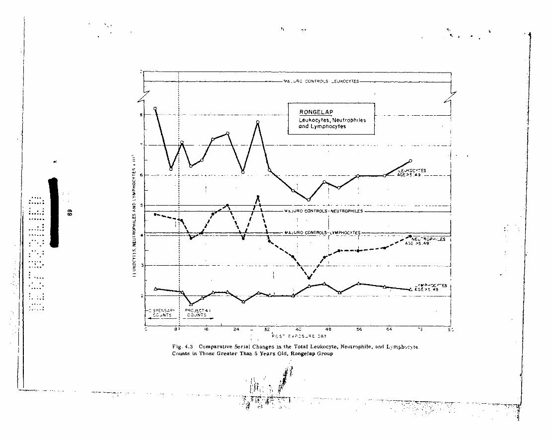
L
. . . . . . . . . . . .
. .
. . . I.....
. . . . . .
. : ..-...
. . . . . .
. . . . . . . . I . . . . 0.
. .
..*a. l .
. . ..I
. . . a..,
. . . . . . . . . . . *. ;.: . . . . * .
..,I..
Q, W
VA.UQi=.
1
CCNTQOLS-1’UNOCYTE~~i
1
i RONGELAP i -- -1_-_d
Leukocytes, Neutrophlles /
and Lymphocytes I
-- -_- .._--~ .._. ~_~ *-__-___._ ----I
0 sr _I___ -- ~~..~~-~~~__~__.~~___
VSJiJRO CONTROLS-NEUTROPHILES
2
- __-___-_ ____-. _-_ _ _..... _ .~ - -.. - --._ -~-_--___
DQCJEC? 4 I COJNTS I
I6 24 :
J
32 Q; 48 56 54 -2 c- __
PCS- ErP,S,;iE 3I.Y * !
Fig. 4.3 Comparative Serial Changes in the Total Leukoqte, Neutrophile, and L~rngh+c:;te Counts in Those Greater Than 5 Years Old, Rongelap Group
. . ,.
.’

400 . . . . . . . . . . . . “‘:‘: . . . . . . . . . . .
. .*.. . .
. . ..I .
. . . . .
. .
. . . . . .
. . . 6 .
. . .
. . . . . .
. . . . . .
. .
.
.,....
. . . . . .
. l .
. .
. . . . . .
. .
. . . . I 300
200
IOC
: , i , : ! :
: . 1 . MP;sRO CONTROLS-AGE <I5 ;
I I I
. : : : : * : : : . : :
RONGELAP
Platelets I I , 1 ,
I I I I .
MAJURO CONTROLS-AGE .15
t
: I . : : * . i -
i i \
3WW~SRY i PROJECT4.1 : COUNTS
-:7 .
89 16
! i *
1 I /
24 32 40 48 56 64 ‘2 I
POST EXPOSUREDAY
Fig. 4.4 Serial Platelet Changes in Those Less Than 15 Years and Greater Than 15 Years of Age of the Rsnge!ap Grv~p
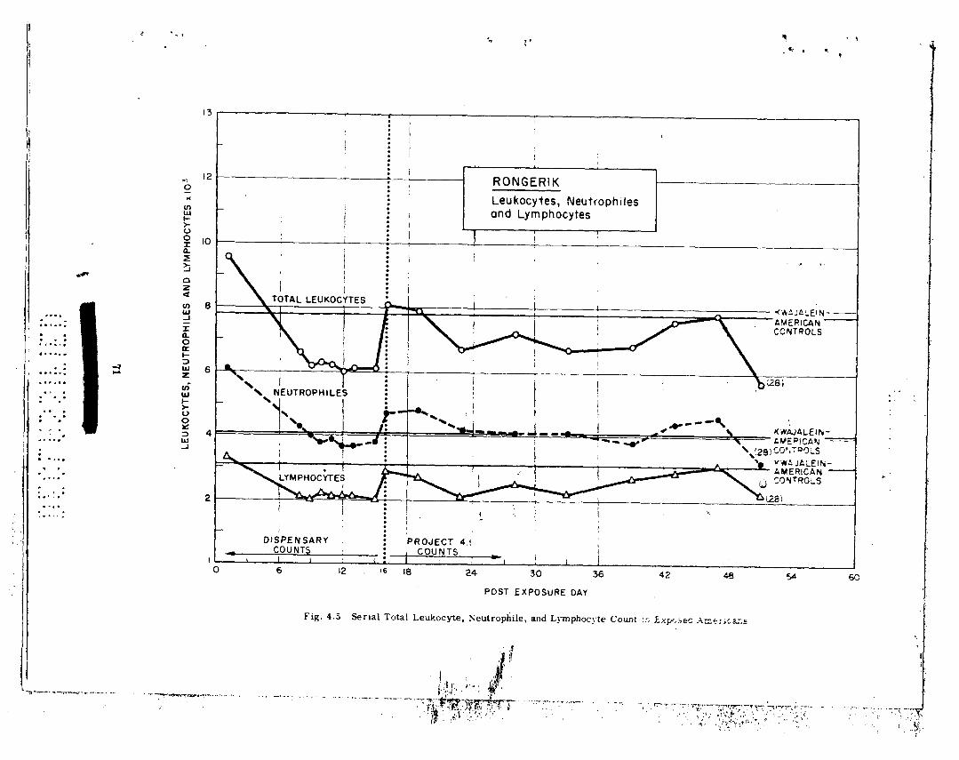
. . . ,
. .,
&
T
_-
/
1 . . ._
. . . . . . . . . . c -_-.. -_ 5 1
0” E
i !I 2 t t
i;l
__ _ ._ -.
- -_-
. . . . . . ...* ---..- 3 .- _ d
_.-.
- . ..a
t- .-
- 4 -
L
t
,-=J ‘-
P
i I i \
- 1 I I
-.
-_-
_.
. . . . . . . .
-.
z z; 2.
I-
- -
-_ - ._.
. . . .
. . . ...*.
. . . . . . . . . . . . .
----. -i
b..
.
~01 x S31A
ZO
Hd
WA
l O
NV
S
31lHd
OU
ln3N
‘S
31A3O
Mn
31
..,___ ._
___.. _.
-- __
. --
P
?------1___=/
,. _,’
..’

I..,..
. .
. a.
4*.*..
. . .
. .
..,.
. .
. .
. . . . .
.
.
..I..
: .
. . . .
. .
. . .
,. .
.
. :*
. . 6
.,....
1 ::-;:j I ;..:.;
. .
..a... . .
s...
I I .-
250
200
l-7 0
x
IfI
L 150
ti I- 4 n
too
50
F .
1 - :
I I I I ! 1 . 1 !
I ! . KWAJALEIN AMERICAN : . I
CONTROLS I I I
I
: !
. :
7 :
i : : ---- :
_----
:
I5 16 20 25 39 35 40 45 50 55 60
POST EXPOSURE DAY
* . I..
.
.
,
Fig. 4.6 Serial hatelet’lfhunts in Exposed Americans

.
n f _I
‘, , ” : r,
4 d
--_-.- ,
/ _- --I -5
P ‘)
-. L.
0
f ‘4 ._A
. -_
7: . . . . . .
. . . . t
\
-+i -._-.
_ -i-
T
_
”
---. -
-. i
- _
- .._ -.__. /
+ i-
t.
. c_
./
_- -i
c P
! I I
i I 1 .a
a5 a=
-fc
g ,! 5, 00 U
W
. . . . . . . . . . --.__
.- .
I..... . . . . . .
- . . . . _
.__.
.._ .
--
. . . . b
.
-
i. f. !. (r
. . . . . @
-- a
. . . . . . . . . . . . . . t . . . . . . . . . . . . . . . . . . . . . . . . --.
-
\ ‘b 1
I *
m
fu
201 X
S3llH
dO
NIS
03
0
.
.
73
i I . . . . . . ..- . *. . . .
. . . . . . . . .
. . . . . . . . . .
*. . . . I
. .
. .
<.
, :.:
: :
“:I .
. .
. .
- 1.
.*... .
. .
*. .
. .
. . l :..:
*.: i
____.__~
___-__- _-

. . .
. I.
i ! I
I / 1
f&abtf-y!Jarn ~~JD~DI.I!~u!~!~ do(abuo&‘y!~abuo~
I llt1301vvJ3H
---Y-- 0 -C
I . . . . . . . . . . . . , :. . . . . . . .
,..a..
z . . . . . . . . . . . . . . . . . . . . , : . . . . . . . . . . . . . . . . ..a.
. . ..- : . . . . .1..
.
. t :
..* . . .
. . .* . . .I....
* _! 1. I ’ . *

100
90 ‘7 -0’ ..o”
L
,..&
---.
I
MAJUHO (91)
_ _
T
I -_____
-.
NEUTROP.HILES
- Cumulative - Age>5 -1 I . - _.
3 4 5 6 7 a 9 IO II I2 13 14
NEUTROPHILES x 10~
75
! L :_
/ ‘I ' i.
:; v-1 ,
. . . . . . . . . . . . . . . . . . . . . . -I . . . . . . . . . . . . . . : *** * *: *: - :: . . . . . . . . . . : :.: : . *: . : . . . . . * . . . . -.. : : . . . -.- ..: *.: [ . .- _. __._ * :. - .

.- -
. .
. . . .
. .
.
*
;:: i .’
-- %
I L
-,a ***...
p “0 "b __ _.---L
n
: 0 *
iFi
_ - .__~.__
z z G
z 3 _.tQ
-_-_--
L
__
_-_
-.-
--
*-+
--I-- ._s _--
J
E
E
0 N
.\ .\
\ -
\ l
?(
. . -1. *
. .
. .
. .
: :..
: :.*
:.,
. .
. *.-
.*. .
. .
_.
_
. .
. .
l
:. :
:s _.
. .
- e-.
. .
. :
:,
. .
. .
. -.
. .*
. .
. .
-.- -
. L
. .
.
.:: :
:., 1..
- _-
- .

,. . .
_ .-
T.’
” --
- .-
-..- ’
. ‘.
--p.f
: .
, .
. .
,. .,
. .
. *
. .
.G
. i
.
. I , . .
I I
._-- - -
!- -.
1. . .
171 . . . . . . . . . . -- . . . . . . . . . . . . . . . . . . . . . .:
: : ,. l . . - - . . . . . . . . . . . . . --. . . . . . . . . . . . . . .*- .
(. .*
. .
. .
. .
..*:
. -
. .
. .
I .
. .
. .
I..
_-_- .--
- -
.

.
d
(‘nut. NIV.
2 7220 1 I 0 2H70 4uli
:I 7:lu ml 2110 420
h :,IIzu’ 11:) 2670 20D
17 IUYO ioh 33no 2(1:,
19 Ml50 1lB 3010 246
21 47:10 It6 2616 IWl
23 7150 195 4100 2n0
3ii 5450 95 2600 265
33 WOO 86 IUIO 3Sl
42 5600 60 2520 262
54 4740 145 2620 196
A5 6050 105 2520 240
GU 4710 115 1420 311
, 5 3926 200 1476 232
20 5020 120 3020 Ins
24 8620 19.5 3450 197
26 6020 146 3410 232
3 :i 5100 140 2100 215
36 512Q 190 2520 241
39 4720 105 2900 156
47 1220 120 4126 22:,
61 5600 105 25ou 291
67 5120 115 2910 192
72 4100 IRS I600 191
75 4200 110 2x20 112
76 !I760 180 2nOO 287
4 8420 130 2660 365
7 5220 195 2520 252
9 5410 125 2100 242
10 4550 106 2110 -157
11 3120 65 1570 135 12 4670 150 3210 127
13 4050 105 2370 152
14 4570 55 2170 170
16 6100 45 4320 165
22 4470 130 2500 190
25 6250 110 4050 212
27 6620 110 3600 265
30 5100 85 3920 180
34 5900 125 3650 2.95
37 5910 130 Sl20 261
40 5600 140 2490 267
46 4620 136 2350 210
4Y 6620 160 4ouo 222
52 5620 I60 2070 246
:i:, 4400 136 1460 212
!,li 6116 126 3520 266
57 M26 5 6 2020 216
!,H 4lMl 60 2600 I WL Ii0 6wJ I60 4060 241
Ii2 WI0 110 8116 262
ti:t 4270 65 2!lbO 162
Ii4 :I606 10 3210 26.;
(i(; 6100 145 2620 312
66 4600 120 2460 202
71 7950 105 4950 210
13 3970 60 2630 126
14 9900 155 1250 255
70 5400 95 3350 I95
79 1600 10 5120 250
80 5670 100 2920 252
82 3250 130 2620 247
-4
78
.
-.. -. I
----- ---____ .

‘1’:1bl~* 4.7 AILINGINAk: GItOlfP MEAN 131,OoD COIJN’I’S Al‘ ‘I’IME’oI ..--._. -.. ..- --.. . -. . _ --- --. .._ _-.
W.B.C.
(Av~r:Igc* from
IkI\. :&I,, !il) -. ..::
I
I6
2H
2!)
31
41
43
45
IjO
51
59
70
3 I !iO
::trolb
:1 I 30
175
I !)5
II5
115
t45
110
215
1x0
95
170
105
185 -.
(i I70
4li70
6970
6750
5650
5120
6150
5650
7050
7750
12400
5070
xi70
“200
3720
a 100
2950
3050
3700
4170
3970
4620
8120
3000
“970
““70
“270
“220
2450
2270
2000
1470
2900
2950
367Il
_- 1750
4.5 IKINGELAP GROUP _... .
The absolute neulrd)philc count of both the younger and older age groups fell during the sc~:ond week to a value, approximately 70 to 80 per cent of that of the controls (see Fig. 4.1). FollowinK the depression of the total neutrophllr count during the second week. the values wt!re unstable until the fifth week. At this time the beginning of a second drop (P i 0.01) was noted for both age groups, and a low value of approximately 50 per cent of controls was reached. The count was maintained at approximately 75 per cent of control values from the seventh week to the end of the study. Although both age groups followed the same general time pattern of re- sponse, the lower age group was below that of the older group throughout most of the observa-
t ion period. The absolute lymphocyte count of the older age group (Fig. 4.2) had fallen by the third day
to a value approximately 55 per cent of the control group. This value was maintained throuyh- out the study, and there was no definite evidence of an upward trend during the study. The values for the younger age group likewise fell before the third day to a value approximately 25 per cent of the control, following which there was a signif.:cant upward trend. With the total lymphocyte count, there is a conststcnt difference between the two age groups. However, during the first four weeks the difference is accentuated when exbressed as per cent decrease because of the relatively high lymphocyte levels in the lower age control group. After thts period thr differences expressed as per cent are less marked since recovery was more rapid in the younger age group.
The cellular elements chiefly responsible for the fluctuations in total while blood cell counl can be determined by comparing the total white, neutrophile and lymphocyte counts (Fig. 4.3). It is seen that the lymphocyte count remained essentially constant throughout the period of study, while the total neutrophile count fluctuated with a pattern essentially identical to t hiit
: ! ;
,
:
79
. . . . . . . . - .- . . . . . . . . . . :. “: .** ; . .: _ ‘. . : ** :.: : * ‘: : . . . .-. l - . . . - . ). -.. ; : . . . l : ..: *a:
--. -_I_ .
--’ - L___._.. _ _
.

d,. . ;.
. . . a..
l~l~rlc*lt~ls wvrc firs1 c:c~mlt~I;!O days after CXp1surC, at whtch Ilint* plalc*lt~l v:iluc~ of III{* . older :II:(’ ~I*OLIJ~S w(‘I‘(* ;I~‘~~~cIXIIII;I~C‘~~ 70 i1cr ccnl of lht+ control gnnqb (FiR. 4.4). l~‘c1llowilr~: Ihis,
I hc ~~l;llt~lel count fell ru:lcliiu~ a low of iIpp~0Xinlnl~~ly 35 per rent of control VillUC’ durlug tlrta .
burl h week. The platclcl COUIII rose during the 5lh wtsek and reached the Vulut? noted for the
initial COWIS OII th(> 10th day. A second decrease in the platelet count (P - 0.01) developed dur-
ing tl~e ‘7!h and 8th wcbeks, and values rtmained at approximately 70 per cent of the control xr’oup during the rermind(~r of the observation period. The patlern of platelet counts in the be- low 15 -year group was r(\nmrk;tl)ly similar to lh;it nolc-d in the older age groups. Differcures
lwl WWII I ht> ;tgt’ groups wprv IWS apparent if the platelet counts were expressed as per cent of I hv UHII rol ~rcnip
4.6 AIIJNCINAE GIWUP
III lhis groul) I~Ic~I~c* W(‘IY! o111y thrw individu:lIs below ag:r 5. For this reason, remarks will bc ccmfitwti wsc~~~li;~l ly IO I hca older age group.
‘rh(! al)solule neutrophilc count fluctuated around the control value for the first six weeks of observation (Table 4.3). At this time the counts began to fall, and a value approximately 76 per cent of lhe control count was reached and maintained throughout the duration of the obeer- vation period. The lymphocyles in this group fell to a value of 55 per cent of normal during the first week. The counts then fluctuated around this value throughout the period of observaticn, and no definite upward trend of the lymphocyte count was noted during the period of observa- _ . . . lion.
As was noted in the Rongelap group the lymphocyte counts remained at an essentially con- stant low level throughout the period of observation. The total leukocyte count inthis group also reflected changes in the neutrophile count. A.
The platelet counls in the Ailinginae group were low, approximately 75 per cent of normal, when first enumerated on post -exposure day 10. The counts remained at this level during the second and third week; however, a definite fall in count was noted during the fourth week when a low value 45 per cent of control was attained. The counts returned during the fifth week to a value approximately 70 per cent of the control level, where they remalned for the duration of the obsorv;~tion period. A secondary fall, as observed in the Rongelap group was not detected.
4.7 UTIRlK GROUP
111 the greater than 5 age grt,up the total white blood cell and neutrophlle counts were de- pressed slightly below control values during the first and second weeks (Table 4.4). The lymphocyte counts were below control levels consistently, and the total white count equal to the control value obtained on day 29 was due to a neutrophilic leukocytosis.
Platelet counts on the 29th day were significantly lower than on the 19th day and were lower than control values. The 29th day coincides with the time of maxlmum depression for the more heavily exposed groups.
4.8 RONGERIK GROUP (AMERICANS)
. The neutrophile count in general reflected the time course of the total leukocyte count
:.
(Fig. 4.5). Neutrophiles accounted almost entirely for the marked rise in total count on post-
. exposure day one, and the values for absdlute neutrophile count fluctuated near the control !
80
i
- __- - __-_..-_I
.

4.9 MONOCYTES AND EOSINOPHILES, ALL GROUPS
Fr~~In TiIt)tI’ 4.2 it in seen Lhat lhe mean monocyte count for the Rongt*lap group rose ab- ruptly from a11 early value below control levels Lo a well defined peak OII day 12, following which
it’fluctualed at values below lhc control level for the duration of the observation period. A similar Lime trend was noted in the Ailinginae and Utirik proups.
The eosinophile count in Ihe older age individuals, Rongelap group, rose l’rom very low levels observed on day 3 to values approximating 35 per cent of control during the second ,P
week, where il remained from the third to the fifth week (Fig. 4.7). The counts thei; decreased :’ _
(P < O.Ol), and remained at a value approximately 15 per cent ol control throughout the re- .f
mainder of the study. The time trend of response was similar in the younger age individuals, however, changes in the younger age group were relatively greater if considered in terms of 2ti the control values. Similar trends in eosinophile count were not evident in other exposure
groups. _- -+
It is possible that the rise in eosi 8s represents that reported as occurring “. . . two
to three weeks after short-wave-lengt. ;iation”.”
: ;‘.
4.10 HEMATOCRIT, ALL GROUPS _. -. i
The hematocrit values for all exposed groups are shown in Table 4.8 and in Fig. 4.8 (a de- tailed breakdown of hematocril by age and sex for control groups is given in Table 4.1). When hematocrlts were first done on Ihe 22nd day, mean values for the Rongelap and Ailingtilde groups were below those of Ihe control population. A significant trend in values after this time
could not be detected stdtisticaily.
4.11 MORPHOLOGY OF PERIPHERAL BLOOD _. __ .._
Significant morphological cellular changes, with the exception of a small number of ab- normal mononuclear cells* seen in a number of individuals during the period of neutropenia, were not observed. Similarly altered cells have been observed previously.‘6 Complete
evaluation of these changes in the present study would necessitate an exhaustive serial study
of the hematology slides.
*There was considerable difference in opinion in respect to classification of these cells. They were classified as atypical monocytes, degenerating lymphocytes, atypical myelocytes, monocytoid lymphocytes, and lymphocytes in transition to myelocytes. At the time of lhis re- port there was no unanimity of opinion in respect to classification and significance of these cells.
81 I I
!- 0 1 !
., **. ” . . . . . . . . . .
. . . . . . . . . .
. . . .
_* ._ -. . . . .
. . I I . .
. . ..- . . . . . . . . *.- ,.. ‘.:
-- __--.- .

35.x 38.0 X.6 :~!).I 36.5 43.2 3H.O 36.7 37.0 38.9
:14.:i 37.7
x4.11 37.x
:I(;.() :17.ti
:I&2 37.G
:w.:l
dli >5 .5 >5 ..5 >B x5 .a6 t5 x5 _ _ ,__.~.__ -._ _-_ ._ --._. __I
Xj,I 40.7 37.5 40.6 37.0 39.4 37.3 40.6
38.9 42.9 :l6.4 3!).2 3ti.0 44.6 32.0 37.2 114.7 39.7
XL9 :I!).0 36.5 43.6 x1.0 37.2 36.3 39.4
xi.2 40.0 35.0 45.6 35.0 37.6 36.0 40.3
:l5.!) :I!).6 36.0 45.2 35.0 37.0 55.7 39.7
40.3
Cant rcdn X1.5 45.2 :17 . 'I . 4O.G X7.1) 4:l.o 36.5 4h.2 37.3 40.6 37.6 43.0 37.11 4:1.0 _. _- , _ . _ _ __
*/Ips III yvars.
44.9
4.12 COMPARISON OF HEMATOLOGICAL FINDINGS IN CHILDREN AND ADULTS, RONGE- LAP~GROUD
It is seen from Tables 4.2 to 4.4 and Figs. 4.1, 4.2, and 4.4 that differences in the degree of depression of cellular elements were.present between children and adults. In Table 4.9, the mean values of the neutrophile, lymphocyte and platelet counts at time of peak depression for each element are given in terms of absolute count and per cent of appropriate control value (mean platelet counts were calculated for the less than 5 and greater than 5 age groups for this-. comparison).
-- -8
Table 4.9 COMPARISON BY AGE OF MEAN NEUTROPHILE. LYMPHOCYTE AND PLATELET COUNTS IN THE RONGELAP GROUP
: AT THE TIME OF PEAK DEPRESSION
Abndute Count x 10’ Per Cent of Control
Agu ~5 Age .5 Age t5 Age >5 i. : .- -. -. . _. _ _.
2.7 3. I 50 64 r 2.11 2.2 40 54 115 I22 30 36
.._ . _
It IS seen that in terms of absolute counts, the children showed a greater depression of the neutrophile count, and the same degree of depression of the platelet counts and less depression of the lymphocyte count. These differences can be most easily described at the time of peak depression. Expressed as per cent of control, all elements were affected more markedly in the younger age group. These results would indicate that children are more sensitive to radiation, or that other biological or physical factors resulted in a relatively greater effect.
,
4.13 DISCUSSION
4.13.1 General
An estimation of the severity of radiation damage incurred can be attempted by comparing the present results with previous hematological data on total lady exposure. The present data
82

- . . . _ . . y.y;--
.
.
.
In thtb following discus&ion it will bc gc>nt*r:tlly asaumcd 1 hat tlw Ilcllrr;ltol(,~:L(.Irl C~[IT~H
noted wt’rt- due primarily to 1110 ptbnetrating ~:;IIIIINI radi:rliwl rt~rcivctl. Thts Ll<*tir r;rdtati~~u III-
jury of thta skin nmy have &ntributed lo fluc’lualionn in the whitt* ~ounl durtng the pt~rtoti ol active lesions during the third, fourth. and MII h week, buf is considered not to h;tvtb con(ribult!d
significanlly to depression of any peripheral elements (scSe rhaptt-r 3). The titngrtw of inlrrnal
contamination with fission products (see chapter 5) was probably too smilll to cant ributc sig-
nificantly to the early hematoloKica1 effects observed. Although il is not possible to Say with certainty lhat th(bse added fartors did not materially affect the hcmatological pattern seen, it
will become evident in the discussion that the changes observed are not inconsistent with those
to be cxpc>cled from exposure Lo penetrating radiation alone. Thus the IIem~tt~~l~~~i~al ChangCS
noled art (*onsidt>rc!d 10 by the result of a single exposure! to pcnc*lrallug ~anlnla radiation, de- livertad at rapidly decrcrxsing dose rates ovt’r a period of approxlmatcly two days. Unless
olhcrwisc> slitt(*d al1 disc*ussion will be limitr~d to the adult Ron~elap grciup.
The I)rinc*illltb sou~~c~~s of prt*vious data available for comparison, and tlitb rharnc’tcristics ;III~ limitations of each ;lr(a summ;rriszed in Table 4.10. Pcrusnl of the table will makr appar~~u1 tht: difIic:ultic*s involved ill attcmpling slrlct comparisons; howt$ver, somr st;itt~m(~iits can bc m;ld(b dcspitts the obvious limitalions. For easy rcferencu, “normal” v:ilucs for pcriphcral blood COUII~S, from the present data and from thr Uteralure arc presenlcd in Table 4.11.
4.13.2 Comparison with the Japanese Data
The limitations s&cd in Table 4.10 apply lo the Japanese low dose groups+ E to H in par- ticular, in which values given”‘” are pooled and include individuals located at the time of the bombing such that they may not have received significant exposure. Hcnce;~while the pattern of change with respect to time is of value, absolute counts probably are high. The time course of hematological change ii the Rongelap people corresponded most clo&y with these low expo- sure *Japanese groups in which definite signs of severe radiation exposure were present in some individuals but in which essentially no mortality occurred (initial hemotological studies on the Japanese terminated at 15 weeks). The early period up to approximately six weeks was characterized by considerable variation in total white count in both the Rongelap and Japanese
people. This fluctuation may be associated with the presence of thermal or other injuries in the Japanese or the active skin lesions in the Marshalleee, or may correspond to the “abortive
rise” noted for animals following exposure.6’*M From the sixth week until the termination of the acule studies ou the Marshalleve during the tenth week, the Japanese and Marshallese counts remained at similar levels.
The ucutrophi le count in h>th the Japanese and Marshallese in general pa~allckd the total
white count. The lymphocyte count in both groups was depresstad early and rc~tlliriued dcpresscd at valut~s of ;rj)proximat(‘ly 2000 utltll week 10. The high value of 2692 rt*porttld for Ihc JitpaIlcsC for weeks 12 to 15 must bo suspucted of being high for the rt’asons given iu ac~tlon 4.13.2.
Three characteristics of the ,Japanese hcmatological trends should be pointed out: a) while high dose exposure groups wlth signiflcanl morlality showed early depression with a definite low poir,t at four weeks, the lower dose groups showed no definite minimum al four weekti but
rather a continued depresslon unlll the eighth or ninth weeks. b) While the mean values for total white and neutrophile counts for even the heavily exposed groups had returned to within
*The Japanese casualties were divided into groups A to H on the basis of degree of expos- ure as determined roughly by distance from the hypocenter and approximate degree of shield- ing. In groups E to H essentially no mortality ascribable to radiation exposure occurred in the first 3 or 4 months.
83
.............. . .......
................
.. .- ...... .
........ :: . : ._-
....... ...
........................ .-
. . ._ ___, ._
.
.’ I
,
!
I 1
--/

‘,I.(
. .
,*. . :. .: ,....a. . ; .
. . . . . . . . . . . . . . . .
Table 4.10 CHARACTERISI’ICS OF hVAll~Al3l,l$ DATA ON ‘I’IIB li~~MA’I’~lI.lll;Ic’nl.
Ek’FEC’l’S OF I’ENETRATlNG RAUIATION
- ;*. .
i:’
- __. ._
(:llllll!Hl R~idlotlii*rupy’~
. _
Sm111l l)tH)r YWI Frill I
- -- - _ ..__ -
Lubnriitory EXpoHWi l,:Ir~c* Arc.idcnts”“” Mnrnhirllt*~ AII111IIti~*”
_.
Yll~lrll I’lMll YPS (hocl
Lnrge (;ood _ .
>Yt,n t ;ood
l.urgc SInId 1
Yc*s No YCN Yes
None NOW Nom*
YC!S No Yt!s
No No No No
Gamma,
some
neutrons
Ilard x rays, Gamma, neutrons, Gamma, beta gamma x rays, betas to skin
Numkrs In Groups Atltn~llllc!y of (:ol,l.r~l1s ,~Criitl ~:OlJll~X (:01111l IllK
‘I’c~chllqlll*s* t:hunc.cb of binr; dw 10
S:IIIIplIII~ Ic~l~tllliqllc~s
“Not-nut1 ”
individu;lls
Inti!rn:kl ronl:~min;dion
Addilional tr~rn;~
(burns etc.)
Spc,cic*s extrapolation ncccssary
Type of radiation
Dosage estimation Poor
Single exposure! Yes
Dose r.,te Instanlaneous
Body region Total body
Good POllI- Usually no Yes -5r/mln Instantaneous
Falr Yes Vurylng,
-Sr/hr
Total body;
Good Yes -’ I Or/min
Total budv beta to skin
- -_. t-,.
Subletbnl Sublethal &
360. f&d -* luthal
Narrow beam
Essenltally Vnriablc ‘rnpld flat fall off to
flat
Usually partial body
Sublethal
Total and part&l body
Sublethal & 1 olhal
Narrow beam__ Rapld fall off
Dosage Rune
Gcomcrlry Depth dose* curve
Sublclhrtl 91 Icthal
Narrow bcttm Modcralc full
off
Narrow beam Variable
*Same tcchniclans for all counts; rigidly atandardixed techniques throughout etc.
Table 4.11 MEAN PERIPHERAL ULOOD COUNT VALUES FOR SEVERAL CONTROL 1’OPULATIONS (x 10’)
Kwaj- American Controls
M:ljuro Controls
Source of dnta Jupnnesu Kure,
Dctt:rmin:ltion 1947-l!#4H6’ 194H-1949’S Amcricana*Sr Americam?” .-. - _. .- ._- . _ _ _. __ _ _. .-
Total While? Count 9.9 9.5 7.4 7.0 Nc*ulrophilc!n 5.6 5.0 4.4 4.3 I,,ympl~c,ylcx~ 2.9 2.R 2.5 2.1 Monocylc?~ 0.6 0.6 0.3 0.4 ISoHinophilw 0.9 1 .o 0.2 0.2
f3itHophil~:X 0.1 0.6 0.0 1’lHtclet.s 250t
--. - -- -- -.-__._-__ - .-. __ _. _.. -_
*Agu 21 years
tThe mean value for 50 normal young American men, using the technique employed in the present study was 2.i7.000
^
7.8 4.1 3.1 0.3 0.3 0.0 23M
u 1 I
;
. . .... ; . . . ............ ..... .......... ..... ............
.... ; ...........
..... . . .......
. . ... ........ - . , , . ......
I -- -----~ --
4.13.4 Comparison with Data Irom Laboratory Accidents _. _.. .- _-_.__ -
Although in the Los Alamos and Argonne accidents’s*‘0 the type of radiation and the condi-
tions of exposure were markedly different from either the Japanese or the Rongelap situations, a large component of penetrating gamma radiation was received and thus attempts at com- parison may be of value. Some findings in the hematological responses are pointed out: a) a uniform early rise in white and neutrophile counts over the first few days, similar to that seen early in the Rongerik Arilerican group was observed uniformly.$ b) Of three high-exposure but non-lethal cases, the total white and _leukocyte counts continued to show sc,me degree of de- pression into the seventh week or beyond. c) The lymph_cyte coun&?in individuals exposed to as little as 50 rem showed an initial marked depression. In most cases the lymphocyte counts remained at low levels thrrnqhout the period of observation. d) Platelet counts were done by a different method, and absolute counts are therefore not comparable. However, of the three high-dose survivors, times of maximum dcprcssion were not inconsistent with the value or 30
days oblaincd in the present studies. The Argonnc Laboratory accidcnt6’ invofvcd four individuals who were estimated to have
received 136, 127, 60, ;lnd 9 rep, respectively. The findings in the two highest exposed indivi- duals in gcncral wero consistent with those in the present study. An initial &SutrophilIc leuko- csytosis was followed by fluctuations in total count, with low values continuing into the seventh week. I~(?c(JVcry WBS not CWnlJlete b 20th week. The lymphocyte count depression was
rapid and marked, and recovery wa ‘evident by the 20th week. Minimum values for the
platelet count were obtained between the 25th and 31st day.
4.13.5 Comparison with Animal Data
The time trends and severity of peripheral blood count change following total body radia- tion in animals has been critically examined recently,6’ and the following general conclusions
are presented.
*Counts 2 years later were not significantly different from control Japanese values.‘2 tCounts on the Rongelap people 6 months after exposure showed no elevation of the mean
total white count, neutrophile or lymphocyte counts over values obtained during the 10th week. fNo counts were taken on the Rongelap and Ailinginae groups during this early period.
‘5 & . . . . * __ ._ , . ..I .c . .- .,, . . . .
. . ..^ . . .,_I
:, . . . . . . . . . .
,. .-
‘.: L.. , . . . . , . . . . . , . .
- . . ..__
._ : : :
t i’
-:

c
-.. t
f
-. . . .*. . .
1)) The rcbsponse of lymphocytes is essentially identical in all animal species. DepressIon can 1~~1 dctectcd withill a few hours, and recovery from the minimum values (achiyxi in 36 to 48 hours) rrquires Ion~er than does ncutrophil(, reccbvery. Lymphocytes fall to very low Ic!vcls at tlosc~s well below Ihcx lethal r:mgc*, and increasIng dose results in no or mtnimal furthttr dc- (*rc;lsf’ ~II COIJI~~. l,yml~ho~ytc~ dq)rtBssion appears 10 have no causal relallonship with ;rcutc
r;ttli:llion do;lths. .
C) I’lal(~lt~t (:ounls h:~vc~ IWCII studied most c~xtc~~slvely in dogs.” As with nc~ulrophlIcs, thr r:tl)itlily and nlagniludo (II dq~r(~ssion is a fu1w1lo11 or dosqa below 111~ letll;iI range‘. Maximum cl(~l~i’c~ssic,rl oc~iirs I)y th(n !)I11 01’ lOth day with doses in the high Iclhal range, by th(+ lllth to 15th day al Slll~leLllill IPVl!lS. IW*ovc?ry t)cKins during the third week, but is not complctc~ by Lhca 30th ti;~y wl~c:r~ n10s1 slud~c!s II;IW been tc*rminated. Insufficient data are available to Indicale the
time required ror c*e)mpletc> recovery.
Considerable* evidence including studies in the mouse using splenic homogenates, induced bacterial infections and spontaneous infections have indicated that critical neutrophile levels exist below which survival is correlated with the absolute neutrophile count following whole body irradiation. From data on dogs, it appears that survival is likely unless neutrophile
counts remain below 1200 cells for a period of time. z
Platelet data on dogs indicate that animals with external purpura have.platelet counts of
50,000 or below. -4 _- Insufficient data on large animals are not as yet available to quantify the extent of maxi--
mum depression of either the neutrophile or platelet counts as a function of dose in the sub- lethal range. The response of the platelet count in the present study was much less subject to- fluctuation than were the ncutrophile or lymphocyte counts. For the preceding reasons, sys- tematic investigation of Ihe platelet and leukocyte counts in large animals as a function of dose in the sublethal range are indicated.
It is not possible to say at present whether severity of exposure, or of radiation damage corrcaiatcs better with absolute levels of peripheral blood count, or with degree of change from (*ollt 1’01 (II’ IIre-cxitosure 1cvc~ls. SOIIIC evidence on this point can be rained by comparint! the CIC~~:.(V~ 01 tlcl)ressioll or Lhc ncutrophlIc and platcslet counts III the Aillnginat: and Itongi>rik
~:l’c,ll~,s, 1)(,th OI whic’lt Ili\d essc*nlially the same calculated exposure but for which control hema- ioI(q:ic*al V;L~UC!S w(:rt* c*onsiclt!rably dlffercnt (lhi! lymphocyte count Is not suikdbk Ior ~!Ol~U.Wi-
SOII sill(:t! deC:t-tqb or d(~~)rt:soion was essentially the same tn these groups and th higher-dose HOII~~~I~~ Kroup). At the time of peak depression for each element, both the neutrophile and platcl~~ (:ounts wore essentially identical in terms of absolute counts, but considerably diIfer- enl in terms of the respective control values. Thus some evidence is afforded that absolute COUIIIS, rather than counts relative to control values, may be the more reliable index of ex-
posure in this dose range.
: ’
:: :
, ‘
4.13.6 Approximation of Minimal Lethal Dose for Man
Some indication of severity of exposure can be gleaned from a comparison of minimum in- dividual counts in Japanese groups exposed at Hiroshima and Nagasaki in which fatalities oc- curred. In general, a significant number of deaths was encountered only in individuals whose neutrophile count fell below 1000. In the Rongelap group 42 or approximately 50 per cent had
.
:. ‘_ . , :- !
3

_. . . I. br;:. ‘C
t:”
nc~ul rollltil~* counts Ludlow 2000 :iI H~IIII(J I ioIt> during’ill6L ~l&~t.v:~(~;u~ I:,‘~I,,,I , .llltl IO ,“‘I‘ (‘(-Ill 11;1tl
01ulils l~low 10(N). I\y lhis i*rilc-ricm. tlwn, IIw c~ffwlivc~ ti0s~ r~.rc~~v~~(l l~y 111~~ 1(,~~~1~,~~:~~1 IjcS,)I,~,S
:llll’ro;ll~l~c~tl IIIC~ Ic4_h;lI l’;lll~:(‘.
III IIN- doi: AlId IIlolIs~‘. ha ;ll)l)~‘c,xitllnl~~ly 50 II) 100 I’ arc’ rc~qtilrc~tl It) Itjwt*~’ Ih nt~i r1~l~h111~
count by 1000 c*c*lls mi 11” iii tllc~ high sul)l~*lh:tl tlosc~ r;inp~~. If ~llt’s(~ dEtt;l (‘;I11 IN- ;l~‘pllcvl l4l lll;l11.
XII iIddilic~ll;ll 50 I0 100 I’ ti)Ultl Il:lVCB I’lil(‘(‘d lilt’ dOtit’ II\ IIW 1~~lll;ll rilllb:(‘. OII IlIt* 01ll~‘r Il;lII(t,
Ilowc~vc~r, il is clt9r fn~iii 1liv pwtwi~ hki LIlill ncutrol~hllc c’ouills bc*lw~*~*ii 1000 ;intl 2000 III
Ilutllirll kings art’ wctll Ici;t~ralcd. Hum;111 bc%inKs with ~IIC~W ICVC~IS of iwulrolhilt*s sl~ow IIO I-lini c*:rI t!vidciic*c~ of illness. arc l)hysically iictivc~, ;rnd do not nc>c!d ~wo~~II~I~Ic*~ ir :rnlIl~~ol I(’ Iht‘rnpy.
Tho Rong:rl:il~ pc~~~~l~~ arc‘ estimntcd to hvc received 175 r as culculiIt~*d from dos(’ riltc* readinKs rne;lsurcd in ;tir in lht! phnc IISSIOII product flel& From the prccScSding par;tgrraph it
is sec1n lhat an additional 50 to 100 r of laboralory radlntion or an ;IVCI’;I~O of 75 r, Ilrol);rbly would hilve resulted in some fatality. CorrcXcling this average valuc~ firomclry.* it Ioll~~ws Illat
the minimal Icth:il dose (MLD) for man expostd in a flssici6 product flc>ld is :il)l)roxilli;lll’ly 225 r measured in air .
It is possible also to estimate the added Increment of dose lhnt would hilvc’ rcsultc-d in
some mortality among the Rougelap people from considerat ion of tllr minimum ltlalc~lcl hunts
obschrved, thcb pl:rlelct I~w~Is in dogs cxposod in the high subkth;ll r:lapa.66 and 111~ tAinntc>d
rate of dccrmsc~ of pl:~1cli!l Ir~cl with incrc.:ising dose in this dosage rangt’. Such ;III :malysis
leads to the s;lml’ conclusions as those derived from neutrophile data.
4.13.7 Peripheral Counts as an Index of Severity of Exposure .-_ .__ ._._. _ ._.____. _ _.
TIw rclat ivc value of thcl several hemalologlcal determinations in estimating the degree of c’xposurc, ;IS well as lhc approximate dose ranges over which maxlmum sensitlvily for each
dt~termination exists, can bc (astimated by comparing the degree of hcmalological rllatlge among
the several exposure Kroups. The relative degree of change in neulrophiles, lymphocylc?, and
platelets can bc seen in Tables 4.2 to 4.5 and Figs. 4.9 to 4.11. Lymphocyte counts were de,,,
pressed appreciably eve!1 in the low-exposure Utirik group. III the higher-dose groups, how-
ever, with widely different physical estimates of exposure the lymphocyte counts showed es-
sentially identical degrees of depression. The lymphocyte counts of4he Rohgelap and Ail’nginae
groups were constantly depressed at a level of approximately 2000 cells. Thus while sensitive
at very low doses, this endpoint may be a poor index of the degree of exposure at higher doses.
The total neutrophile count of the Rongelap group was consistently more depressed than was
that of the Ailinginae group and the difference was of the order of 500 to 1000 cells. However,
day-to-day wide fluctuations in the neutrophile counts occurred. Accordingly this endpoint ap-
peared to be of limited usefulness as an index of relative exposure severity except when counts
on groups to be compared are performed at the same time.
The platelet count showed a more systematic trend than did the neutrophile count. Differ-
t~ces between the low-dose Utirik group and controls at the time of maximum depression for
alI groups could be deterted, and appreciable differences existed between the means for the
Marshalleso hil$or cxl)osurc qoups. Platelet counting Is as easily carried out and more I‘(:-
producible lh;111 I~+ukocylc~ coun1s.36*s7 Thus the plateiet cc;unt may prove@ to IN* :I usc~lul index of
tl(~~:rc:c~ of (~xl~~sur~ lhrc~~ghoul the sublelhal range.
‘1’11(~ abovt~ c’c,liNid(‘r;lliorls ;Ir<’ in accord wit11 previous findings OII ~U~I;\II I)cGjis and aIll - IlI:lls.
*From gcoinctric and depth dose considerations set forth in chapter 1, 1 r measured in air in a fission product field is equivalent in its effect on man to approximately 1.5 r of penetrating x- or gamma radiation under geometric conditions usually used in the laboratory. Thus the minimal lethal dose for man exposed to penetrating radiation under the usual laboratory con- ditions would be approximately 335 r.
i
I..
..-r. 4*._ _-
i.
; _
i
87 I
i i t L
! . . . . , . . . . . . . . . . . . . . . .
. . 1. . . . ..* :.. ..I
. . . . . . . . . . . . . . . . .
. . . . . ..‘. -.. . . . . . . . . . .: ., .I .‘ . . . ,. . . , . . . . . . . . . . . . . . i
-’

-7 _ _ --
. .;p--
. ., . . . . . . ) .:..;... . ‘-
4.13.8 Conclusions . . . . . .
.
.
!.
1. Consideration of the drgrc>cb of depression of periphc>r;ti ct%iiui;1r c*lrmrnts indic*;1ttBs Ihat
;I) ‘l’h(b loI;lI wllilcb WUII( in~rt~as(*s tiurlnji the first two or more* duyv and 1holl dt*crt~;lst~s IN~IOW tlorm;il Ic*vcrl.s. ‘I’htx Iot;II c’ounl then fluc*tu;ltcs over the next flvc> or nix wccsks. with no
dc:firlilc* minimum and with some v;~luc!s aLH)ve norm11 (the yrt~sencc of thc~rmal or belit lesions, or oII~(br ;~c.ul(~ proc(tsst!s during this lime may account in Ilarl for these fluclualions). The
count IW~OIII~~S stabilizrd during the seventh or ci#th weeks at low levels, and minimum counts
Iu-obal)ly o(‘cur al this lime. A definite trend upward is apparent in the ninth or Icnth weeks;
however, complete recovrry may require several months or more.
I)) The neulrophile count parallels the total white blood cell count. Complete return to nor-
ma1 values does not occur for several months or more. The initial rise in total white count is
due to a neutrophiiic leukocytosis. t
c) The drop in lymphocytes is early and profound. No evidence of recovery may be appar- > 6
ent several months after exposure, and return to normal levels may not occur for months or
years. . .. d) The> platelet count, unlike the fluctuating total leukocyte count, falls in a regular fashiort . ._A * i;~ p+..
and rcachcs a low on Ihe 30th day. Some recovery is evident early; however, as with Ihe other;_..
elements, recovery may nol be comI>iete several monlhs after exposure.
4. As an index of severity of exposure, parlicuiariy in the sublethal r..n#e, 1fie total white ._’
or noutrophi1c: counts are of limited usefulness bccausc of wide fluctuations and because sev- eral wcbt1k.s m;ly LW rtaquirt>d for maximum depression to become evident. The lymphocyte
count is of more v;~iuc~ in this regard particularly in the low dose range, since depression oc-
curs within hours of c’xposure. HowcIvet-, since a marked depression of lymphocyte counts oc-
curs with low doses and since further increase in dose produces little more depression, this
index is of little value ;II the higher doses. 5. PIitteIet counts showed a regular pattern of change in the present studies, with the same
time of m;lximum depression in all exposure groups and with the degree of depression roughly
proI)ort~cm;il to the calculated doses. It appears, therefore, that the platelet count has con-
sidoreble I’romise in the sublethal range as a convenient and relatively easy direct method of
dcttctrmining the degret> of exposure.
88 i
\ 1 . . . . . . . . , . . . . . , . .** :* : :” : :” l = . - . . . ‘ . : : ;.: l .
. . ,-- -. ** . * :* : :’ : : . . -.. . . . . : : .: .: : : : :.. :: !
- ..--__. ;. L___ -.- .-. - __II- _. B

I
. .
..‘.
l
:
CHAPTER 5
INTERNAL RADIOACTIVE CONTAMINATION
5.1 INTRODUCTION _..
. ,
.
A study of the nature and extent of the internal radioactive cont:tmin;ltion in the exposed human beings was initiated on the 16th day post-detonation, with the c*ollection of 24-hour urine samples from the Rongelap, Ai!inginae and American groups. Additional information on the extent and nature of the radioactive contamination was obtained from domestic animals, food- stuffs, water and soil collected on the contaminated atolls and sent to the U. S. Naval Radio- logical Defense Laboratory, the Naval Medical Research Institute, the New York Operations Office of the Atomic Energy Commission, and Ihe Los Alamos Scientific Laboratory for gross activity measurements and radiochemical analyses. Data on soil, water, and plants were ^ -,< necessary to determine the feasibility of utilizing the foodstuffs and living area of the contami- nated atolls. Long term studies of the domestic animals obtained are being conducted at the
NHDL, in order lo obtain information on the I, ssiblc acute and delayed hakard from internal . radiation. _.-
The main findin& of Ihe internal contamination study in the human beings and their en- vironment are presented in this report. A detailed report on the nature and extent of the iu- : ternal radiation hazard in human beings and animals, as well as the contnmination of the environment will be presented in an addendum report.
5.2 PHYSICAL ENVIRONMENT STUDIES _~ ._-._ __--._-__-I_
A comparison of the food, water and soil samples from Rongelap, Utirik and Rongerik indicated a much higher level of contamination on Rongelap than on the other islands. The activity appeared to be distributed in the same manner on all the islands. In general, the con- lamination was associated with very fine particulate matter and was uniformly distributed throughout samples of earth, thatch, and grass. Significant amounts of beta activity (1 pc per plant) as well as fissionable material 11 x lo-’ lgm) were present on the exlernal surfaces of plants ;It 42 days, bul only small amounts of beta and no alpha activity were detected in the edible porlions. Coc’onut tree sap, an importanl item in the native diet, was found to have beta :ictivities of the level of 1 pc/liter on Rongelap at two months after detonation. Fish collec’tod from the Rongelitp lagoon were found to have very high levels of activity (0.3 to 3 IIC per fish)
as late as 120 days post-detonation. The activity found in soil was determined to be associated mainly with fallout particles
of relatively large diameter. Rongelap water samples from both cisterns and a well (Figs. 5.1 and 5.2) had activities of about 1 PC/liter at 30 days. Samples of thatch roofing exhibited the highest levels of activity \n the physical environment. The presence of fissionable mate- rial was detected on thatch, grass and on plant food.
89
‘, - .
. . . . . . . . . . . . . . 0 . . . . . .
. . . . ,. . ..* :.. . . .
. . .* . . . . . , :_: : : .: : ; . . . . .L. .
. . ,. ,-. . :: ,_,.*. l *:.,: . . . . . . . . . . 1.
i : ;-. ’
! _
.-
;
/
- -._ ,

Flg. 6.1 Well, Rongelap
i f-.-r /- ‘._ I
Fig. 5.2 Cistern, Rongelap
90 i , . . . !‘* . . . . :
.I_. . .
_** l . . l . . : :.. . . . . :.*
. . . . . :.: *. *, : :’ ::. . . / ..- . . . . . . . . . I . . . . . .: .: : :
: : : : . . . . . .
- ._-._ --- _ _ ..___- _ -.___- ---- --ye .- ___ ___ _.. ._ _---

i r L
.c
3 z=-
p-
.
. .* . . . ‘- l -• ’ --- ’ --
-.. . ,. 5.3 ANIMAL STUDIES .
The studiefi on the Ron@?lap and Uttrik animal8 included measurement of I~~trra;~l radhb-
active cont:tmin;~Ilon ;Ind rndloc*hemlcal ;~n:~ly~i~ of excruta and ttsnues. The! :ICW~C hc*rn;llol~,Kt t.
(‘~1 ;lnd p;Lll\<~lo~ic*;ll c?ffec*tu‘ III the CX/)OLI~I’P , ;IH well 3s Irmg term ellctrta OII IIIWV ;III~I~II~H, :.
;I IT Iwir,c: slutlic!d. In addilion, fc~rtlllty and h;~~c*habillty sludlctn nn wt!ll ati 1~;1dloc~l~c-1~1~~~~11 ;III:I~ - j
ysiy of cogs laid IJY hens from iIongel;q were pvformed. Thea> studies have prkided informittion on the exlent and nature of the internal co&lmi-
i.. 1 i
nation 111 the expsed hum;ln belags. For ex:tmpIe, radiouutographe of anim;rl bones })rcpiIri\d . 1. .: :
by NRDL. and Argonne National Laboratory”” (Ei~s. 5.3 and 5.4) indicate the patttvn of skel- eta1 distribution of fission products, particularly the high concentrations of rudioelt*nients
! ,? >’ ~.
in the epiphyseal region of the long bones. Further, information on these animals (which re- [I ,,. -~
ccived a much higher external radiation dose than was received by the native group and over I ;:
10 times the internal deposition) should be of considerable prognostic value for the human beings.
- 6
Fig. 5.3 Autoradiograph of Rcngelap Chicken Tibia, 1’4 x, 21 hr Exposure. Animal Sacrificed 45 Days Post-exposure (Argonne National Laboratory Photo).
. . . . . . . . . . . . . . -:
.* : _., .-. .* _ , _ -- - - . . .- . . . .- ::. . . . . . . . . . . . . . ..: : :.: ._: ___ . . .- ‘.: . . . . . . . . l . . ’ . . . . ..-
. .
-
. _ ____ ____~._~
Y... _... ,_ . _ ______ ._ ___ -_c

Fig. 5.4 Autoradiograph of Rungelap Pig Tlbla, 42 hr Exposure. Animal Sacrtftccd 58 I);W
I’ost-c!xpc~surc. -I ,-- -.
5.4 HUMAN STLJI$ES ___ -_- .- ,. w_ =
5.4.1 Excretion ._--- .-.
Gross beta activity measurements on the urine samples from Rongelap, Ailinginae, and Rongerik indicate significant contamination in the majority of samples. The variation in beta activity clxc*reted, 24 hours among individuals in any one group is quite large (Table 5.1). If the mran values of urine activity of the adults of the three groups six weeks after exposure ;I w c.trrnp;lred, it will be seen that the Rongelap group had the highest activity, 1208 d m 24 110\11~5. ‘fkt Ailinginae group had 553 d/‘mi24 hours, ;lnd the American group had 309 d, m 24
hours. A group of the .Jap;rnese fishermen exposed to fallout from the same detonation had a urine ;lc*tivity c.omp:lrahle to that of Lhe Rongelap adult group.
.
‘T’hc mean :Ic*tivity of the individuals under 15 years of age appears to be c*onsidernbly
lower lh;tn th;rI 01 thr ;ldult group. An examIn:ltion of the group collections an;~lyzc~d by lhe
-V;LI’IOIIS Ial~r-;~tori~ involved showed fairly conslsfttnt levels of a(‘livity [or the indlvidu;tl from day to d;ly. The-variation which occurs is not considered to be excessively I;\rge for this type ()I’ study. The activity as function of time fell off rapidly due to the large component of ml40 _. T2a140 and other shcrt lived radioisotopes. For example the Rongelap group had a mean activity of 1208 d/m:24 hours at 46 days and activity of 339 d/m/24 hours at 90 days. At six months post-exposure the Rongelap urine was found to contain a barely detectable amount of beta activity. The results of radiochemical studies of urine indicate that Sr”‘, Ba’“‘, and the rare earth group apparently constitute 77 per cent of the total beta activity at 46 days. Stron- tium-89contributes 40 per cent of the total beta activity, Ba”‘- 11 per cent and the rare earth
92
. . -. . . . . ~ I , _ . _ _ _ ,-- _.. . . . . . . . . . .
. . . . - . _ . .^. : :. : :. : :
.._ .: .‘: : **: l *
. . -.. ._- . _ . . .: : _ : : : :.. . .
_
.
i k.
f :
. I
. -

Cat-w No.
ABC 5 years
2
3
5
23
33
54 69
Mcrm 1 ti5 404
Age6to 1.5
years
20
24
26 35
36 39
47
67 72
75
76
Moan
‘l’Ollll
Volunlc!
-74 hr *
120 712
150 894
155 313
40 223
260 0
80 286 465 301
265 1900 550 0
650 1032
255 0
190 236
280 1100
650 1705
450 674 110 507 440 0
980 1180 - ._ .-_ -__
43!l 758
-
Cawu No. _. :_
24 hr
&J ’ 16 ynara 4 455
7 UIO
9 355
10 960
11 450
13 340 14 7HO 18 455
22 47 30 960
34 750
:17 480
40 550
46 330
49 425
G2 780
55 320
56 700
57 550
58 750
60 810
62 980
63 636
66 -655
48 300
71 290
73 230 78 965 79 465
80 540
62 670 -- ._____-..- --
MWII 581 _._-._ -_ _ -. _.
-.
-
ii34
i. j‘
17lJo :. i
201
549 I- :’ t.
15H3 1677 :
2460 ( 1670
77 438
!
570
79%
1450
495
0
0
1080 3220 :.
1095
2170
580 -=- I:_. _ 1985 *.G-_ _.
2260
1715
2010
1450 0 _-
.I _- / ._
52 i -‘- 2038 1363
2140 - - -.____.
1206
group-25 per cent (of which 11 per cent is La”*, in equilibrium with Ba”’ at this time). Fis- sionable material was not found in significant amounts in any of the urine samples analyzed. Iodine-131 was found only in samples analyzed at early time intervals, due to its relatively short physical half life.
5.4.2 Estimate of Body Burden _M ---~
For estimating the human body burden of the various fission products, a semi-empirical approach, involving extrapolation from animal data, was used. Two Rongelap pigs were killed after their average 24 hour urinary excretion of fission products was determined. All tissues were analyzed for total beta activity, as we11 as for Sr”, Ba”O, and the rare earth content c (Table 5.2). By applying the ratio of animal body burden/urine excretion activities to the
. Rongelap native urine samples analyzed at about the same post-detonation time, a total human body burden of 0.33 UC, for post-exposure +y 82 was obtained. If this total human body burden
i

a.. 4 * . . *.. * . . . . , : . , .
Table 5.2 RADIOCHEMICAL ANALYSIS OY TISSUES AND URINE OIZ LIt,Nt;b:l.AI’ l’lt;s
.
GroHN
Aotlvlty .wllph ‘7% (X 10-T
. _. ._
Pig 124 (25.6 kgm) Skclc~tcm ‘9,690 l,lvc*r 31 Colon & Contcmts 12 Lung (Alvoolnr) 1.5 Stomach . 12 Intostint. (Small) 2.3 Kidney :?.3 Rcmilining Tissues 690
Total 9630
Urine: .Samplc, 24 hr (s/27) 13
llriw Sample, 24 hr 9.6
Avcrngc for dayti H2 lo
HH powl-uxponurc
l’lg #25 (22.7 kpn)
Skoiclon (total) 6600 Liwr 27 Colon XI Cc>ntonts 16 l.ung (Alvwlar) 1.1 Stomach 2.0 Intcwtinc (Smull) 2.6 Kidney 3.1 Rctmaining tissues 220
Total 6870
Urine Sample, 24 hr (5/27) 6.2 Urine Sample, 24 hr 6.1
Average for days 62
lo H6 post-exposure
5660 660 0.49 0.33 6.0 2.4
0.22 0.20
0.22 1.1 0.62 0.50 0.21 0.42
5667 665 1020
6.7 1.2 1.6
5100 530 690 0.63 0.20 5.5 5 3.2 4.9 0.26 0.23 0.33 0.29 0.13 0.30 0.83 O.HR O.HH
0.14 0.19 0.52
5107
4.4 -534
0.40
IOltl
6.4 3.2
0.H
1.3 0.51
0.74
-- _)
702
0.64
Total
Average Per cent of Individual RadIoelements Sr@ Ba”’ Rare Earth
Skeleton 62 6.8 9.7
Total Body 56 6.5 9.0
II rinc (24 hr) 69 7.9 10.5 -- .-.. - .-_-._--. _._. ~- _... -._. -
is separaled into its components using the ratio of Individual isotope activities in the urine or in the lotal body of the animals at thts time, the following values for the Kongelap group, 82 days poet-detonation were ohbtned:
Sr” - 0.19 pc Fla”O - 0.021 PC
Rare Earth Group - 0.030 pc Others - 0.09 j~c
i
The excretion rate of Sr and Ba can be described empirically by a power function, At = A,t’” where n is 0.4 and At is the activity remaining in the body at time t (days). The ex- ponent was derived from Cowan,” SrBo human inhalation data, and it corresponds very closely
94
3 --
-_ .!
‘. .,.
. . . . . . . *_ .‘ . . . . . . .
.P---cI_F”j_ T I
. . . i ,. . . .
.I . . . a. *. . . . .; . . .:: ..: .
L lo the exponent found in the hilhl;ifi~;tdfilhl’cr.il;t ‘31 Ni)rri#” ~~~d’l&il~cy”‘. -1JSlllc: IIllS I~,I~Illrll;l-
tion the individual skeletal body burdens extrapolated back to day one are ;~s follows:
SP .- 1.6 I’(’ . . . Eh”o 2.7 j1c
Fl~slonablc! nraterlal negative
E&sed on the SrBD annlysiq the Hunter-Ballou fission data”’ and the rotentlon data ol Hamllton.‘s the following estimates c;f skeletal body burden In the ROnKelap group on day 1 were made:
a) rare earth group - 1.2 pr b) I’“’ - 6.4 mc
The integrated dose to the thyrold from I”’ and the shorter lived iodine isotopes (I”‘, I”‘, and
11s6), assuming a 20 per cent uptake/24 hours was 180 rep. The Ailinginae values were then
approximately one-half and the American were one-fourth of these values. On the basis of a radiochemical analysis on pooled urine samples collected from a cross
section of the Rongelap and American populations, 16- 18 M;lrch, Los Alamoe reported” the following estimate of fissions associated with material inhaled and/or ingested by the Rongelap native group:
I”’ - 5.5 x 10” fissions Sr” - 1 2 x 10” fissions .
Ba”’ - 1.0 x 10” fissions
.
. -.
From these data, using certain assumptions as to the uptake and retention of these radioele- ments by the body, the following estimate of body burden at one day was derived:
Sr” Ba “’ Ru’Os Ca”
I”‘, and short lived P’ equivalents
Rongelap
W
2.2 0.34-. 0.013 0.19
5.1 mc
American
(PC)
fz-42 -* 0.27 0.015 0.04
1.9 mc
Fissionable material 0.016 pgm
The initial body burden of I”’ and short lived iodine isotopes, eneigetically equivalent to I”’ is 5.1 mc. The estimated total integrated dose to the thyroid from the lodlne isotopes assuming i1 20 per cent uptake/24 hours and with corrections for decay of the very short lived isotopes was calculated to be 150 rep for the Rongelap natives and 50 rep for the Americans.
It can be seen that with widely different approachee to the estimation of the body burden, the results obtained are very sinrllar with the exception of the Ba”’ estimate.
An attempt lo detect bone-fixed radioactive emitters by means of sensitive film badges
taped below the knee over the epiphysie of the tibia on 40 exposed Marshallese yielded nerd-
tive results.
5.4.3 Internal Radioactive Decontamination
Ethylene diamine tetracetic acid (EDTA) has been shown to be the most effective chemical agent to date for mobj.lizing fiqsion products from the skeleton and for increasing their ex- cretion rate.6S*66~6’ This chelating agent was therefore used in a decontamination therapy attempt on a group of seven Rongelap individuals having relatively large amounts of internally deposited radioelements. Oral administration of calcium EDTA, 1 gm/25 lb body weight daily
95

9
: !I c
3
for three days increased theiiveF~a11 niean ‘actNilj’i11 W 24’hMr WIIWS of the tre;~l~*d p~~~11)l~. by a factor of 2.5 times compared with the pretreatment control excreti&. Since the rate of excretion of activity al this time wxs about 0.1 per cent; 24 hours, the observed inrreasc in
cxc-relion did not significantly alter the total body burden. This study and previous animal work
suggest that EDTA, perhaps ad&inistered 1.V. and ;II t.!arly inlervals, might have been of (*on-
sidcr;~My greiiter value in inc*rensing the excretion of some of the internally depwited fission
prodwls.
5.5 IMXXJSStON _
.3
.
.
‘1’1~ I;rlloul m:lIcr’i;tI I,rOl)iIl)ly cnlcred the cxposcd people through both Ihe lnh;tl;ltion and ingcslicm rcn~lcn. ‘1%~~ r(!l:~l ~vc*ly large t)i\rt tcte alm tudtcalea lhnl tht* t)roh;\bltlly ctl inhatattou
of :t irt)cmlo c~c~rtf.;rrni11:tlic~ri W;lS Yt71il t I. This Is baned on the obrrervat tou Ihal III gc~c?r:11 only
pi rt iclos 0.1 to 3 11 it) cti;~mclc~~~ rcac*h the atvt~~~li on Lnl~l;ltlon and that larger par1l~le~ art*
littcrcd out by lhr IIOW and upper respiratory passagt’ and swallowt~d.“” In addltic~n, the lungs and air sacs of c:hic*kcns autopnied in the period 24 to 100 days post-detonation showed tow lcvsls ol c*ontamination, while the g:astrointestinal tract and its contents were relatively active. The high Icvels of activity found in the liver are also compatible with this hypothesis indicating ingestion as the route of entry. Autopsy findings on five pigs indicate substantially the same results.
III general the mean gross beta activities in the urine of Rongelap and Ailinginae groups
were found to he roughly proportional to the calculated external dose and therefore to the ron- centrations of airborne fission products. However, a comparison of mean beta activities of the Ailinginae and American groups show the latter to have a somewhat lower amount of internal contamination than the former though the groups received approximately the same calculated external dose. The difference may well lie in the fact that the Ailingtnae people continued to eat contaminated food and drink contaminated water up to the time of evacuation. The American personnel probably ingested less contaminated food. Their water was distilled and stored in _ closed containers which precluded the possit$lity of radioactive contamination. It is also poe- ” sible that indoc:trin;ttion of the American group as to the inhalation hazard and perhaps their more adequatr shelter was responsible for the lower am, tint of internal cmtamhtation re- coived. Radioxn;llysis of urine samples of the Japanese fishermen, who were exposed to the same I;lltout, indic*ated that they had received approximalely the same amount of internal con- lamination as did the Rongelap adult group. The lower urine activities found in the Marshallese individuals under 15 years mily indicate that with active bone growth occurring, the internally deposited fission products were more firmly fixed and therefore less readily excreted. It does not seem likely that the children were subjected to any less internal radioactive conhmination than were the adults.
5.6 CONCLUSlON -.-- --. ._ .__-_
The degree of internal radiation hazard was too low to have contributed significantly to the
acute radiation syndrome observed. Efforts to correlate individual body burdens with their clinic;11 or hematological findings (platelet and white blood cell levels) were unsuccessful. Due
to the ability of the skeletal system to concentrate fission products, any injury to the body from these internal emitters would most likely be’ determined by the radiation effect on the bone. The concentration and type of internal radioactive contaminants, however, minimize the proba- bility of any significant long term effects from the internal radiation.
The possibility of synergistic effects from the combined exposure to external and near tolerance doses of internal radiation is also very slight. The occurrence of such a synergism has been demonstrated where animals were subjected to relatively high doses of internal emit- Lers (2 Ilcjgm) in addition to high external doses. The only suggestion of such a combined ef-
fect in this situation was from the hematological response otserved in these exposed human beings, which did not follow precisely the time course chara&eristically seen in lower mam- mals after short exposure to external radiation.
96
: ,

. . . L. . . . I
. ‘ 1 .
. _ . . , . .
.a .4 . . . . . .
. . . . . . . . . . . . . . . . . . .
\ .I I
\ !
. 1. _i .
.3 I:. 1; .- f -.. I-.
CHAPTER 8 11.: :, 1 :
RECOMMENDATIONS I.., > !
.
1. It is recommended that establishment of a medical team on a continuing basis, prepared to handle emergency situations connected with radiation, be considered. Qualified personnel should be designated by name; minimal clinical and research procedures considered essential should be established and necessary equipment should be stockpiled.
2. It is recommended that steps be taken to obtain additional data ln the laboratory in the following categories:
> -
(a) The hematologlcal response of large animolr, and preferably man, exposed to radiation producing the depth-dose curve seen with fallout gamma radlation from a plane field. Data well beyond thta usual 30-day period, and preferably for at least a year after exposure, are neces- sary . - -..‘-.,+
(b) The response of human skln to beta radiatlon under conditions of different beta ener- gies, source sizes, and source geometries. _- -’
97

1. 2. 3. 4.
5.
6.
7.
a. 9.
10.
11.
12.
13.
14. 15. 16. 17.
18.
19. 20. 21. 22.
23.
24.
25.
26.
. .
-3
REFERENCES
licwr iques, J., .J. Lab. Invest., Vol. 1, No. 2: 153, 1952. Glcndcnin, L. E., Nucleon&x, Vol. 2, No. 1: 12, 1948. Warren, Shields, Arch. of Palh. 35: 304, 1953. Knowlton, N. P., Leifcr, E., Hogness, T. R., Hempleman, L. H., Blaney, L. F., Gill, D. C., Oaks, W. R., and Shafer, C. L., J.A.M.A., Vol. 141, No. 4: 239, 1949. Comar, C. L., Hansard, S. L., Hobbs, C. S., Hood, S. L., Paysinger, J., Plumlee, M. P., Sikes, D., West, J. L., UT-AEC-1, 1953. Lushbaugh, C. E., Spalding, J. F., and Hale, D. B., Report on sheep losses adjacent to Nevada Proving Grounds, AEC report Jan. 6, 1954. Robbins, L. L., Aub, J. C., Cope, O., Cogan, D. G., Langohr, J. L., Cloud, R. W., and Merrill, 0. E., Radiology 46: 1, 1946. Crawford, S., Arch of Dermat. and Syph. 27: 579, 1933. Low-Beer, B.V.A., Radiology 47: 213, 1946. Wirth, J. E., and Raper, J.. R., Biological effects of external beta radiation, Zirkie, R. E., .% Chapter 12, McGraw-Hill Book Co., In&., First Edition, New York, 1951. Morton, J. J., Lewis, J. J., Shimomura, S., Fujii, T., Sears, M.;and-TsucMtori, K., Atomic Bomb Casualty Commission, Preliminary Report; March 26, 1954. Morton, J. J., Lewis, J. J., and Shimomura, S., Atomic Bomb Casualty Commission, Sup- plementary Report, Apr. 19, 1954. MacKee, G. M., Clpollaro, A. C., and Montgomery, H. M., X-Ray and radium treatment of diseases of the skin, Fourth Edition, Lea and Febiger, Philadelphia, 1947. Wilhelmy, E., Strahlentherapie, 55: 498, 1936. Harris, M., Leddy, E. T., and Sheard, C., Radiology 19: 233, 1932. Halkin, H., Arch. of Dermat. and Syph., 65: 201, 1903. Goldberg, S. V., Cited in Das Radium in der Biologic und Medizin by London, J.S., Leip- zig, 1911, page 56. Bloom, W., and Bloom, M. A., Radiation Biology, Hollander, Vol. I, Part II, pg. 1119, McGraw-Hill Book Co., Inc., New York,’ 1954. Warren, Shields, Physio. Rev., 24: 225, 1944. N&U, F., Strahlentherapie, 92: 576, 1953. Walbach, S. W., J. of Med. Res., 16: 415, 1909. Raper, J. R., Zirkle, R. E., and Barnes, K. K., Atomic Energy Commission Report MDDC- 439, Feb. 12, 1946. Snider, R. S., Histopathology of Irradiation, Bloom, W., First Edition, Chapter 4, McGraw- Hill Book Co., Inc., New York, 1948. Raper, J. R., Henshaw, P. S., and Snider, R. S., Atomic Energy Commission Report MDDC-580, June 6, 1946. Raper, J. R., and Barnes, U. K., Biological effects of external beta radiation, Zirkle, R. E., Chapter 4, First Edition, New York, 1951. Raper, J. R., Henshaw, P. S., and Snider, R. S., Atomic Energy Commission Report MDDC-578, April 25, 1946.
98
. . . . . . . .
. , - . . . . , l . .
. . . . . .
. r -- : ’ : : : *. -. : :. : :.
. . . . . . . . . : : . . ”
. . . . . 0. . . . -: ,.. * ’ . . .: . . . . .-. LD ::
l . . . . .
r
i
--~.---_---- ----_I.
_...i _. _________----___-
. _,
27. 28. 29. 30. 31.
32.
33.
34. 35. 36. 37. 38.
39.
40.
41.
42.
43.
44.
45.
46.
47. 48.
49.
50.
51. 52.
53.
54.
55.
56. 57.
. . . . I . ,. . . . . .,
. . . * . . . .
. .:-; .:. . . :‘; ’
. . . . . . ’ . . . ..I ,.. l . . . . . . . . . . .
Henshaw, P. S., Snider, R. S., and Riley, E. ;.:.Ra&ol0g{ ;2:‘401, 1949. Lushbaugh, C. C., Stover, J. B., and Hale, D. B., Cancer, Vol. 6, No. 4: 671, 1953. Trum, B. F., Personal communication of work in progress. Moritz, A. R., and Henriques, F. W., J. Lab. Invest., Vol. 1, No. 2: 167, 1952. Raper, J. R., Wirth, J. E., and Barnes, K. K., Biological effects of externai beta radiation, Zirkle, R. E., Chapter 2, First Edition, New York, 1951. Sulzberger, M. B., Baer, R. L., and Borota, ,A:, Atomic Energy Commission Report AECU-1616. No date’. .I ,’ Dorn, H. F.,-Illness from cancer in the United States, Publid Health Report, Reprint’No.’ 2537, Govt. Printing Office, Jan. 1944. Sutton, R. L., Jr., J.A.M.A., Vol. 150, No. 3: 210, 1852. Brecher, G., and Cronkite, E. P., Journal of Applied Physiology, 3: 365, 1860. Wintrobe, M. M., Clinical Hematology, Lea and Febiger, Phil., 1961. Standard values in Blood, Air Force TechnIcal Report No. 6039, 1961. Walsh, J. E., “Applications of some significant teete for the medlan which are valid under very general conditions,” Jour. Amer. Stat. ABBOC., Sept. Vol. 44 (lQ4Q), pp 342-385. Rosenbaum, S., “Tables for a nonparametric test of location,” Annals of Math. Stat., March, Vol. 25 (1954) pp 146-150. Rosenbaum, S., “Tables for a nonparametric test of disperions,” Annals of Math. Stat., December, Vol. 24 (1953) pp 663-668. Dixon, W. J., “The statistical sign test,” Jour. Amer. Stat. Assoc., December, Vol. 41, (1946) pp 557-566. Quenouille, M. H., Chapter 3, “Ordering Test,” Academic Press Inc., Publishers, New York, 1952. - - White, C., “The use of ranks in a test of significance for comparing two treatments,” Bio- metrics, 8, 33 (1952). Moses, L. E., Nonparametric statistics for psychological research, Psych. Bull., 49: 122, _ March 1952. Minot, G. R., and Spurling, R. G., The effect on the blood of irradiation, especially short wave length Roentgen Ray Therapy, Am. J. Med. Sci., 168: 215+Q24.-’ Oughterson, A. W., Leroy, G. V., Liebow, A. A., Hammond, E. C., Barrett, H. L., Rosen- baum, J. D., and Schneider, B.A., Medical effects of atomic bombs. The Report of the Joint Commission for the Investigation of the Effects of the Atomic Bomb in Japan. Vols. III and V. Atomic Energy Commission Documents, NP3038 and NP3040, 1651. LeRoy, G. V., Hematology of Atomic Bomb Casualties. Arch. Internal Med., 86: 6Q1, 1950. Dunlap, C. E., III, Effects of radiation on the blood and hematopoietic tissues, including the spleen, the thymus and the lymph nodee, in’warren, S.: Effects of radiation on normal tissues. Arch. Path., 34: 562, 1942. Hempelmann, L. H., Lisco, H., and Hoffman, J. G., The acute radiation syndrome: A study of nine cases and a review of the problem. Ann. Internal Med., 36: 279, 1952. Hasterlik, R. J., Clinical report of four individuals accidentally exposed to gamma radia- tion and neutrons. Argonne National Laboratory, Jan. 1953. Cronkite, E. P., The protective effect of granulocytee in radiation injury. To be published. Snell, Fred &and Neel, James V., Hematologic ‘studies in Hiroshima and a control city two years after the atomic bombing. Arch. Internal Med., 84: 569, 1949. Yamasoiva, Y., Iiematologic studies of irradiated survivors in Hiroshima, Japan. Arch. Internal Med., 91: 310, 1953 Jacobson, L. O., E. R. Marks and E. Lorenz. Hematological effect8 of ionizing radiations, Radiology, 52: 371, 1949. Cronkite, E. P., and G. Brecher, Defects in hemostasis produced by whole body irradia- tion. Josiah Macy, Jr. Foundation, Transactions of the fifth conference on blood coagula- tion, New York, 1952. Carter, R. E., Cronkite, E. P., and Bond, V. P. Unpublished data. Brecher, G., Schneiderman, B. S., and Cronkite, E. P., The reproducibility and constancy of the platelet count. Am. J. Clin. Path., 23: 15, 1953.

59.
k r-- 61.
i F
62.
63.
64. 65.
66.
67.
68.
. . _. . . . . . . . Norris, W. P., L. A. Woodruff, P. F. G&&son and’i: M. B&s, Report on biologic;~l
specimens collected on Rongelap Atoll In March 1954, ANL-HDY 731. Cowan, F. P., L. B. Farabec and R. A. Love, Ht~1t.h physics and medical aspects of iI strontium-90 inhalation incident, Am. J. Roent.. Rad. Ther. & Nut. Med., 67: 805, 1952. Norrts, W. P., T. W. Spwkman and P. F. Gustafoon, Studies on the nwtat)olinm of II radium in humann. Unpublished. Looney, W. B., Late effects (25-40 yews) of thct early mcdlcal and lndwtrlal uatn of ra- dloacllve malcrinls, Yrclrcntcd in part at the 35th annual,~eoalon of Amcrlc1111 College of’ Surgeons, Chicago, Illinois, April 9, 1954. Hunter, H. F., and N. E. I~allou, Simultaneous slow neutron Iielilon of U’* atoms;. 1. Indl- vidual and total rates of decay of the flseion products, USNRDL-65, Aprll 1949. Hamilton, J. ct., The metabolic propertien of the fieeion products and actinidr elementa, Rev. Mod. Physictr, 20: 716, 1948. Harris, P. S., Personal communication. Foreman, H., and J. G. Hamilton, The use of chelating agents for accelerating excretion of radioelements, AECD-3247, 1951.
Cohn, S. H., J. K. Gong and M. C. Fishler, Studies on EDTA treatment of internal radio-
active contamination, Nucleonics, 11: 56, 1953. :. ;
Gong, J. K., W. L. Milne and S. H. Cohn, Studies on the treatment of internal radioactive contamination. IV. Effect of zirconium citrate and EDTA on the distribution and excretion of a mixture of long-lived fission products, USNRDL-426, Dec. 1953. Patty, F. A., Indust., Hyg. and Toxicol., Interscience Publishers, Inc., N. Y., 1948.
.
r
.
100

. Y DISTRIBUTION
Milifary Distribution Category S-50
ARMY ACTIVITIES
Asst. Chief of Staff, G-3, D/A, Washington 25, D. C., ATTN: Dep. CofS, G-3 (RR&SW) Deputy Chief of Staff for Logistics, D/A, Washington 25, D. C., ATTN: Director of Hcaearch
and Development Chief of Ordnance, D/A, Washington 25, D. C., ATTN: ORDTX-AR Chief Signal Officer, D/A, P&O Division, Washington 25, D. C., ATTN: SIGOP The Surgeon General, D/A, Washington 25, D. C., ATTN: Chief, R&D Dlvlsion Chief Chemical Officer, D/A, Washington 25, D. C. The Quartermaster General, CBH, Liaison Officer, Rosearch and Development DIV., D/A,
Washington 25, D. C.
I
.
Chief of Engincern, D/A, Washtngton 25, D. C., ATTN: ENGNB Chief of TranHpOrtatiOn, Military Planning and lntelllgenoe Div., Washington 25, D. C. Chief, Army Field Forces, Ft. Monroe, Va. President, Board ff 1, OCAFF, Ft. Bragg, N. C. l’resldcnt, Board #2, OCAFF, Ft. Knox,.Ky. President, Board (3, OCAFF, Ft. Benning, Ga. Commanding Gcneral, Firet Army, Governor‘s Island, New York 4, N. Y. -- -’ Commanding General, Second Army, Ft. George 0. Meade, Md;‘ Commanding General, Third Army, Ft; McPherson, Ga., ATTN: ACofS, G-3 Commanding General, Fourth Army, Ft. Sam Houston, Tex., ATTN: G-3 Section Commanding General, Fifth Army, 1660 E. Hyde Park Blvd., Chicago 15, Ill. Commanding General, Sixth Army, Presidio of San Francisco, Calif., ATTN: AMGCT-4 Commanding General, U. S. Army Caribbean, Ft. Amador, C. Z., ATTN: Cml. Off. Commanding General, USARFANT & MDPR, Ft. Brooke, Puerto Rico Commanding General, U. S. Forces Austria, APO 166, c/o PM, New York, N. Y,, ATTN:
ACofS, G-3 Commander-in-Chief, Far East Command, APO 500, c/o PM, San Francisco, Callf.,
ATTN: ACofS, J-3 Commanding General, U. S. Army Foroee Far East (Main), APO 343, c/o PM, San
Francisco, Calif., ATTN: ACofS, G-3 Commandlng General, U, S. Army Alaska, APO 942, c/o PM, Seattle, Wash. Commanding Gcneral, U. S. Army Europe, APO 403, c/o PM, New York, N. Y., ATTN:
OPOT Div., Combat Dev. Br. Commundlng General, U. S. Army Pactfic, APO 956, c/o PM, Bon Franoisoo, Callf.,
ATTN: Cml. Off. Commandant, Command und General Staff College, Ft. Leavenworth, Kan., ATTN:
ALLLS(AS) Commandant, The Artillery School, Ft. Sill, Okla.
. Secretary, The AA&GM Branch, The Artillery School, Ft. Bliss, Tex., AT’I’N: Lt. Col.
. Albert D. Epley, Dept. of Tactics and Combined Arms b Commanding General, Medical Field Service School, Brooke Army Medical Center, Ft. Sam
. Houston, Tex. Director, Special Weapons Development Office, Ft. Bliss, Tex., ATTN: Lt. Arthur Jaskierny
. Commandant, Army Medical Service Graduate School, Walter Reed Army Medical Center, . Washington 25, D. C.
101
1
2
3 4-6 7-8 9-10
11-12 13-16 17 16-20 21 22 23- 24 25 26 27 28 29 30 31
32
33-34
35 36
37-38
39-40
41 42
43
44 45
46
L _ _ _._ _ . _ _ - . . . . e... __z. ., e.. ._ ----_------_E~--_~_~_._.__ _ ---_-_.,- ---

Communding Gwwrnl, 7%~ Engintkr (‘(*nk*r, Ft. Uelvoir, Vu.. ATTN: Awl. Comnuwiunt. . ., Engineer School
Commnnding Ol’fincr, XlI:incc*r Rwwnwh and I)cvcloprnont Lrborutory, Ft. Hrlvolr. VII., AT’I’N: (:hicsl’, ‘I’whnlwl Intt~ll igcnrc: Hrnnch
C~,mnumding O~fiwr, I%~ntinny A rwnnl, I)cwt*r, N. J.. hl”l’N: ORDBB-TK (:ommunding <)l’fiw*r, Army Mc*tlic*aI Hc!nt*nrch Labornb,ry. Ft. Knox. Ky.
NAVY ACTIVI’I’IES
Chief of N:~val Opcr;ktionx, D/N. Washington 25, D. C., ATTN: OP-36
Chief, Bureau of Medicine: and Surgery, D/N, Washington 25, D. C., ATTN: Special Weapons
Dcfcnsc Div.
Chief of Naval Personnel, D/N, Washington 25, D. C.
Chief, Bureau of Ships, D/N, Washington 25, D. C., ATTN: Code 348
Chief, Bureau of Supplies and Accounts, D/N, Washington 25, D. C.
Chief, Bureau of Aeronautics, D/N, Wnshington 25, D. C.
Commander-in-Chief. U. S. Pacifjc Fleet, Fleet Post Office, San Francisco, Calif. , , Commander-in-chief, U. S. Atlantic Flee& U. S. Navnl Base, Norfolk 11, Va. Commandant, II. S. Marine Corps, Wushington 25. D. C., ATTN: Code A03H _- T’ Su~!rintenclcnt. U. S. Nuvxl Postgraduate School, Monterey, Calif. ___ Commanding Offiwr, 11. S. Nwnl Schools Command, U. S. Naval Station, Treasurct Island,
San E’ranciwo, (::tlil.
Commanding: Offiwr, Cl. S. vIcc!t Training Center, Navul Base, Norfolk 11, Vu., ATTN: Spucinl Wc!al>ons Schc,ol
C’~~mm:~nding Ol’l’iwr, II. S. c’lc*c!t ‘l’rutnlng Center, Navel Stiltion, San Dlogo 3ti, Cltilf.. AT’I’N:
tSI’Wl’ Schoc,l)
Conim:lntlin): Oll’ic-c&r, II. S. N:lv;tl I)ntnagu Control ‘frxining Center, Navnl Huse, Phlladrlphln
12, I’u., AT’I‘N: A I%C Dofcnsc Coursl
C’onrni:iiiti~ii~ Oll’icc!r, U. S. Nuv;ll Unit, Chemical Corps School, Army Chemical Trnining
C’cwcr, 0. Mc~!leilan. Ala.
Joint L:inding Forw Board, Marine Burrack8, Camp Lejeune, N. C.
c’omm:mder, U. S. Nnwl Ordnance Laboratory, Silver Spring 19, Md., ATTN: R
Commnndcr, 11. S. Naval Ordnance Test Station. Inyokrrn, China Lake, Calif. Commzmding Officer, U. S. Naval Medical Research Inst., National Naval Medical Center,
Bcthosdn 14, Md.
Director, ‘I’hc Material Laboratory, New York Navnl Shlpynrd, Brooklyn, N. Y. Commanding Officer and Director, U. S. Navy Electronics Laboratory. Snn Diego 52, Callf.,
AT’I’N: Code 4223
Commanding Officer, U. S. Naval Radiological Defense Laboratory, San Francisco 24, Calif., ATTN: Technical Information Division
Commander, U. S. Naval Air Development Center, JohnSville, Pa.
Director, Office of Naval Research Branch Office, 1000 Geary St., San Francisco, Calif. Officer-in-Charge, U. S. Naval Clothing Factory,. U. S. Naval Supply Activities, New York,
3rd Avenue and 29th Street, Brooklyn, N. Y., ATTN: R&D Division Commandant, U. S. Coast Guard. 1300 E. St. N.W., Washington 25, D. C., ATTN: Cap’;. J. R.
Stew:1 rt
102 4 ‘f..
:I I 5” .I
:,:I 55
!iti 5 7 :,H
65 - 66
67 -66 69 70 71 72 -73 74 75 76 77
7x
7!)
no
Hl
H:! 83 84 x5
86 87
88
89-92 99 94
95
96
.C ,J’ 3
I
*
2 4. -- - --_ I_.- ___..~____.___ -- ----- - -----

c AIR FORCE ACTWTIES
l * Asst. for Atomic Energy,
2 Director of OP!ration~~-itt,rldquortcrH, USAF, Wu~hington 25, D. C.. ATTN: Oporatlone Analysis 98 Director of Plann, Hc;tdqu:lrtcrs, IJSAF, Wnshin&w 25, D. C., ATTN: War PIann Div.
Director of >le~c:~rch and I)cvcloprncnt, llcadqu:irtcr8, USAF, Wanhlngton 25, D. C., ATTN:
Comhnt Compmc~nts Div.
Dirwtor of Intulll~cnri~, H~!Hdquurters, USAF, Wltshlngton 26, D. C., ATTN: AFOIN-lB2 The Surgeon Gctntlral, Iluttdquartom, USAF, Waahlugton 25, D.,,:.; ATTN: Hio. Def. Br..
Pro. Med. Div. ‘I
Deputy Chief of &ff, Int~*lllgcncc, Hoadqunrtcr~, U. 8. Air Fades Europe, APO 633, c/o PM, New Yclrk, N. Y ., AI’TN: Dlrt~ctorato of Atr Targets
Comrnunder, 4!)7th Heconnni~~enct~ Technical Squadron (Augmnnted), APO 693, U/O PM, New York, N. Y.
Commander. k’rir Eaet Air ForcoB, APO 926, c/‘o PM, San Franoiroo, Calif. Commander, Strategic Air Command, Offutt Air Foroe Bane, Omaha, Nebraska, ATTN:
Special Weapon8 Brnnch, Inspection DIV., Inspector Choral Commander, Tactlcal Air Command, Langley AFB, Va., ATTN: Documents Seourity Branoh Commander, Air Defense Command, Ent AFB, Colo. Commander, Air Materiel Command, Wrlght-Patterson AFB, Dayton, 0.. ATTN: MCAIDS Commander, Air Training Command, Scott AFB, Belleville, Ill., ATTN: DCS/O GTP Commander, Air Research and Development Command, PO Box 1395, Baltimore, Md., ATTN:
RDDN
99
. I
(9 ri ;
.
Commander, Air Proving Ground Command, Eglin AFB, Fla., ATTN: AG/TRB Commander, Air University, Maxwell AFB, Ala. Commander, Flying Training Air Force, Waco, Tex., ATTN: Director of Obeerver Training Commander, Crew Training Air Foroe, Randolph Field, Tex., ATTN: OGTS, DCS/O Commander, Headquarters, Technical Training Air Foroe, Gulfport, MLr., A’I’TN: TAQD Commandant, Air Force School of Aviation Medicine, Randolnk AFB, Tex. Commander, Wright Air Development Center, Wright-Patteraon AFB, Dayton, O., ATTN:
WCOESP Commander, Air Force Cambridge Rerearch Center, 230 Albany Street, Cambridge 39, MM&,
ATTN: CRQST-2 Commander, Air Force Special Weapon8 Center, Kirtland AF7, N. Mex., ATTN: Library Commandant, USAF lnstltute of Technology, Wright-PatterronAFB, Dayton, O., ATTN:
Resident College Commander, Lowry AFB, Denver, Colo., ATTN: Department of Armament Training Commander. 1009th Special Weapon8 Squadron, Headquarters, USAF, Wuhington 25, D. C. The RAND Corporation, 1700 Main Street, Santa Monica, Calif., ATTN: Nuclear Energy
Division
OTHER DEPARTMENT OF DEFENSE ACTIVITIES
Asst. Secretary of Defense, Research and Development, D/D, Warhington 25, D. C. U. S. National Military Representative, Headquarters, SHAPE, APO 55, o/o PM, New York,
N. Y., ATTN: Col. J. P. Healy Director, Weapons Systems Evaluation Group, OSD, Rm 2E1006, Pentagon, WaaNngton 25,
D. C. Commandant, Armed Forces Staff College, Norfolk 11, Va., ATTN: Secretary Commanding General, Field Command, Armed Forces Special Weapons Project, PO Box 6100,
Albuquerque, N. Mex. Commanding General, Field Command! Armed Forces, Special Weapons Projeot, PO Box
5100, Albuquerque, N. Mex., ATTN: Technical Training Group Chief, Armed Forces Special Weapon8 Project, Washington 25, D. C. Commanding General, Military District of Washington, Room 1543, Building T-7, Gravelly
Point, Va.
ATOMIC ENERGY COMMISSION ACTIVITIES
U. S. Atomic Energy Commlssion, Classified Technical Library, 1901 ConrUtuUon Ave.,
Washington 25, D. C., ATTN: Mrs. J. M. O’Leary (for DMA) Los Alamos Scientific Laboratory, Report Library, PO Box 1663, Loa Alamoe, N. Mex.,
ATTN: Helen Redman
i 100 101~-102 r t
103
104
106 106
107
108
109 110-111 112
113 114 115-116 l17- 124 125 126 127-126
131-132 133-135
139- 140
141 ‘.
142
143 144
145-150
151-152 153- 161
162
163- 165
166- 170

Marlin 1,ucc.w
*, ’ Uniwreity of C:rIilfornIiI I~adl111l~~n Lnl)or~~tory, PO Ilox HOH, Livnrmore, Cullf., A’ITN: , r ! . M:krKnrut Edlund - . i.
I a
\ (1 Wwpln L)alu Swllon, ‘l’cchnlcul lnformaliun !St+rvloe, Ouk HI&e, TOM. 175 1, . ;.. Technical Informillon Scwlce, Onk Rltlgu, Tenn. (surplus) 176-~400,
w ;.. .
i' *
', b
i' r 3’ : r-c_ ::_ j: )’ ,- !. .._I t.. .i
. .,. . t,.
i
:
_. --