final report for microbial diversity course 2009 · final report for microbial diversity course...
TRANSCRIPT

Antimicrobial activity of bioactive compound(s) produced by Bacillus species
By
Mobolaji A. Okulate
Final Report
For
Microbial Diversity Course 2009

Abstract
Antibiotics are metabolic by-products
of complex biosynthetic pathways in microorganisms. They are usually produced by
aerobic spore-forming bacteria in the genera Bacillus and Streptomyces and in the Fungi
Penicillium and Cephalosporium. Antimicrobials such as bacteriocins are inhibitory
peptides or proteins, which have bactericidal effects on micro-organisms closely related
to the producer. The aim of this study was to investigate the characteristic of bioactive
compound produced by a Bacillus sp. against Paenibacillus and the influence of co-
culturing on antibiotic production. Pure colonies of Bacillus and Paenibacillus sp. were
isolated from a soil sample in Woods Hole Massachusetts. Partial characterization of the
bioactive compound from the isolate of Bacillus sp. was conducted by extraction from
agar and culture supernatant. Bacillus colony was grown in Tryptic Soy Broth medium
overnight at 30OC. Centrifuged and filtered supernatant was tested against the indicator
organism by the well diffusion method to examine the antibacterial activity of the
isolate against selected indicator bacteria colony. For partial purification, cell-free
supernatant from an overnight TSB culture was collected by centrifugation and
filtration. Supernatant proteins were recovered by ammonium sulfate precipitation and
dialyzed against Tris-HCl buffer pH 7.5 for 20h. The resultant solution was tested for
bioactivity. Organic extraction of bioactive compounds was also attempted according to
available protocol. The antimicrobial property the purified bioactive compounds from
the Bacillus and Bacillus-Paenibacillus supernatants on selected culturable microbes from
the hindgut of termites was also investigated. Bioactivity was detected against the
indicator organism with precipitated and dialyzed co-culture supernatant of Bacillus-
Paenibacillus on Nutrient Agar plate but not with Bacillus-only supernatant, suggesting
that the presence of Paenibacillus induces the production of bioactive compound by
Bacillus. No bioactivity was observed against termite hindgut microbes 6h post
incubation. Further characterization of the bioactive compound and the mechanism of
this cell-cell interaction need to be further investigated.

Introduction
Antimicrobial substances are widely produced among bacteria. Bacteriocins and
bacteriocin-like inhibitory substances (BLIS) are ribosomally synthesized antimicrobial
peptides produced by a number of different bacteria that are often effective against
closely related species (Riley and Wertz 2002, Cherif et. al 2003). They have received
increasing interest, especially those produced by lactic acid bacteria (LAB), because of
their potential use as food additives and their efficiency for the biological control of
spoilage and pathogenic organisms (Delves-Broughton, 1990). Bacteriocins are classified
into different groups (Klaenhammer 1993). Class I bacteriocins (lantibiotics) are small
peptides that undergo extensive post-translational modification to produce the active
peptide. Nisin, the most studied bacteriocin, belongs to class I bacteriocins, which are
active against a broad spectrum of food spoilage and pathogenic bacteria, including
Listeria monocytogenes (Maisnier-Patin et al. 1992). Class II bacteriocins are heat-stable,
low molecular weight, membrane-active peptides. Members of class III are large heat-
labile proteins, and a fourth class (complex bacteriocins) has also been suggested,
requiring nonprotein moieties for activity (Klaenhammer 1993). Bacteriocins from a
variety of Gram-positive species have been biochemically and genetically characterized,
including staphylococci (Navaratna et al. 1998; Oliveira et al. 1998) and coryneform
bacteria (Valde´s-Stauber et al. 1991; Motta and Brandelli 2002).
The genus Bacillus encompasses a number of bacteriocinogenic species, such as B.
subtilis which produces subtilin, (Jansen and Hirschmann 1944) and subtilosin (Zheng
and Slavik 1999), B. coagulans which produces coagulin (Hyronimus et al. 1998), and B.
megaterium which produces megacin (von Tersch and Carlton 1983). Bacillus
thuringiensis is widely used in agriculture for the control of many insect pathogens. It is
characterized by the production of crystal proteins (d-endotoxins) with a specific
activity against certain insect species (Beegle and Yamamoto 1992), nematodes, mites
and protozoa (Feitelson et al. 1992). Moreover, a number of extracellular compounds are
produced by B. thuringiensis, including phospholipases, chitinases, proteases (Lovgren
et al. 1990), b-exotoxins, secreted vegetative insecticidal proteins and antibiotic
compounds with antifungal activity (Stabb et al. 1994). The objective of this study was

to evaluate the potential antimicrobial activity of a bioactive compound produced by a
Bacillus sp. isolated from soil sample in Woods Hole Massachusetts.
Materials and Methods
Isolation of Pure Colonies: Approximately 1 gram of soil sample collected near the
Marine Biological Laboratory Woods Hole Massachusetts was placed in 5 ml distilled
water and vortexed vigorously to dissolve the particles. The soil sample was boiled for
10 min in water bath and allowed to cool. 10-fold, 100-fold and 1000-fold dilutions of
the re-suspended soil sample were made and 0.1 ml from each sample, including the
undiluted solution was plated onto Nutrient agar plates. The plates were incubated
overnight at 30OC and examined at 12, 24 and 36 hrs. Individual colonies of Bacillus and
lawn of Paenibacillus were re-streaked on fresh nutrient agar plates.
Colony Identification: Samples of pure colonies of Bacillus and Paenibacillus were used
for amplification of 16S ribosomal RNA (16S rRNA) gene by performing colony PCR
analysis. The following primers pairs were used: 8F / 1492R. PCR conditions for
amplification of 16S rRNA genes were: 95° for 5 min; 35x (95° for 30 sec; 46° for 30 sec;
72° for 1.5 min); 72° for 5 min; store at 4°C. PCR products were resolved on 1% agarose
gels and visualized with a UV-trans-illuminator. ExoSAP-cleaning of PCR products was
performed according to the manufacturer’s protocol. The purified PCR products were
sequenced on a capillary sequencer. The deduced nucleotide sequences were used to
confirm the identification of the colonies on the Ribosomal Database Project website.
Partial Purification of Bioactive compound
Bioactive Compound Extraction from Agar: In order to extract the bioactive compound
from Bacillus, pure colonies of the isolate were grown on Nutrient agar plate and
incubated overnight at 30OC. Bacteria cells were carefully removed from agar plate and
1 cm2 slices of the agar were placed in five test tubes containing 2ml of different organic
solvents as follows – Acetone, Chloroform, Ethyl Acetate, Methanol, and 2-Propanol. A
sixth test tube with 2ml of de-ionized distilled water was used for aqueous extraction

from agar. 1 ml sample from each tube was analyzed on CARY UV spectrophotometer.
The same samples were also analyzed on High Performance Liquid Chromatography
(HPLC) and Thin Layer Chromatography plates. The remaining agar pieces were
soaked in 20 ml of de-ionized distilled water for two hours. The agar was removed and
the liquid was passed through a 0.22 µm filter. Solid ammonium sulfate was slowly
added to 80% saturation at 4°C with constant stirring overnight. Precipitated proteins
were pelleted by centrifugation (4100 rpm for 30 min, 4°C), resuspended in 5 ml of 10
mmol Tris-HCl buffer (pH 7.5), and extensively dialyzed against 0.5 liter of 10 mmol
Tris-HCl buffer (pH 7.5) for 20 h in Fisher brand no. 3 dialysis tubing (molecular weight
cut-off, 3500; Fisher Scientific , Pittsburgh PA, USA). The resultant solution was
designated as the partially purified bioactive compound. Both the well diffusion
method and the spotting assay were used to examine the antibacterial activity of the
bioactive compound against Paenibacillus which was chosen as the indicator strain.
Bioactive Compound Extraction from Culture Supernatant: To extract bioactive
compound from culture supernatant during the growth cycle, the producer strain
Bacillus was either inoculated by itself or co-inoculated with Paenibacillus (1%, v/v) into
200 ml sterile TSB medium and incubated on a shaker at 30°C overnight. Cells were
collected from a 24 h culture by centrifugation (4100 rpm for 15 min) and the
supernatant recovered and passed through a 0.22 µm filter. Solid ammonium sulfate
was slowly added to 80% saturation at 4°C with constant stirring overnight.
Precipitated proteins were pelleted by centrifugation (4100 rpm for 30 min, 4°C),
resuspended in 10 ml of 10 mmol Tris-HCl buffer (pH 7.5), and extensively dialyzed
against 0.5 liter of 10 mmol Tris-HCl buffer (pH 7.5) for 20 h in Fisher brand no. 3
dialysis tubing (molecular weight cut-off, 3500; Fisher Scientific , Pittsburgh PA, USA).
The resultant solution was designated as the partially purified bioactive compound.
Both the well diffusion method and the spotting assay were used to examine the
antibacterial activity of the bioactive compound against Paenibacillus which was chosen
as the indicator strain. Protein quantification was done using the Bradford reagent.
Termite hind-gut culture: Hindgut from the Common Eastern Subterranean termite
Reticulitermes flavipes was dissected to remove microbial colony. Recovered hindgut
microbial colonies were cultured in TSB aerobically and anaerobically and the culture
tested against the partially purified bioactive compound.
Results and Discussion
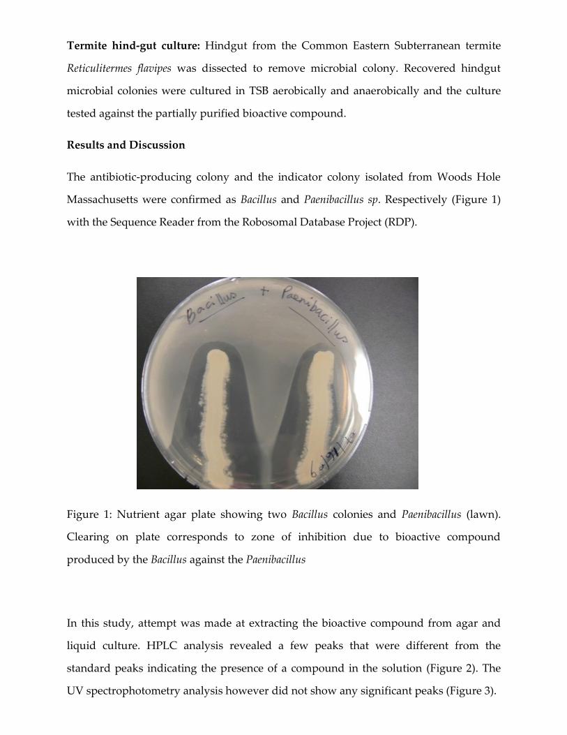
The antibiotic-producing colony and the indicator colony isolated from Woods Hole
Massachusetts were confirmed as Bacillus and Paenibacillus sp. Respectively (Figure 1)
with the Sequence Reader from the Robosomal Database Project (RDP).
Figure 1: Nutrient agar plate showing two Bacillus colonies and Paenibacillus (lawn).
Clearing on plate corresponds to zone of inhibition due to bioactive compound
produced by the Bacillus against the Paenibacillus
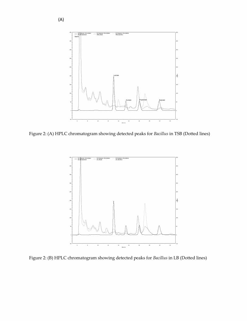
In this study, attempt was made at extracting the bioactive compound from agar and
liquid culture. HPLC analysis revealed a few peaks that were different from the
standard peaks indicating the presence of a compound in the solution (Figure 2). The
UV spectrophotometry analysis however did not show any significant peaks (Figure 3).
(A)
Minutes
6 8 10 12 14 16 18 20 22 24
mV
olts
-50
0
50
100
150
200
250
300
350
400
450
mV
olts
-50
0
50
100
150
200
250
300
350
400
450
Lactate
Acetate Propionate Butyrate
UV Detector Ch1-214nm
20 mM standard
Name
UV Detector Ch1-214nm
TSB_Blank
UV Detector Ch1-214nm
TSB_Bacillus
Figure 2: (A) HPLC chromatogram showing detected peaks for Bacillus in TSB (Dotted lines)
Minutes
6 8 10 12 14 16 18 20 22 24
mV
olts
-50
0
50
100
150
200
250
300
350
400
450
mV
olts
-50
0
50
100
150
200
250
300
350
400
450UV Detector Ch1-214nm
20 mM standard
UV Detector Ch1-214nm
LB_Blank
UV Detector Ch1-214nm
LB_Bacillus
Figure 2: (B) HPLC chromatogram showing detected peaks for Bacillus in LB (Dotted lines)

Acetone Chloroform
Ethyl Acetate Methanol
2-Propanol Water
Figure 3: UV spectrophotometry analysis of bioactive compound from Nutrient agar.
No significant peak was shown for bioactive compound detection
When organic and aqueous extracts were spotted unto Thin Layer Chromatography
plate, no significant signal was detected, however there was indication of presence of
compound in Acetone, methanol and water lanes on the TLC plate.
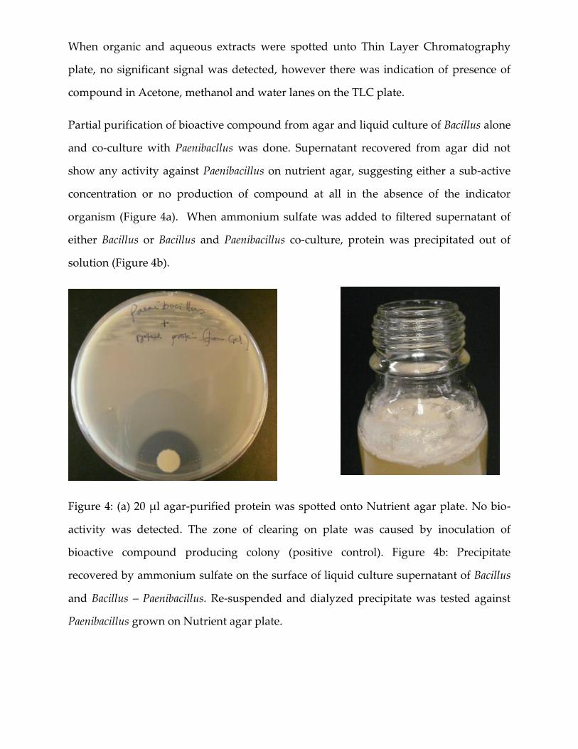
Partial purification of bioactive compound from agar and liquid culture of Bacillus alone
and co-culture with Paenibacllus was done. Supernatant recovered from agar did not
show any activity against Paenibacillus on nutrient agar, suggesting either a sub-active
concentration or no production of compound at all in the absence of the indicator
organism (Figure 4a). When ammonium sulfate was added to filtered supernatant of
either Bacillus or Bacillus and Paenibacillus co-culture, protein was precipitated out of
solution (Figure 4b).
Figure 4: (a) 20 µl agar-purified protein was spotted onto Nutrient agar plate. No bio-
activity was detected. The zone of clearing on plate was caused by inoculation of
bioactive compound producing colony (positive control). Figure 4b: Precipitate
recovered by ammonium sulfate on the surface of liquid culture supernatant of Bacillus
and Bacillus – Paenibacillus. Re-suspended and dialyzed precipitate was tested against
Paenibacillus grown on Nutrient agar plate.

We performed Bradford Assay to determine protein concentration (Figure 5).
Figure 5: Protein standard curve for protein recovered from Bacillus-only culture (B) and
Bacillus-Paenibacillus co-culture (M). BSA standard in shown in blue.
When dialyzed protein from Bacillus-only culture was spotted on Paenibacillus indicator
plate, we detected no activity. However, a zone of inhibition was seen (Figure 6-white
arrow) when protein from Bacillus-Paenibacilus co-culture was tested the indicator
organism, suggesting that the presence of Paenibacillus was necessary for the production
of the bioactive compound from Bacillus. No activity was seen in 1:10 dilution (yellow
arrow) and Tris-HCl buffer alone (not shown).
Figure 6: Nutrient agar plate of Bacillus-Paenibacillus co-culture spotted with 20µl of
protein. Zone of inhibition on plate is shown with arrow.
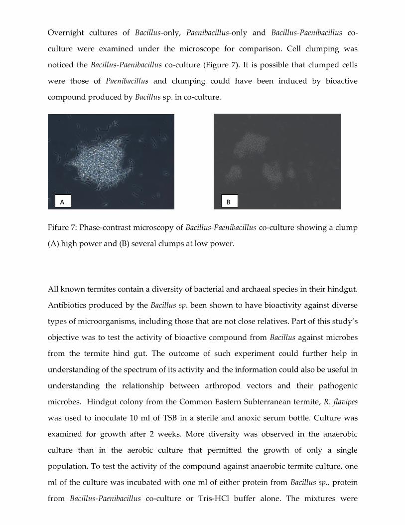
Overnight cultures of Bacillus-only, Paenibacillus-only and Bacillus-Paenibacillus co-
culture were examined under the microscope for comparison. Cell clumping was
noticed the Bacillus-Paenibacillus co-culture (Figure 7). It is possible that clumped cells
were those of Paenibacillus and clumping could have been induced by bioactive
compound produced by Bacillus sp. in co-culture.
Fifure 7: Phase-contrast microscopy of Bacillus-Paenibacillus co-culture showing a clump
(A) high power and (B) several clumps at low power.
All known termites contain a diversity of bacterial and archaeal species in their hindgut.
Antibiotics produced by the Bacillus sp. been shown to have bioactivity against diverse
types of microorganisms, including those that are not close relatives. Part of this study’s
objective was to test the activity of bioactive compound from Bacillus against microbes
from the termite hind gut. The outcome of such experiment could further help in
understanding of the spectrum of its activity and the information could also be useful in
understanding the relationship between arthropod vectors and their pathogenic
microbes. Hindgut colony from the Common Eastern Subterranean termite, R. flavipes
was used to inoculate 10 ml of TSB in a sterile and anoxic serum bottle. Culture was
examined for growth after 2 weeks. More diversity was observed in the anaerobic
culture than in the aerobic culture that permitted the growth of only a single
population. To test the activity of the compound against anaerobic termite culture, one
ml of the culture was incubated with one ml of either protein from Bacillus sp., protein
from Bacillus-Paenibacillus co-culture or Tris-HCl buffer alone. The mixtures were
A B

incubated at 30OC and examined microscopically at two and six hours after incubation.
The results are shown in Table 1 below.
Culture 2hr 6hr
Bacillus + Paenibacillus cells Alive, clumpy same
Protein + Bacillus Alive, happy same
Protein + Paenibacillus Alive, slow Fwere
Mixed protein + Bacillus Alive, slow same
Mixed protein + Paenibacillus Alive, slow same
Buffer + Bacillus Alive, happy same
Buffer + Paenibacillus Alive, happy same
Protein + termite culture Alive, happy same
Protein + termite culture Alive, happy same
Buffer + termite culture Alive, same same
From the table, there was evidence of life in all cultures six hours after incubation
although cell movement was slower when Bacillus and Paenibacillus was co-cultured.
Higher concentration of the bioactive compound and longer incubation times may give
a better picture.
The identity of the bioactive compound produced by the Bacillus sp. is still unknown.
Further analysis by protein electrophoresis and MS/MS mass spectrometry may help to
reveal the identity of the protein.
References
1. Beegle, C.C. and Yamamoto, T. (1992) History of Bacillus thuringiensis
Berliner research and development. Canadian Entomologist 124, 587±616.
2. Cherif, A.. Ouzari, H., Daffonchio, D., Cherif, H., Slama, K.B., Hassen, A., Jaoua, S.,
and Boudabous, A. (2001) Thuricin 7: a novel bacteriocin produced by Bacillus
thuringiensis BMG 1.7, a new strain isolated from soil Letters in Applied
Microbiology 32, 2434–2247.
3. Cherif, A.. Chehimi, S., Limem, F., Hansen, B.M., Hendriksen, N.B., Daffonchio, D.
and Boudabous, A. (2003) Detection and characterization of the novel bacteriocin
entomocin 9, and safety evaluation of its producer, Bacillus thuringiensis ssp.
Entomocidus HD9 Journal of Applied Microbiology 95: 990 – 1000
4. Delves-Broughton, J. (1990) Nisin and its uses as food preservative. Food Technology
44, 100±117.
5. Feitelson, J.S., Payne, J. and Kim, L. (1992) Bacillus thuringiensis: insects and beyond.
Bio/Technology 10, 271±275.
6. Hyronimus, B., Le Marrec, C. and Urdaci, M.C. (1998) Coagulin, a bacteriocin-like
inhibitory substance produced by Bacillus coagulans I4. Journal of Applied
Microbiology 85, 42–50.
7. Jansen, E.F. and Hirschmann, D.J. (1944) Subtilin, an antibacterial substance of
Bacillus subtilis: culturing condition and properties. Archives of Biochemistry 4,
297±309.
8. Klaenhammer, T.R. (1993) Genetics of bacteriocins produced by lactic acid bacteria.
FEMS Microbiology Reviews 12, 39–86.
9. Lovgren, A., Zhang, M.Y., Engstrom, A., Dalhammar, G. and Landen, R. (1990)
Molecular characterization of immune inhibitor
A, a secreted virulence protease from Bacillus thuringiensis. Molecular Microbiology 4,
2137±2146.
10. Maisnier-Patin, S., Deschamps, N., Tatini, S.R. and Richard, J. (1992) Inhibition of
Listeria monocytogenes in Camembert cheese made with a nisin-producing
starter. Lait 72, 249–263.
11. Motta, A.S. and Brandelli, A. (2002) Characterisation of an antimicrobial
peptide produced by Brevibacterium linens. Journal of Applied Microbiology 92,
63–70.
12. Navaratna, M.A.D.B., Sahl, H.G. and Tagg, J.R. (1998) Twocomponent
anti-Staphylococcus aureus lantibiotic activity produced by Staphylococcus
aureus C55. Applied and Environmental Microbiology 64, 4803–4808.
13. Oliveira, S.S., Abrantes, J., Cardoso, M., Sordelli, D. and Bastos, M.C.F. (1998)
Staphylococcal strains involved in bovine mastitis are inhibited by
Staphylococcus aureus antimicrobial peptides. Letters in Applied Microbiology
27, 287–291.
14. Riley MA, Wertz JE (2002) Bacteriocins: evolution, ecology and application. Annu
Rev Microbiol 56:117–137
15. Stabb, E.V., Jacobson, L.M. and Handelsman, J. (1994) Zwittermycin A producing
strains of Bacillus cereus from diverse soils. Applied and Environmental
Microbiology 60, 4404±4412.
16. Valde´s-Stauber, N., Go¨tz, H. and Busse, M. (1991) Antagonistic effect of
coryneform bacteria from red smear cheese against Listeria species. International
Journal of Food Microbiology 13, 119–130.
17. von Tersch, M.A. and Carlton, B.C. (1983) Bacteriocin from Bacillus megaterium
ATCC 19213: comparative studies with megacin A-216. Journal of Bacteriology
155, 872±877.
18. Zheng, G. and Slavik, M.F. (1999) Isolation, partial puri®cation and characterization
of a bacteriocin produced by a newly isolated Bacillus subtilis strain. Letters in
Applied Microbiology 28, 363±367.