final report on aloe vera - images-na.ssl-images-amazon.com · 2 cosmetic ingredient review dermal...
TRANSCRIPT

International Journal of Toxicology, 26(Suppl. 2):1–50, 2007Copyright c© American College of ToxicologyISSN: 1091-5818 print / 1092-874X onlineDOI: 10.1080/10915810701351186
Final Report on the Safety Assessment of Aloe AndongensisExtract, Aloe Andongensis Leaf Juice, Aloe Arborescens LeafExtract, Aloe Arborescens Leaf Juice, Aloe Arborescens LeafProtoplasts, Aloe Barbadensis Flower Extract, AloeBarbadensis Leaf, Aloe Barbadensis Leaf Extract, AloeBarbadensis Leaf Juice, Aloe Barbadensis LeafPolysaccharides, Aloe Barbadensis Leaf Water, Aloe FeroxLeaf Extract, Aloe Ferox Leaf Juice, and Aloe Ferox LeafJuice Extract1
Plant materials derived from the Aloe plant are used as cosmeticingredients, including Aloe Andongensis Extract, Aloe Andongen-sis Leaf Juice, Aloe Arborescens Leaf Extract, Aloe ArborescensLeaf Juice, Aloe Arborescens Leaf Protoplasts, Aloe BarbadensisFlower Extract, Aloe Barbadensis Leaf, Aloe Barbadensis Leaf Ex-tract, Aloe Barbadensis Leaf Juice, Aloe Barbadensis Leaf Polysac-charides, Aloe Barbadensis Leaf Water, Aloe Ferox Leaf Extract,Aloe Ferox Leaf Juice, and Aloe Ferox Leaf Juice Extract. Theseingredients function primarily as skin-conditioning agents and areincluded in cosmetics only at low concentrations. The Aloe leaf con-sists of the pericyclic cells, found just below the plant’s skin, and theinner central area of the leaf, i.e., the gel, which is used for cosmeticproducts. The pericyclic cells produce a bitter, yellow latex con-taining a number of anthraquinones, phototoxic compounds thatare also gastrointestinal irritants responsible for cathartic effects.The gel contains polysaccharides, which can be acetylated, par-tially acetylated, or not acetylated. An industry established limitfor anthraquinones in aloe-derived material for nonmedicinal useis 50 ppm or lower. Aloe-derived ingredients are used in a wide vari-ety of cosmetic product types at concentrations of raw material thatare 0.1% or less, although can be as high as 20%. The concentra-tion of Aloe in the raw material also may vary from 100% to a lowof 0.0005%. Oral administration of various anthraquinone compo-nents results in a rise in their blood concentrations, wide systemicdistribution, accumulation in the liver and kidneys, and excretionin urine and feces; polysaccharide components are distributed sys-temically and metabolized into smaller molecules. aloe-derived ma-terial has fungicidal, antimicrobial, and antiviral activities, and hasbeen effective in wound healing and infection treatment in animals.Aloe barbadensis (also known as Aloe vera)–derived ingredientswere not toxic in acute oral studies using mice and rats. In par-enteral studies, the LD50 using mice was >200 mg/kg, rats was
Received 30 November 2006; accepted 5 March 2007.1Reviewed by the Cosmetic Ingredient Review Expert Panel.
Address correspondence to F. Alan Andersen, Cosmetic Ingredient Re-view, 1101 17th Street, NW, Suite 412, Washington, DC 20036, USA.
>50 mg/kg, and using dogs was >50 mg/kg. In intravenous stud-ies the LD50 using mice was >80 mg/kg, rats was >15 mg/kg, anddogs was >10 mg/kg. The 14-day no observed effect level (NOEL)for the Aloe polysaccharide, acemannan, in the diet of Sprague-Dawley rats, was 50,000 ppm or 4.1 to 4.6 g/kg day−1. In a 3-monthstudy using mice, Aloe vera (extracted in ethanol) given orally indrinking water at 100 mg/kg produced reproductive toxicity, in-flammation, and mortality above that seen in control animals. Aloevera extracted in methanol and given to mice at 100 mg/kg in drink-ing water for 3 months caused significant sperm damage comparedto controls. Aloe barbadensis extracted with water and given topregnant Charles Foster albino rats on gestational days (GDs) 0through 9 was an abortifacient and produced skeletal abnormal-ities. Both negative and positive results were found in bacterialand mammalian cell genotoxicity assays using Aloe barbadensis–derived material, Aloe Ferox–derived material, and various an-thraquinones derived from Aloe. Aloin (an anthraquinone) did notproduce tumors when included in the feed of mice for 20 weeks, nordid aloin increase the incidence of colorectal tumors induced with1,2-dimethylhydrazine. Aloe-emodin (an anthraquinone) given tomice in which tumor cells had been injected inhibited growth ofmalignant tumors. Other animal data also suggest that compo-nents of Aloe inhibit tumor growth and improve survival. Vari-ous in vitro assays also demonstrated anticarcinogenic activity ofaloe-emodin. Diarrhea was the only adverse effect of note with theuse of Aloe-derived ingredients to treat asthma, ischemic heart dis-ease, diabetes, ulcers, skin disease, and cancer. Case reports includeacute eczema, contact urticaria, and dermatitis in individuals whoapplied Aloe-derived ingredients topically. The Cosmetic Ingredi-ent Review Expert Panel concluded that anthraquinone levels inthe several Aloe Barbadensis extracts are well understood and canconform to the industry-established level of 50 ppm. Although thephototoxicity anthraquinone components of Aloe plants have beendemonstrated, several clinical studies of preparations derived fromAloe barbadensis plants demonstrated no phototoxicity, confirm-ing that the concentrations of anthraquinones in such prepara-tions are too low to induce phototoxicity. The characterization ofaloe-derived ingredients from other species is not clear. In the ab-sence of well-characterized derivatives, biological studies of thesematerials are considered necessary. The studies needed are 28-day
1

2 COSMETIC INGREDIENT REVIEW
dermal toxicity studies on Aloe Andongensis Extract, Aloe Andon-gensis Leaf Juice, Aloe Arborescens Leaf Extract, Aloe ArborescensLeaf Juice, Aloe Ferox Leaf Extract, Aloe Ferox Leaf Juice, andAloe Ferox Leaf Juice (ingredients should be tested at current useconcentrations). In Aloe-derived ingredients used in cosmetics, re-gardless of species, anthraquinone levels should not exceed 50 ppm.The Cosmetic Ingredient Review Expert Panel advised the industrythat the total polychlorobiphenyl (PCB)/pesticide contamination ofany plant-derived cosmetic ingredient should be limited to not morethan 40 ppm, with not more than 10 ppm for any specific residueand that limits were appropriate for the following impurities: ar-senic (3 mg/kg maximum), heavy metals (20 mg/kg maximum), andlead (5 mg/kg maximum).
INTRODUCTIONAloe Andongensis Extract, Aloe Andongensis Leaf Juice,
Aloe Arborescens Leaf Extract, Aloe Arborescens Leaf Juice,Aloe Arborescens Leaf Protoplasts, Aloe Barbadensis FlowerExtract, Aloe Barbadensis Leaf, Aloe Barbadensis Leaf Extract,Aloe Barbadensis Leaf Juice, Aloe Barbadensis Leaf Polysac-charides, Aloe Barbadensis Leaf Water,Aloe Ferox Leaf Extract,Aloe Ferox Leaf Juice, and Aloe Ferox Leaf Juice Extract arecosmetic ingredients derived from Aloe. This review presentsinformation relevant to the safety of these Aloe-derived ingre-dients as considered by the Cosmetic Ingredient Review (CIR)Expert Panel.
The general Aloe CAS number is 8001-97-6. Aloe barbaden-sis extract has the CAS number 85507-69-3. As described in theInternational Cosmetic Ingredient Dictionary and Handbook(Gottschalck and McEwen 2004), these ingredients function pri-marily as skin-conditioning agents or have no specific functionlisted. In most cases, the definitions of these ingredients are tau-tologies, e.g., Aloe Barbadensis Flower Extract is an extract ofthe flowers of Aloe barbadensis.
CHEMISTRY
DefinitionPlant material derived from the aloe plant is characterized
by its source (e.g., what part of the plant), the species of plant,the physical description of the material, and by the constituentsfound in the material.
The definitions of each ingredient, according to the In-ternational Cosmetic Ingredient Dictionary and Handbook(Gottschalck and McEwen 2004), are:
Aloe Andongensis Extract is the extract of the leaves of Aloe’andongensis.
Aloe Andongensis Leaf Juice is the liquid expressed from theleaves of Aloe andongensis.
Aloe Arborescens Leaf Extract is the extract of the leaves ofAloe arborescens.
Aloe Arborescens Leaf Juice is the juice expressed from theleaves of Aloe arborescens.
Aloe Arborescens Leaf Protoplasts are the protoplasts ob-tained form the leaves of Aloe arborescens.
Aloe Barbadensis Flower Extract is the extract of the flowersof Aloe barbadensis.
Aloe Barbadensis Leaf is a plant material derived from theleaves of Aloe barbadensis.
Aloe Barbadensis Leaf Extract is an extract of the leaves ofAloe barbadensis.
Aloe Barbadensis Leaf Juice is the juice expressed from theleaves of Aloe barbadensis.
Aloe Barbadensis Leaf Polysaccharides is the polysaccharidefraction isolated from the leaf of Aloe barbadensis.
Aloe Barbadensis Leaf Water is an aqueous solution of theodoriferous principles distilled from the leaves of Aloe bar-badensis.
Aloe Ferox Leaf Extract is an extract of the leaves of Aloeferox.
Aloe Ferox Leaf Juice is the juice expressed from the leaves ofAloe ferox.
Aloe Ferox Leaf Juice Extract is an extract of the juice of theleaf of Aloe ferox.
Table 1 gives a list of synonyms for these ingredients as givenin the International Cosmetic Ingredient Dictionary and Hand-book (Gottschalck and McEwen 2004). Aloe vera is a frequentlyused synonym for Aloe barbadensis.
Composition and Physical and Chemical PropertiesGjerstad (1969) described Aloe as the dried juice of the lower
portion of the leaves of any three geographical varieties of theAloe genus. The juice appears blackish-brown, opaque, andsmooth.
Ghannam et al. (1986) described the solid residue of Aloebarbadensis (native to Mediterranean countries and the SaudiArabian peninsula) obtained by evaporating the sap drained fromthe cut leaves. This drained latex solidifies and turns brown onexposure to air. It contains anthraquinone glycosides, barbaloin,and β-barbaloin, the hydrolysis of which yields aloe-emodin andd-arabinose.
Natow (1986) stated that Aloe is the name given to the genusthat includes more than 300 plants, grown all over the world.This report considers ingredients from four species: andongen-sis, arborescens, barbadensis, and ferox.
Klein and Penneys (1988), Briggs (1995), and the M.D. An-derson Cancer Center (2003) described the leaf of Aloe plantsas consisting of two main parts. One part, the pericyclic cells, isfound just below the plant’s skin. The pericyclic cells produce abitter, yellow latex known as Aloe juice, or latex. When this juicedries it forms a dark brown solid material. The latex containsthe anthraquinone, emodin, a gastrointestinal irritant responsi-ble for cathartic effects. The second part, the inner central areaof the leaf, contains the thin walled parenchymal cells that pro-duce the clear slightly viscous (mucilaginous) fluid known asAloe gel or inner gel. This gel contains the polysaccharides.There are generally more than one polysaccharide found in the

ALOE 3
TABLE 1List of synonyms (Gottschalck and McEwen 2004)
Ingredient Synonyms
Aloe Andongensis Extract Aloe ExtractExtract of Aloe andongensis
Aloe Andongensis Leaf Juice Aloe andongensisAloe Arborescens Leaf Extract Aloe arborescens
Kidachi AloeAloe Arborescens Leaf Juice Aloe arborescens
Kidachi Aloe KajyuKidachi Aloe YoujyuKidachi Aloe Youjyu Masto
Aloe Arborescens Leaf Protoplasts Aloe arborescensAloe arborescens CRS
Aloe Barbadensis Flower Extract Aloe barbadensisAloe barbadensis ExtractAloe Flower ExtractExtract of Aloe barbadensis FlowerExtract of Aloe Flowers
Aloe Barbadensis Leaf AloeAloe barbadensisAloe Leaf PowderAloe vera
Aloe Barbadensis Leaf Extract Aloe barbadensisAloe ExtractBarbados Aloe ExtractCuracao Aloe ExtractExtract of AloeExtract of Aloe barbadensisExtract of Aloe leavesAloe vera
Aloe Barbadensis Leaf Juice Aloe barbadensisAloe GelAloe barbadensis GelAloe JuiceAloe vera Gel
Aloe Barbadensis Leaf Polysaccharides Aloe barbadensisAloe Barbadensis Leaf Water Aloe barbadensisAloe Ferox Leaf Extract Aloe ferox
Extract of Aloe feroxCape Aloe
Aloe Ferox Leaf Juice Aloe feroxCape Aloe
Aloe Ferox Leaf Juice Extract Aloe feroxCape Aloe Ekisu
gel; they can be acetylated, partially acetylated, or not acety-lated. The gel portion (Aloe leaf juice) is the part of the plantgenerally used for emollient and moisturizing effects desired incosmetics, and the healing action desired for certain medicinalproducts.
The International Aloe Science Council (IASC) distinguishedbetween the anthraquinone found in the outer cell layer of thealoe leaf and the rest of the plant (IASC 2003). According tothis group, the maximum allowable aloin content in aloe-derivedmaterial for nonmedicinal use is 50 ppm or lower.

4 COSMETIC INGREDIENT REVIEW
TABLE 2Description of Aloe products (Carrington Labs 2001)
Product name Appearance Odor Taste pH
Aloe vera Whole Leaf 1:1 Light amber liquid Light vegetable Slight bitter vegetable 3.0–4.0Aloe vera gel 1:1 decolorized Clear, slightly translucent Light vegetable Slight characteristic 3.5–5.0
vegetableAloe vera gel 10:1 decolorized Semiviscous, light to medium amber Light vegetable Slight vegetable 3.5–5.0Aloe vera gel 40:1 decolorized Semiviscous, light to medium amber Light vegetable Slight vegetable 3.5–5.0Aloe vera Gel Freeze Dried White to light tan powder Not given Not given Not given
Powder 200:1
Danhof (1998) stated that aloe polysaccharides consist oflinear chains of β-1-4-linked glucose and mannose molecules, inwhich mannose predominates. Chain lengths may be as small asfrom a few molecules up to several thousand, but by conventionthe lower end of the molecular weight is usually considered to be1000 daltons, consistent with being a polysaccharide. Dependingon the particular chain length, this material will have differentphysical properties.
The IASC (1998) maintains a certification program in which“whole leaf aloe vera gel” is expected to adhere to the followingspecifications: solids between 0.46 to 1.31%; pH between 3.5and 4.7; calcium between 98.2 and 448 mg/L; magnesiumbetween 23.4 and 118 mg/L; and malic acid between 817.8 and3427.8 mg/L. Consistent with these specifications, Rowe andParks (1941) determined the pH of fresh Aloe vera leaves to be4.7.
TABLE 3Constituents of Aloe Vera (Shelton 1991; Vogler and Ernst 1999)
Nonessential Inorganic EssentialAnthraquinones Saccharides Vitamins amino acids compounds Enzymes amino acids Miscellaneous
Aloin Cellulose B1 Histidine Calcium Cyclooxygenase Lysine CholesterolBarbaloin Glucose B2 Arginine Sodium Oxidase Threonine TriglyceridesIsobarbaloin Mannose B6 Hydroxyproline Chlorine Amylase Valine SteroidsAnthranol L-Rhamnose Choline Aspartic Acid Manganese Catalase Leucine β-SitosterolAloetic Acid Aldopentose Folic Acid Glutamic Acid Zinc Lipase Isoleucine LigninsCinnamic Acid C Proline Chromium Alkaline Phenylalanine Uric Acid
Ester phosphataseAloe-emodin α-Tocopherol Glycine Copper Carboxypeptidase Methionine GibberellinEmodin β-Carotene Alanine Magnesium Lectin-like
substanceChrysophanic Tyrosine Iron Salicylic Acid
AcidResistannol Arachidonic AcidAnthracene Potassium SorbateEthereal Oil
Table 2 presents descriptions and chemical properties of com-mercial Aloe products from one company analyzed by Carring-ton Labs (2001). All products listed in the table are consid-ered acceptable for use in cosmetics, and are derived from Aloebarbadensis.
Active Organics (2002) described a trade name product, Ac-tiphyte, of Aloe Vera 10 Fold, as consisting of Propylene Glycoland Aloe Barbadensis Leaf Extract. The product was a clear topale yellow liquid, with a characteristic odor, and a flash point of228◦F, boiling point of 225◦F, specific gravity of 1.02 to 1.05 (at25◦C), complete solubility in water, refractive index of 1.3620to 1.3700 (at 25◦C), and a pH of 4.0 to 6.5 (at 25◦C).
As noted earlier, aloe-derived ingredients also are character-ized by their specific constituents. The constituents reported byShelton (1991) and Vogler and Ernst (1999) found in Aloe veraare listed in Table 3.
ALOE 5
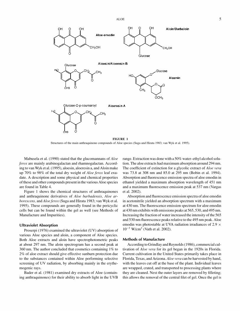
FIGURE 1Structures of the main anthraquinone compounds of Aloe species (Suga and Hirata 1983; van Wyk et al. 1995).
Mabusela et al. (1990) stated that the glucomannans of Aloeferox are mainly arabinogalactan and rhamnogalactan. Accord-ing to van Wyk et al. (1995), aloesin, aloeresin a, and Aloin makeup 70% to 98% of the total dry weight of Aloe ferox leaf exu-date. A description and some physical and chemical propertiesof these and other compounds present in the various Aloe speciesare found in Table 4.
Figure 1 shows the chemical structures of anthraquinonesand anthraquinone derivatives of Aloe barbadensis, Aloe ar-borescens, and Aloe ferox (Suga and Hirata 1983; van Wyk et al.1995). These compounds are generally found in the pericycliccells but can be found within the gel as well (see Methods ofManufacture and Impurities).
Ultraviolet AbsorptionProserpi (1976) examined the ultraviolet (UV) absorption of
various Aloe species and aloin, a component of Aloe species.Both Aloe extracts and aloin have spectrophotometric peaksat about 297 nm. The aloin spectrogram has a second peak at360 nm. The author concluded that cosmetics containing 1% to2% of aloe extract should give effective sunburn protection dueto the substances contained within Aloe performing selectivescreening of UV radiation, by absorbing mainly in the erythe-mogenic rays.
Bader et al. (1981) examined dry extracts of Aloe (contain-ing anthraquinones) for their ability to absorb light in the UVB
range. Extraction was done with a 50% water–ethyl alcohol solu-tion. The aloe extracts had maximum absorption around 294 nm.The coefficient of extinction for a glycolic extract of Aloe verawas 73.8 at 308 nm and 85.0 at 295 nm (Bobin et al. 1994).Absorption and fluorescence emission spectra of aloe emodin inethanol yielded a maximum absorption wavelength of 451 nmand a maximum fluorescence emission peak at 537 nm (Vargaset al. 2002).
Absorption and fluorescence emission spectra of aloe emodinin acetonitrile yielded an absorption spectrum with a maximumat 430 nm. The fluorescence emission spectrum for aloe emodinat 430 nm exhibits with emissions peaks at 565, 530, and 495 nm.Increasing the fraction of water increased the intensity of the 565and 530 nm fluorescence peaks relative to the 495 nm peak. Aloeemodin was photostable at UVA radiation irradiances of 2.9 ×10⁻³ W/cm2 (Vath et al. 2002).
Methods of ManufactureAccording to Grindlay and Reynolds (1986), commercial cul-
tivation of Aloe vera for its gel began in the 1920s in Florida.Current cultivation in the United States primarily takes place inFlorida, Texas, and Arizona. Aloe vera can be harvested by hand,with the leaves cut off at the base of the plant. Individual leavesare wrapped, crated, and transported to processing plants wherethey are cleaned. Next the outer layers are removed by filleting;this allows the removal of the central filet of gel. Once the gel is

TA
BL
E4
Phys
ical
and
chem
ical
prop
ertie
sof
aloe
cons
titue
nts
UV
abso
rptio
nM
eltin
gC
ompo
nent
Che
mic
alID
MW
peak
spo
int
Des
crip
tion
Ref
eren
ce
Ace
man
nan
β-(
1,4)
acet
ylat
edpo
lym
anno
sew
ithin
ters
pers
edO
-ace
tylg
roup
s80
,000
——
Lon
gch
ain,
poly
disp
erse
dM
cAna
lley
1990
aka
Car
n75
0,C
arn
1000
,Po
lym
anno
acet
ate,
Alim
inas
e,A
love
x,C
arri
syn
Ace
tyla
ted
Man
nan
—40
,000
——
—Y
agie
tal.
1977
Alo
ctin
AL
ectin
s18
,000
——
—Su
zuki
etal
.197
9A
loct
inB
24,0
00—
——
Alo
e-em
odin
1,8-
Dih
ydro
xy-3
-hyd
roxy
met
hyl-
9,10
-ant
hrac
ened
ione
270.
2322
1,25
3,26
6,28
9,48
3nm
inet
hano
l22
0–22
1W
ater
solu
ble;
depe
ndin
gon
the
conc
entr
atio
nco
lor
can
bere
dth
roug
hbr
own
tobl
ack
Hir
ata
and
Suga
1977
;B
udav
ari1
989;
Stri
ckla
ndet
al.2
000
1,8-
Dih
ydro
xy-3
-(hy
drox
ymet
hyl)
-9,
10-a
nthr
acen
edio
ne1,
8-D
ihyd
roxy
-3-
hydr
oxym
ethy
lant
hraq
uino
ne3-
Hyd
roxy
met
hylc
hrys
azin
,1,
8-di
hydr
oxy-
3-(h
ydro
xym
ethy
l)an
thra
quin
one
1,8-
Dih
ydro
xy-3
-hyd
roxy
met
hyl-
anth
raqu
inon
eC
15H
10O
5
Alo
efer
on—
70,0
00—
——
Mad
iset
al.1
989
Alo
eman
nan
Glu
com
anna
n37
4,00
0—
——
Gow
daet
al.1
979
Alo
eM
anna
nPa
rtia
llyac
etyl
ated
β-D
-man
nan
15,0
00—
—W
ater
solu
ble
Yag
ieta
l.19
77A
loen
in(a
loea
rbon
asid
e)4-
Met
hoxy
-6[1
0-hy
drox
y-12
-met
hyl-
8-O
- D-g
luco
pyra
nosy
l]ph
enyl
-py
rone
-2
—23
2,24
7,30
7in
etha
nol;
302
inm
etha
nol
145–
147
Bitt
ergl
ucos
ide
Suga
and
Hir
ata
1972
;M
akin
oet
al.1
973;
Hir
ata
and
Suga
1977
;H
irat
aet
al.1
981;
Gut
term
an20
00O
-glu
cosi
deC
19H
22O
10·H
2O
Alo
eres
inB
Syno
nym
for
Alo
esin
——
——
Mak
ino
etal
.197
3A
loer
esin
A5-
Met
hylc
hrom
one
C-g
lyco
side
——
148–
150
O-p
-cou
mar
oyl
deri
vativ
eof
Alo
esin
Gra
mat
ica
etal
.198
2
Alo
erid
ePo
lysa
ccha
ride
4–7
×10
6—
—So
lubl
ew
hite
pow
der
Mad
iset
al.1
989
6

Alo
esin
2-A
cety
lony
l-8-
β-D
-glu
copr
anyo
syl-
7-hy
drox
y-5-
met
hylc
hrom
one
394
248,
254,
297
inet
hano
l;21
6,34
0in
met
hano
l14
2–14
4W
ater
solu
ble
McC
arth
yan
dH
ayne
s19
67;H
olds
wor
th19
71;H
irat
aan
dSu
ga19
77;v
anW
yket
al.
1995
;Gut
term
an20
00C
-glu
cosy
lchr
omon
eC
17H
18O
9
Alo
in10
β-D
-glu
copr
yano
syl-
1,8-
dihy
drox
y-3-
hydr
oxym
ethy
l-9(
10H
)an
thra
ceno
neC
21H
22O
9
418.
3625
0–29
0in
met
hano
l,pe
akat
260
148–
149
Bitt
erju
ice
that
drie
sto
aye
llow
pow
der;
can
behy
drol
yzed
toal
oe-e
mod
in;i
nclu
des
Alo
inA
and
Alo
inB
dias
tere
omer
s;sl
ight
lyso
lubl
ein
wat
eran
dor
gani
cso
lven
ts
Hay
and
Hay
nes
1956
;Jo
intC
omm
ittee
1967
;M
cCar
thy
1969
;H
irat
aan
dSu
ga19
77;
Bud
avar
i198
9;Sh
elto
n19
91;v
anW
yket
al.1
995;
Koc
h,19
96;V
iljoe
net
al.
2001
Alo
esp
ecie
sex
trac
t;ak
aA
loe
spec
ies
resi
nD
ried
Alo
eba
rbad
ensi
sM
iller
orA
loe
fero
xM
iller
——
—W
ater
extr
acte
d;br
own
fine
pow
der;
tapp
edde
nsity
0.5g
/ml;
part
icle
size
notl
ess
than
90%
<30
0µ
m
Ham
mer
Phar
ma
2002
Arb
oran
sA
Gly
cans
with
asm
alla
mou
ntof
O-a
cety
lgro
ups
1.2
×10
4—
——
Hik
ino
etal
.198
6
Arb
oran
sB
5.7
×10
4
Ver
ectin
Gly
copr
otei
n29
,000
——
—Y
agie
tal.
1997
,200
0
7

8 COSMETIC INGREDIENT REVIEW
removed, the cell walls, lignified fibers, and other contaminantsare removed by either squeezing and filtering or by a decantationprocess. When removing the gel, care needs to be taken to notcontaminate the gel with the green rind. Because the gel’s activ-ity becomes unstable after removal from the leaves, a numberof processes have been developed to overcome this instability.One method used to stabilize the gel is to expose the gel to hightemperatures for a short time (3 min). Ultraviolet stabilization,chemical oxidation with hydrogen peroxide, and preservativesand additives are other methods of retaining the gel’s activity.
A method of manufacture patented by McAnalley (1990)isolates the gel filet of Aloe vera and can further isolate the activeingredient carrisyn (also known as acemannan) (see Table 3).According to this method, the leaf, cut from the base of the Aloeplant, is washed in a bactericidal solution and the end portion ofthe leaf is removed to allow the anthraquinone-rich sap to drainout, additionally the rind is removed. McAnalley’s method alsoallows for the leaf of the plant to be crushed and then dialyzedto remove the anthraquinones. The gel filet that is left behind isground and homogenized to produce aloe juice. The juice is thenfiltered to remove fibrous material. At this point the juice canhave preservatives, flavors, excipient carriers, and/or colorantadded to it. To further isolate the active ingredient, an aliphaticpolar solvent is added to precipitate out carrisyn. Carrisyn isthen sterilized and eventually dried by lyophilization.
The process used by Aloecorp involves soaking the justharvested leaves in a food-grade sanitizer to reduce microbialcounts, the gel is then extracted, flash cooled to 5◦C, and thenconcentrated while under vacuum. The concentrated gel is thenfreeze dried (Aloecorp 2001).
Agarwala (1997) stated that the primary difference betweenwhole leaf processing versus inner gel processing from the whole
TABLE 5Comparison of inner gel and whole leaf material (Agarwala 1997)
Property Inner gel Whole leaf
Soluble solids 0.62% 1.3–3.5%pH 4.5 4.2Nitrogen (as ammonia) 12 6Ca2+ (mg/L) 340 600Mg2+ (mg/L) 60 100K+ (mg/L) 390 750Conductivity 1200–2300 µ 1900–2500 µ
Taste Bland SaltyMethanol precipitable solids (MPS) 0.12–0.16% 0.45–1.3%Ratio of MPS to total solids 25–35 30–40Ratio of ethanol precipitable solids to total solids 6–10 1–10HPLC peaks E-peak E-peak; unidentified peak at 21–31 minUV absorption peak 224 nm 254 nmMonosaccharide Galactan and glucomannan Glucomannan
leaf is that for the inner gel, leaves are manually or machinefilleted before further processing. In both processes, the wholeleaf is cut and quickly transported to the processing center. Afterwashing, the whole leaves are either ground through a hammermill and directed into a heating vat, or filleted to remove the innergel. Heating is done to reduce the amount of slime. Heating cantake place one of two ways: at low temperature for a long time, orat high temperature for a short time. After heating, the slurry isfiltered through a screw press or cloth press. After partial coolingtakes place, activated charcoal is added (decolorizes and adsorbsthe bitter components). For complete clarity, the gel is filteredthrough diatomaceous earth.
For the inner gel, this process results in 0.5% solids, of which0.1% are large molecules such as polysaccharides, glycopro-teins, and proteins, and 0.4% of which are small moleculessuch as simple sugars, organic and inorganic salts, and nitroge-nous compounds. For the whole leaf, the process results in1.5% solids, of which 0.05% are large molecules and 1.45%are small molecules. The practical ramification of these twotechniques is that for each ton of whole leaves, processing thewhole leaf could produce around 25 to 30 pounds of total solu-ble solids from the gel component, while filleting the leaf beforeprocessing could produce around 5 pounds. The author statesthat some manufacturers do process the whole leaf, taking carein the process to remove bitter anthraquinones from the finalproduct.
Table 5 shows the different properties of material derivedfrom the inner gel versus the whole leaf (Agarwala 1997).
Meadows (1980) used 2% sodium benzoate and 0.15% ascor-bic acid to preserve aloe gel. Agarwala (1997) stated that themost commonly used preservatives are sodium benzoate andpotassium sorbate with pH adjusted to <4.6 with citric acid.

ALOE 9
Aloe extracts and aloe gel extracts can be freed of an-thraquinones by means of activated charcoal and filleting pro-cesses (UNITIS 2003).
Aloe ferox suitable for cosmetic purposes can be manufac-tured by two methods: (1) percolation with hot water at 60◦Cand concentrated under vacuum to dryness, or (2) percolationwith propylene glycol and concentrated under vacuum (Patri andSilano 2002).
Analytical MethodsNakamura and Okuyama (1990) detected aloenin, a com-
ponent of Aloe arborescens, in cosmetics (lotions and creams)by gas chromatography with mass fragmentography. Kuzuyaet al. (2001) determined the presence of aloenin, barbaloin,
and isobarbaloin (all components of Aloe arborescens) in com-mercial products using micellar electrokinetic chromatogra-phy. Reverse-phase high-performance liquid chromatography(HPLC) and paper or thin-layer chromatography (TLC) havebeen used to detect barbaloin content within Aloe plants (Ishiiet al. 1984).
Reverse-phase HPLC and paper or thin-layer chromatogra-phy have been used to detect the various components of Aloebarbadensis (Hutter et al. 1996; Holdsworth 1971). Ross et al.(1997) used size exclusion chromatography to examine variousproducts (cosmetic and non-cosmetic) for the levels of Aloe veramucilaginous polysaccharide.
According to the IASC (2001a), polymerase chain reactionfor the detection of Aloe DNA can allow for identification ofplant species.
Leaf exudate samples from Aloe ferox were collected byapplying slips of filter paper to a cut leaf and were analyzedby HPLC. The major leaf exudate compounds detected werealoesin, aloeresin, aloin B, aloin A, aloinoside B, and aloinosideA (Viljoen et al. 2001). Van Wyk et al. (1994) analyzed com-mercial samples (type not described) by HPLC and found theconcentration of Aloe ferox constituents in the products were notconsistent.
Waller et al. (1978) showed that Aloe barbadensis leaves con-tain various amino acids, with highest concentration of arginine(449 µmol/100 g), followed by asparagine (344 µmol/100 g),glutamate (294 µmol/100 g), aspartate (237 µmol/100 g), andserine (224 µmol/100 g). The authors also found that mannoseand glucose were in a molar ratio of 5:4 and trace amounts ofxylose, rhamnose, galactose, and either arabinose or fructosewere also present.
Robson et al. (1982) performed a detailed chemical analysisof 99.5% pure Aloe vera extract and found organic compoundssuch as glucose (13 mg/dl), uric acid (0.5 mg/dl), salicylicacid (3.6 mg/dl), creatinine (1.9 mg/dl), alkaline phosphatase(1 IU/L), creatinine phosphokinase (10 IU/L), cholesterol(11 mg/dl), triglycerides (374 mg/dl), lactate (14.8 mg/dl), andprotein (0.2 mg/dl). Inorganic constituents found were sodium(19 mEq/L), potassium (21.5 mEq/L), inorganic phosphorus
(14 mg/dl), and chloride (1 mEq/L). Trace metals present inthe extract were calcium (23.5 mEq/L), magnesium (4.6 mg/dl),copper (0.2 mg/dl), and zinc (0.02 mg/dl). Aloe contains a num-ber of anthraquinone glycosides, the principal one of which isbarbaloin (aloe-emodin anthrone C-10 glucoside) (Tyler et al.1988). O-Glycosides of barbaloin with an additional sugar alsohave been isolated from certain samples of Cape aloe. Thesecompounds have been designated aloinosides. Free (nonglycosi-dal) aloe-emodin and a free and combined anthranol are alsopresent. Chrysophanic acid has been detected in certain typesof aloe. The active constituents of aloe vary qualitatively andquantitatively according to the species from which the mate-rial is obtained. In addition to the physiologically active com-pounds (10% to 30%), aloe contains inactive ingredients includ-ing large amounts (16% to 63%) of a resinous material plus avolatile oil.
Aloe-emodin can be determined using HPLC and ranges be-tween 2.9 and 3.7 ppm in Aloe arborescens (UNITIS 2003).
ImpuritiesDanof and McAnalley (1983) were able to show that the yel-
low sap (containing the anthraquinones) was lethal in humanfibroblast assays; care should be taken to keep it out of commer-cial products. Examples of anthraquinones and anthraquinonederivatives include aloin, aloe-emodin, aloesin/aloeresin B, alo-eresin A, aloin/barbaloin, and aloenin.
CTFA (2002) reported a study where the amount of aloe-emodin present in Aloe Arborescens CRS was determined. AloeArborescens CRS was filtered to remove insoluble componentsand the liquid component was analyzed using high pressure liq-uid chromatography and compared to a standard aloe-emodinsolution. It was found that samples 1 and 2 from the filteredAloe Arborescens CRS contained an average (of two runs) of2.9 and 3.7 ppm aloe-emodin, respectively.
The International Aloe Science Council (2001b) considersthe presence of maltodextrin, lactobacillus, lactic acid, or aceticacid to be a sign of bacterial contamination of commercial prod-ucts.
Nakano et al. (1985) detected various levels of aloenin in15 commercial cosmetic lotions by HPLC analysis. Table 6lists the results of the HPLC analysis on 5 µl of the com-mercial lotions. Nakamura and Okuyama (1990) detected theanthraquinone aloenin in several commercial skin care lotions,creams, etc. (0.5-g samples). The average recoveries were 94%with a relative standard deviation of 4% to 7% and a detectionlimit of 0.02 µg/g. The aloenin content of the cosmetic productsare shown in Table 7.
The values in Tables 6 and 7 may be compared with the IASC(1998) stated limit of 50 ppm (equivalent to µg/ml and µg/g inthe tables).
Kim et al. (1998) developed simple and accurate methods todetect adulteration of commercial Aloe gel products by otherplant polysaccharides. Isolation of crude polysaccharides from

10 COSMETIC INGREDIENT REVIEW
TABLE 6Aloenin levels in commercial lotions
(Nakano et al. 1985)
Product Aloenin (µg/ml)
A 32.4B 17.7C 4.8D 1.2E 13.0F 0.8G 0.8H 0.7I 0.6J 0.5K 0.4L 0.3M <0.2N <0.2O <0.2
commercial aloe gel powders was performed by precipitationwith ethyl alcohol. Analyses of free sugars was by gas chro-matography and quantification of total hexose was by the DuBoisassay. Maltodextrin was detected by two methods, TLC andHPLC (maltodextrin in Aloe products is considered to be a con-taminant).
1H nuclear magnetic resonance (NMR) can be used to detectthe presence of lactic acid, acetic acid, and formic acid in Aloevera products. All three acids are undesirable ingredients in Aloevera products (Diehl and Teichmuller 1998).
TABLE 7Aloenin levels in commercial products
(Nakamura and Okuyama 1990)
Sample Aloenin content (µg/g)
Lotion 1 42Lotion 2 1.2Lotion 3 0.80Milky Lotion 1 1.7Milky Lotion 2 0.96Cream 1 2.3Cream 2 3.5Cream 3 4.0Cream 4 0.25Cream 5 1.8Face Pack 1 2.3Face Pack 2 4.8Hair Rinse 1.6Hair Tonic 6.2
USE
CosmeticAs given in the International Cosmetic Ingredient Dictio-
nary and Handbook (Gottschalck and McEwen 2004), Aloe An-dongensis Extract and Aloe Andongensis Leaf Juice functionas a skin-conditioning agent—humectant; Aloe ArborescensLeaf Juice, Aloe Barbadensis Flower Extract, Aloe BarbadensisLeaf Juice, Aloe Ferox Leaf Extract, Aloe Ferox Leaf Juice,Aloe Ferox Leaf Juice Extract and Aloesin all function asskin-conditioning agents—miscellaneous in cosmetic formu-lations. Aloe Barbadensis Leaf Extract functions as an exter-nal analgesic, humectant, oral care agent, skin-conditioningagent—miscellaneous, and skin-conditioning agent—emollientin cosmetic products. Aloe Barbadensis Leaf Polysaccharidesfunctions as film former, humectant, skin-conditioning agent—emollient, and skin conditioning agent—humectant. Aloe Bar-badensis Leaf Water functions as a fragrance ingredient. Thefunction of Aloe Arborescens Leaf Extract and Aloe Barbaden-sis Leaf in cosmetic formulations was not reported.
The frequency of Aloe use reported to the Food and DrugAdministration (FDA) in 2001 is shown in Table 8 (FDA 2001).Unfortunately, the terminology of aloe-derived ingredients inthe categories in the FDA database do not correspond to thoseused by industry (CTFA 2002). To merge the two pieces of in-formation, the following assumptions were made: (1) Aloe Ex-tract reported to FDA meant Aloe Barbadensis Leaf Extract; (2)Aloe reported to FDA meant Aloe Barbadensis Leaf; (3) AloeArborescens reported to FDA meant Aloe Arborescens Leaf Ex-tract; (4) Aloe Flower Extract reported to FDA meant Aloe Bar-badensis Flower Extract; (5) Aloe Gel reported to FDA meantAloe Barbadensis Leaf Juice; Aloe, Powdered reported to FDAmeant all ingredients referred to as powders in the CTFA (2002)submission; and (6) that there was nothing reported to FDA com-parable to the current industry data on Aloe Ferox Leaf Extract.
According to the Japan Ministry of Health, Labor and Welfare(MHWL), the Aloe ingredients reviewed in this report are notincluded on the list of ingredients that must not be combined incosmetic products that are marketed in Japan (MHLW 2001a)or on the restricted ingredient list for cosmetic products thatare marketed in Japan (MHLW 2001b). In addition, none ofthe Aloe ingredients found in this report are restricted from usein any way under the rules governing cosmetic products in theEuropean Union (European Commission 2002).
NoncosmeticThe FDA has determined that Aloe perryi, Aloe barbadensis,
Aloe ferox, and hybrids of this species with Aloe africana andAloe spicata are food additives permitted for direct addition tofood for human consumption as natural flavoring substances (21CFR 172.510).
According to Duke and Beckstrom-Sternberg (1994) the Fla-vor and Extract Manufacturers’ Association acceptable level forAloe vera was 5 to 2000 ppm.

ALOE 11
TABLE 8Product formulation data for aloe-derived ingredients
Product category(number of formulations
in each category)(FDA 2002)
Formulationscontaining ingredient
(FDA 2002)
Concentration ofraw material inproduct (%)a
(CTFA 2002)
Concentration of Aloein raw material
(% solids)(CTFA 2002)
Aloeb
Baby lotions, oils, powders, and creams (60) 2 — —Other Baby Products (34) 1 — —Bath Oils, Tablets, and Salts (143) 3 — —Bubble Baths (215) 2 — —Other Bath Preparations (196) 7 — —Eyeliner (548) 3 — —Eye Lotion (25) 1 — —Eye Makeup Remover (100) 1 — —Other Eye Makeup Preparations (152) 3 — —Other Fragrance Preparations (173) 1 — —Hair Conditioners (651) 7 — —Shampoos (Non-Coloring) (884) 7 — —Hair Tonics, Dressings, etc. (598) 1 — —Other Hair Preparations (277) 1 — —Blushers (All Types) (245) 6 — —Face Powders (305) 3 — —Lipstick (962) 1 — —Makeup Bases (141) 8 — —Bath Soaps and Detergents (421) 7 0.0005 100Deodorants (Underarm) (247) 3 — —Feminine Deodorants (4) 2 — —Other Personal Cleanliness Products (308) 2 0.05 200 × (45% solids)c
Aftershave Lotion (231) 7 — —Shaving Cream (134) 6 — —Other Shaving Preparation Products (63) 7 — —Skin Cleansing Creams, Lotions, etc. (775) 10 0.01 100Depilatories (34) 2 — —Face and Neck Skin Care Preparations (310) 5 0.05 100Body and Hand Skin Care Preparations (840) 11 0.05 100Body and Hand Sprays (35) — 0.01 0.25Moisturizers (905) 24 — —Night Creams, Lotions and Powders (200) 2 — —Paste Masks (Mud Packs) (271) 3 — —Skin Fresheners (184) 4 — —Other Skin Care Preparations (725) 15 0.04 100Suntan Gels, Creams, and Liquids (131) 9 — —Indoor Tanning Preparations (71) 4 — —
Total 181 0.005–0.05Aloe Extractb
Baby Lotions, Oils, Powders, and Creams (60) 2 0.009–0.1 Not available0.1 51 1×c
0.1 40×c
(Continued on next page)
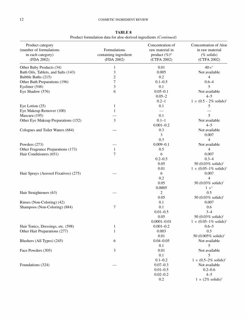
12 COSMETIC INGREDIENT REVIEW
TABLE 8Product formulation data for aloe-derived ingredients (Continued)
Product category(number of formulations
in each category)(FDA 2002)
Formulationscontaining ingredient
(FDA 2002)
Concentration ofraw material inproduct (%)a
(CTFA 2002)
Concentration of Aloein raw material
(% solids)(CTFA 2002)
Other Baby Products (34) 1 0.01 40×c
Bath Oils, Tablets, and Salts (143) 3 0.005 Not availableBubble Baths (215) 2 0.2 4Other Bath Preparations (196) 7 0.1–0.5 0.6–4Eyeliner (548) 3 0.1 5Eye Shadow (576) 6 0.05–0.1 Not available
0.05–2 4–50.2–1 1 × (0.5 – 2% solids)c
Eye Lotion (25) 1 0.1 5Eye Makeup Remover (100) 1 — —Mascara (195) — 0.1 5Other Eye Makeup Preparations (152) 3 0.1–1 Not available
0.001–0.2 4–5Colognes and Toilet Waters (684) — 0.3 Not available
3 0.0070.3 4
Powders (273) — 0.009–0.1 Not availableOther Fragrance Preparations (173) 1 0.5 4Hair Conditioners (651) 7 6 0.007
0.2–0.5 0.3–40.05 50 (0.03% solids)c
0.01 1 × (0.05–1% solids)c
Hair Sprays (Aerosol Fixatives) (275) — 6 0.0070.2 4
0.05 50 (0.03% solids)c
0.0005 1 ×c
Hair Straighteners (63) — 2 0.50.05 50 (0.03% solids)c
Rinses (Non-Coloring) (42) 0.1 0.007Shampoos (Non-Coloring) (884) 7 0.1 0.6
0.01–0.5 3–40.05 50 (0.03% solids)c
0.0001–0.01 1 × (0.05–1% solids)c
Hair Tonics, Dressings, etc. (598) 1 0.001–0.2 0.6–5Other Hair Preparations (277) 1 0.003 0.5
0.01 50 (0.005% solids)c
Blushers (All Types) (245) 6 0.04–0.05 Not available0.1 5
Face Powders (305) 3 0.01 Not available0.1 5
0.1–0.2 1 × (0.5–2% solids)c
Foundations (324) — 0.07–0.3 Not available0.01–0.5 0.2–0.60.02–0.2 4–5
0.2 1 × (2% solids)c

ALOE 13
TABLE 8Product formulation data for aloe-derived ingredients (Continued)
Product category(number of formulations
in each category)(FDA 2002)
Formulationscontaining ingredient
(FDA 2002)
Concentration ofraw material inproduct (%)a
(CTFA 2002)
Concentration of Aloein raw material
(% solids)(CTFA 2002)
Lipstick (962) 1 0.4–6 Not available0.1–5 0.6–51–6 250.3 1 × (2% solids)c
Makeup Bases (141) 8 0.2 Not available0.5 40.2 1 × (2% solids)c
Rouges (28) — 1 Not availableOther Makeup Preparations (201) — 2 Not available
0.001 4Cuticle Softeners (19) — 0.01 4Nail Polish and Enamel (123) — 0.0005 Not availableOther Manicuring Preparations (55) — 0.03 Not available
5 97Bath Soaps and Detergents (421) 7 0.1 0.007
0.05 1 (0.0005% solids)c
0.01–0.5 4–50.1 1 × (0.05–1% solids)c
Deodorants (Underarm) (247) 3 <0.01–0.5 2–40.1 40 × (16% solids)c
Feminine Deodorants (4) 2 — —Other Personal Cleanliness Products (308) 2 0.1 0.007Aftershave Lotion (231) 7 0.5 0.6
0.3 4Preshave Lotion (all types) (14) — 0.1 0.6Shaving Cream (134) 6 0.0003 Not available
0.003–1 0.6–4Other Shaving Preparation Products (63) 7 0.05 Not available
0.2 0.6Skin Cleansing Creams, Lotions, etc. (775) 10 0.002–0.5 0.6–4
0.05 1 (0.0005% solids)c
0.0005 1×c
0.01 40×c
Depilatories (34) 2 0.1 0.6Face and Neck Skin Care Preparations (310) 5 0.00009–1 0.6–4Body and Hand Skin Care Preparations (840) 11 0.03 Not available
0.1–3 0.1–40.1 1 (0.001% solids)c
Body and Hand Sprays (35) — 0.06 0.6Moisturizers (905) 24 0.1–0.5 0.6–4
0.4 40Night Creams, Lotions, and Powders (200) 2 0.1–0.5 0.6–4Paste Masks (Mud Packs) (271) 3 0.5 4
1 50 (0.5% solids)c
Skin Fresheners (184) 4 0.0006–0.5 0.3–40.05 97
(Continued on next page)
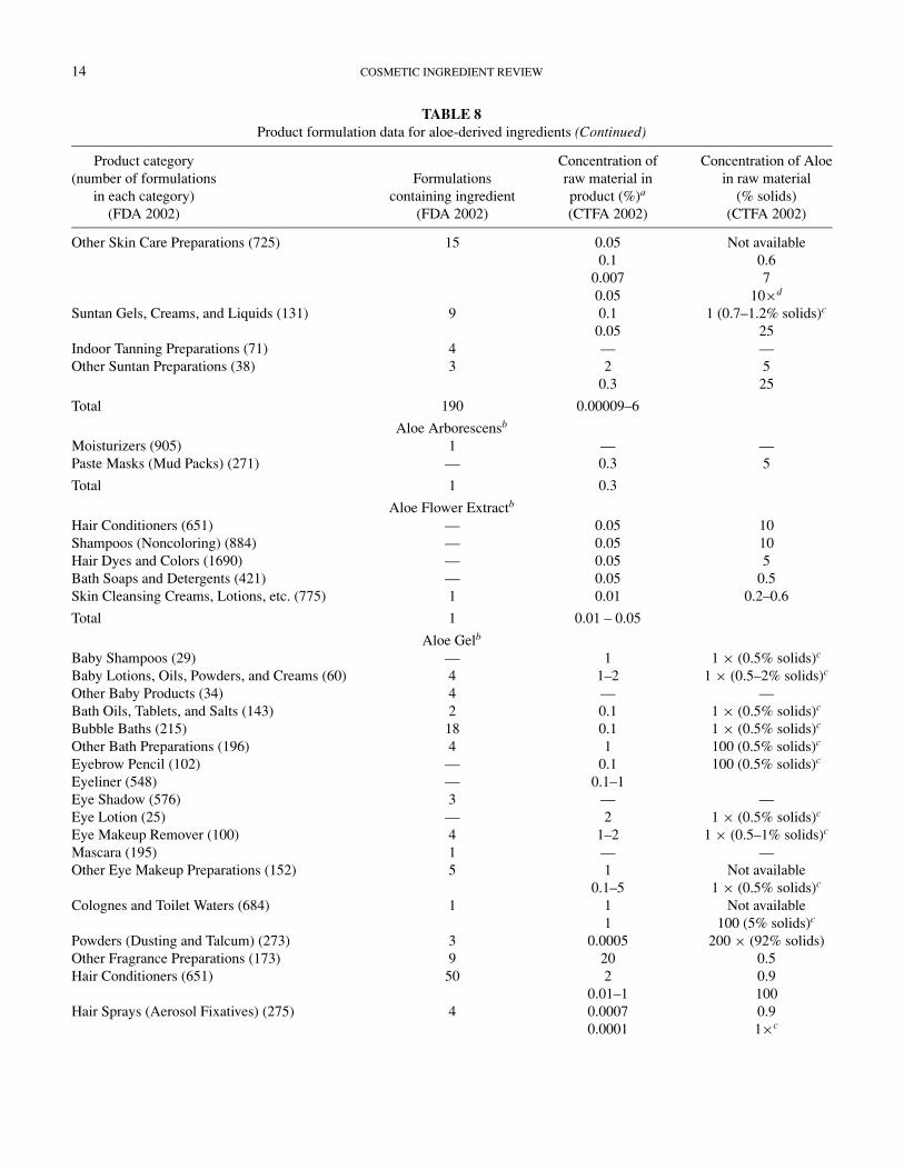
14 COSMETIC INGREDIENT REVIEW
TABLE 8Product formulation data for aloe-derived ingredients (Continued)
Product category(number of formulations
in each category)(FDA 2002)
Formulationscontaining ingredient
(FDA 2002)
Concentration ofraw material inproduct (%)a
(CTFA 2002)
Concentration of Aloein raw material
(% solids)(CTFA 2002)
Other Skin Care Preparations (725) 15 0.05 Not available0.1 0.6
0.007 70.05 10×d
Suntan Gels, Creams, and Liquids (131) 9 0.1 1 (0.7–1.2% solids)c
0.05 25Indoor Tanning Preparations (71) 4 — —Other Suntan Preparations (38) 3 2 5
0.3 25
Total 190 0.00009–6
Aloe Arborescensb
Moisturizers (905) 1 — —Paste Masks (Mud Packs) (271) — 0.3 5
Total 1 0.3
Aloe Flower Extractb
Hair Conditioners (651) — 0.05 10Shampoos (Noncoloring) (884) — 0.05 10Hair Dyes and Colors (1690) — 0.05 5Bath Soaps and Detergents (421) — 0.05 0.5Skin Cleansing Creams, Lotions, etc. (775) 1 0.01 0.2–0.6
Total 1 0.01 – 0.05
Aloe Gelb
Baby Shampoos (29) — 1 1 × (0.5% solids)c
Baby Lotions, Oils, Powders, and Creams (60) 4 1–2 1 × (0.5–2% solids)c
Other Baby Products (34) 4 — —Bath Oils, Tablets, and Salts (143) 2 0.1 1 × (0.5% solids)c
Bubble Baths (215) 18 0.1 1 × (0.5% solids)c
Other Bath Preparations (196) 4 1 100 (0.5% solids)c
Eyebrow Pencil (102) — 0.1 100 (0.5% solids)c
Eyeliner (548) — 0.1–1Eye Shadow (576) 3 — —Eye Lotion (25) — 2 1 × (0.5% solids)c
Eye Makeup Remover (100) 4 1–2 1 × (0.5–1% solids)c
Mascara (195) 1 — —Other Eye Makeup Preparations (152) 5 1 Not available
0.1–5 1 × (0.5% solids)c
Colognes and Toilet Waters (684) 1 1 Not available1 100 (5% solids)c
Powders (Dusting and Talcum) (273) 3 0.0005 200 × (92% solids)Other Fragrance Preparations (173) 9 20 0.5Hair Conditioners (651) 50 2 0.9
0.01–1 100Hair Sprays (Aerosol Fixatives) (275) 4 0.0007 0.9
0.0001 1×c

ALOE 15
TABLE 8Product formulation data for aloe-derived ingredients (Continued)
Product category(number of formulations
in each category)(FDA 2002)
Formulationscontaining ingredient
(FDA 2002)
Concentration ofraw material inproduct (%)a
(CTFA 2002)
Concentration of Aloein raw material
(% solids)(CTFA 2002)
Hair Straighteners (63) 1 0.0001 0.5Permanent Waves (207) 4 — —Rinses (Noncoloring) (42) 2 — —Shampoos (Noncoloring) (884) 47 0.01–0.05 Not available
2 0.90.01 100
Hair Tonics, Dressings, etc. (598) 26 0.1 0.9Wave Sets (53) 4 — —Other Hair Preparations (277) 2 0.0002 0.9Hair Dyes and Colors (1690) 21 0.1 5Hair Color Sprays (Aerosol) (5) 1 — —Hair Bleaches (120) 7 — —Face Powders (305) 1 0.1 Not available
0.1 1 × (0.5% solids)c
Foundations (324) 3 0.5–4 1 × (0.5% solids)c
Lipstick (962) — 0.5 92Makeup Bases (141) 1 — —Rouges — 0.05 1 × (0.5% solids)c
Other Makeup Preparations (201) 3 0.2 1 × (0.5% solids)c
Cuticle Softeners (19) 1 — —Nail Creams and Lotions (15) — 0.01–2 1 × (2–5% solids)c
Other Manicuring Preparations (55) 3 — —Mouthwashes and Breath Fresheners (46) 1 — —Bath Soaps and Detergents (421) 15 0.5 5
0.0001 0.10.05–1 1 × (0.5–1% solids)c
0.01 200 × (∼90% solids)c
Deodorants (Underarm) (247) 5 — —Douches (5) — 1 1 × (0.5% solids)c
Feminine Hygiene Deodorant (4) — 0.1 Not availableOther Personal Cleanliness Products (308) 29 1–2 1 × (0.5–5% solids)c
Aftershave Lotions (231) 8 20 Not available0.5 50.1 92
1–20 1 × (0.5–2% solids)c
Shaving Cream (134) 10 0.5 Not available0.1–0.5 100
Other Shaving Preparation Products (63) 9 1–2 1 × (0.5–5% solids)c
Skin Cleansing Creams, Lotions, Liquids (775) 22 0.5 50.01–0.5 0.1–5
0.5–7 1 × (0.5–5% solids)c
Depilatories (34) 3 — —Face and Neck Skin Preparations (310) 14 0.5 5
0.1–0.5 0.1–10.05–2 1 × (0.5–1% solids)c
(Continued on next page)

16 COSMETIC INGREDIENT REVIEW
TABLE 8Product formulation data for aloe-derived ingredients (Continued)
Product category(number of formulations
in each category)(FDA 2002)
Formulationscontaining ingredient
(FDA 2002)
Concentration ofraw material inproduct (%)a
(CTFA 2002)
Concentration of Aloein raw material
(% solids)(CTFA 2002)
Body and Hand Skin Care Preparations (840) 38 0.5 50.2 923 0.9
1–20 1 × (0.5–1% solids)c
0.01 200 × (∼90% solids)c
Body and Hand Sprays (35) — 0.001 0.1Foot Powders and Sprays (35) — 0.1 100 (5% solids)c
Moisturizers (905) 59 0.5 51–3 1 × (0.5–5% solids)c
Night Creams, Lotions and Powders (200) 3 0.5 921–5 0.5–1
Paste Masks (Mud Packs) (271) 8 1 Not available5 0.5
Skin Fresheners (184) 14 0.1–2 1 × (0.5–1% solids)c
0.03 5Other Skin Care Preparations (725) 32 2 Not available
0.01 920.1–3 1 × (0.5–5% solids)c
0.1 40 × (21% solids)c
Suntan Gels, Creams, and Liquids (131) 8 3–4 1 × (0.5% solids)c
0.1 40 × (21% solids)c
Indoor Tanning Preparations (71) 9 0.02 92Other Suntan Preparations (38) 3 5 1 × (0.5% solids)c
Total 529 0.0001–20
Aloe, Powderedb
Bath oils, tablets, and salts (143) — 0.1 100 (powder)Eye Lotion (25) — 0.05 100 (powder)Other Eye Makeup Preparations (152) — 0.01 100 (powder)Fragrance Powders (273) — 0.1 100 (powder)Blushers (All Types) (245) 2 0.1 100 (powder)Face Powders (305) — 0.05 100 (powder)Foundations — 0.01 100 (powder)Bath Soaps and Detergents (421) 1 — —Other Personal Cleanliness Products (308) — 0.01 100 (powder)Body and Hand Skin Care Preparations (840) 1 0.0003 200× (powder)Moisturizers (905) — 0.2 100 (powder)Other Skin Care Preparations (63) — 0.00001 200× (powder)
— 0.01 100 (powder)— 0.1 100 (powder)
Other Suntan Preparations (38) — 0.0003 200× (powder)
Total 4 0.00001–0.2
Aloe Ferox Leaf Extractb
Colognes and Toilet Water (684) — 5 0.002Rinses (Noncoloring) (42) — 0.1 0.002Shampoos (Noncoloring) (884) — 0.1 0.002

ALOE 17
TABLE 8Product formulation data for aloe-derived ingredients (Continued)
Product category(number of formulations
in each category)(FDA 2002)
Formulationscontaining ingredient
(FDA 2002)
Concentration ofraw material inproduct (%)a
(CTFA 2002)
Concentration of Aloein raw material
(% solids)(CTFA 2002)
Bath Soaps and Detergents (421) — 0.1 0.002Other Personal Cleanliness Products (308) — 0.1 0.002Aftershave Lotions (231) — 0.08 2.5Body and Hand Skin Care Preparations (840) — 5 0.002Body and Hand Sprays (35) — 5 0.002Moisturizers (905) — 0.001 3
0.1 0.6Other Skin Care Preparations — 0.1 0.002
Total None 0.001–5
aIn a given product category, there may be more than one industry report of a concentration of raw material in such products. All data providedare given, but the number of products at each concentration is unknown.
bReports to FDA did not correspond to current ingredient terminology. See text.C As reported.
Aloe-derived materials, not specified, are used as drug prod-ucts containing certain active ingredients offered over thecounter as orally administered menstrual drug products (21 CFR310.545). Aloin, an active ingredient in Aloes, is used as a drugproduct containing certain active ingredients offered over thecounter as a stimulant laxative (21 CFR 310.545). Aloe ex-tract and aloe flower extract used in over-the-counter (OTC)drug products as a stimulant laxative were not recognized andare not generally recognized as safe (GRAS) and effective orare misbranded due to a lack of carcinogenicity data (21 CFR310.525(a)12(iv)(C)).
Sturm and Hayes (1984) stated that Aloe may be used in med-icated liners in immediate dentures, and as an anti-inflammatoryagent after brushing or oral surgery, in addition to mouth rinsesand toothpastes.
GENERAL BIOLOGY
Absorption, Distribution, Metabolism, and ExcretionHirata et al. (1981) administered 10 mg 14C-labeled aloenin
(see Figure 1), derived from Aloe arborescens, in water orally to2-month-old rats after an overnight fast. The feces and urinewere collected 24 and 48 h after administration. Methanol(MeOH) extraction of the feces and urine indicated that mostof the 14C-labeled aloenin was excreted within the first 24 h.Only a small amount was excreted within the next 24-h pe-riod. Thin-layer chromatography indicated that aloenin wasmetabolized to 4-methoxy-6-(2,4-dihydroxy-6-methylphenyl)-2-pyrone, 2,5-dimethyl-7-hydroxychromone, and glucose. The
distribution and/or accumulation of aloenin in the stomach, liver,and kidneys indicated that aloenin and its metabolites accumu-lated in the liver and the kidneys.
Ishii et al. (1987) administered barbaloin (see Figure 1), dis-solved in distilled water at 20 mg/ml, orally to male Wistar Ratsat a dose of 100 mg/kg. At specific time points after administra-tion, blood was withdrawn from the rats to measure the serumlevels of barbaloin. Barbaloin was first seen in the serum 30 min(0.092 µg/ml) after administration with maximum concentration(0.337 µg/ml) occurring at 90 min. After the 90-min time point,concentrations of barbaloin decreased smoothly with detectionstill possible at 6 h after administration.
Barbaloin (31.1 mg/5 ml/kg in 5% gum arabic solution)administered by cecal intubation to male Wistar rats (150 to200 g), produced aloe-emodin-9-anthrone 1 h after adminis-tration. Aloe-emodin-9-anthrone, a decomposition product ofbarbaloin, was measured by thin-layer chromatography. Aloe-emodin-9-anthrone peaked after 4 h of administration of thebarbaloin and was found in the cecum at levels of 508 µg/ratand in the colon at 83 µg/rat (Ishii et al. 1994).
Lang (1993) administered aloe-emodin (see Figure 1) to SPFBrown-Norway rats (numbers not given) orally in a tragacanth(0.3%) suspension at a dose of 4.5 mg/kg. The aloe-emodin hadbeen previously labeled with 14C. Blood, feces, urine, and 24 or-gans were collected at specific time points to elucidate the dis-tribution of aloe-emodin. Maximum blood concentrations werereached 1.5 to 3 h post administration and were 248 ng (males)and 441 ng (females) equivalents aloe-emodin/ml. During thefirst 6 h post administration there were no differences in organconcentrations between the males and females; however, in later

18 COSMETIC INGREDIENT REVIEW
samples females had higher concentrations than the males. Theliver and kidney were the only organs that had higher concen-trations of aloe-emodin than plasma. Three-fourths of the dosewas excreted in the feces within the first 2 days. Plasma proteinbinding of aloe-emodin was determined in vitro and ex vivo.The results were an in vitro binding of 98% to 99% and an exvivo binding of 86% to 96%. No sex differences were found.
Heidemann et al. (1996) reported that NMRI and DBA micetreated with a single oral dose (2000 mg/kg) of aloe-emodinhad blood plasma concentrations ranging from 3 to10 µg/ml.NMRI mice and Wistar rats treated with a single oral dose ofaloe-emodin (1500 or 2000 mg/kg) had maximum blood plasmaconcentrations of 17 µg/ml after 3 h.
Yagi et al. (1999) administered aloemannan (a polysaccharideof Aloe barbadensis) to mice at 120 mg/kg orally or by intra-venous injection (number and strain of mice were not given).Prior to administration, the aloemannan had been labeled withfluorescein isothiocyanate (FITC-aloemannan). Examination ofthe urine and feces showed the FITC-aloemannan was metabo-lized into smaller molecules that accumulated in the kidneys. In-travenous administration resulted in a greater quantity of FITC-aloemannan in the urine within the first 24 h, with minimalamounts found in the feces over the 48-h period. Oral adminis-tration resulted in a greater quantity of FITC-aloemannan in thefeces within the first 24-h period when compared to the urine.
Fungicidal ActivityFujita et al. (1978) tested a whole leaf powder of Aloe ar-
borescens and an Aloe arborescens leaf homogenate (high-molecular-weight) lyophilized powder against three strains ofTrichophyton mentagrophytes. Both powders were shown tohave fungicidal activity against T. mentagrophytes. The min-imum inhibitory concentration (MIC) was 25 mg/ml for thewhole-leaf powder and 10 mg/ml for the high-molecular-weightpowder. Both the whole-leaf powder and the high-molecular-weight component powder induced various morphological ab-normalities in spores and hyphae by the inhibition of spore ger-mination and development of hyphae.
Ali et al. (1999) screened extracts of fresh leaves of Aloearborescens and Aloe barbadensis for their antifungal activityagainst Aspergillus niger, Cladosporium herbarum, and Fusar-ium moniliforme. The solvent used for the extraction affectedfungicidal activity. Ethanol extraction was the most effective fol-lowed by chloroform, benzene, and water. All extracts of Aloearborescens and Aloe barbadensis had some fungicidal activity.
Antimicrobial ActivityLorenzetti et al. (1964) made a preparation of Aloe vera leaves
by cutting them at the base and standing them upright to al-low the juice to drain out. This juice was heated for 15 minat 80◦C and then freeze dried. The freeze-dried juice was re-constituted in distilled water (20 mg/ml) and tested in the agardiffusion test against: Staphylococcus aureus 209, Escherichia
coli, Streptococcus pyogenes, Corynebacterium xerose, Shigellaparadysenteriae, Salmonella typhosa, Salmonella schottmuel-leri, and Salmonella paratyphi. Significant inhibition of growthoccurred with S. aureus 209, S. pyogenes, C. xerose, and S.paratyphi.
Northway (1975) used an Aloe vera product, in three forms,topically to treat a variety of infections in 76 animals. The Aloeproducts were 100% Aloe vera gel (Aloe 99 Gel), 75% Aloevera gel in a cream (Aloe 99 Creme), and 82% Aloe vera gelin a lotion (Aloe 99 Lotion). The active ingredient was derivedfrom the mature leaves of 4-year-old Aloe vera plants. Treatmentwas from 1 to 4 weeks depending on condition. Except in thecases of abscesses, treatment consisted of thoroughly rubbingthe medication into the lesion two to four times daily. Abscesseswere infused with medication twice daily. In otitis externa, theear was cleansed once a day and the medication applied andworked into the canal twice daily. The medication was workedinto the staphyloma four times daily for a month. Results aresummarized in Table 9.
The author concluded that the aloe products appeared to re-tard exuberant granulation tissue and that pain and itching wererelieved promptly. No toxic reactions or other adverse effectswere seen in any animal (Northway 1975).
Heggers et al. (1979) tested Aloe vera gel and Dermaide Aloe(a commercially prepared purified extract) against 10 bacterialstrains. Sixty percent to 90% concentrations were used to inoc-ulate Staphylococcus aureus, Streptococcus pyogenes, Strepto-coccus agalactiae, Escherichia coli, Serratia marcescens, Kleb-siella sp., Enterobacter sp., Citrobacter sp., Bacillus subtilis, andCandida albicans. At 90%, Aloe vera gel was effective againstall organisms, but at the 70% concentration only S. pyogenes wasinhibited. Dermaide Aloe was effective against all organisms ata concentration of 70%.
Heck et al. (1981) tested a preserved Aloe gel extract (froma commercial provider) and an unpreserved Aloe extract thatresembled the one that could be obtained from a householdplant against Pseudomonas aeruginosa, Enterobacter aero-genes, Staphylococcus aureus, and Klebsiella pneumoniae. Con-centrations of each of the extracts (10%, 20%, 40%, 70%, 80%,90%) were inoculated by 102 and 106 concentrations of eachbacterium. The preserved Aloe gel extract was effective in con-trolling bacterial growth at a concentration of 40%: none of thecultures of Pseudomonas, Enterobacter, or Klebsiella had anygrowth, Staphylococcus had growth in two out of nine cultures.Only the 90% concentration of the unpreserved Aloe extracthad any effect on the bacterial cultures and even that was notconsistent.
Robson et al. (1982) studied the antibacterial effects of Aloevera extract and found that concentrations as low as 60% werebactericidal against 7 of the 12 species of organisms studied.These were Citrobacter sp., Serratia marcescens, Enterobac-ter cloacae, Klebsiella pneumoniae, Pseudmonas aeruginosa,Streptococcus pyogene, and Streptococcus agalactiae. Concen-trations between 80% and 90% were bactericidal for the above

ALOE 19
TABLE 9Aloe vera treatment for various conditions (Northway 1975)
Species Response∗
Condition treated Number of animals Dog Cat Other E G P N
Ringworm∗∗ 14 4 10 — 4 10 — —Atopy (allergy) 12 10 2 — 6 6 — —Abscess 12 1 11 — — 12 — —Otitis externa 11 8 3 — — 9 — 2Hot spots 11 9 2 — — 10 1 —Misc. fungal infections 9 7 1 — 2 7 — —Lacerations∗∗∗ 4 — — 1 (rodent) — 4 — —Lip fold dermatitis 1 1 — 4 (horses) — — 1 —Inflamed cyst 1 1 — — — 1 — —Staphyloma 1 1 — — — 1 — —
∗E = Excellent (better than other drugs on market); G = Good (equal to the best drugs on market); P = Poor (not as good as other drugs onmarket); N = No response.
∗∗Fluorescent under ultraviolet light.∗∗∗Exuberant granulation tissue removed surgically in two horses, then Aloe vera Gel applied.
species and the other species of the organisms studied, viz.Staphylococcus aureus, Escherichia coli, Streptococcus fea-calis, Bacillus subtilis, and Candida albicans (yeast).
Antiviral ActivityKahlon et al. (1991a) tested acemannan (concentrations
ranged from 3.2 to 1000 µg/ml) in a variety of cell lines(human peripheral blood mononuclear cells [PBMCs], CEM-SS and MT-2) for antiviral activity against human immunod-eficiency virus [HIV]-1. Maximum inhibitory effect was ob-served in CEM-SS cells (infected with HIV-1); it was concen-tration dependent. Fifty percent inhibition occurred at concen-trations of 45 to 48 µg/ml, whereas 100% inhibition occurredat 1000 µg/ml; at this second concentration no cytotoxic effectswere observed. Acemannan, at a concentration of 31.25 µg/ml,suppresses viral-induced syncytia formation, with inhibition oc-curring at 62.5 µg/ml. In PBMCs, acemannan caused a con-centration dependent inhibition of HIV-1 replication. Maximumsuppression occurred at concentrations of 62 or 125 µg/ml.
Kahlon et al. (1991b) tested acemannan, at various concentra-tions, alone or in conjunction with either azidothymidine (AZT)or acyclovir. Acemannan (concentrations ranged from 15.6 to250 µg/ml), with or without AZT, was tested in vitro in maturehuman T4 lymphocytes infected with HIV-1. Acemannan, at aconcentration of 125 µg/ml, inhibited replication of HIV-1 cells;however, 15.62 µg/ml of Acemannan and 0.001 or 0.01 ng/ml ofAZT produced 96% and 100% inhibition, respectively. Aceman-nan (0 to 100 µg/ml concentration) was tested with or withoutacyclovir, in vitro, in herpes simplex virus (HSV)-1–infectedVero cells. Acemannan alone was not able to significantly re-duce replication of HSV-1–infected cells; however Acemannan
(40 µg/ml) combined with 0.025 µg/ml of acyclovir inhibitedreplication by ≥90%.
Sydiskis et al. (1991) tested hot glycerin extracts of Aloe bar-badensis against a variety of viruses, including herpes simplexvirus (HSV-1 and HSV-2), pseudorabies virus (PSV), varicella-zoster virus (VZV), influenza virus (INF), rhinovirus (RH), andadenovirus (AD). The active hot glycerin extract of Aloe bar-badensis was determined to contain aloe-emodin, so that com-pound was tested in the same assays. The hot glycerin extracts ofAloe barbadensis were virucidal to HSV-1 within 15 min of incu-bation. The effect of the hot glycerin Aloe barbadensis extract onHSV-1 inactivation was concentration dependent and occurredmore rapidly at 37◦C than 4◦C. Aloe-emodin at a concentra-tion of 0.1 mg/ml in 50% glycerin was active against HSV-1,HSV-2, pseudorabies virus, varicella-zoster virus, and influenzavirus. However, at the highest concentration tested, rhinovirusand adenovirus were not affected when compared to the controls(50% glycerin). Aloe-emodin–treated Vero or WI-38 cells didnot exhibit altered morphology, indicative of cytotoxicity, whencompared to the controls.
CytotoxicityBrasher et al. (1969) incorporated solutions of prednisolone,
indomethacin, and Aloe vera gel into cell maintenance mediumto look for cytotoxicity in HeLa cells and rabbit kidney fibrob-lasts. Aloe vera gel at the 5 × 10⁻¹ dilution (no concentrationunits given) was toxic to both cell lines at all hours examined.Aloe vera gel that was diluted 10−1, 10−2, and 10−3 did not haveany significant effect on the cell lines.
Winters et al. (1981) conducted a study in which fresh leavesfrom Aloe barbadensis were minced, homogenized, and cen-trifuged to give two different fractions, the supernatant and the
20 COSMETIC INGREDIENT REVIEW
pellet. Aloe vera gel was also purchased off the shelf from alocal store and underwent the same procedure. The supernatantand the pellet from both sources (concentrations were not given)were used in four different assays (hemagglutination titration,immunodiffusion, cell attachment and growth, and wounded cellmonolayer) to evaluate cytotoxicity. The supernatant fraction ofthe fresh leaves and the commercial preparation were foundto have high levels of lectin-like substances as seen with thehemagglutination titration and immunodiffusion assays. The su-pernatant fraction from Aloe barbadensis was found to markedlypromote cellular attachment and growth of normal human cellsbut not cancer cells; however, the commercial Aloe vera gel(fractions not specified) was cytotoxic for human normal andtumor cells in vitro. Additionally, the fractions from Aloe bar-badensis enhanced the healing of wounded cell monolayers.
Danof and McAnalley (1983) tested four commercial prepa-rations (description not given) of stabilized Aloe vera gel sam-ples for their cytotoxicity in human endothelial cells and fibrob-lasts. In addition, the yellow sap from fresh Aloe vera was tested.The yellow sap at all concentrations was lethal to human fibrob-lasts. Two of the four products were cytotoxic to both cell types;one showed significant toxicity.
Bouthet et al. (1995) studied the effect of aloe on cultures ofhuman lung embryonic (HEL) cells and rat adrenal pheochro-mocytoma (PC12) cells. Liquid extract of whole leaf Aloe (Aloebarbadensis) and the gel filet portion of Aloe leaves (both pur-chased commercially) were lyophilized and then reconstitutedwith sterile water. Both the liquid extract of whole leaf Aloeand the gel filet portion of Aloe were added to cell cultures tomeasure their effects on cell proliferation. Control cells receivedan equal amount of media. The liquid extract of whole leaf Aloesignificantly stimulated the growth of the PC12 cells at concen-trations of 0.162 and 0.312 µg/ml. The liquid extract of wholeleaf Aloe stimulated PC12 cell growth quicker and better thanthe HEL cells. The gel filet portion of Aloe (0.162 and 0.625µg/ml) significantly stimulated the growth of OC12 cells after2 days. The gel filet portion of Aloe had little or no effect onHEL cell proliferation. Differentiation of PC12 cells did not oc-cur with either the liquid extract of whole leaf Aloe or gel filetportion of Aloe.
Avila et al. (1997) isolated three fractions from the leaves ofAloe barbadensis: native gel (mucilaginous parenchymous tis-sue scraped from Aloe leaves), purified gel (removal of debris),and a low-molecular-weight fraction (LMWF). The authors usedthe three fractions, along with purchased aloin, to evaluate thecytotoxicity of the LMWF (50 µg/ml). The cell injury assaywas performed with fibroblasts that were cultured from chickeneggs. A 1:10 dilution of native gel (diluted with Dulbecco’s min-imal essential medium) caused cell injury, whereas the purifiedgel did not cause injury when tested at the same dilution. TheLMWF behaved as the native gel did. Aloin also damaged thecells.
Tello et al. (1998) cultured commercially available humanfibroblasts with formulations of acemannan in preservative
(ratios ranged from 20:1 to 150:1). Details of the formulationswere not given. A 1% concentration of the solutions wasincubated for 3, 6, 12, and 24 h. Formulations of acemannanhad significant cytotoxicity, especially in the formulations witha high level of preservative, although one formulation hadminimal cytotoxicity.
Enzyme ActivityYagi et al. (1985) isolated a 40,000 molecular weight (MW)
glycoprotein from Aloe arborescens that stimulated DNA syn-thesis of BHK-21 cells at 5 µg/ml. The cells did not have anymorphological changes.
Yagi et al. (1987), repeating the work of Fujita et al. (1976), re-ported that an Aloe glycoprotein isolated from Aloe arborescensappeared to have enzymatic activity against bradykinin. Extractsof fresh leaves of Aloe arborescens were homogenized and cen-trifuged to isolate a glycoprotein of 40,000 MW. The glycopro-tein (1 mg) was incubated with 10 µg of bradykinin for 10 min.The bradykinin solution (0.2 ml) was incubated with isolatedguinea pig ileum for 5 min. The contractile response of the ileumwas measured. The Aloe glycoprotein degrades bradykinin.
Norton et al. (1990) examined the outer green rind of freshAloe barbadensis leaves for possible glyoxalase activity. Purifi-cation was done by affinity ligand-enzyme binding. GlyoxalaseI, a basic protein with a molecular weight of 44,000, and gly-oxalase II, an acidic protein with a molecular weight of 27,000,were isolated.
Sabeh et al. (1993) isolated glutathione peroxidase from theinner mucilaginous parenchymal tissue of Aloe barbadensis.The enzyme has a molecular weight of 62 kDa and is composedof four identical subunits. Sabeh et al. (1996) identified sevensuperoxide dismutases in the inner mucilaginous parenchymaltissue (produces the gel).
Lee et al. (1997) reported that aloesin has cell-growth stim-ulatory activity. DNA synthesis of SK-HEP-1 cells treated withaloesin was stimulated in a dose-dependent manner. At the 1 to50 µM concentration range, aloesin stimulated DNA syntheticactivity two- to fourfold over controls. The authors further de-termined that aloesin significantly increased intracellular levelsof cyclin E, CDK2, and CDC 25A in SK-HEP-1 cells.
Yagi et al. (1997) prepared two fractions from the leaf gelof Aloe barbadensis by column chromatography; one frac-tion was a 29-kDa glycoprotein (later named verectin) andthe other was a neutral polysaccharide. Both fractions werethen tested with either BHK-21 or normal human dermalcells. The glycoprotein fraction promoted cell growth; how-ever, the neutral polysaccharide fraction did not stimulate cellgrowth.
Esteban et al. (2000) tested commercial Aloe vera gel prepara-tions and the inner aqueous leaf parenchyma of Aloe barbadensisfor possible peroxidase activity. Peroxidase activity was detectedin the Aloe barbadensis preparation and the commercial prepara-tion. Both preparations lost their peroxidase activity when heated

ALOE 21
for 5 min in a water bath at 100◦C. The peroxidase activity ofthe commercial preparations varied.
Immunological Effectst’Hart et al. (1988) excised mucilaginous parenchymal tis-
sue from fresh Aloe vera leaves, lyophilized, reconstituted withHanks’ balanced salt saline (HBSS) and centrifuged to obtaina high-molecular-weight and a low-molecular-weight fraction.Each fraction was tested with pooled human serum for com-plement activity. The high-molecular-weight fraction depletedthe classical and alternative complement systems. The low-molecular-weight fraction inhibited the production of oxygenfree radicals by polymorphonuclear leukocytes.
Marshall et al. (1993) incubated acemannan, in concentra-tions ranging from 0 to 100 µg/ml, overnight with human pe-ripheral blood mononuclear cells. Cytokine concentrations wereassayed by enzyme-linked immunosorbent assay (ELISA). Ace-mannan stimulates tumor necrosis factor (TNF)-α, interleukin(IL)-1β, and IL-6.
Strickland et al. (1994) investigated the ability of Aloe bar-badensis gel extract to prevent the suppression of contact hyper-sensitivity (CHS) and delayed-type hypersensitivity (DTH) re-sponses in mice by ultraviolet (UV) radiation. Treatment groupscontained five C3H/Hen(MTV−) female mice. The mice in thelocal suppression group received four daily doses of 400 J/m2
of UVB on their abdomens. The systemic suppression groupreceived either a single dorsal exposure of 5 kJ/m2 (DTH sup-pression) or 10 kJ/m2 (CHS suppression). Control mice did notreceive UVB irradiation. Irradiated mice were treated with theAloe barbadensis gel extract (in Aquaphor) (0.167%, 0.5%, or1.67%) within 5 min after irradiation. Approximately 75 mgof vehicle control (Aquaphor) or Aloe barbadensis gel extractin vehicle was applied to each mouse. CHS was induced by asolution of 0.5% fluorescein isothiocyanate (FITC) and DTHwas induced with formalin-fixed Candida albicans. Treatmentof UV-irradiated skin with the Aloe barbadensis gel extract (allconcentrations) prevented the induction of local and systemicsuppression of both CHS and DTH. The Aloe barbadensis gelextract treatment partially preserved the number and morphol-ogy of Langerhans and Thy-1+ dendritic epidermal cells in skin,compared to those in the skin of mice given only UV radiationor UV radiation plus the vehicle control.
Tyler (1994) postulated that some of the beneficial effectsof aloe are the result of a carboxypeptidase that inhibits thepain-producing agent bradykinin. The enzyme is also believedto hinder the formation of thromboxane, the activity of which isdetrimental to burn wound healing. Antiprostaglandin activitywas also suggested.
Cultures of normal chicken spleen cells and HD11 line cellsproduce nitric oxide in response to Acemannan extracted fromAloe vera plant (Karaca et al. 1995). The nitric oxide induc-ing effect of Acemannan on spleen cells was dose dependent.Acemannan-induced nitric oxide synthesis may be mediated
through macrophage mannose receptors, and macrophage ac-tivation may be accountable for some of the immunomodula-tory effects of Acemannan in chickens. Macrophage activationmay also account for some of the wound healing capabilitiesattributed to Acemannan.
Lee et al. (1995) tested dichloromethane extracted Aloe veragel (yielded two low-molecular-weight fractions) for angiogenicactivity in the chick embryo chorioallantoic membrane assay.Both fractions (100 and 250 µg/egg) produced a significant an-giogenic effect. The effect was similar to phorbol-12-myristate-13-acetate (PMA) (positive control).
Egger et al. (1996) injected CARN 750 (dissolved in dis-tilled phosphate-buffered saline [D-PBS]), isolated from Aloebarbadensis, subcutaneously into female C57BL/6 mice (num-ber not given) at doses of 0.05, 1, or 2 mg/animal for 18days. Negative-control animals received PBS on a similar sched-ule and positive-control animals received 3 µg of granulocytecolony-stimulating factor (G-CSF). Bone marrow and spleencells were obtained from the euthanized animals. The adminis-tration of CARN 750 significantly increased splenic and periph-eral blood cellularity as well as hematopoietic progenitors in thespleen and bone marrow. In myelosuppressed mice, CARN 750had activity equal to or greater than G-CSF.
Zhang and Tizard (1996) tested acemannan, from the centralgel of the leaf of Aloe barbadensis, for its effects on the mousemacrophage cell line RAW 264.7. The cells were treated witheither acemannan alone (100 µg/ml), interferon (IFN)-γ alone(10 U/ml; positive control), or acemannan/IFN-γ (acemannan100 µg/ml/ IFN-γ 10 U/ml) for 24 h. Acemannan stimulatedmacrophage cytokine production, nitric oxide release, surfacemolecule expression, and cell morphologic changes. The pro-duction of the cytokines IL-6 and TNF-α were dependent on thedose of acemannan.
Lee et al. (1997) prepared epidermal cells from mouse earskin and exposed them, in vitro, to a single dose of 180 J/m2
of UVB radiation. The epidermal cells were used to determinewhat fraction of Aloe vera gel would be able to prevent UV-B–induced impairment of Langerhans cells. Lyophilized Aloe veragel (provided by the Aloe Research Foundation) was dissolvedin distilled water and then separated into four fractions basedon molecular size: <500, between 500 and 1000, between 1000and 3000, and >3000. After testing the four fractions with themouse epidermal cells, the fraction that was found to be in the500 to 1000 MW range was further fractionated by a Bio-GelP-2 column into five fractions: G1C2F0, G1C2F1, G1C2F2,G1C2F3, and G1C2F4. The fraction G1C2F1 (low-molecular-weight fraction) gave the strongest response in preventing UV-B–induced impairment of Langerhans cell function.
In the course of their investigation of Aloe barbadensis gel’sability to restore CHS and DTH that had been suppressedby UV radiation, Byeon et al. (1998) discovered that the re-sponses varied between lots. Four lots were obtained from theAloe Research Foundation (manufactured by Aloecorp, Harling-ton, TX): ARF’91, ARF’94B, ARF’94G, and ARF’94K. Three
22 COSMETIC INGREDIENT REVIEW
months after its manufacture, lot ARF’91G (this lot was not men-tioned previously in the methods and materials section) was onlyable to restore 43% of the CHS response, whereas lot ARF’91Acompletely restored the CHS response. After 9 months of stor-age as a lyophilized powder, none of the extracts from the lotsprevented UV-induced suppression of CHS. It was also discov-ered that commercially prepared Aloe barbadensis gels wereuniformly inactive even when tested within 1 month of theirmanufacture.
In contrast, all lots of Aloe barbadensis gel prevented sys-temic suppression of DTH. The immunoprotective activity re-mained unchanged after 12 months of storage. The differentdecay rates observed in the CHS and DTH protective activity in-dicated that Aloe gel contains multiple factors that act on the twodifferent models of immune suppression. The labile of the factorthat prevents suppression of CHS responses varies in differentlots of the specifically prepared gel extract and was influencedby the manufacturing process used to produce the extracts. Thefinding that biologically active components in Aloe can be labilehelps to explain some of the difficulties that investigators havereported in reproducing results using unfractionated materialsfrom the plant (Byeon et al. 1998).
Ramamoorthy and Tizard (1998) treated raw 264.7 cells (amouse monocytic-macrophage cell line) with acemannan (50µg/ml), IFN-γ (1 unit/ml), or a combination of acemannan andIFN-γ to investigate acemannan’s ability to induce apoptosis.After 18 h of treatment with acemannan, apoptosis was evident,and by 36 and 48 h, 65% and 100%, respectively, of the cellshad undergone apoptosis.
Lee et al. (1999) exposed pathogen-free male C57BL/6mice (number not given) to UVB radiation. The dose used(2.4 kJ/m2) was the optimal dose needed to suppress con-tact hypersensitivity (CHS). The irradiated skin was imme-diately treated with G1C2F1 cream (isolated component ofAloe vera mixed with polyethylene glycol to form a cream).A single dose of 0.1 ml/mouse reduced contact hypersensitiv-ity suppression significantly. The percentage recovery of UVB–suppressed CHS response was 52.3%, 77.3%, and 86.6% whenthe irradiated skin was treated once with 0.1, 0.5, and 2.5mg/ml of G1C2F1 cream, respectively. Topical application ofG1C2F1 prevented UVB–induced suppression of contact hy-persensitivity response by possible restoration of the immuneresponse.
Strickland et al. (1999) exposed pathogen-free femaleC3H/HeN and BALB/C mice (5 animals/group) to 5to 30 kJ/m2dose of UVB radiation. Aloe barbadensispoly/oligosaccharides in phosphate-buffered saline or controlpolysaccharides (methylcellulose or dextran) were applied im-mediately after UVB exposure. The poly/oligosaccharides ofAloe barbadensis prevented the UV-B–induced suppression ofthe DTH response whereas the control polysaccharides didnot. Additionally, Pam212 keratinocytes were exposed to 300J/m2 UVB radiation and then treated with Aloe barbadensispoly/oligosaccharide. Control cells were similarly treated but
not exposed to the UVB radiation. The poly/oligosaccharide ofAloe barbadensis decreased (50%) IL-10 production.
Qui et al. (2000) tested a modified aloe gel extract (MAP)(MW 80 Da) from Aloe barbadensis for its immunomod-ulatory activity. Peritoneal macrophages isolated from ICRmice were incubated overnight with MAP, native Aloe gel, orlipopolysaccharide (LPS). MAP stimulated TNF-α release fromthe macrophages in a dose-dependent manner, with the greatestrelease of TNF-α occurring at a concentration of 250 µg/ml.The LPS data mimicked the MAP data. The native Aloe gel wasunable to stimulate TNF-α release from macrophages. The MAPwas also tested for its ability to prevent UVB irradiation–inducedimmune suppression. SPF female C3H/HeN mice were exposedto 2000 J/m2 of UVB; control animals did not receive irradiation.Immediately after exposure, the mice were either dosed with thevehicle (water in Aquaphor) or MAP in Aquaphor. The micewere sensitized on their abdomens 3 days later with 0.3% dini-trofluorobenzene (DNFB), with challenge on the ears with 0.2%DNFB occurring 6 days after the sensitization. In the controlgroups, the vehicle and MAP did not affect the challenge; how-ever, MAP had, in a dose-dependent manner, UV-B–protectivecontact hypersensitivity activity.
Lee et al. (2001) reported that acemannan induced matu-ration of dendritic cells (DCs) isolated from C57BL/6 mousebone marrow in vitro. Phenotypic maturation was evident withthe up-regulation of class II major histocombatibility complex(MHC) molecules and costimulatory molecules such as B7-1,B7-2, CD40, and CD54. There was also increased allogenic T-cell stimulatory activity and production of cytokine IL-12. TheDC maturation was dose dependent (0, 10, 100 µg/ml aceman-nan). The higher exposure group had a more potent allogenicT-cell stimulatory activity compared to the low exposure. Man-nose (acemannan is a polymer of acetylated mannose) did nothave a significant effect on the acemannan-induced phenotypicand functional maturation of immature DCs. However, mannosesuppressed the mitogenic activity.
Pugh et al. (2001) performed a macrophage activation as-say on aloeride (a polysaccharide) to determine if this fractionfrom Aloe barbadensis was the fraction responsible for the im-munostimulatory properties that have been attributed to Aloevera. Aloeride activated the transcription factor nuclear factor(NF)-κB at a concentration of 200 µg/ml.
HYPOGLYCEMIC/HYPERGLYCEMIC ACTIVITYDixit and Joshi (1983) studied the effect of an aqueous ex-
tract of Aloe barbadensis in Triton WR–induced hyperlipidemiain the Presbytis monkey. Fresh leaves of Aloe barbadensis wereground at room temperature and the yellow mucoid fluid wasdecanted, dried, and used for this study. Ten adult male Pres-bytis entellus monkeys (11 to 16 kg) were deprived of foodfor 12 to 16 h and then injected with Triton WR (200 mg/kgbody weight) to induce a hyperlipidemic state. After the mon-keys were confirmed to be hyperlipidemic, they were orally

ALOE 23
administered 200 mg/kg body weight of the aqueous extractof Aloe barbadensis or clofibrate, a comparative control drug(five monkeys per group). Blood was withdrawn 4, 8, 12, and24 h after dose administration. Immediately after the 24-h bloodsampling, a second dose of the aqueous extract of Aloe bar-badensis and clofibrate (same dose) were administered. Bloodwas withdrawn 4, 8, 12, and 24 h after the second dose. A signif-icant decrease of total cholesterol was observed 28 h after drugadministration.
The aqueous extract of Aloe barbadensis decreased the totalcholesterol level by 61.6%, triglyceride level by 37.8%, phos-pholipid level 51.2%, and nonesterified free fatty acids by 45.5%.In comparison, clofibrate decreased the total cholesterol levelby 47.6%, triglyceride by 50%, phospholipid by 41.7%, and23% for nonesterified free fatty acids. A significant decreaseof low-density lipoprotein (LDL) cholesterol (79%) and very-low-density lipoprotein (VLDL) cholesterol (37.5%) occurredin the monkeys treated with the aqueous extract of Aloe bar-badensis as compared to the control monkeys. The high-densitylipoprotein (HDL)/total cholesterol ratio in the aqueous extractof Aloe barbadensis–dosed monkeys was significantly increasedat all intervals compared to their initial values (Dixit and Joshi1983).
Ghannam et al. (1986) studied two groups of five fastingnormal Swiss albino mice to determine the effect of the solidresidue obtained after evaporating the sap of various species ofaloe on fasting plasma glucose (FPG) level. The negative-controlgroup received a single oral dose of normal saline. The testgroup received an oral aqueous suspension of Aloes, 500 mg/kg.The aqueous suspension consisted of the dried sap of Aloespurchased from a local market. Plasma glucose was measured at0 (before treatment), 2, 4, and 6 h after treatment. A single oraldose of the Aloes failed to lower the FPG levels of the mice after2, 4, or 6 h after treatment. No adverse effects were reported.
These authors also orally dosed 25 normal Swiss albino micetwice daily with 10 mg/kg of glibenclamide (two groups of fiveanimals) or 500 mg/kg glibenclamide (two groups of five ani-mals). A control group of five animals received water only. Thefasting plasma glucose was measured on day 0 (control group),and on days 3 and 5 (treated groups). In the Aloes groups, FPGlevels were decreased after 5 days of treatment. The mice didnot experience any adverse effects (Ghannam et al. 1986).
Hikino et al. (1986) reported that methanol-water extracts andwater extracts of Aloe arborescens yielded two polysaccharidefractions, named arborans A and B. The arborans were admin-istered intraperitoneally to normal mice (strain and number notgiven). It was found that both arborans decreased plasma glucoselevels in a dose-dependent manner. In mice with alloxan-inducedhyperglycemia, both arborans decreased plasma glucose levels.
Ajabnoor (1990) examined the possible hypoglycemic effectof aloe in hyperglycemic induced mice. Induction was via a sin-gle tail injection of alloxan monohydrate, 70 mg/kg. Male MFImice were placed into four groups: a control group (0.9% saline10 ml/kg, intraperitoneal injection); a reference group dosed
with glibenclamide (10 mg/kg in 0.9% saline, oral administra-tion); a third group dosed with the bitter principal from aloe(5 mg/kg in 0.9% saline, intraperitoneal injection); and a fourthgroup orally dosed with 500 mg/kg of aloe in 0.9% saline. Alldoses were a single administration. The 500 mg/kg dose of Aloedid not reduce the FPG, whereas the bitter principal reducedthe FPG up to 24 h after administration. No mortality occurredduring the study.
This author also conducted a 5-day study of the same design;however, the dosing was twice daily for 5 days. The twice-a-daydosing of aloe (500 mg/kg) decreased FPG starting on day3, with the greatest decrease occurring by day 5. As in thesingle-administration study, the bitter principal significantlyreduced FPG. No mortalities occurred over the 5-day study(Ajabnoor 1990).
Koo (1994) induced diabetes mellitus in male Sprague-Dawley rats by a subcutaneous injection of alloxan monohydrate(120 mg/kg) to study how a commercial health product contain-ing Aloe vera gel would affect plasma glucose levels. One groupof eight rats was treated with the Aloe vera gel preparation andanother group of eight was treated with distilled water (bothdosed once at 2 ml/kg). Three groups of 10 rats each were treatedtwice daily for 10 days. One group was orally administered theAloe vera gel preparation, the second group was orally adminis-tered distilled water, and the third group received no treatment.None of the Aloe vera treated groups saw a decrease in theirplasma glucose levels, although the levels were significantly in-creased compared to controls. The author concluded that an Aloevera preparation is very likely to be ineffective in the treatmentof diabetes mellitus.
Gastrointestinal EffectsGreen (1941) tested aloin, isolated from Aloe barbadensis
and Aloe ferox, against United States Pharmacopeia (USP) aloinfor its effect on the large intestine of guinea pigs (strain andnumber not given). A 5-cm loop of large intestine was isolatedfrom anaesthetized guinea pigs and injected with either 0.5 cc ofUSP aloin or aloin isolated from Aloe barbadensis. The concen-trations of each injection were 1.0 to 1.5 mg/cc. Aloin extractedfrom Aloe barbadensis caused a higher degree of inflammationwhen compared to the USP aloin. Aloin extracted from Aloeferox had the same degree of inflammation as the USP aloin.
Barbaloin, an anthrone C-glycoside found in aloe, some-times referred to as vegetable laxative, is used as a treatment forconstipation (de Witte 1993). Barbaloin decomposes to aloe-emodin-9-anthrone and aloe-emodin in the large intestine ofrat, causing a significant increase in the water content. Ishiiet al. (1990) found that aloe-emodin-9-anthrone inhibited ratcolonic Na+,K+-adenosine triphosphatase (ATPase) in vitro andincreased the paracellular permeability across the rat colonicmucosa. Thus the increased water content in the large intes-tine is due to multiple mechanisms of action of aloe-emodin-7-anthrone such as the inhibition of absorption and stimulation ofsecretion without stimulation of peristalsis.

24 COSMETIC INGREDIENT REVIEW
WOUND HEALINGAloe vera extract (preparation not given) was used to treat
corneal ulcers in rabbits. The extract was applied as eye dropsto the left eye of the rabbits while the right received saline. TheAloe vera–treated eyes had less signs of irritation, less cellularreaction, and more rapid healing (Hegazy et al. 1978).
Parmar et al. (1986) found both the exudate and the gel ofAloe vera did not provide a protective effect for gastric andduodenal ulcers. Koo (1994) confirmed the results of Parmarwith oral administration of a health product containing Aloevera gel to rats that did not protect against ulcer formation anddid not promote healing of previously induced ulcers.
Contrary to these results, Saito et al. (1989) and Teradairaet al. (1993) found that extracts from Aloe arborescens do havesuppressive and healing effects on gastric-induced ulcers in rats.Aloctin A (from Aloe arborescens) administered intravenouslyinhibited gastric juice, acid, and pepsin output in pylorus-ligatedrats (Saito et al. 1989).
Davis et al (1994a) showed that mannose-6-phosphate, a ma-jor sugar in Aloe gel, was effective as an anti-inflammatoryagent at 300 mg/kg in adult male ICR mice. Wound healingwas also improved at this dose. Glucose-6-phosphate, anothermajor sugar in Aloe gel, was not found to be effective.
Using HSD-ICR male mice Davis et al. (1994b) showed thatmice receiving 1 mg/kg hydrocortisone had a 46.7% reductionin wound healing, but those treated with 100 or 300 mg/kg ofaloe along with hydrocortisone showed 66.7% and 100% in-crease in wound healing, respectively. Sterols in aloe, especiallylupeol, campesterol, and β-sitosterol, were found to be anti-inflammatory. The authors stated that aloe inhibited hydrocorti-sone’s anti–wound-healing activity as well as increased woundtensile strength above the aqueous control. The authors proposedthat the wound-healing properties of aloe could be attributed tostimulation of macrophages and fibroblasts.
Tizard et al. (1994) reported that 30 µg of freeze-dried ace-mannan (reconstituted in saline) injected at the site of wounds inyoung (3-month-old) F344 × BNF1 hybrid male rats decreasedwound healing time of approximately 4 days when compared tountreated controls. When 2-year-old rats were treated in the sim-ilar manner, wound-healing time was decreased from 21 to 14days. Acceleration of wound healing was also observed whenAcemannan was administered by either intraperitoneal or in-tracardiac routes. The authors believe the effects of Acemannanare via activation of macrophages resulting in release of fibro-genic cytokines. The other possibility proposed by the authorswas that the growth factors may bind directly to Acemannan,thereby increasing the stability and stimulating effects on gran-ulation tissue formation.
Udupa et al. (1994) showed that in Wistar albino male andfemale rats, fresh juice (0.2 ml/100 g, intraperitoneal [IP]) ofAloe vera significantly increased breaking strength in skin andgranuloma tissues, enhanced wound contraction and decreasedepithelialization period. They also noted an increase in lysyloxidase activity, mucopolysaccharide content in the wound area,
and anti-inflammatory activity without much effect on chronicinflammation. The authors concluded that fresh juice of aloecould increase tensile strength by increasing cross-linking incollagen and interactions with the ground substance.
Roberts and Travis (1995) exposed the right leg of male C3Hmice to single doses of gamma radiation ranging from 30 to 47.5Gy. Acemannan-containing gel was applied beginning on day−7, 0, or +7 relative to the day of irradiation (day 0) and con-tinuing for 1, 2, 3, 4, or 5 weeks. Unlike aloe-based ointments,wound-dressing gel maintains the healing properties associatedwith the fresh pulp of the plant but in a stable form with stan-dardized potency. The right inner thigh was scored on a scaleof 0 to 3.5 for severity of radiation reaction from the 7th to35th days after irradiation. The left leg served as a control. Theauthors found that the average peak skin reaction of the wound-dressing gel–treated mice was lower than that of the untreatedmice at all radiation doses. A reduction in skin reaction wasgreatest in the groups that received wound dressing for at least2 weeks beginning immediately after irradiation. There was nosignificant effect if the gel was applied only before irradiationor beginning 1 week after irradiation. The authors also con-cluded that the Acemannan-containing wound-dressing gel, butnot personal lubricating jelly or healing ointment, reduced acuteradiation-induced skin reactions in C3H mice if applied dailyfor at least 2 weeks beginning immediately after irradiation.
Chithra et al. (1998) anesthetized male Wistar rats and cut a2 × 2-cm wound extending into the subcutaneous tissue. Aloevera gel was prepared from mature plants with the rind removed.Then the colorless parenchyma was ground in the blender andcentrifuged at 10,000 g to remove fibers. This was lyophilizedat room temperature. There were six rats per treatment: groupI, untreated; group II, treated topically (30 mg/ml of Aloe verain water); and group III, treated orally (gavage) with 30 mg/mlof Aloe vera in water. Aloe vera increased total collagen ofthe granulation tissue by 93% in topical treatments and 67%in the oral treatment. The degree of cross-linking increasedas seen by an increase in aldehyde content (42% topical and30% oral) and decrease in acid solubility for both treatmentgroups (5% topical and 4.8% oral). Type I/III collagen ratiosof treatment groups were lower than the untreated controls,indicating enhanced levels of type III.
According to Reynolds and Dweck (1999), healing in rats andmice, wounded by punch biopsy, occurred more quickly whenthe animals were treated with decolorized Aloe vera gel (with-out anthraquinones). Daily injection of the Aloe vera gel reducedwound diameter and increased skin circulation. Topical applica-tion of Aloe gel healed cuts more rapidly than untreated controlsor application of antimicrobials. Acemannan healed wounds ofelderly and obese rats.
In rats and mice, Aloe vera has been used to decrease theinflammatory effects induced by kaolin, carrageenan, albumin,gelatin, mustard, and croton oil. Application of the Aloe vera gelwas either topical or injected. Topically applied Aloe vera geldecreased croton oil–induced inflammation 67% in mice ears.

ALOE 25
However, in pigs, Aloe vera had no effect on wound healing andno healing properties were seen in rabbits with corneal punchwounds (Reynolds and Dweck 1999).
Burn HealingRobson et al. (1982) reported that a cream base containing
70% of Aloe vera extract was very effective in preserving thedermal microcirculation after thermal injury. It also inhibitedsome of the products of arachidonic acid metabolism such asthromboxane B2 and limited the production of prostaglandin(PG) F2α , thus preventing progressive dermal ischemia. The au-thors concluded that Aloe vera extract was very beneficial in aburn wound.
McCauley et al. (1990) stated that Aloe vera can be used inthe treatment of frostbite. The rationale was that both frostbiteblisters and burns contain thromboxane and if Aloe vera reversesprogressive ischemia in burns, it should also be effective forfrostbite.
Aloe vera gel was tested in a variety of burn models in guineapigs and rabbits (Reynolds and Dweck 1999). Burns induced byeither heat or electrical means healed more rapidly with Aloevera gel than with other treatment. Aloe vera gel was shown topenetrate tissue, relieve pain, reduce inflammation, and increaseblood supply to the burn area. Aloe vera gel application to hotplate induced burns on guinea pig skin resulted in faster healing.In mice with gamma-radiation burns, Aloe vera gel preparationsimproved the recovery time of first- and second-degree burns.
Protection Against Aspirin-Induced DamageMaze et al. (1997) reported that Aloe vera extracts provided
protection against acute aspirin-induced mucosal injury in maleSprague-Dawley rats. Rats were fasted for 24-h and five pergroup were exposed: group 1 received 1 ml of saline by gav-age followed 30 min later by gavage of 1 ml of vehicle; group2 received 1 ml of Aloe vera extract (at a dose of 100 mg/kgbody weight) by gavage followed 30 min later by gavage of 1ml of vehicle; group 3 received 1 ml of saline by gavage fol-lowed 30 min later by gavage of 1 ml of aspirin (100 mg/kg);group 4 received 1 ml of Aloe vera extract (at a dose of 100mg/kg body weight) by gavage followed 30 min later by gav-age of 1 ml of aspirin (100 mg/kg); and group 5 received heatinactivated (boiled 30 min) 1 ml of Aloe vera extract (at a doseof 100 mg/kg body weight) by gavage followed 30 min later bygavage of 1 ml of aspirin (100 mg/kg). Pretreatment with Aloevera extract reduced aspirin-induced acute gastric mucosal in-jury (recorded as number and severity of gross mucosal lesion instomach and mucosal PGE2 synthesis) by 70% (p < .005 versussaline pretreatment). Heat-inactivated aloe did not significantlyreduce gastric lesions. Prostaglandin levels were not altered withpretreatment with native or heat inactivated Aloe vera.
Liver EffectsArosio et al. (2000) divided 24 male Sprague-Dawley rats
into four equal groups to investigate whether aloe-emodin has a
protective effect on the liver. Group 1 was not treated. Group 2was dosed with a single intraperitoneal injection of carbon tetra-chloride (CCl4) to induce liver damage. Group 3 received theCCl4 injection, and an intraperitoneal injection of aloe-emodin(1 ml/kg) 2 hours before the CCl4 injection and 2 hours after.Group 4 received only two aloe-emodin injections of 50 mg/kgeach. All rats were killed within 24 h of dosing and their liverswere examined. CCl4 induced liver damage within 24 h in group2. It also induced liver damage in group 3; however, the lesionswere less marked. The control and group 4 livers did not haveany lesions.
Norikura et al. (2002) reported that various solvent-extractedaloe powders (mixtures of Aloe ferox Miller, Aloe africanaMiller, and Aloe spicata Baker) had a protective effect against1,4-naphthoquinone and carbon tetrachloride hepatotoxicity inprimary cultured rat hepatocytes (Sprague-Dawley). Cells incu-bated with 100 µg of 1,4-naphthoquinone (NQ) and 0, 25, 50,and 100 µg/ml of aloe had significant improvement of viabilityin a dose-dependent manner. Ethanol and ethyl acetate extractedaloe was the most protective (p < .05), whereas chloroform andbutanol extracted aloe had no effect. Lactose dehydrogenase(LDH) activity decreased significantly. Glutathione (GSH) andthiol protein levels increased significantly with the treatmentof ethanol and ethyl acetate aloe extracts on NQ-induced cells.Carbon tetrachloride (3 µl/2 ml)–induced cells had a significantincrease in viability with the addition of 100 µg/ml of ethylacetate extracted aloe (p < .01).
Woo et al. (2002) reported that aloe-emodin significantly in-hibited the proliferation of activated primary hepatic stellatecells collected from Sprague-Dawley rats. The aloe-emodin ex-posures were 0, 1, 2.5, 5.0, and 10 µg/ml. Smooth muscleα-actin gene expression was inhibited and the production ofcollagen type I protein was decreased in a dose-dependent man-ner. Smooth muscle α-actin gene expression was inhibited 80± 10.7% (p0/01), and 99% (p < .001) of control levels by 5.0and 10 µg/ml of aloe-emodin, respectively. Type I collagen wasdecreased to 59 ± 7% (p < .05), 37 ± 14% (p < .001), and 13± 12% (p < .001) of control levels by 2.5, 5.0, and 10 µg/mlof aloe-emodin respectively.
Hormonal EffectsCapasso et al. (1983) tested aloin for prostaglandin activity
in the rat colon. Rats were treated with aloin (100 mg/kg) 4 hbefore sacrifice. Following death, the rat colons were excised,suspended in a medium of Tyrode solution for 30 min, and thenassayed for prostaglandin activity. Colons from rats treated withaloin produced significantly more prostaglandin than the controlgroup. Additionally, rats treated with indomethacin (4 mg/kgsubcutaneous) 12, 24, and 48 h prior did not have an increase inprostaglandin production.
Kar et al. (2002) reported that Aloe vera gel was found toinhibit serum levels of thyroid hormones thyroxine (T4) andtriiodothyronine (T3), thus suggesting their potential use as

26 COSMETIC INGREDIENT REVIEW
TABLE 10Summary of acute toxicity studies of injectable acemannan (Fogelman et al. 1992a)
Species No. of each sex Route Dose (mg/kg) Result LD50
Mouse 5 IV 20, 40, 80 No effects >80 mg/kg5 IP 100, 200 No effects >200 mg/kg
Rat 5 IV 7.5, 15 No effects >15 mg/kg5 IP 25, 50 No effects >50 mg/kg
Dog 2 IV 10 Emesis, loose stool in 2/4,2–4 h post dose
>10 mg/kg
2 IP 50 Emesis, loose stool,decreased activity, 2–4 hpost dose
>50 mg/kg
antithyroidal agents. The Aloe vera gel was prepared by remov-ing the outer rind and the colorless parenchyma was ground ina blender and centrifuged to remove fibers. Swiss albino malemice were dosed with 125 mg/kg of Aloe vera by gastric intu-bation. Levels of hepatic lipid peroxidation (LPO), superoxidedismutase (SOD) and catalase (CAT) were not affected by ad-ministration of 125 mg/kg of Aloe vera as compared to vehiclecontrols.
Hypotensive EffectSaleem et al. (2001) reported that aloe-emodin and aloin A
extracted from Aloe barbadensis had a hypotensive effect onSprague-Dawley rats. The mean arterial blood pressure was re-duced by 26%, 52%, and 79% at corresponding doses of 0.5, 1,and 3 mg/kg.
ANIMAL TOXICOLOGY
Acute Oral ToxicityShah et al. (1989) administered Aloe vera extracted in ethanol
orally to Swiss albino male mice at doses of 500 mg/kg, 1 g/kg,and 3 g/kg. No signs of toxicity were observed, except in thehigher dose groups, in which there was a decrease in centralnervous system (CNS) activity noted (CNS activity measure-ment not reported).
Lagarto Parra et al. (2001) administered an aloe extractto Swiss albino mice (number not given) for estimation ofLD50. Aloe vera leaves were dried, chopped, and extracted inmethanol. The extracts (concentrations were not given) weregiven orally. The estimated LD50 for Aloe vera 24 h after dosingwas 120.65 mg/kg.
MDS Pharm Services (2000) reported a study in which AloeBarbadensis Leaf Water (2.01 ml volume of 100% Aloe) wasadministered by gavage to 10 Sprague-Dawley rats (5 females,5 males). There were no clinical signs of toxicity, mortality,weight changes, and no gross findings at necropsy (at day 14).
Acute Parenteral ToxicityDhar et al. (1968) conducted a study using the entire Aloe
barbadensis plant, ground up into a powder, extracted with 50%
ethanol, and dried. The dried extract was suspended in either0.1% agar solution or 1.0% gum acacia in distilled water. Thisextract was administered intraperitoneally to adult albino mice(either sex, two to three animals) at an initial dose of 400 to500 mg/kg. The dose was increased or decreased by a factor oftwo. The maximum tolerated dose (MTD) was 100 mg/kg bodyweight and the LD50 was 250 mg/kg.
Fogelman et al. (1992a) administered acemannan (in saline)once intravenously by tail vein to Sprague-Dawley rats andCD-1 mice or by cephalic vein to purebred Beagle dogs orby intraperitoneal injection to rats, mice, and dogs at volumesconsidered to be maximal without undue discomfort and thatdid not interfere with the interpretation of results. Each animalwas observed during the 1st hour following dosing, at 4 h, anddaily thereafter for 14 days. Body weights were recorded. Allanimals surviving to day 14 were euthanized and necropsied.Organ weights were recorded for liver, kidney, pancreas,thymus, thyroid, and spleen. No significant adverse effects,deaths or gross pathology occurred in any of the animals. Studyresults are summarized in Table 10.
Acute Dermal ToxicityAloe Ferox Leaf Extract was applied to shaved skin of male
white New Zealand rabbits. The shaved skin was scarified (exci-sion with a scalpel cutting the epidermis only) or not damaged.The two skin sites were exposed to 0.5 ml of the products andoccluded with hydrophilic patches. One of the six rabbits hada reaction of a very slight erythema on the scarified tissue thatcleared after 72 h and Aloe Ferox Leaf Extract was considereda nonirritant (0.04:8) (Institut Francais de Recherches et EssaisBiologoques 1981).
Short-Term Oral ToxicityAjabnoor (1990) reported that male albino mice (MFI strain)
were orally administered the bitter principal of Aloes in 0.9%saline (up to 20 mg/kg) for 2 weeks. Further experimental detailswere not provided. No mortality was observed.
Fogelman et al. (1992b) mixed acemannan in the basal diet ofSprague-Dawley rats at dose levels of 0, 6000, 12,500, 25,000,

ALOE 27
or 50,000 ppm for 2 weeks. Animals were observed twice daily.Five randomly selected rats/sex/group were evaluated for hema-tologic and serum chemistry parameters at termination (day 14).All survivors were necropsied and brain, heart, liver, kidney,adrenals, and testes weights were recorded. No mortalities oc-curred. Body weights, body weight gains, and food consumptionwere comparable in all groups. Hematology and serum chem-istry values were comparable between treated and control ani-mals. At necropsy, three males in the 12,500 ppm dose grouphad red/brown foci on the stomach; all other males were nor-mal. A female in the 50,000 ppm group had a reddened stomach.These observations were sporadic and not dose-related. Abso-lute and relative organ weights were not significantly differentfrom controls. The 14-day NOEL (no observed effect level) foracemannan in the diet, in rats, was 50,000 ppm, equivalent to4068 mg acemannan/kg/day in male rats and 4570 mg aceman-nan/kg/day in female rats.
Short-Term Parenteral ToxicityMale albino mice (MFI strain) were administered the bitter
principal of Aloes in 0.9% saline (up to 20 mg/kg) intraperi-toneally for 2 weeks. No mortalities were observed (Ajabnoor1990).
Fogelman et al. (1992a) reported that no significant toxicitieswere seen in rats, mice, and dogs after eight doses of acemannan(in saline) administered intravenously or intraperitoneally atfour-day intervals over 30 days. The results are summarized inTable 10.
Dose levels were 4 to 50 mg/kg for Sprague-Dawley rats,20 to 200 mg/kg for CD-1 mice, and 0.3 to 50 mg/kg for Bea-gle dogs. Rats and mice were distributed into groups of 10 persex and Beagle dogs were distributed into groups of 2 per sex.All animals were observed daily, and 1 and 4 hours after dosing.Body weights for dogs were recorded prior to each injection andweekly for the rats and mice. Food consumption was measuredweekly for rats and mice. In addition, all animals had ophthalmo-logic examinations prior to study initiation and at termination.All animals had hematologic and serum chemistry evaluations.Tissues obtained at necropsy were examined. In addition, selectorgan weights were recorded.
Of mice intravenously administered acemannan, 3/20 at the40 mg/kg/day and 6/20 at the 80 mg/kg/day dose level died.Three of these deaths were judged to be unrelated to the treat-ment. Clinical signs seen in the mice were limited to decreasedactivity, abnormal gait and stance, flaccid body tone, piloerec-tion, and tremors. These signs appeared to be transient as re-covery was apparent within 24 h. Mean body weights of the80 mg/kg/day acemannan group were significantly lower thancontrols on days 8, 15, and 22. Hematology and serum chemistryvalues were comparable to controls. Treatment related histolog-ical changes were found in the lungs at all dose levels and werecharacterized as minimal to slight multifocal accumulations ofmacrophages.
Acemannan administered intravenously to rats did not af-fect body weight and food consumption or produce any clinicalsigns of toxicity. Three rats died while on study but the causeof death was not apparent. Histologically, the test rats had thesame observations in the lungs as the mice.
Body weights of dogs treated intravenously with acemannanwere not affected. Clinical signs were limited to emesis seensporadically among the groups. Hematologic and serum chem-istry values were within normal ranges for Beagles (comparedto historical data). Histological findings in the dogs treated in-travenously were similar to those seen in the rats and mice.
In both rats and mice (intraperitoneal administration), bodyweights and food consumption were similar to the controls. Clin-ical signs of toxicity were not seen in the rats; however, themice had abnormal gait and stance, poor grooming, piloerec-tion, and ptosis. Five mice died from treatment related causes;no rats died while on study. In the rats and mice, hematology andserum chemistry values were generally similar to the controls.However, male mice at the 100 mg/kg/day dose and femalesat the 200 mg/kg/day dose levels had a significant increase intotal leukocytes. Significant differences in absolute or relativeorgan weights were seen in the mid- and high-dose male mice,high-dose female mice, and the mid- and high-dose female rats.Histological changes for both the mice and rats were limited tomicrogranulomas seen on the surfaces of the peritoneal organs.
As seen in the intravenous study, dogs treated with aceman-nan intraperitoneally did not have any changes in their bodyweights. The intraperitoneally treated dogs experienced emesis,decreased activity, and abdominal discomfort. Hematology andserum chemistry values were not significantly different fromcontrols; however, there were increases in the monocyte lev-els. No significant differences occurred in organ weights of thetreated dogs. Histological responses were similar to those seenin the rats and mice.
Maximum dose levels with no observed adverse effects were20 mg/kg intravenous or intraperitoneal in mice, and 4 mg/kgintravenous and 50 mg/kg intraperitoneal in rats. The no adverseeffect level for dogs was observed with 1 mg/kg intravenousand the lowest observed adverse effect level for intraperitonealadministration was 5 mg/kg (Fogelman et al. 1992a).
Subchronic Oral ToxicityShah et al. (1989) conducted a study in which 20 Swiss al-
bino male mice were orally administered Aloe vera (extractedin ethanol) in their drinking water at a dose of 100 mg/kg for3 months. Control animals were dosed with distilled water. Allanimals were observed for signs of toxicity and mortality, andbody weights were determined during the study. Blood and or-gans were analyzed from five animals in the control and Aloevera treatment groups at study termination. Body weights andorgan weights in treated animals were not different from con-trols. No abnormalities of the viscera were observed in treatedor control animals. Six of the twenty Aloe vera–treated mice

28 COSMETIC INGREDIENT REVIEW
died while on study; this was significant when compared to thecontrols. Alopecia of the genital region and degeneration andputrification of the sex organs occurred in 20% of the animals.Ten percent of the animals had inflammation of the hind limb.Red blood cell counts were considerably decreased in the Aloevera–treated animals.
Fogelman et al. (1992b) mixed acemannan in a predeter-mined quantity of feed to deliver doses of 0, 100, 400, or 1500mg/kg/day for each purebred Beagle dog over a 90-day dos-ing period. The dogs were observed twice daily for clinicalsigns of toxicity. Detailed examinations were done prior to ini-tiation of dosing and at weeks 4, 5, 8, 9, 10, 11, 12, and 13.Body weights were recorded weekly. Ophthalmic examinationswere conducted prior to study initiation and at termination ofthe study. Serum chemistry, hematologic, and urinalysis datawere recorded at day 45 and at termination. The animals wereeuthanized at day 90 and subjected to a complete necropsy.
There were no significant signs of systemic toxicity. Bodyweights and food consumption were comparable between treatedand nontreated dogs. Serum chemistry, hematology, and urinal-ysis data from the treated dogs were all within the normal limitsat all times evaluated. No significant gross or microscopic le-sions were attributed to the ingestion of acemannan. The NOELfor acemannan, orally administered to Beagle dogs, was at least1500 mg/kg/day (estimated), which is equivalent to 1,170 mgacemannan/kg/day.
In a six month study by these authors, acemannan was mixedin the basal diet of Sprague-Dawley (20 rats/sex/group) rats toprovide acemannan doses of 0, 200, 650, or 2000 mg/kg/day.Individual body weights were determined initially, weekly for15 weeks, biweekly thereafter, and at study termination. Foodconsumption was measured during the week prior to initiationand concurrently with body weights throughout the study. Twicedaily observations were made for mortality and clinical signs oftoxicity. Concurrent with the body weight data collection, de-tailed physical examinations were performed. Ophthalmic ex-aminations were performed initially and at study termination.At 1, 3, and 6 months, 10 rats/sex/group underwent hematology,serum chemistry, and urinalysis determinations. Complete grossnecropsy examinations were conducted on all animals that diedduring the study, 10 rats/sex/group at day 90, and all survivorsat 6 months. Organ weight data were obtained and select tissuesfrom the control and high-dose groups were examined.
Body weights for males and females receiving acemannanwere comparable to controls throughout the study. Male ace-mannan consumption ranged from 193 to1986.3 mg/kg/day andfemale acemannan consumption ranged from 200.9 to 1994.8mg/kg/day. Adverse clinical signs were randomly distributedamong the dosage groups and sexes, and were not consideredto be due to acemannan. Ophthalmological examinations re-vealed no significant changes among the rats. All serum chem-istry, urinalysis, and hematological values were normal. Organweights and gross and microscopic pathology for the treated ratswere normal and similar to the corresponding controls. No gross
lesions attributable to the ingestion of acemannan were noted inany of the rats.
Six female rats died during the study; four deaths were at-tributed to the bleeding procedure. Of the two unexplaineddeaths, one female at the 2000 mg/kg/day dose died on day30 and the gross necropsy revealed enlarged and mottled kid-neys. The other unexplained death occurred on day 139 in afemale receiving the 650 mg/kg/day dose. The cause of deathwas confirmed histologically as chronic pyelonephritis. The 6-month NOEL in rats was 2000 mg/kg/day, which was equivalentto 1549 mg acemannan/kg/day for males and 1555 mg aceman-nan/kg/day for females (Fogelman et al. 1992b).
Herlihy et al. (1998a) administered Aloe barbadensis to fourgroups of SPF Fischer 344 male rats (20 rats per group) for 1.5or 5.5 months. Aloe barbadensis was processed into four for-mulations. Formulation A1 was a 1% preparation of Aloe filetsthat had been homogenized, lyophilized, and frozen. Formula-tion A10 was the same preparation, at a 10% concentration. B1and B10 formulations were prepared the same way as A1 andA10; however, the homogenate was charcoal filtered prior tolyophilization (removal of barbaloin) to more closely resemblecommercial products. The four groups of rats received one of theAloe preparations in their diet; a fifth, the control group, was fednormal rat chow. Data were collected for food consumption, wa-ter consumption, body weights, clinical signs, blood chemistry,histopathological analysis, and gastrointestinal transit time.
Due to diarrhea and restricted growth rates seen in therats fed A10, this group was eliminated from the study.Formulations A1, B1, and B10 did not have any effect on bodyweights, food consumption, organ weights, or gastrointestinaltransit time when compared to the controls. B10 increasedwater consumption; however, analysis of the rat chow/Aloemixture revealed increased salt levels that arose from theconcentration of the salts during lyophilization. In the groupsfed the Aloe preparations, pathology and blood chemistrywere not significantly different from the controls, except fordecreased serum cholesterol, HDL, aspartate transferase, andalanine transferase (Herlihy et al. 1998a).
Ocular ToxicityAloe Ferox Leaf Extract was instilled in the conjunctival sac
of the eye of male New Zealand white rabbits. Six healthy malerabbits received 0.1 ml (no concentration specified) of AloeFerox Leaf Extract in the conjunctival sac of the eye and wereobserved 1 h, 24 h, 2 days, 3 days, and 7 days after administra-tion. No significant changes were seen except for 5/110 com-bined Draize score after 1 h (all six animals exhibited minimalchemosis and 5/6 had slight corneal erythema), a 2/110 com-bined Draize score after 1 day, and 0/110 after day 2 (InstitutFrancais de Recherches et Essais Biologoques 1981).
The Institut D’Expertise Clinique (2000) administered AloeBarbadensis Leaf Water (300 µl; no concentration specified) tothe vascular chorionic-allantoic membrane of White Leghorn

ALOE 29
PA12 eggs in an alternative ocular irritation assay. After contactfor 20 s, the membrane was washed with 5 ml of physiologicalserum. There were four eggs per treatment; a negative controlof 0.9% physiological serum; and positive control of sodiumdodecyl sulfate (SDS) 0.5% w/v. The mean irritation index forSDS was 12.0 (irritant) and the Aloe irritation index was 1.3(slight irritant).
REPRODUCTIVE AND DEVELOPMENTAL TOXICITYShah et al. (1989) reported that Swiss albino male mice were
orally administered Aloe vera (extracted in methanol) in theirdrinking water for three months at a dose of 100 mg/kg. The ex-tracted Aloe vera caused significant sperm damage (megacephaliand flat head > swollen achrosome > rotated head) when com-pared to the controls (dosed with distilled water).
Parry and Matambo (1992) studied the reproductive toxicityof three Aloe species (Aloe chabaudii, Aloe globuligemma, Aloecryptopoda) in Wistar rats and mice (strain not given). No in-formation was available on the similarity of extracts from thesespecies and those addressed in this report. The three Aloe plantswere prepared the same way: pounded with a mortar and pestle,collection of the juices, filtered through muslin cloth, and freezedried. The dried powder was reconstituted with distilled water.Ten rats received 500 mg/kg of A. chabaudii (eight were dosedintraperitoneally and two were dosed orally) on days 14, 15,and 16 of their pregnancy. Eight rats received 500 mg/kg of A.cryptopoda intraperitoneally and eight rats received 500 mg/kgintraperitoneally of A. globuligemma. Control rats were admin-istered saline intraperitoneally. The mice were only adminis-tered A. chabaudii either orally (four) or intraperitoneally (five)(500 mg/kg) on days 14, 15, and 16 of their pregnancy. Controlgroups received normal saline either intraperitoneally or orally.
Even though a number of rats died due to the toxicity ofthe Aloe, no resorptions of the fetuses occurred. Additionally,no abortions were noted. Rats and mice that survived deliverednormal sized, healthy litters (Parry and Matambo 1992).
Nath et al. (1992) extracted ground up leaves of Aloe bar-badensis with water and orally administered (vehicle was 1%acacia) the material to five pregnant Charles Foster albino rats.Dose administration took place from gestation day 0 though ges-tation day nine. Five control rats were given the vehicle only.Cesarean delivery was performed on gestation day 20. A total of51 fetuses was examined for macroscopic effects, 25 for visceralabnormalities, and 26 for skeletal abnormalities.
In the treatment group, per animal, the implantations were13.0 ± 1.0 (8.6 ± 1.1 in controls); resorptions were 2.8 ± 1.1(none in controls); live births were 10.2 ± 1.3 (8.6 ± 1.1 incontrols); fetal body weight was 2.60 ± 0.85 (4.83 ± 0.44in controls); and fetal body length was 2.87 ± 0.42 (3.87 ±0.22 in controls). Overall 21.5% abortifacient activity (resorp-tions/implantations) was calculated (0% in controls). No macro-scopic, visceral, or skeletal deformities were reported in controlfetuses. Macroscopic effects in treatment fetuses included kink-
ing of tail (5.9%), clubbing of right hind limb (11.8%), andleft wrist drop (19.6%). No visceral abnormalities were seenin treatment fetuses. Skeletal deformities in the treatment fe-tuses included nonossification of skull bones (15.4%), nonossi-fied ribs (15.4%), fused tarsal (15.4%), and intercostal space inribs (11.5%). The authors noted that traditional medicinal plantsused as abortifacient agents can have an effect on the fetus if not100% effective (Nath et al. 1992).
GENOTOXICITYBoth negative and positive results were found in bacterial
and mammalian cell genotoxicity assays using Aloe barbaden-sis, Aloe ferox and various components of Aloe. The data aresummarized in Table 11.
CARCINOGENICITYSiegers et al. (1993) reported that male NMRI mice were
administered different diets to test if aloin was carcinogenic. Thesubjects were randomized into five groups (20 per group), andeach was fed a different diet for 20 weeks. Group 1 was injectedsubcutaneously with 20 mg/kg of dimethylhydrazine (DMH) toinduce tumors weekly for 10 weeks plus 0.03% (equivalent to100 mg/kg/day) sennosides in diet for 20 weeks. The secondgroup was injected subcutaneously with 10 mg/kg of EDTA(solvent for carcinogen) plus 0.03% sennosides in diet for 20weeks. Group 3 was injected subcutaneously with 20 mg/kgDMH weekly for 10 weeks plus normal diet for 20 weeks. Group4 was injected subcutaneously with 20 mg/kg DMH weekly for10 weeks plus 0.03% aloin in diet for 20 weeks. The final groupwas injected subcutaneously 10 mg/kg EDTA weekly for 10weeks plus 0.03% aloin in diet for 20 weeks. The mice weresacrificed by decapitation at the end of the experimental period.Tumors were detected only in the distal segment of the colonand rectum.
The authors concluded that aloin did not significantly alterthe incidence of colorectal tumors. Group 3 (normal diet plusDMH) produced 10/19 tumor-bearing animals, group 4 (aloinplus DMH) had 7/20 tumor-bearing animals, and group 5 (con-trol for aloin) had 1/ 20 tumor-bearing animals. The mean tumorrate in tumor-bearing animals was 1.50 ± 0.22, 1.0, and 2.0 ±0.58 for groups 1, 2, and 3, respectively (Siegers et al. 1993).
Strickland et al. (2000) reported a study in which semi-synthetic and extracted aloe-emodin (from Aloe barbadensis),in 25% ethanol, were applied to specific pathogen free femaleC3H/HeNCr (MTC−) mice. Mice were exposed to 15 kJ/m2
UVB radiation three times weekly. Treatment was with eitheraloe-emodin or 25% ethanol (1 ml per mouse) and took placebefore UVB exposure. Control mice were not irradiated butwere treated with either aloe-emodins or ethanol. Two addi-tional groups were treated with either a total of 90 kJ/m2 UVradiation for 2 weeks then aloe-emodin three times weekly for31 weeks or a total of 60 µg aloe-emodin and then UV radi-ation for 31 weeks. The mice administered either aloe-emodin

30 COSMETIC INGREDIENT REVIEW
TABLE 11Genotoxicity of aloe compounds
Material tested Assay type Treatment Results Reference
Bacterial Cell AssaysAloe-emodin Salmonella
TA1537With and without
activation,100 µg/plate
61 His revertants per plate,0.22 per nmole of aloeemodin; activation hadno effect
Brown et al. 1977
Aloe-emodin With and withoutactivation,50 µg/plate
330 His revertants perplate, 0.42 per nmole ofemodin; significantactivation
Aloe-emodin SalmonellaTA1535,TA100,TA1537,TA153,TA1538, TA98
With and without S9activation
Negative in all strainsexcept for TA1537without S9
Brown and Dietrich1979
Aloin Salmonella/microsomeTA1535,TA100,TA1537TA1538, TA98
With and without S9,50–250 µg/plate
Negative all strains Brown and Dietrich1979
Aloe ferox waterextracted
Bacillus subtilisrec-assay
With and withoutmetabolic activationconcentrations of 5,10, 20, 50, 100 mg/ml
Positive Morimoto et al. 1982
SalmonellaTA98, TA100
Negative
Aloe feroxethanolextracted
Bacillus subtilisrec-assay
” Negative
SalmonellaTA98, TA100
Negative
Aloe-emodin SalmonellaTA1537,TA1538, TA98
With and withoutactivation1–1000 µg/ml
Mutagenic in all strainswith and withoutactivation
Westendorf et al. 1990
Aloe-emodin Salmonella TA1537, TA1538,TA98
10–5000 µg/platewith/without S9
TA 1537 and TA 98negative with S9,positive without S9;TA1538 positivewith/without S9
Heidemaan et al. 1993
Aloe-emodin Micronucleus test NMRI mice,1500 mg/kg
Negative Heidemaan et al. 1993
Aloe-emodin ChromosomeAberration test
Wistar Rats, 2000mg/kg
Negative Heidemaan et al. 1993
Crushed leaves ofAloebarbadensis
SalmonellaTA100, TA98
TA98 with and withoutmetabolic activation,6 mg/ml
All strains negative Badria 1994

ALOE 31
TABLE 11Genotoxicity of aloe compounds (Continued)
Material tested Assay type Treatment Results Reference
Aloe-emodin SalmonellaTA1535,TA1537,TA1538, TA98
10–5000 µg/plate withor without S9
TA1535 negativewith/without S9;TA1537 and TA 98negative with S9,positive without S9;TA1538 positivewith/without S9
Heidemaan et al. 1996
Mammalian Cell AssaysAloe-emodin C3H/M2
fibroblasts1–30 µg/ml Positive reaction in
transformation of cellsWestendorf et al. 1990
Aloe-emodin V79-HGPRTassay
5–30 µg/ml Weakly mutagenic Westendorf et al. 1990
Aloe-emodin ChromosomeAberration test
CHO cells with andwithout S9, at18.75–75.00 µg/ml
With S9, positive at37–75 µg/ml
Heidemaan et al. 1993
Without S9, positive at18.75–75 µg/ml
Aloe-emodin MouseTFT-resistanceL5178Y tk+/−
mutation assay
37–111 µM tk mutations were inducedin mouse lymphomacells L5178Y
Mueller et al. 1996
Aloe-emodin V79-HGPRT 1.6 × 10⁶ cells exposedwith and without S9,5–350 µg/ml
Negative with/without S9 Heidemaan et al. 1996
Animal AssaysAloe-emodin Mouse Spot test NMRI females crossed
with DBA malesgiven 200 and 2000mg/kg on gestationday 9
Negative Heidemaan et al. 1996
Aloe-emodin Micronucleus test NMRI mice,1500 mg/kg
Negative Heidemaan et al. 1996
Aloe-emodin ChromosomeAberration test
Wistar rats, 2000 mg/kg Negative Heidemaan et al. 1996
In Vitro AssaysAloe-emodin UDS test In vitro and in vivo in
Wistar rats, 100 and1000 mg/kg
Negative Heidemaan et al. 1993
alone or vehicle alone did not develop tumors by study termi-nation. Sixteen out of 20 mice dosed with either 2 µg or 5 µgof either aloe-emodin and then UV radiation had tumors bystudy termination; however, the number was not significantlydifferent from the ethanol-treated/UV-radiated mice. However,aloe-emodin significantly increased the incidence of melanomatumors when compared to the unirradiated controls. Mice thatwere exposed to UV radiation first and then aloe-emodin didnot develop tumors. Mice that were exposed to aloe-emodin and
then the UV radiation did develop tumors, 30% of which weremelanoma.
AnticarcinogenicityAloe barbadensis
Harris et al. (1991) administered acemannan to 32 dogs and11 cats that had spontaneous malignant tumors that had failedconventional treatment. Each animal received 1 mg/kg of ace-mannan intraperitoneally and 2 mg of acemannan injected into
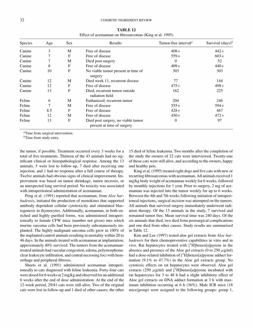
32 COSMETIC INGREDIENT REVIEW
TABLE 12Effect of acemannan on fibrosarcomas (King et al. 1995)
Species Age Sex Results Tumor-free intervala Survival (days)b
Canine 3 M Free of disease 408+ 442+Canine 7 F Free of disease 559+ 603+Canine 7 M Died post surgery 0 52Canine 8 F Free of disease 409+ 440+Canine 10 F No viable tumor present at time of
surgery303 303
Canine 12 M Died week 11, recurrent disease 77 144Canine 12 F Free of disease 475+ 498+Canine 13 F Died; recurrent tumor outside
radiation field162 225
Feline 6 M Euthanized; recurrent tumor 204 246Feline 7 M Free of disease 555+ 594+Feline 8.5 F Free of disease 428+ 467Feline 12 M Free of disease 450+ 472+Feline 13 F Died post surgery, no viable tumor
present at time of surgery0 97
aTime from surgical intervention.bTime from study entry.
the tumor, if possible. Treatment occurred every 3 weeks for atotal of five treatments. Thirteen of the 43 animals had no sig-nificant clinical or histopathological response. Among the 13animals, 5 were lost to follow-up, 7 died after receiving oneinjection, and 1 had no response after a full course of therapy.Twelve animals had obvious signs of clinical improvement. Im-provement was based on tumor shrinkage, tumor necrosis, oran unexpected long survival period. No toxicity was associatedwith intraperitoneal administration of acemannan.
Peng et al. (1991) reported that acemannan, from Aloe bar-badensis, initiated the production of monokines that supportedantibody dependent cellular cytotoxicity and stimulated blas-togenesis in thymocytes. Additionally, acemannan, in both en-riched and highly purified forms, was administered intraperi-toneally to female CFW mice (number not given) into whichmurine sarcoma cells had been previously subcutaneously im-planted. The highly malignant sarcoma cells grew in 100% ofthe implanted control animals resulting in mortality within 20 to46 days. In the animals treated with acemannan at implantation,approximately 40% survived. The tumors from the acemannan-treated animals had vascular congestion, edema, polymorphonu-clear leukocyte infiltration, and central necrosing foci with hem-orrhage and peripheral fibrosis.
Sheets et al. (1991) administered acemannan intraperi-toneally to cats diagnosed with feline leukemia. Forty-four catswere dosed for 6 weeks at 2 mg/kg and observed for an additional6 weeks after the end of dose administration. At the end of the12-week period, 29/41 cats were still alive. Two of the originalcats were lost in follow-up and 1 died of other causes; the other
15 died of feline leukemia. Two months after the completion ofthe study the owners of 22 cats were interviewed. Twenty-oneof those cats were still alive, and according to the owners, happyand healthy pets.
King et al. (1995) treated eight dogs and five cats with new orrecurring fibrosarcomas with acemannan. All animals received 1mg/kg body weight of acemannan weekly for 6 weeks, followedby monthly injections for 1 year. Prior to surgery, 2 mg of ace-mannan was injected into the tumor weekly for up to 6 weeks.Between the 4th and 7th weeks following initiation of intraperi-toneal injections, surgical incision was attempted on the tumors.All animals that survived surgery immediately underwent radi-ation therapy. Of the 13 animals in the study, 7 survived andremained tumor free. Mean survival time was 240 days. Of thesix animals that died, two died from postsurgical complicationsand one died from other causes. Study results are summarizedin Table 12.
Kim and Lee (1997) tested aloe gel extracts from Aloe bar-badensis for their chemopreventive capabilities in vitro and invivo. Rat hepatocytes treated with [3H]benzo[a]pyrene in theabsence and presence of the Aloe gel extracts (0 to 250 µg/ml)had a dose-related inhibition of [3H]benzo[a]pyrene adduct for-mation (9.1% to 47.7%) in the Aloe gel extracts group. Nocytotoxic effects on rat hepatocytes were observed. Aloe gelextracts (250 µg/ml) and [3H]benzo[a]pyrene incubated withrat hepatocytes for 3 to 48 h had a slight inhibitory effect ofAloe gel extracts on DNA adduct formation at 3 h with max-imum inhibition occurring at 6 h (36%). Male ICR mice (18mice/group) were assigned to the following groups: group 1,

ALOE 33
control; group 2, daily oral treatment with 50 mg/mouse/day ofAloe gel extracts in distilled water; group 3, single oral dose ofbenzo[a]pyrene (10 mg/mouse in corn oil); group 4, single oraldose of benzo[a]pyrene (10 mg/mouse in corn oil) then dailyoral treatment with Aloe gel extracts (10 mg/mouse/day) for16 days; and group 5, oral pretreatment with Aloe gel extracts(10 mg/mouse/day) for 16 days followed by a single oral dose ofbenzo[a]pyrene (10 mg/mouse in corn oil) and then continuoustreatment with 50 mg/mouse/day of Aloe gel extract for 16 days.
DNA adduct formation was significantly decreased in groups4 and 5 when compared to group 3. Glutathione S-transferaseactivity was slightly increased in the liver but cytochrome P450content was not affected by Aloe gel extract. The authors con-cluded that the inhibitory effect of the Aloe gel extract onbenzo[a]pyrene diol epoxide–DNA adduct formation might havea chemopreventive effect by inhibition of benzo[a]pyrene ab-sorption (Kim and Lee 1997).
Corsi et al. (1998) conducted a study in which male Fisherrats (number not given) previously injected with Yoshida AH-130 ascite hepatoma cells (2 × 10⁵ in a saline suspension) intothe pleural space received Aloe vera (20 µg suspended in 0.5ml sterile saline) injections daily for 2 weeks. Injections of Aloevera were also administered into the pleural space. At speci-fied times (days 7 and 14) the growth of the tumor cells in thepleural space was evaluated. The use of Aloe vera significantlydecreased tumor cell growth with respect to nontreated controlanimals. The survival of rats was progressively prolonged whenAloe vera was used as a therapy in the tumor-bearing animals.
Shamaan et al. (1998) studied the effects of an aloe derivativein rats. Male Sprague-Dawley rats were divided into six groupsof five to eight rats per group. Groups 1 to 3 received a basaldiet: group 1 was the control group and received no additionaltreatment; group 2 received vitamin C in their drinking water;group 3 received 0.1 g Aloe vera gel extract/100 ml in theirdrinking water for the 9-month study. A single dose of diethyl-nitrosamine (DEN) 200 mg/kg body weight and a diet contain-ing 0.02% (w/w) 2-acetylaminofluorene (AAF)-induced chem-ical hepatocarcinogenesis in groups 4 to 6. Group 5 receivedvitamin C and group 6 received Aloe vera gel extract as pre-viously described. DEN/AAF treatment significantly increasedthe GGT (γ -glutamyltransferase) concentration in carcinogen-induced rats; however, the addition of Aloe vera gel extract sig-nificantly decreased the activity of GGT. Both GGT- and GSTP(placental glutathione S-transferase)-positive foci were detectedin the carcinogen treated rats; however, the addition of Aloe veragel extract to the drinking water of group 6 caused a significantdecrease in number of foci on their livers. The livers of the ratsin group 6 were devoid of neoplastic nodules whereas cellulardamage was seen in group 4. Aloe vera gel extract supplemen-tation was found to be able to reduce the severity of chemicalhepatocarcinogenesis.
Kim et al. (1999) screened a polysaccharide of Aloe gel ex-tract of Aloe barbadensis for its chemopreventive effects us-ing biomarkers involved in chemical carcinogenesis. In the
benzo[a]pyrene (B[a]P)-DNA binding assay, a concentration de-pendent inhibition of DNA adduct formation occurred with theAloe polysaccharide (180 µg/ml). Oxidative DNA damage by 8-hydroxyoxyguanosine was significantly decreased by the Aloepolysaccharide (180 µg/ml). Additionally, the Aloe polysac-charide had a positive effect in a dose-dependent manner oninhibition of phorbol myristic acetate (PMA)-induced tyrosinekinase activity in HL-60 cells and PMA-induced ornithine decar-boxylase activity in BALB/3T3 cells. The Aloe polysaccharidesignificantly inhibited superoxide anion formation.
Pecere et al. (2000) screened aloe-emodin in vitro and invivo for its anticarcinogenicity activity against highly malig-nant tumors: neuroectodermal tumors, Ewing’s sarcoma, andneuroblastoma. The in vitro study was conducted first cultur-ing neuroblastoma cells, pPNET cells, Ewing’s sarcoma cells,T-cell leukemia cells, vinblastine-resistant cells, colon adeno-carcinoma cells, and doxorubicin-resistant cells. Cytotoxicityactivity was determined by growing cells in the presence ofaloe-emodin for 72 h, using many different concentrations ofaloe-emodin. After 24 and 48 h, the cells were scraped, washed,fixed overnight in a buffer solution, and processed for viewing.The results showed that aloe-emodin decreased the survival oftumor cells and normal fibroblasts. There was a large difference,however, between the two. The reduction in the neuroectodermaltumor cell lines was much more significant than in the differenttumor cell lines and normal fibroblasts.
The results of the in vitro study led to another antitumor studythat used 6-week-old female severely compromised immunode-ficient (SCID) mice. The mice were organized into three differ-ent groups. The first two groups both received IP injections in thedorsal region of 10 × 106 human neuroblastoma cells; however,the difference between the two groups was when they startedtreatment. Drug treatment started immediately in the first groupthat continued for 5 days for a total of 5 doses. Five animalsreceived 0.4 ml/day of DMSO (saline solution—control), andthe other mice were treated with 50 mg/kg/dose of aloe-emodin.The second group received the same doses, except treatmentdid not start until 15 days after they were injected with tumorcells. All of the mice were sacrificed in groups 1 and 2 when themean tumor volume in the control was 1.5 cm3. In group 3, thetreatment was exactly the same as group 1; however, these micereceived the same amount of IP injections of colorectal adeno-carcinoma a cells instead of neuroblastoma cells. All of the micewere sacrificed at day 30. In all three groups, the tumors weremeasured with a micrometer caliper twice a week throughoutthe study.
The tumors in group 1 (immediate treatment of aloe-emodin)were very sensitive to the drug, showing a significant reduc-tion of growth in the animal hosts. When aloe-emodin treatmentwas delayed (group 2) until a palpable tumor mass had devel-oped (day 15), tumor growth was halted throughout the periodof drug administration. The group that was injected with col-orectal adenocarcinoma cells was refractory to the treatment.No appreciable signs of acute or chronic toxicity were observed

34 COSMETIC INGREDIENT REVIEW
in any of the treated animals; weight, neurological and intestinalfunctions, and hematological parameters were normal (Pecereet al. 2000).
Singh et al. (2000) investigated the cancer chemopreventivecapabilities of Aloe vera leaf pulp extract by analyzing Aloe’seffects on the enzymes that play a role in carcinogen metabolismand those involved in cellular antioxidant activity. Random-bredSwiss albino male mice were divided into four groups: group 1was fed a normal diet and sham treated with distilled water byoral gavage for 14 days; group 2 was also fed a normal diet; how-ever, they were treated with 30 µl of Aloe vera leaf pulp extractper animal by oral gavage for 14 days; group 3 was also fed a nor-mal diet; however, their treatment amount was 60 µl of Aloe veraleaf pulp extract per animal, also delivered by oral gavage; group4 was fed a diet containing 0.75% butylated hydroxyanisole(BHA) for 14 days. The last group served as a positive controlbecause BHA is known to be an anticarcinogenic compound.The animals were killed and their livers homogenized. The re-sulting supernatant from the livers was used to assay for totalcytosolic glutathione S-transferase, DT-diaphorase, lactate de-hydrogenase, and antioxidant enzymes. The pellet was used forassaying cytochrome P450, cytochrome b5, cytochrome P450reductase, cytochrome b5 reductase, and lipid peroxidation.
During the treatment phase of the experiment there were noanimal deaths or decreases in body weight. Additionally, therewas no indication of cellular damage. Aloe vera leaf pulp de-creased the levels of cytochromes P450 and b5, but increased theactivities of cytochrome P450 and b5 reductases. There were sig-nificant increases in the activity of glutathione S-transferase andDT-diaphorase in the high-dose group as compared to the con-trol. In the low-dose group, the enzyme activity was comparablewith that of the control group. The activity of lactate dehydroge-nase was decreased in a dose-dependent manner. The activitiesof the antioxidant enzymes (glutathione, glutathione peroxidase,glutathione reductase, superoxide dimutase, and catalase) wereall increased in a dose-dependent manner. The extent of lipidperoxidation was decreased significantly in the high-dose group;the low-dose group was also inhibited but it was not significant(Singh et al. 2000).
Lee (2001) reported aloe-emodin–induced apoptosis in lungcell carcinoma cell lines CH27 (human lung squamous carci-noma cells) and H460 (human lung non–small cell carcinomacells). During apoptosis increases in cytochrome c and activationof caspase-3 were observed and expression of protein kinase Cappeared to occur downstream of caspase-3.
Kuo et al. (2002) reported that aloe-emodin inhibited cell pro-liferation and induced apoptosis in two liver cancer cell lines,Hep G2 and Hep 3B. Hep G2 cells induced p53 (tumor suppres-sor gene) expression and was accompanied by induction of p21(cell cycle regulator) expression that was associated with a cellcycle arrest in G1 phase. Fas/APO 1 (type one I membrane pro-tein) receptors and Bax (proapoptotic protein) expression wasincreased in Hep G2 with aloe-emodin. In Hep 3B cells, theinhibition of cell proliferation by aloe-emodin was mediated
through p21, and neither cell cycle arrest nor an increase in thelevel of Fas/APO1 receptor was observed. Bax expression wasincreased by aloe emodin in Hep 3B cells.
Wasserman et al. (2002) described Merkel cell carcinoma(MCC) as a rare and aggressive tumor of the skin, also known asa primary neuroendocrine carcinoma of the skin. A free floatingcell line was established from a metastasis from a MCC patient.Aloe-emodin decreased the viability of carcinoma cells after 72 hof treatment. The results are statistically significant (p < .02)for 10 µmol and higher concentrations (100 µmol) (p < .001).Aloin was also investigated at the same concentration range andno effect was detected.
Aloe arborescensYagi et al. (1977) injected aloemannan, a partially acetylated
β-d-mannan isolated from the leaves of Aloe arborescens, in-traperitoneally into ICR mice (10 animals/group) that were im-planted with sarcoma-180 cells. Doses of 5 and 100 mg/kg wereadministered daily for 10 days and produced inhibition ratios of38.1% and 48.1%, respectively. No toxicity was observed.
Imanishi et al. (1981) administered aloctin A, a glycoproteinof Aloe arborescens, intraperitoneally to BALB/c mice (5 to 6weeks of age) once daily for 5 days after tumor implantation.Tumors were induced by methylcholanthrene. At 10 mg/kg/dayfor 5 days, aloctin A significantly inhibited the growth of themethylcholanthrene-induced fibrosarcomas. To determine if thealoctin A had direct cytotoxicity on tumor cells or was due tohost-mediated effects, the authors tested the effect of aloctin Aon various tumor cell lines. Aloctin A had almost no inhibitoryeffect on the growth of tumor cell lines up to a concentration of200 µg/ml.
Uehara et al. (1996) measured the formation of 2-amino-3-methylimidazo[4,5-f]quinoline (IQ)-DNA adducts by 32P-postlabeling analysis. Cytochrome P450 (CYP) 1A1 and 1A2protein levels were analyzed by ELISA (enzyme-linked im-munosorbent assay) to assess the mechanism of chemopreven-tion of hepatocarcinogenesis by freeze-dried whole leaves ofAloe arborescens. Ten male F344 rats were fed a diet contain-ing 30% Aloe arborescens over an 8-day period. The 11 controlanimals were fed a basal diet only. On day 7, all animals weresubjected to a two-third partial hepatectomy (PH). Twelve hoursafter the PH, the rats received a single intragastric dose of thecarcinogenic food pyrolysate (IQ) (100 mg/kg) to initiate hepa-tocarcinogenesis. Rats were killed 6, 12, 24, and 48 h after IQadministration.
The levels of adducts, expressed as relative adduct labelingvalues, in rats treated with Aloe arborescens were decreased ascompared with the control group at hour 24 (36 h after PH) andhad further decreases at hour 48. The levels of CYP1A2, knownto be responsible for the activation of IQ, also were decreased athour 48. The authors concluded that Aloe arborescens has thepotential to decrease IQ-DNA adduct formation, presumably asa result of decreased formation of active metabolites (Ueharaet al. 1996).
ALOE 35
Shimpo et al. (2001) examined freeze-dried powder of whole-leaf Aloe arborescens Miller var. natalensis Berger (Japanesename Kidachi aloe) for its modifying effect on azoxymethane(AOM)-induced aberrant crypt foci (ACF), putative neoplasticlesions, in male F344 (4-week-old) rats. Rats were fed a basaldiet (groups 1 and 5), experimental diet with 1% (groups 2, 3,and 6), or 5% (groups 4 and 7) aloe for 5 weeks. On days 7, 14,and 21, groups 1 to 4 were given a subcutaneous injection of15 mg/kg of AOM and groups 5 to 7 received a subcutaneousinjection of normal saline (4 ml/kg).
No rats died. The 5% aloe diet rats had a “mud-like” or softfeces. Body weight was reduced significantly in all the AOM-dosed rats compared to the AOM-untreated rats. ACF developedonly in the rats treated with AOM (with or without aloe) and noACF were present in the untreated rats. ACF were decreased by35% and 14% with the 1% and 5% aloe treatments, respectively.AOM had no effect in the quinone reductase (QR) levels in theliver, although QR activity was significantly greater in the liverof rats fed 5% aloe with or without AOM treatment as comparedto the rats fed a basal diet (Shimpo et al. 2001).
Furukawa et al. (2002) reported that freeze-dried whole leafpowder of Aloe arborescens modified the initiation phase ofcancer in female Syrian hamsters (total = 90; group = 30).N -nitrosobis(2-oxopropyl)amine (BOP) was administered sub-cutaneously for 4 weeks at a dose of 10 mg/kg. Aloe was givenin the diet at 0%, 1%, or 5% for 5 weeks. At 54 weeks all surviv-ing animals were killed and the development of neoplastic andpreneoplastic lesions was assessed histopathologically.
Aloe at 5% decreased significantly the number of animalswith pancreatic adenocarcinomas (58.6%) compared to BOPalone (86.6%) and decreased significantly the number of ani-mals with atypical hyperplasia (27.5%) compared to BOP alone(56.6%). Adenocarcinomas per animal were decreased signif-icantly by Aloe at 5% (0.69 ± 0.66) compared to BOP alone(1.20 ± 0.76) and atypical hyperplasias per animal were de-creased significantly (0.34 ± 0.61) compared to BOP alone(0.76 ± 0.77). The decreases seen in the group treated withAloe at 1% were significant only for the number of animalswith adenocarcinomas and the number of adenocarcinomas peranimal, compared to BOP alone. The authors concluded thattreatment with the Aloe arborescens derivative has an inhibitoryeffect during the induction phase for pancreatic neoplastic andpreneoplastic lesions (Furukawa et al. 2002).
Shimpo et al. (2002) reported that the ethyl acetate extractof an acetone soluble Aloe arborescens Miller var. natalensisBerger (Japanese name Kidachi aloe) fraction inhibited 12-O-tetra-decanoylphorbol-13-acetate (TPA)-induced ear edema (anearly marker of tumor promotion) in ICR mice (n: control =4; TPA = 6; TPA + Aloe extract = 6). The effect of topicaltreatment of 1 mg of aloe with 1.6 nmol topically applied TPAto the ear was 40% reduction of edema (p < .0005). Aloe ex-tract itself did not induce edema. The 20 mg of aloe extracttogether with 5 nmol TPA also inhibited the TPA-induced in-crease in epidermal putrescine level by 30% (p < .005). Aloe
extract affected the promotion of skin papillomas by TPA in7,12-dimethylbenz[a]anthracene (DMBA)-initiated mice (n =20/group). The extract lowered the percentage of tumor-bearingmice by 45% after 10 weeks (p < .005) and average number oftumors decreased (no tumors in solvent control, 25.3 tumor inTPA-treated, and 7.7 tumors in aloe-treated mice) (p < .001).
PHOTOTOXICITYVath et al. (2002) exposed human skin fibroblast cell lines
(ATCC CRL 1634) to 1 µM aloe-emodin and UVA radiation(320 to 400 nm) from two GE F40BL bulbs filtered through 3mm of soft glass. The irradiance of this source was 2.9 × 10⁻³
W/cm2. UVA exposure alone did not affect cell survival up to ex-posures of 5 J/cm2. UVA-plus aloe-emodin reduced survival to50% at around 3 J/cm2. UVA plus aloe-emodin in D2O (extendsthe lifetime of singlet oxygen) reduced survival to 50% at around1 J/cm2. The authors suggested that this finding supports a pho-totoxic mechanism that involves formation of singlet oxygenby aloe-emodin in the presence of UVA. Cells exposed to aloe-emodin in the dark had no effect on survivability. Aloe-emodinwas shown to bind to DNA, although single strand breaks inplasmid (E. coli) DNA were not observed.
Vargas et al. (2002) exposed human red blood cells to aloe-emodin in phosphate-buffered saline (PBS) solution (20 to80 µg/ml) and UVA-irradiated the cells under a Rayonet pho-tochemical reactor equipped with 16 phosphor lamps (340to 500 nm) or alternatively with an Osram HQL 250 Wattmedium-pressure Hg lamp in a pyrex immersion-well photore-actor (radiation dose of 4.5 J/cm2). Aloe-emodin caused cell ly-sis (>50%) within 70 min after irradiation of erythrocyte suspen-sion. Hemolysis did not occur in the dark controls. Free radicalscavengers reduced glutathione (GSH), and superoxide dismu-tase (SOD) had little protective effect on the photohemolytic pro-cess. Photohemolysis was decreased 20% by addition of serumproteins. Preirradiated aloe-emodin was responsible for 60%hemolysis during 80 min. When the photohemolysis tests weredone under argon there was a 54% decrease in the effect. Pho-toperoxidation was observed in irradiated aloe-emodin/linoleicacid in PBS solution. Significant amounts of hydroperoxideswere formed. When this test was performed under argon a 34%reduction in lipid peroxidation was seen.
The effect of 5 × 10⁻⁶ M aloe-emodin on acetyl-cholinesterase (ACE) activity on human erythrocyte membranesirradiated with UV light (340 to 500 nm) was determined. ACEactivity was inhibited by 50% with respect to controls; other frac-tions were incubated with reactive oxygen scavengers butylatedhydroxyanisole (BHA) (1 mM), sodium azide (1 mM), diazabi-cyclooctane (DABCO) (1 mM), and SOD (0.01 mg/ml). TheACE activity recovered by 34.1 ± 5.4%, 21.0 ± 4.8%, 21.9 ±3.8%, and 3.8 ± 3.0% (not significant), respectively. The au-thors thought the radical species and singlet oxygen productionand subsequent peroxyl radical formation are involved in thephotoinhibitory effects of aloe-emodin on ACE activity (Vargaset al. 2002).

36 COSMETIC INGREDIENT REVIEW
Shiseido Safety Research Labs (2002) reported photoaller-genicity of Aloe Arborescens CRS was evaluated by Adjuvantand Strip test method using female Hartley strain albino guineapigs (five subjects per group). During the induction phase of theexperiment, 0.1 ml of emulsified Freund’s complete adjuvant(FCA) was prepared and injected intradermally at 4 corners ofa clipped and shaved nuchal area of the subject. Then, 0.1 ml of3% Aloe Arborescens CRS in distilled water was applied to thearea and irradiated with 10.2 J/cm2 of UVA. Similar applicationand irradiation continued for 4 days, and control groups weretreated in the same manner, only using distilled water. During thechallenge phase of the experiment (followed a 2-week restingperiod), 0.02 ml of Aloe Arborescens CRS—6%, 3%, 0.6%, and0.3% in distilled water—were applied to a clipped and shavedflank symmetrically in duplicate. One side was irradiated with10.2 J/cm2 of UVA, and the other side was covered with alu-minum foil to prevent exposure to the light. The reactions withrespect to erythema, eschar formation, and edema were scoredat 24, 48, and 72 h and followed a scoring system to evaluate.
It was concluded that Aloe Arborescens CRS does not pos-sess photosensitizing potential under the test conditions sinceno reactions were observed in the animals of both the treatedand control groups. One animal in the control group, however,was excluded from the estimation due to persistent light reaction(Shiseido Safety Research Labs 2002).
Shiseido Safety Research Labs (2002) reported on anotherstudy analyzing skin phototoxicity of Aloe Arborescens CRS inHartley strain male albino guinea pigs. There were five subjectsin each of the treatment and control groups. The test materialfor the treatment group was 100% and 3% Aloe ArborescensCRS in distilled water and for the control group it was 0.02%8-methoxypsoralen (8-MOP) in ethanol. The dorsum of the sub-jects was shaved and depilated, and 24 h later, 0.02 ml of the testmaterials was applied to the hairless area symmetrically in du-plicate. One side was irradiated with 14 J/cm2 of UVA, and theother side was covered in aluminum foil to prevent exposure tothe light. The skin reactions with respect to erythema and edemawere evaluated at 24, 48, and 72 h after irradiation with a scoringsystem. There were no skin reactions for Aloe Arborescens CRSat the irradiated and unirradiated sites. However, the assessmentscore of 8-MOP (positive control) at concentration 0.02% wasseverely phototoxic. The Shiseido Safety Research Lab con-cluded that Aloe Arborescens CRS does not possess phototoxicpotential under the test condition.
CLINICAL ASSESSMENT OF SAFETY
Dermal Irritation/SensitizationThe Institut D’Expertise Clinique (2000) applied Aloe Bar-
badensis Leaf Water to the back of 10 healthy female adults. Thetest substance (0.02 ml) was applied to the back on a 50-mm2
area of skin and occluded for 48 h. No reactions or pathologicalirritation were observed other than a very slight erythema in oneperson.
Photoallergy/PhotosentizationIvy Laboratories (1987a) applied a 0.1% Aloe extract (suntan
product) to the lower back of 25 healthy Caucasian subjectswith no history of sun sensitivity. Each subject underwent threephases: pretesting, induction, and challenge. In the pretestingphase the minimal erythema dose (MED) was established onthe midback (1-cm diameter) in 25% increments from thexenon arc solar simulator (150-watt compact xenon arc sourceequipped with UV-reflecting dichroic mirror and 1-mm-thickSchott WG-320 filter to produce simulation of solar UVBspectrum 290 to 320 nm). The MED was the area with distincterythema after 20 to 24 h.
The induction phase involved application of (10 µl/cm2) ofaloe extract to a 2 × 2-cm section of the lower back and coveredfor 24 h. The site was then wiped and exposed to the MED andleft open for 24 h and occluded for another 24 h. The patcheswere removed and exposed to 3 MEDs of solar simulator radi-ation. This sequence was repeated for the same test sites twiceweekly for 3 weeks. The challenge phase occurred 10 to 14 daysafter the last induction exposure. The aloe was reapplied to thesame area and in the same manner but in duplicate to a newlydesignated skin site of the same dimensions on the opposite sideof the lower back and occluded for 24 h. Each site was irra-diated with 4 J/cm2 of UVA (UVB was filtered from the solarsimulator with a 1-mm-thick UG5 filter). The duplicate site wasunirradiated; another skin site untreated with aloe was exposedto 4 J/cm2 of UVA. No adverse reaction at any phase of theexperiment was seen (Ivy Laboratories 1987a).
Ivy Laboratories (1987b) applied a suntan preparation con-taining 0.1% aloe extract to the lower back of 10 healthy,Caucasian adults with fair skin (skin type I: always burns eas-ily, never tans; II: always burns easily, tans minimally; andIII: burns moderately, tans gradually). All subjects were overthe age of 18, with no medical or dermatological illness. Aloe(50 µl) was applied to a 2 × 2-cm area in duplicate, allowedto dry, and covered. Six hours later one site was exposed to30 J/cm2 of UVA radiation. An adjacent skin site was treatedwith vehicle (hydrophilic ointment USP) and exposed to theUVA. Reactions were graded immediately, and at 24 and 48hours.
No abnormal reactions were observed. One subject had min-imal erythema reaction immediately. One individual had a min-imal erythema to the vehicle control and the aloe immediatelythat persisted at 24 h and cleared by 48 h (Ivy Laboratories1987b).
Ivy Laboratories (1988) applied a 0.01% Aloe BarbadensisFlower Extract (suntan product) to the lower back of 25 healthyCaucasian subjects with no history of sun sensitivity. Each sub-ject underwent three phases: pretesting, induction, and chal-lenge. In the pretesting phase the minimal erythema dose (MED)was established on the midback (1-cm diameter) in 25% incre-ments from the xenon arc solar simulator (150-watt compactxenon arc source equipped with UV-reflecting dichroic mirrorand 1-mm-thick Schott WG-320 filter to produce simulation of

ALOE 37
solar UVB spectrum 290 to 320 nm). The MED was the areawith distinct erythema after 20 to 24 h.
The induction phase involved application of aloe extract,10 µl/cm2, to a 2 × 2-cm section of the lower back and coveredfor 24 h. The site was then wiped and exposed to the MED andleft open for 24 h and occluded for another 24 h. The patcheswere removed and exposed to 3 MEDs of solar simulatorradiation. This sequence was repeated for the same test sitestwice weekly for 3 weeks. The challenge phase occurred 10 to14 days after the last induction exposure. The aloe was reappliedto the same area and in the same manner but in duplicate toa newly designated skin site of the same dimensions on theopposite side of the lower back and occluded for 24 h. Each sitewas irradiated with 4 J/cm2 of UVA (UVB was filtered from thesolar simulator with a 1-mm-thick UG5 filter). The duplicatesite was unirradiated; another skin site untreated with aloe wasexposed to 4 J/cm2 of UVA.
No adverse reactions were noted, except for mild to moderateerythema, scaling, and tanning (Ivy Laboratories 1988).
The Consumer Product Testing Co. (1996) applied a 0.1%Aloe extract (suntan product) to the lower back of 26 healthyCaucasian subjects with no history of sun sensitivity. Each sub-ject underwent three phases: pretesting, induction, and chal-lenge. One subject dropped out for personal reasons. In thepretesting phase the minimal erythema dose (MED) was es-tablished on the midback (1-cm diameter) in 25% incrementsfrom the xenon arc solar simulator (150-watt compact xenonarc source equipped with UV-reflecting dichroic mirror and 1-mm-thick Schott WG-320 filter to produce simulation of solarUVB spectrum 290 to 320 nm).
The induction phase involved application of aloe extract(0.2 g) to a 3/4 × 3/4-inch area of the lower back and cov-ered for 24 h. The site was then wiped and exposed to the MEDand left open for 24 h and occluded for another 24 h. The patcheswere removed and exposed to two times the MED as determinedfor each subject. This sequence was repeated for the same testsites twice weekly for 3 weeks. The challenge phase occurred10 to 14 days after the last induction exposure. The product wasreapplied to the same area and in the same manner but in du-plicate to a new designated skin site of the same dimensions onthe opposite side of the lower back and occluded for 24 h. Eachsite was irradiated for 3 min with UVA (UVB was filtered fromthe solar simulator with a 1-mm-thick UG5 filter). The dupli-cate site was unirradiated; another skin site untreated with theproduct was exposed to 4 J/cm2 of UVA.
No adverse reactions at any phase of the experiment wereobserved (Consumer Product Testing Co. 1996).
Ivy Laboratories (1996) applied a 0.5% Aloe extract (othersuntan product) to the lower back of 31 healthy Caucasian sub-jects with no history of sun sensitivity. Each subject underwentthree phases: pretesting, induction, and challenge. In the pretest-ing phase the minimal erythema dose (MED) was establishedon the midback (1-cm diameter) in 25% increments from thexenon arc solar simulator (150-watt compact xenon arc source
equipped with UV-reflecting dichroic mirror and 1-mm-thickSchott WG-320 filter to produce simulation of solar UVB spec-trum 290 to 320 nm). The MED was the area with distinct ery-thema after 20 to 24 h.
The induction phase involved application of 80 mg of theproduct to a 2 × 2-cm area of the lower back and covered for24 h. The site was then wiped and exposed to the MED and leftopen for 24 h and occluded for another 24 h. The patches wereremoved and exposed to 3 MEDs of solar simulator radiation.This sequence was repeated for the same test sites twice weeklyfor 3 weeks. The challenge phase occurred 10 to 14 days after thelast induction exposure. The product was reapplied to the samearea and in the same manner but in duplicate to a new designatedskin site of the same dimensions on the opposite side of the lowerback and occluded for 24 h. Each site was irradiated with 4 J/cm2
of UVA (UVB was filtered from the solar simulator with a 1-mm-thick UG345 filter). The duplicate site was unirradiated;another skin site untreated with the product was exposed to 4J/cm2 of UVA.
A total of 25 subjects exhibited, at most, mild erythema,desquamation, and tanning at any phase of the experiment. Sixsubjects dropped out of the study for personal reasons (Ivy Lab-oratories 1996).
KGL Inc. (2000) applied a product containing 0.1% aloe ex-tract to the lower back of 26 Caucasian adults with fair skin (skintype I: always burns easily, never tans; II: always burns easily,tans minimally; and III: burns moderately, tans gradually) overthe age of 18 with no medical or dermatological illness and nohistory of sun sensitivity. The subjects underwent three phases:pretesting, induction, and challenge. In the pretesting phase theminimal erythema dose (MED) was established on the midback(1-cm diameter) in 25% increments from the xenon arc solar sim-ulator (150-watt compact xenon arc source equipped with UV-reflecting dichroic mirror and 1-mm-thick Schott WG-320 filterto produce simulation of solar UVB spectrum 290 to 400 nm).The MED was the area with distinct erythema after 20 to24 h.
The induction phase involved application of 40 mg of theproduct to a 2 × 2-cm area of the lower back and covered for24 h. The site was then wiped and exposed to the MED andleft open for 48 h and occluded for another 24 h. The patcheswere removed and exposed to 3 times the MED as determinedfor each subject. This sequence was repeated for the same testsites twice weekly for 3 weeks. The challenge phase occurred 12days after the last induction exposure. The product was reappliedto the same area and in the same manner but in duplicate to anew designated skin site of the same dimensions on the oppositeside of the lower back and occluded for 24 h. Each site wasirradiated with 4 J/cm2 of UVA (UVB was filtered from the solarsimulator with a 1-mm-thick UG5 filter). The duplicate site wasunirradiated; another skin site untreated with the product wasexposed to 4 J/cm2 of UVA.
No adverse reactions at any phase of the experiment wereseen (KGL Inc. 2000).

38 COSMETIC INGREDIENT REVIEW
KGL Inc. (2001) applied a product containing 0.5% aloe ex-tract to the lower back of 26 healthy Caucasian subjects with nohistory of sun sensitivity. Subjects were of skin type I: alwaysburns easily, never tans; II: always burns easily, tans minimally;and III: burns moderately, tans gradually and over the age of18 with no medical or dermatological illness. The subjects un-derwent three phases: pretesting, induction, and challenge. Onesubject dropped out of the study for personal reasons. In thepretesting phase the minimal erythema dose (MED) was es-tablished on the midback (1-cm diameter) in 25% incrementsfrom the xenon arc solar simulator (150-watt compact xenonarc source equipped with UV-reflecting dichroic mirror and 1-mm-thick Schott WG-320 filter to produce simulation of solarUVB spectrum 290 to 320 nm). The MED was the area withdistinct erythema after 20 to 24 h.
The induction phase involved application of 40 mg of aloeextract to a 2 × 2-cm section of the lower back and covered for24 h. The site was then wiped and exposed to 3 MEDs and leftopen for 48 h and occluded for another 24 h. The patches wereremoved and exposed to 3 MEDs of solar simulator radiation.This sequence was repeated for the same test sites twice weeklyfor 3 weeks. The challenge phase occurred 16 days after the lastinduction exposure. The product was reapplied to the same areaand in the same manner but in duplicate to a new designated skinsite of the same dimensions on the opposite side of the lowerback and occluded for 24 hours. Each site was irradiated with4 J/cm2 of UVA (UVB was filtered from the solar simulator witha 1-mm-thick UG345 filter). The duplicate site was unirradi-ated; another skin site untreated with the product was exposed to4 J/cm2 of UVA.
No adverse reactions at any phase of the experiment wereseen (KGL Inc. 2001). Table 13 summarizes the results of theseproduct studies.
In addition to the above studies, CTFA (2003) provided re-sults of testing of a number of products containing differentaloe-derived ingredients for photosensitization and phototox-icity on human volunteers. The subjects were categorized ashaving skin type I (always burns easily, never tans), skin type II
TABLE 13Photoallergy and photosensitization studies containing aloe-derived ingredients
Product Concentration of ingredient No. of subjects Results Reference
Suntan product 0.1% Aloe extract 25 No adverse reaction observed Ivy Laboratories 1987aSuntan product 0.1% Aloe extract 10 No adverse reaction observed Ivy Laboratories 1987bSuntan product 0.01% Aloe Barbadensis 25 No adverse reaction observed Ivy Laboratories 1988
Flower extractSuntan product 0.1% Aloe extract 26 No adverse reaction observed Ivy Laboratories 1996Suntan product 0.1% Aloe extract 25 No adverse reaction observed KGL Inc. 2000Other suntan 0.5% Aloe extract 31 No adverse reaction observed Ivy Laboratories 1996
preparationsOther suntan 0.5% Aloe extract 26 No adverse reaction observed KGL Inc. 2001
preparations
(always burns easily, tans minimally), and skin type III (burnsmoderately, tans gradually) and the ultraviolet dosage was ad-justed according to the skin type. Semioccluded patches of testmaterials were employed for each type of study. The compoundsstudied and results obtained are provided in Table 14.
Effects on PregnancyIn the CRC Handbook of Medicinal Herbs (Duke 1985), Aloe
barbadensis is cited as being considered abortive. Briggs (1995)mentions that aloe latex or juice should not be ingested duringpregnancy due to the potent cathartic action of anthraquinonesthat may stimulate reflex contractions of the uterus, possiblyresulting in premature labor or miscarriage; and nursing mothersshould not take aloes as a laxative because the anthraquinonesmay be secreted into their milk, resulting in diarrhea in the baby.
Kacew (1993) reported that Aloe ingestion by the mother(from a laxative) increased intestinal peristalsis in the fetus, re-leased meconium in uterus, and caused kidney damage in thefetus. In addition, Aloe administration to the infant through thebreast milk caused increased bowel activity in the infant.
Treatment UsesAsthma
Shida et al. (1985) reported that 33 patients received an Aloeextract (species not indicated) orally for 6 months for the treat-ment of adult bronchial asthma. The clinical trial was conductedfor 24 weeks, preceded by a 4-week basal period. Five millilitersof a 20% solution of Aloe extract in saline was administeredorally twice a day. No adverse effects were reported throughoutthe 24-week trial period.
Heart DiseaseAgarwal (1985) treated 5000 patients with evidence of is-
chemic heart disease with 100 g of the fresh flesh gelatin ofAloe vera and 20 g of Husk of Isabgol. The two ingredientswere mixed with wheat flour to make a bread and eaten dailyat lunch and dinner for 5 years. The scope of this study was to

ALOE 39
TABLE 14Further photosensitization and phototoxicity studies containing aloe-derived ingredients (CTFA 2003)
Product and ingredient Concentration Type of study No. of subjects Results
Toner—Aloebarbadensis leafextract
0.05% Phototoxicity 10 No reactions on either irradiated ornon-irradiated test material contactsites were observed. No dermalphototoxicity observed
Dermal photoallergy 26 No dermal photoallergy or dermalsensitization in human subjects wasobserved
Lotion—Aloebarbadensis innerleaf gel concentrate
0.1% Phototoxicity 10 No reactions on either irradiated ornonirradiated test material contactsites were observed. No dermalphototoxicity observed
Dermal photoallergy 30 No dermal photoallergy or dermalsensitization in human subjects wasobserved
Hand lotion—Aloeextract Veracontainingpolysaccharide & nodetectableanthraquinone
0.1% Phototoxicity 10 No reactions on either irradiated ornonirradiated test material contactsites were observed. No dermalphototoxicity observed
Dermal photoallergy 29 No dermal photoallergy or dermalsensitization in human subjects wasobserved
Lotion—Aloebarbadensis leafextract
0.05% Phototoxicity 10 No reactions on either irradiated ornonirradiated test material contactsites were observed. No dermalphototoxicity observed
Dermal photoallergy 26 No dermal photoallergy or dermalsensitization in human subjects wasobserved
Powder—Aloe vera gel,powder with aloinlevels 3–5 ppm inreconstituted product
0.01% Phototoxicity 10 No reactions on either irradiated ornonirradiated test material contactsites were observed. No dermalphototoxicity observed
Dermal photoallergy 28 No dermal photoallergy or dermalsensitization in human subjects wasobserved
Oil—Aloe ferox leafextract
0.2% Phototoxicity 10 No reactions on either irradiated ornonirradiated test material contactsites were observed. No dermalphototoxicity observed
Dermal photoallergy 29 No dermal photoallergy or dermalsensitization in human subjects wasobserved
Gel—Aloe barbadensisleaf extract withanthraquinone lessthan 5 ppm
0.1% Phototoxicity 10 No reactions on either irradiated ornonirradiated test material contactsites were observed. No dermalphototoxicity observed

40 COSMETIC INGREDIENT REVIEW
TABLE 14Further photosensitization and phototoxicity studies containing aloe-derived ingredients (CTFA 2003) (Continued)
Product and ingredient Concentration Type of study No. of subjects Results
Dermal photoallergy 25 No dermal photoallergy ordermal sensitization inhuman subjects wasobserved
VP40:1 or biostaticAloe Vera
Aloe vera gel65% and Aloeconcentrate0.75%
Phototoxicity 10 No reactions on eitherirradiated or nonirradiatedtest material contact siteswere observed. No dermalphototoxicity observed
Dermal photoallergy 28 No dermal photoallergy ordermal sensitization inhuman subjects wasobserved
Dermal photoallergy 30 No dermal photoallergy ordermal sensitization inhuman subjects wasobserved
observe the effects of the two ingredients in treating heart dis-ease and associated complications. There were no adverse sideeffects noted in this study.
DiabetesGhannam et al. (1986) studied five otherwise healthy, non–
insulin-dependent diabetic patients in a study of the antidiabeticactivity of Aloes (see previous definition under miscellaneousbiology). Apart from the blood sugar levels, all routine hema-tological and biochemical indices were normal. After 2 to 24weeks without any treatment, the patients were weighed. Serumfrom the fasting patients was obtained for measurement of glu-cose (FSG) and insulin (FSI). After the patients had been takingAloes for 4 to 14 weeks, FSG estimations were repeated in allpatients and FSI values in three patients. The Aloes, in the formof dried resin, were taken with water by three patients and swal-lowed with food by two patients. The dose was half a teaspoondaily. The patients followed their normal diet throughout thestudy. The FSG levels of every patient fell during the consump-tion of the Aloes from a mean of 273 ± 25 (SE) to 151 ± 23mg/dl, p < .001. The patients’ body weights and serum insulinlevels were unchanged. No patient had side effects, although allnoted a bitter taste produced by the Aloes. The amount con-sumed was insufficient to cause abdominal cramps or diarrhea.No electrolyte disturbances were noted on routine screening.
Bunyapraphatsara et al. (1996) enrolled 49 men and 23women in a study to evaluate the effect of Aloe vera juice ondiabetes. The treatment group received one tablespoon full ofAloe vera juice twice a day and two tablets of glibenclamide(5 mg). The control group received a placebo (made to lookand taste like the Aloe vera juice) and the two tablets of gliben-
clamide. The control group had stable levels of fasting bloodglucose, cholesterol, and triglycerides. The treatment group hada significant decrease in blood sugar levels within 2 weeks andtriglycerides within 4 weeks. The Aloe vera juice did not causeany adverse effect throughout the study.
UlcersBlitz et al. (1963) treated 12 patients diagnosed with peptic
ulcers with 2 to 2.5 fluid drams of Aloe vera gel in a heavy liquidpetrolatum emulsion (duration of treatment not specified). Allpatients recovered completely.
Atherton (1998) treated 7 patients with chronic leg ulcerswith an Aloe vera gel orally (60 ml of fluid) and a topicalAloe jelly (amount not given). One patient withdrew due tothe stinging sensation she felt when the Aloe jelly was applied;otherwise, none of the other patients had side effects. Threeof the remaining six wounds healed completely, one healedpartially and one showed no improvement. The six patientswere pleased with the cleansing effect of the Aloe jelly report-ing that there was less exudate and odor from their wounds.The author stated that swabs taken from the leg ulcers demon-strated that the Aloe jelly destroyed infecting bacteria, especiallyPseudomonas.
Garnick et al. (1998) conducted a study in which 40 patientsdiagnosed with aphthous stomatitis ulcers of the oral mucosalsurfaces were randomly assigned to receive either control gel(carrier gel), carrier gel with Aloe vera, carrier gel with silicondioxide and Aloe vera, carrier gel with Aloe vera and allantoin, orcarrier gel with silicon dioxide, Aloe vera, and allantoin. Eachsubject applied the gel product with a cotton swab and thenbrushed their teeth with the gel, spreading the gel throughout

ALOE 41
the mouth. The subjects using the carrier gel with silicon diox-ide, Aloe vera, and allantoin had significant decrease in lesionduration. There were no other significant results in the othergroups in comparison with the control group.
Thomas et al. (1998) reported a study in which 30 subjectswith pressure ulcers were randomized into two groups: a salinedressing control group and the acemannan (isolated from Aloebarbadensis) hydrogel experimental group. Acemannan hydro-gel dressing was purchased commercially. No differences wereobserved in complete healing between the experimental and con-trol groups. The authors concluded that acemannan hydrogeldressing is as effective as but not superior to a moist salinegauze dressing for pressure ulcer management.
Skin DiseaseZawahry et al. (1973) applied gauze soaked with gel that had
been extracted from the center of Aloe vera leaves locally tochronic ulcers three to five times daily on three patients. All pa-tients had chronic leg ulcers that had not responded to any othertreatment. All three patients had an increase in the epithelializa-tion of their ulcers. The only side effect reported was increasedpain at the start of treatment; the pain diminished by the 2ndweek. Additionally, the authors treated three patients with acne,three patients with seborrheic alopecia, and three patients withalopecia areata with the Aloe vera gel. All three ulcer patientsshowed improvement with their Aloe vera treatment. There wasincreased vascularization of all ulcers and removal of necrotictissue. The three patients with seborrheic alopecia had somehair regrowth; the three acne patients had facial drying followedby improvement in their acne. Two of the three were acne freewithin 1 month. The three patients with alopecia areata had hairgrowth. None of the patients reported contact dermatitis or anytype of irritation.
Syed et al. (1996) randomly allocated 60 psoriasis patients(36 males and 24 females) aged 18 to 50 years with slight tomoderate chronic-type psoriasis to two parallel groups. A 0.5%Aloe vera preparation in a hydrophilic cream, using a combina-tion of mineral oil and castor oil as vehicles, was provided topatients in the treatment group. A matching placebo cream thatdid not contain Aloe vera was given to the control group.
At the start and end of the study, full thickness 6-mm punchskin biopsies were taken under local anaesthesia from one lesionof each patient and were stained for the evaluation of acanthosis,parakeratosis, thinning, papillary vessel dilatation, and inflam-matory infiltration. Patients were given a precoded 100-g tube(active or placebo) for 1 week of use. Additionally, patients wereshown how to apply the trial preparation topically to their lesionsthree times daily (without occlusion and without exposure of le-sions to sunlight) for 5 consecutive days, for a maximum of 15applications per week. The study was restricted to four weeksof active treatment. To assess the clinical efficacy and toleranceof the trial preparation, patients were examined once a week forup to 16 weeks and thereafter followed up on a monthly basisfor 8 months.
The trial preparation was well tolerated (all patients received4 weeks of treatment) and no dropouts occurred. All 60 pa-tients were available for efficacy analysis. During the 4-weektrial period progressive decrease of desquamation, followed bydecreased erythema and infiltration, resulting in moderate toexcellent improvement or complete resolution of psoriatic le-sions, was noted. By the end of the 4-week active treatment,27/60 (45%) patients and 356/762 (46.7%) of psoriatic plaqueswere cleared. The Aloe vera cream cleared a significantly largernumber of patients than the placebo (25/30 [83.3%] versus 2/30[6.6%]). The Aloe vera group had a higher number of healedchronic plaques (328/396 [82.8%] versus 28/366 [7.7%]). Pe-riodic laboratory tests, including blood cell count and urinaly-sis, were all within normal range. Biopsy analysis revealed thatcured lesions had a decrease in the levels of epidermal acan-thosis, parakeratosis, thinning, papillary vessel dilatation, andinflamation. Patients experienced no treatment-related adversesymptoms, either local or systemic, and there were no reports ofhypersensitivity or dermatitis (Syed et al. 1996).
Skin DamageHeck et al. (1981) randomly assigned 18 burn patients with
second-degree burns to be treated with either Aloe vera creamor Silvadene ointment. Burn wounds were first debrided andthen covered with gauze containing the treatment material. Pa-tients were seen on an alternate day schedule for wound care andevaluation; cultures were taken of the wounds. None of the pa-tients had any significant bacterial colonization. The Aloe veracream group had a mean healing time of 13 days whereas theSilvadene group had a mean healing time of 16.15 days. Thedifference was not significant. No adverse reactions occurredduring the duration of the study.
Fulton (1990) studied the effects of Aloe vera gel on woundhealing in 18 full-face dermabrasion patients. Following der-mabrasion, the abraded face was divided in half. One side wastreated with the standard polyethylene oxide gel wound dressing.The other side was treated with polyethylene oxide gel dress-ing saturated with stabilized Aloe vera gel. The polyethyleneoxide dressing provided an excellent matrix for the release ofAloe vera gel during the initial 5 days of wound healing. By 24to 48 h there was dramatic vasoconstriction and accompanyingdecrease in edema on the Aloe vera gel–treated side. By the3rd to 4th day, there was less exudate and crusting at the Aloevera gel site, and by the 5th to 6th the reepithelialization at theAloe vera gel site was complete. Overall, wound healing wasapproximately 72 h faster at the Aloe site.
Visuthikosol et al. (1995) treated 27 patients with recent burninjuries and no previous treatment using a treatment protocol thatdivided their burn area into two treatment sites. The distal partof the burn site was dressed with Aloe vera gel (85%) and theother site was treated with Vaseline gauze. The Aloe vera gelsite was also wrapped with gauze. Both burn wound areas hadtheir dressings changed twice daily. The Aloe vera gel–treated

42 COSMETIC INGREDIENT REVIEW
site had a significantly quicker healing time when comparedto the Vaseline-treated sites. Side effects from the Aloe veragel–treated sites were minor, some discomfort and pain. An in-depth description of the side effects was not presented in thepaper.
Williams et al. (1996) studied Aloe barbadensis gel for its ef-fect on ionizing radiation–induced skin toxicity. An initial 194patients, previously diagnosed with breast cancer and with aplanned course of radiation therapy to the breast and/or chestwall, were randomized in a double-blind manner to receive ei-ther an Aloe barbadensis gel or a placebo gel. Following analysisof these initial patients, another 108 patients were randomizedto another trial to receive either the Aloe barbadensis gel orno treatment. The Aloe barbadensis gel was 98% pure. It wasobtained from decorticated leaves of Aloe barbadensis plants.An inert gel was added to provide a gel consistency and wasused as the placebo. The study medication (Aloe barbadensisgel or placebo) was applied lightly to the treatment field twicedaily starting within 3 days of initiation of the radiation. Theplanned target radiation dose, including boost, ranged from 45to 60 Gy. Patients were to follow “usual” skin care precautionsgiven at the start of radiation therapy. No other prophylacticcreams/gels/lotions were to be topically applied to the radia-tion field. However, treatment of established radiation-induceddermatitis was allowed. Patients with pruritus and/or markederythema were to use a 1% hydrocortisone cream two to threetimes daily. The patient’s chest wall skin was examined at studyinitiation and at weekly intervals. Scoring was as follows: 0 =normal; 1 = mild erythema; 2 = marked erythema with or with-out dry desquamation; or 3 = moist desquamation and/or ul-ceration. Skin dermatitis scores were virtually identical on bothtreatment arms during both of the trials. The only toxicity notedwas allergic reactions that occurred in three patients treated withAloe barbadensis gel and one treated with the placebo.
Olsen et al. (2001) reported on 70 patients (male and female),randomized to receive either Aloe vera or no treatment, to exam-ine Aloe vera’s (purchased commercially from a local supplier)ability to prevent skin reactions in patients undergoing radiationtherapy. Patients in the Aloe vera group began therapy on the1st day of radiation therapy. They were instructed to wash witha mild soap and to apply the gel liberally to the affected areafollowing their radiation treatment. Patients were encouraged toreapply the gel throughout the day. Aloe vera did not significantlyprotect against radiation induced skin changes. In addition, noallergic contact dermatitis occurred in the Aloe vera group.
Genital HerpesSyed et al. (1997) randomized 60 adult males into two groups
for treatment of genital herpes. Groups received either Aloe veraextract (0.5% by weight in a hydrophillic cream) or placebo (hy-drophillic cream without Aloe vera). Treatment involved appli-cation of preparation three times daily for 5 consecutive days.The Aloe vera extract group had significantly shorter healingtimes when compared to the placebo group (4.9 days versus
12 days) and a higher number of patients healed in the Aloevera extract group compared to the placebo group (20/30 versus2/30). No drug-related adverse symptoms were noted; however,five patients did experience nonobjective itching which resolvedwithin 24 h.
CancerLissoni et al. (1998) treated 50 patients with locally advanced
or metastatic solid tumors, for whom no other effective standardtherapy was available, with either melatonin or melatonin plusAloe vera. Melatonin was administered orally daily at a dose of20 mg/day, whereas Aloe vera was administered as a tincture(Aloe vera leaves and alcohol) at a dose of 1 ml twice a day(mornings and evenings). Patients treated with melatonin alonedid not see any tumor regression. In the melatonin/Aloe vera–treated group 12/24 patients had their disease stabilized, whereasonly 7/26 in the melatonin only group had stabilized disease.The melatonin/Aloe vera–treated group had significantly highersurvival rates for 1 year when compared to the melatonin onlygroup. Diarrhea occurred in 17% of the melatonin/Aloe vera–treated group and was limited to day 1.
Case ReportsTreatment Cases
Collins and Collins (1935) reported that roentgen dermatitiscan be treated using whole Aloe leaf. Their patient, a womanaged 31 was suffering with severe roentgen dermatitis and didnot respond to various ointments including boric acid and phenolin olive oil. However, 24 h after a 1-h application of fresh Aloevera leaf extract, the sensation of itching and burning had en-tirely subsided. After 5 weeks of treatment, there was completeregeneration of the skin, new hair growth, complete restorationof sensation, and absence of scar.
Wright (1936) reported that a 60-year-old woman who hadreceived prolonged x-ray treatment for eczema developed fissur-ing and ulceration of the palmar surface of the right forefingerand thumb. She was treated with Aloe vera ointment. She appliedthe macerated inner gel liberally and covered the affected areawith waxed paper. Within 3 months the hand had completelyhealed. This author also reported that a doctor diagnosed withx-ray ulceration of the left hand was treated with the whole leafof Aloe vera and Aloe vera ointment (preparation unknown).Within 3 days of starting the treatment there was a marked im-provement in the appearance of the fingers and in 3 weeks theskin was virtually healed.
Loveman (1937) reported two cases of dermatitis that werecured by fresh aloe leaf extract. A man aged 40 had severe roent-gen ray dermatitis involving the back of both hands. After someother methods of treatment failed, fresh whole leaf extract ofAloe vera was used on the large ulcer and aloe leaf ointmentwas employed on the rest of the hand. This patient did not showany response for 2.5 weeks, but the condition significantly im-proved after 5 weeks of application. After 8 months of treatment,
ALOE 43
the entire ulcer had healed and the skin texture had improved.The slow response of this patient was attributed to the fact thatonly certain parts of the edge of the ulcer were capable of re-generating epithelium, therefore it takes a longer time for thehealing to start. Another case involved a 46-year-old man suf-fering with severe dermatitis on both hands who was treatedwith fresh whole leaf ointment as well as ointment. This patientreported that the pain subsided within 24 to 36 h and the healingwas complete within 3 months. The authors noted that in boththe cases, fresh leaf extract proved to be more effective thanthe ointment. Further, the ointment proved fairly satisfactory inthe treatment of small erosions, ulcers, and keratoses along withgeneral improvement of the skin texture.
Mandeville (1939) treated a 54-year-old male diagnosed withsquamous cell epithelioma of the right side of his tongue andfloor of his mouth with radium therapy and external deep x-ray.The patient developed a large ulcer on the tongue and floor ofthe mouth (5 × 1.5 cm). For treatment the patient held withinhis mouth the jelly-like substance of fresh Aloe vera leaf for anaverage of 7 hours a day. Treatment was for 10 weeks; by theend of the 10th week the ulcer was completely healed.
Sauchak (1977) reported the case of a 40-year-old male whoself treated a boil on his right forearm with aloe leaves (speciesnot given). After treatment, the right forearm and hand reddened,major edema occurred, and a large blister appeared. By the nextday, the patient had erythema and blisters on the anterior surfaceof his chest, left shoulder, forearm, and chin. The patient wastreated with dexamethasone, pipolfen, and oxycort; within 17days the patient had recovered.
Adverse Reaction CasesHamilton (1932) noted the case of a 49-year-old woman with
varicose eczema of both legs for 15 years who ingested twoherbal capsules over a period of 12 h. Within 24 h she had arash over her body, swollen eyelids, the face, arms, legs, groin,area under the breasts, and front of chest were covered withminute vesicles. An acute eczema resulted. The substance in thecapsules was identified as Curacao Aloe (Curacao Aloe Extractis synonymous with Aloe Barbadensis Leaf Extract).
Morrow et al. (1980) stated that hypersensitivity, manifestedby generalized nummular eczematous and papular dermatitis,and presumably by contact urticaria developed in a 47-year-oldman after 4 years of using oral and topical Aloe. Patch tests forAloe were positive in the patient.
Shoji (1982) reported a 66-year-old man with a 20-day his-tory of itchy dermatitis. The man was using a topical Aloe jellyfor treatment. He had a history of allergic contact dermatitisto an anti-inflammatory analgesic plaster. Initial examinationshowed large, erythematous, eczematous plaques and papulesover most of the shoulders, waist, abdomen, and extremities. Apunch biopsy revealed crusting, spongiosis, and a mild edemawith a moderate perivascular infiltrate of lymphocytes, histi-ocytes, and eosinophils. Topical application of the Aloe jelly
preparation was stopped and the eruption was treated with 0.12%betamethasone 17-valerate ointment. A patch test was made 1week later for the Aloe jelly (Aloe arborescens). At 48 and 96h the Aloe jelly had a positive result. Eight subjects functionedas the control for the patch test and they all had negative resultsfor Aloe arborescens.
Nakamura and Kotajima (1984) reported on a 7-year-old boywho had no personal or family history of contact dermatitis,eczema, asthma, or hay fever. Four days after using the leafjelly Aloe arborescens on his perioral region because of a scalyeruption, he had an itchy, erythematous, burning, papular, ede-matous eruption around the mouth. A patch test with the freshleaf jelly was positive after 48 h. Six control subjects were allnegative. The boy made a rapid recovery following the use oftopical corticosteroids.
Hogan (1988) reported that after 3 years of stasis dermatitisinvolving the right ankle, a 66-year-old woman began treatmentwith jelly from an Aloe vera plant. A moderately erythematous,scaly, papular eruption developed in the “V” of her neck , on herarms, and on the right ankle. Prominent varicose veins were seenin the lower right leg. She was advised to stop using the jellyand to apply 0.1% amcinonide cream twice daily to the affectedareas for 2 weeks. A patch test performed with Aloe vera jellygave a strong reaction at 48 and 96 h.
Hunter and Frumkin (1991) reported the case of a 65-year-oldwoman who underwent a perioral chemical peel with standardBaker’s solution without taping. The treated area healed welland the face looked pink but had no crusting or secretions. Thepatient thought she could improve the results further by apply-ing Aloe vera. She cut the leaf of the plant, squeezed the juice,and applied it to her nasolabial folds. She felt an immediatesensation of burning and the area around the nasolabial foldsbecame red and swollen. The patient was immediately startedon oral diphenhydramine and a 1% hydrocortisone cream. Asevere erythema occurred and crusting returned, which took ap-proximately 10 weeks to resolve. After 5 months, the dermatitishad completely resolved.
Adverse Reaction ReportsIn 1998, 27 adverse events due to Aloe vera were reported to
the Food and Drug Administration (FDA) Special NutritionalsAdverse Event Monitoring System (FDA 1998). Most productsthat caused adverse events contained ingredients listed eitheras Aloe vera or Aloe vera extract. Events that were reportedwhere Aloe vera was listed as the main ingredient were stom-ach problems, depression, low thyroid, attempted suicide, tenderand sore kidneys, blackout, faint feeling, stroke, death, tiredness,dizziness, buzzing or tingling in the ears, red itchy rash, asepticmeningitis, and severe headache. Other events occurred; how-ever, Aloe vera was not listed as the main ingredient. Some ofthose events were liver failure, kidney failure, death, seizure andstroke, nervousness, panic attacks, inability to concentrate, andventricular fibrillation.

44 COSMETIC INGREDIENT REVIEW
SUMMARYPlant material derived from the Aloe plant is character-
ized by the plant species, the plant part, the material derived,and the constituents in that material. The materials currentlydescribed as cosmetic ingredients include Aloe AndongensisExtract, Aloe Andongensis Leaf Juice, Aloe Arborescens LeafExtract, Aloe Arborescens Leaf Juice, Aloe Arborescens LeafProtoplasts, Aloe Barbadensis Flower Extract, Aloe Barbaden-sis Leaf, Aloe Barbadensis Leaf Extract, Aloe Barbadensis LeafJuice, Aloe Barbadensis Leaf Polysaccharides, Aloe Barbaden-sis Leaf Water,Aloe Ferox Leaf Extract, Aloe Ferox Leaf Juice,and Aloe Ferox Leaf Juice Extract. These ingredients functionprimarily as skin conditioning agents and are included in cos-metics only at low concentrations.
The leaf of Aloe plants consists of two main parts: the per-icyclic cells, found just below the plant’s skin, and the innercentral area of the leaf, i.e., the gel, which is used for cos-metic products. The pericyclic cells produce a bitter-yellow la-tex containing a number of anthraquinones, including emodin,a phototoxic compound that is also a gastrointestinal irritantresponsible for cathartic effects. The gel contains polysaccha-rides. There are generally more than one polysaccharide foundin the gel; they can be acetylated, partially acetylated, or notacetylated. An industry established limit for anthraquinonesin aloe-derived material for nonmedicinal use is 50 ppm orlower.
Processing the Aloe leaf may be done in two ways. In bothprocesses, the whole leaf is cut and quickly transported to theprocessing center. After washing, the whole leaves are eitherground through a hammer mill and directed into a heating vat,or filleted to remove the inner gel. Heating is done to reducethe amount of slime and to stabilize the gel material. Filtration,addition of activated charcoal, and dialysis may be used tofurther clarify the material and remove anthraquinones thatmay be present.
The terminology used by industry in reporting the type ofAloe-derived material used in cosmetic products does not matchthe current terminology used to describe individual Aloe-derivedcosmetic ingredients. Nonetheless, it is clear that Aloe-derivedingredients are reported to be used in a wide variety of cos-metic product types. Aloe-derived ingredients generally containconcentrations of raw material that are 0.1% or less, althoughseveral uses report raw material concentrations around 5%. Theconcentration of Aloe in the raw material also may vary from100% to a low of 0.0005%.
No data were available on the dermal absorption ofAloe-derived ingredients. Oral administration of various an-thraquinone components results in a rise in blood concentra-tions, wide systemic distribution, accumulation in the liver andkidneys, and excretion in urine and feces. Intravenous injectionor oral administration of aloemannan (a polysaccharide com-ponent) resulted in systemic distribution and metabolism intosmaller molecules. Intravenous injection resulted in excretionprimarily in urine over the first 24 h, whereas oral administra-
tion resulted in excretion in the feces at levels higher than inurine over the first 24 h.
Aloe-derived material has fungicidal, antimicrobial, and an-tiviral activities, and has been effective in wound healing andinfection treatment in animals. Polysaccharide material derivedfrom the inner gel is not cytotoxic, but the latex material is cyto-toxic. Glutathione peroxidase and superoxide dismutase activ-ities are detected in Aloe-derived material. Other componentshave been shown to degrade bradykinin; increase levels of cy-clin E, CDK2, and CDC 25A; stimulate TNF-α, IL-1β, and IL-6;reduce IL-10 production; inhibit tyrosinase; and suppress bothcontact hypersensitivity and delayed-type hypersensitivity.
Aloe barbadensis–derived ingredients (also known as Aloevera) were not toxic in acute oral studies using mice (at dosesup to 3 g/kg) and rats (dose not given). In parenteral studies, theLD50 using mice was >200 mg/kg, using rats was >50 mg/kg,and using dogs was >50 mg/kg. In intravenous studies the LD50
using mice was >80 mg/kg, using rats was >15 mg/kg, andusing dogs was >10 mg/kg.
The 14-day NOEL for the Aloe polysaccharide, acemannan,in the diet of Sprague-Dawley rats, was 50,000 ppm or 4.1 to4.6 g/kg day−1. No significant toxicity was seen in mice, rats,or dogs given acemannan intravenously or intraperitoneally at4-day intervals over 30 days, at maximum dose levels of 200,50, and 50 mg/kg, respectively.
In a 3-month study using mice, Aloe vera (extracted inethanol) given orally in drinking water at 100 mg/kg pro-duced reproductive toxicity, inflammation, and mortality abovethat seen in control animals. Acemannan, at doses up to1500 mg/kg day−1 in the diet of dogs and at doses up to 2000mg/kg in the diet of mice for 180 days, had no observableeffect.
Filtered and unfiltered Aloe barbadensis–derived material,fed to SPF Fischer 344 male rats for 1.5 or 5.5 months at 1%and 10%, with and without charcoal filtration (a common stepin producing cosmetic ingredients), in a rat chow diet resultedin severe diarrhea and reduced growth rates for the unfiltered10% preparation, such that this arm of the study was terminated.The filtered 10% and the unfiltered and filtered 1% preparationresulted in no adverse effects.
Aloe Ferox Leaf Extract instilled into the eye of male NewZealand white rabbits produced no significant changes, and thesmall changes that were seen at 1 h post instillation were reducedstill further after 1 day, and not present at 2 days.
Aloe vera, extracted in methanol and given to mice at100 mg/kg in drinking water for 3 months, caused significantsperm damage compared to controls. Aloe barbadensis extractedwith water and given to pregnant Charles Foster albino rats ongestational days (GDs) 0 through 9 was an abortifacient andproduced skeletal abnormalities.
Both negative and positive results were found in bacterial andmammalian cell genotoxicity assays using Aloe barbadensis–derived material, Aloe Ferox–derived material, and various an-thraquinones derived from Aloe.
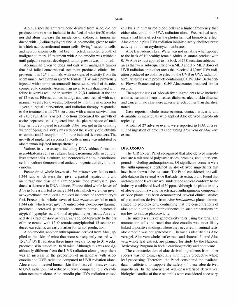
ALOE 45
Aloin, a specific anthraquinone derived from Aloe, did notproduce tumors when included in the feed of mice for 20 weeks,nor did aloin increase the incidence of colorectal tumors in-duced with 1,2-dimethylhydrazine. Aloe-emodin, given to micein which neuroectodermal tumor cells, Ewing’s sarcoma cells,and neuroblastoma cells had been injected, inhibited growth ofmalignant tumors. If treatment with Aloe-emodin was withhelduntil palpable tumors developed, tumor growth was inhibited.
Acemannan given to dogs and cats with malignant tumorsthat had failed conventional treatment produced clinical im-provement in 12/43 animals with no signs of toxicity from theacemannan. Acemannan given to female CFW mice previouslyinjected with murine sarcoma cells increased survival of the micecompared to controls. Acemannan given to cats diagnosed withfeline leukemia resulted in survival in 29/41 animals at the endof 12 weeks. Fibrosarcomas in dogs and cats, treated with ace-mannan weekly for 6 weeks, followed by monthly injections for1 year, surgical intervention, and radiation therapy, respondedto the treatment with 7/13 survivors with a mean survival timeof 240 days. Aloe vera gel injections decreased the growth ofascite hepatoma cells injected into the pleural space of maleFischer rats compared to controls. Aloe vera gel in the drinkingwater of Sprague-Dawley rats reduced the severity of diethylni-trosamine and 2-acetylaminofluorene induced liver cancers. Thegrowth of implanted sarcoma 180 cells in mice was inhibited byaloemannan injected intraperitoneally.
Various in vitro assays, including DNA adduct formation,neuroblastoma cells in culture, lung carcinoma cells in culture,liver cancer cells in culture, and neuroendocrine skin carcinomacells in culture demonstrated anticarcinogenic activity of aloe-emodin.
Freeze-dried whole leaves of Aloe arborescens fed to maleF344 rats, which were then given a partial hepatectomy andan intragastric dose of a carcinogenic food pyrolysate, pro-duced a decrease in DNA adducts. Freeze-dried whole leaves ofAloe arborescens fed to male F344 rats, which were then givenazoxymethane, produced a reduced incidence of aberrant cryptfoci. Freeze-dried whole leaves of Aloe arborescens fed to maleF344 rats, which were given N -nitroso-bis(2-oxopropyl)amine,produced decreased pancreatic adenocarcinomas, pancreaticatypical hyperplasias, and total atypical hyperplasias. An ethylacetate extract of Aloe arborescens applied topically to the earof mice treated with 12-O-tetradecanoylphorbol-13-acetate re-duced ear edema, an early marker for tumor production.
Aloe-emodin, another anthraquinone derived from Aloe, ap-plied to the skin of mice that were subsequently treated with15 J/m2 UVB radiation three times weekly for up to 31 weeks,produced skin tumors in 16/20 mice. Although this was not sig-nificantly different from the UVB radiation alone group, therewas an increase in the proportion of melanomas with Aloe-emodin and UVB radiation compared to UVB radiation alone.Aloe-emodin–treated human skin fibroblast cell lines, exposedto UVA radiation, had reduced survival compared to UVA radi-ation treatment alone. Aloe-emodin plus UVA radiation caused
cell lysis in human red blood cells at a higher frequency thaneither aloe-emodin or UVA radiation alone. Free radical scav-engers had little effect on the photochemical hemolytic effect.Aloe-emodin plus UVA radiation inhibited acetylcholinesteraseactivity in human erythrocyte membranes.
Aloe Barbadensis Leaf Water was not irritating when appliedto the back of 10 healthy female adults. A suntan product with0.1% Aloe extract applied to the back of 25 Caucasian subjects inareas that were subsequently given MED and 3 × MED doses ofUVB radiation or in other areas that received 4 J/cm2 UVA radi-ation produced no additive effect to the UVB or UVA radiation.Similar studies with products containing 0.01% Aloe Barbaden-sis Flower Extract and up to 0.5% Aloe extract produced similarresults.
Therapeutic uses of Aloe-derived ingredients have includedasthma, ischemic heart disease, diabetes, ulcers, skin disease,and cancer. In no case were adverse effects, other than diarrhea,noted.
Case reports include acute eczema, contact urticaria, anddermatitis in individuals who applied Aloe-derived ingredientstopically.
A total of 27 adverse events were reported to FDA as a re-sult of ingestion of products containing Aloe vera or Aloe veraextract.
DISCUSSIONThe CIR Expert Panel recognized that aloe-derived ingredi-
ents are a mixture of polysaccharides, proteins, and other com-pounds including anthraquinones. Of significant concern werethe anthraquinones identified in aloe-derived ingredients thathave been shown to be toxicants. The Panel considered the avail-able data on the several Aloe Barbadensis extracts and found thatanthraquinone levels are well understood and can conform to theindustry-established level of 50 ppm. Although the phototoxicityof aloe-emodin, a well-characterized anthraquinone componentof Aloe plants, has been demonstrated, several clinical studiesof preparations derived from Aloe barbadensis plants demon-strated no phototoxicity, confirming that the concentrations ofaloe-emodin, or other anthraquinones, in such preparations aretoo low to induce phototoxicity.
The mixed results of genotoxicity tests using bacterial andmammalian cells indicated that aloe-emodin was most likelylinked to positive findings, where they occurred. In animal tests,aloe-emodin was not geneotoxic. Chemicals identified as Aloevera gel, Aloe vera whole leaf extract, and charcoal filtered Aloevera whole leaf extract, are planned for study by the NationalToxicology Program in both a carcinogenicity and photocarc.
The characterization of aloe-derived ingredients from otherspecies was not clear, especially with highly productive wholeleaf processing. Therefore, the Panel considered the availabledata insufficient to support the safety of these aloe-derivedingredients. In the absence of well-characterized derivatives,biological studies of these materials were considered necessary.

46 COSMETIC INGREDIENT REVIEW
The studies needed are 28-day dermal toxicity studies on AloeAndongensis Extract, Aloe Andongensis Leaf Juice, AloeArborescens Leaf Extract, Aloe Arborescens Leaf Juice, AloeFerox Leaf Extract, Aloe Ferox Leaf Juice, and Aloe FeroxLeaf Juice Extract (ingredients should be tested at current useconcentrations). Such data may be developed on one ingredientfrom each species if it can be demonstrated that the tested ma-terial is representative of the group. In aloe-derived ingredientsused in cosmetics, regardless of species, anthraquinone levelsshould not exceed 50 ppm.
The Panel indicated that data indicating the presence or ab-sence of pesticide residues in Aloe plants harvested are limited.The Panel advised the industry that the total polychlorobiphenyl(PCB)/pesticide contamination of any plant-derived cosmeticingredient should be limited to not more than 40 ppm, with notmore than 10 ppm for any specific residue. The Panel also ad-vised that limits were appropriate for the following impurities:arsenic (3 mg/kg maximum), heavy metals (20 mg/kg maxi-mum), and lead (5 mg/kg maximum).
It was noted that inhalation exposure was of concern giventhe lack of inhalation toxicity findings in the report. The fewingredients used in aerosol formulations, however, are at verylow concentrations. In addition, the Panel expects that theaerosolized cosmetic formulations in which these ingredientsare found would not contain aerosolized particles of a size thatare respirable.
CONCLUSIONThe CIR Expert Panel concluded that Aloe Barbadensis
Flower Extract, Aloe Barbadensis Leaf, Aloe Barbadensis LeafExtract, Aloe Barbadensis Leaf Juice, Aloe Barbadensis Poly-saccharides, and Aloe Barbadensis Leaf Water are safe ascosmetic ingredients in the practices of use and concentrationsas described in this safety assessment, if anthraquinone levelsin the ingredients do not exceed 50 ppm. The available data areinsufficient to support the safety of Aloe Andongensis Extract,Aloe Andongensis Leaf Juice, Aloe Arborescens Leaf Extract,Aloe Arborescens Leaf Juice, Aloe Ferox Leaf Extract, AloeFerox Leaf Juice, or Aloe Ferox Leaf Juice Extract in cosmeticproducts.
REFERENCESActive Organics, Inc. 2002. Material Safety Data Sheet. Actiphyte©R of Aloe
Vera 10 Fold. www.activeorganics.comAgarwal, O. P. 1985. Prevention of atheromatous disease. Angiology 36:485–
492.Agarwala, O. M. 1997. Whole Leaf aloe gel vs. standard Aloe gel. Drugs and
Cosmeic Industry Februayy 22–28.Ajabnoor, M. A. 1990. Effects of aloes on blood glucose levels in normal and
alloxan diabetic mice. J Ethnopharmacol. 28:215–220.Ali, M. I., N. M. Shalaby, M. H. Elgamal, et al. 1999. Antifungal effects of
different plant extracts and their major components of selected aloe species.Phytother Res. 13:401–407.
Aloecorp. 2001. Time, TEMPERATURE AND Sanitation. www.aloecorp.comArosio, B., N. Gagliano, L. M. Fusaro, et al. 2000. Aloe-emodin quinone pretreat-
ment reduces acute liver injury induced by carbon tetrachloride. PharmacolToxicol. 87:229–233.
Atherton, P. 1998. Aloe vera: Magic or medicine. Nurs Stand. 12:49–54.Avila, H., J. Rivero, F. Herrera, and G. Fraile. 1997. Cytotoxicity of a low molec-
ular weight fraction from Aloe vera (Aloe barbadensis Miller) gel. Toxicon.35:1423–1430.
Bader, S., L. Carineli, R. Cozzi, and O. Cozzoli. 1981. Natural hydroxyan-thracenic polyglycosides as sunscreens. Cosmet. Toilet. 96:67–74.
Badria, F. A. 1994. Is man helpless against cancer? An environmental approach:antimutagenic agents from Egyptian food and medicinal preparations. CancerLett. 84:1–5.
Blitz, J., J. W. Smith, and J. R. Gerard. 1963. Aloe vera gel in peptic ulcertherapy: Preliminary report. J. Am. Ost. Assoc. 62:731–735.
Bobin M. F., M. Raymond, and M. C. Martin. 1994. UVA/UVB absorptionproperties of natural products. Cosmet. Toilet. 109:63–70.
Bouthet, C. F., V. R. Schirf, and W. D. Winters. 1995. Stimulation of neuron likecell growth by aloe substances. Phytother Res. 9:185–188.
Brasher, W. J., E. R. Zimmermann, and C. K. Collings. 1969. Effects of pred-nisolone, indomethacin and Aloe vera gel on tissue culture cells. Oral Surg.Oral Med Oral Pathol. 27:122–128.
Briggs, C. 1995. Herbal medicine: Aloe. Can. Pharmaceut. J. 128:48–50.Brown, J. P., and P. S. Dietrich. 1979. Mutagenicity of anthraquinone and ben-
zanthrone derivatives in the Salmonella/microsome test: Activation of an-thraquinone glycosides by enzymic extracts of rat cecal bacteria.Mutat. Res.66:9–24.
Brown, J. P., P. S. Dietrich, and R. J. Brown. 1977. Frameshift mutagenicity ofcertain naturally occurring phenolic compounds in the Salmonella/Microsometest: Activation of anthraquinone and flavonol glycosides by gut bacterialenzymes. Biochem. Soc. Trans. 4:1489–1492.
Budavari, S., ed. 1989. The Merck Index. An encyclopedia of chemicals, drugs,and biologicals, 11th ed. Rahway, NJ: Merck & Co.
Bunyapraphatsara, N., S. Yongchaiyudha, V. Rungpitarangsi, O. Chokechai-jaroenporn. 1996. Anti-diabetic activity of Aloe vera juice. Clinical trial indiabetes mellitus patients in combination with glibenclamide. Phytomedicine3:245–248.
Byeon, S. W., R. P. Pelley, S. E. Ullrich, et al. 1998. Aloe barbadensis extractsreduce the production of interleukin-10 after exposure to ultraviolet radiation.J. Invest. Dermatol. 110:811–817.
Carrington Labs. 2001. Product specifications for aloe. www.carringtonlabs.comCapasso, F., F. Borrelli, R. Capasso, et al. 1998. Aloe and its therapeutic use.
Phytother. Res. 12:S124–127.Capasso, F., N. Mascolo, G. Autore, and M. R. Duraccio. 1983. Effect of
Indomethacin on Aloin and 1,8 Dioanthraquinone-induced production ofprostaglandin in rat isolated colon. Prostaglandin 26:557–562.
Chithra, P., G. B. Sajithlal, and G. Chandrakasan. 1998. Influence of Aloe vera oncollagen charactersitics in healing dermal wounds in rats. Mol. Cell. Biochem.181:71–76.
Collin C. E., and C. Collins. 1935. Roentgen dermatitis treated with fresh wholeleaf of Aloe vera. Am. J. Roentgen. 33:396–397.
Consumer Product Testing Co. 1996. Photoallergy test procedure: Aloe extract0.1% in product type 77. Unpublished data. 9 pages.2
Corsi, M. M., A. A. Bertelli, G. Gaja, et al. 1998. The therapeutic potential ofAloe vera in tumor bearing rats. Int. J. Tissue React. 20:115–118.
Cosmetic, Toiletry, and Fragrance Association (CTFA). 2002. Determinationof Aloe-emodin in Aloe Arborescens CRS. Unpublished data submitted byCTFA. 2 pages.2
CTFA. 2003. Photosensitization and phototoxicity studies of products containingAloe-derived ingredients. Unpublished data. 233 pages.2
Danhof, I. E., and W. McAnalley. 1983. Stabilized Aloe vera: Effect on humanskin cells. Drug and Cosmetic Industry 133, 52, 54, 105–106.
2Available for review: Director, Cosmetic Ingredient Review, 110117th Street, NW, Suite 412, Washington, DC 20036, USA.
ALOE 47
Danhof, I. E. 1998. Position statement on polysaccharides. http://www.gothica.com.iasc.poly.html.
Davis, R. H., J. J. Di Donato, G. M. Hartman, and R. C. Haas. 1994a. Anti-inflammatory and wound healing of growth substances in Aloe vera. J. Am.Pediatr. Med. Assoc. 84:77–81.
Davis, R. H., J. J. DiDonato, R. W. S. Johnson, and C. B. Stewart. 1994b. Aloevera, hydrocortisone and sterol influence on wound tensile strength and anti-inflammation. J. Am. Pediatr. Med. Assoc. 84:614–621.
de Witte, P. 1993. Metabolism and pharmacokinetics of anthranoids. Pharma-cology. 47(suppl.1):86–97.
Dhar, M. L., B. N. Dhar, B. N. Dhawah, et al. 1968. Screening of Indian plantsfor biological activity Part 1. Ind. J. Exp. Biol. 6:232–247.
Diehl, B., and E. E. Teichmuller. 1998. Aloe vera, quality inspection and iden-tification. Agro-Food Industry Hi-tech. 9:14–16.
Dixit, V. P., and S. Joshi. 1983. Effect of Aloe barbadensis and clofibrate onserum lipids in Triton-induced hyperlipidaemia in Presbytis monkeys. Ind. J.Med. Res. 78:417–421.
Duke, J. A. eds. 1985. Aloe barbadensis Mill. CRC handbook of medicinal herbs,31–32. Boca Raton, FL: CRC Press.
Duke, J. A., and S. M. Beckstrom-Sternberg. 1994. “Acceptable” levels of fla-voring ingredients? Dev. Food Sci. 34:741–757.
Egger, S. F., G. S. Brown, L. S. Kelsey, et al. 1996. Hematopoietic augmentationby a β-(1,4)-linked mannan. Cancer Immunol. Immunother. 43:195–205.
European Union 2000. Opinion concerning the first update of the inventory ofingredients employed in cosmetic products. Section II: Perfume and aromaticraw materials. http://europa.eu.int/comm/food/fs/sc/sccp/out131 en.pdf
European Commission. 2002. The rules governing cosmetic products inthe European Union. Volume 1.Cosmetic Legislation—Cosmetic products.http://dg3.eudra.org/F3/home.html.
Esteban, A., J. M. Zapata, L. Casano, M. Martin, and B. Sabater. 2000. Per-oxidase activity in Aloe barbadensis commercial gel: Probable role in skinprotection.Planta Med. 66:724–727.
Food and Drug Administration (FDA). 1984. Cosmetic product formulation andfrequency of use data. FDA Database. Washington, DC: FDA.
FDA. 1992. Modification in voluntary filing of cosmetic product ingredient andcosmetic raw composition statements. Final rule. Fed. Register 57:3128–3130.
FDA. 1998. The Special Nutritionals/Adverse Event Monitoring System WebReport for aloe. http://www.cfsan.fda.gov.
FDA. 2001. Frequency of use of cosmetic ingredients. FDA Database.Washington, DC: FDA.
Fogelman, R. W., J. M. Chapdelaine, R. H. Carpenter, and B. H. McAnalley.1992a. Toxicologic evaluation of injectable acemannan in the mouse, rat anddog. Vet. Hum. Toxicol. 34:201–205.
Fogleman, R. W., T. E. Sheelenberger, M. F. Balmer, et al. 1992b. SubchronicOral administration of acemannan in the rat and dog. Vet. Hum. Toxicol.34:144–147.
Fujita, K., R. Teradaira, and T. Nagatsu. 1976. Bradykinase activity of aloeextract.Biochem. Pharmacol. 25:205.
Fujita, K., Y. Yamada, K. Azuma, and S. Hirozawa. 1978. Effect of leaf extractsof Aloe arborescens Mill subsp natalensis Berger on growth of Trichophytonmentagrophytes. Antimicrob. Agents Chemother. 14:132–136.
Fulton, J. E. 1990. The stimulation of postdermabrasion wound healing with sta-bilized Aloe vera gel–polyethylene oxide dressing. J. Dermatol. Surg. Oncol.16:460–467.
Furukawa F., A. Nishikawa, T. Chihara, K. Shimpo, H. Beppu, H. Kuzuya, I. S.Lee, and M. Hirose. 2002. Chemopreventive effects of Aloe arborescens on N -nitrobis(2-oxypropyl)amine-induced pancreatic carcinogenesis. Cancer Lett.25:117–122.
Garnick, J. J., B. Singh, and G. Winkley. 1998. Effectiveness of a medicamentcontaining silicon dioxide, aloe, and allantoin on aphthous stomatitis. OralSurg. Oral Med. Oral Pathol. Oral Radiol. Oral Endod. 86:550–556.
Ghannam, N., M. Kingston, I. A. Al-Meshaal, M. Tariq, N. S. Parman, and N.Woodhouse. 1986. The antidiabetic activity of aloes: Preliminary clinical andexperimental observations. Horm. Res. 24:288–294.
Gjerstad, G. 1969. An appraisal of the Aloe vera juice. Am. Perfum. Cosmet.84:43–46.
Gottschalck, T. E., and G. N. McEwen, Jr., eds. 2004. International cosmeticingredient dictionary and handbook, 10th ed., Vol. 1, 76–81. Washington,DC: CTFA.
Gowda, D. C., B. Neelisiddaiah, and V. Anjaneyalu. 1979. Structural studies ofpolysaccharides from Aloe vera. Carbohydr. Res.72:201–205.
Gramatica, P. D. Monti, G. Speranza, and P. Manitto, 1982. Aloe revisited, thestructure of Aloeresin A. Tetrahedron Lett. 23:2423–2424.
Green, M. W. 1941. The irritant effect of Aloin. Preliminary note. J. Am. Phar-maceut. Assoc. 30:186–187.
Grindlay, D., and T. Reynolds. 1986. The Aloe vera phenomenon: A review of theproperties and modern uses of the leaf parenchyma gel. J. Ethnopharmacol.16:117–151.
Gutterman, Y., and E. Chauser-Volfson. 2000. The distribution of the phenolicmetabolites barbaloin, aloeresin and aloenin as a peripheral defense strategyin the succulent leaf parts of Aloe arborescens. Biochem. Syst. Ecol. 28:825–838.
Hamilton, I. 1932. Acute vesicular dermatitis caused by curacao aloes. Med.J.Aust.. 1:302–303.
Hammer Pharma. 2000. Hamma Pharma S.p.A. Herbal extracts. Aloe dry extract.http://www.hammerpharma.it/specifications/1/htm.
Harris, C., K. Pierce, G. King, et al. 1991. Efficacy of acemannan in treatmentof canine and feline spontaneous neoplasms. Mol. Biother. 3:207–213.
Hay, J. E., and L. J. Haynes. 1956. The aloins. Part I. The structure of barbaloin.J. Chem. Soc. 3141–3147.
Heck, E., M. Head, D. Nowak, et al. 1981. Aloe vera (gel) cream as a topicaltreatment for outpatient burns. Burns 7:291–294.
Hegazy, M. A. A. Mortada, M. R. Hegazy, and M. Helal. 1978. The use of Aloevera extract in the treatment of experimental corneal ulcers in rabbit. J. DrugRes. Egypt 10:199–209.
Heggers, J. P., G. R. Pineless, and M. C. Robson. 1979. Dermaide Aloe/Aloe veraGel: Comparison of the antimicrobial effects. J. Am. Med. Technol. 41:293.
Heidemaan, A., H. G. Miltenburger, and U. Mengs. 1993. The genotoxicity ofSenna. Pharmacology 47:179–186, Suppl 1.
Heidemaan A., W. Voelkner, and U. Mengs. 1996. Genotoxicity of aloe emodinin vitro and in vivo. Mutat. Res. 367:123–133.
Herlihy, J. T., H. A. Bertrand, J. D. Kim, J. Ikeno, and B. P. Yu. 1998. Effectsof Aloe vera ingestion in the rat. I. Growth, food and fluid intake and serumchemistry. Phytother. Res. 12:183–188.
Hikino, H., M. Takahashi, M. Murakami, C. Konno, et al. 1986. Isolation andhypoglycemic activity of arborans A and B, glycans of Aloe arborescens varNatalensis leaves. Int. J. Crude Drug Res. 24:183–186.
Hirata, T., S. Sakano, and T. Suga. 1981. Biotransformation of aloenin, a bitterglucoside constituent of Aloe arborescens, by rats. Experientia 37:1252–1253.
Hirata, T., and T. Suga. 1977. Biologically active constituents of leaves and rootsof Aloe arborescens var natalensis. Z Naturforsch. 32:731–734.
Hogan, D. J. 1988. Widespread dermatitis after topical treatment of chronic legulcers and stasis dermatitis. Can. Med. Assoc. J. 138:336–338.
Holdsworth, D. K. 1971. Chromones in Aloe species. Planta Med. 19:322–325.Hunter, D., and A. Frumkin. 1991. Adverse reactions to vitamin E and Aloe vera
preparations after dermabrasion and chemical peel. Cutis 47:193–196.Hutter, J. A., M. Salman, W. B. Stavinoha, et al. 1996. Anti-inflammatory C-
glucosyl chromone from Aloe barbadensis. J. Nat. Prod. 59:541–543.Imanishsi, K., T. Ishiguro, H. Saito, and I. Suzuki. 1981. Pharmacological
studies on a plant lectin, Aloctin A. I. Growth and inhibition of mousemethylcholanthrene-induced fibrosarcoma (Meth A) in ascites form byAloctin A. Experientia 37:1186–1187.
Institut D’Expertise Clinique. 2000. Ocular tolerant assessment and evaluationof the primary cutaneous tolerance. Unpublished data. 12 pages.2
Institut Francais de Recherches et Essais Biologoques. 1981.Ocular irritation inthe rabbit and cutaneous primary irritation in the rabbit. Unpublished data. 32pages.2

48 COSMETIC INGREDIENT REVIEW
International Aloe Science Council (IASC). 2001a. Polymerase chain reaction:A new method for the identification of aloe. http://www.iasc.org/orndorf.html.
IASC. 2001b. Aloe vera, Quality inspection and identification. www.iasc.org.IASC. 2003. IASC addresses aloe nomenclature for CTFA. Inside Aloe Septem-
ber, 2003. www.iasc.org.Ishii, Y., H. Tanizawa, and Y. Takino. 1984. Fluorophotometry of barbaloin in
aloe. Chem. Pharm. Bull. (Tokyo). 32:4946–4950.Ishii, Y., H. Tanizawa, and Y. Takino. 1987. Determination of barbaloin in rat
serum. Chem. Pharm. Bull. (Tokyo). 35:4642–4644.Ishii, Y., H. Tanizawa, and Y. Takino. 1990. Studies of Aloe. III. Mechanism of
laxative effect. Chem. Pharm. Bull. 38:197–200.Ishii, Y., H. Tanizawa, and Y. Takino. 1994. Studies of Aloe. V. Mechanism of
cathartic effect. Biol. Pharm. Bull. 17:651–653.Ivy Laboratories. 1987a. The determination of the photocontact allergenic po-
tential of 0.1% Aloe extract in product type 77. Unpublished data. 13 pages.2
Ivy Laboratories. 1987b. The determination of the phototoxic potential of 0.1%Aloe extract in product type 77. Unpublished data. 15 pages.2
Ivy Laboratories. 1988. The determination of the photocontact allergenic poten-tial of 0.01% Aloe Barbadensis flower extract in product type 77. Unpublisheddata. 15 pages.2
Ivy Laboratories. 1996. The determination of the photocontact allergenic poten-tial of 0.5% Aloe extract in product type 79. Unpublished data. 12 pages.2
Joint Committee of the Pharmaceutical Society and the Society for AnalyticalChemistry on Recommended Methods for the Evaluation of Drugs. 1967. Thechemical assay of Aloes. Analyst 92:593–596.
Karaca, K., J. M. Sharma, and R. Norgen. 1995. Nitric oxide production bychicken macrophages activated by acemannan, a complex carbohydrate ex-tracted from Aloe vera. Int. J. Immunopharmacol. 17:183–188.
KGL, Inc. 2000. The determination of the photocontact allergenic potential of0.1% Aloe extract in product type 77. Unpublished data. 16 pages.2
KGL, Inc. 2001. The determination of the photocontact allergenic potential of0.5% Aloe extract in product type 79. Unpublished data. 18 pages.2
Kacew, S. 1993. Neonatal toxicology. Gen. Appl. Toxicol. 2:1047–1068.Kahlon, J. B., M. C. Kemp, R. H. Carpenter, et al. 1991a. Inhibition of AIDS
virus replication by acemannan in vitro. Mol. Biother. 3:127–135.Kahlon, J. B., M. C. Kemp, and N. Yawei. 1991b. In vitro evaluation of the
synergistic antiviral effects of acemannan in combination with azidothymidineand acyclovir. Mol. Biother. 3:211–220.
Kar, A., S. Panda, and S. Bharti. 2002. Relative efficacy of three medicinal plantextracts in the alteration of thyroid hormone concentrations in male mice. J.Ethnopharmacol. 81:281:285.
Kim, H. S., S. Kacew, and B. M. Lee. 1999. In vitro chemopreventive ef-fects of plant polysaccharides (Aloe barbadensis Miller, Lentinum edo-des, Ganoderma lucidum and Coriolus versicolor). Carcinogenesis 20:1637–1640.
Kim, H. S., and B. M. Lee. 1997. Inhibition of benzo [a] pyrene-DNA adductformation by Aloe barbadensis Miller. Carcinogenesis 18:771–776.
Kim, K. H., J. G. Lee, D. G. Kim, et al. 1998. The development of a new methodto detect the adulteration of commercial aloe gel powders. Arch. Pharm. Res.21:514–520.
King, G. K., K. M. Yates, P. G. Greenlee, et al. 1995. The effect of acemannanimmunostimulant in combination with surgery and radiation therapy on spon-taneous canine and feline fibrosarcomas. J. Am. Anim. Hosp. Assoc. 31:439–447
Klein, A. D., and N. S. Penneys. 1988. Aloe vera. J. Am. Acad. Dermatol.18:714–20.
Koo, M. W. 1994. Aloe vera: Anti-ulcer and anti-diabetic effects. Phytother. Res.8:461–464.
Kuo, P.-L., T.-C. Lin, and C.-C. Lin. 2002. The antiproliferative activity of aloe-emodin is through p53-dependent and p21-dependent apoptotic pathway inhuman hepatoma cell lines. Life Sci. 16:1879–1892.
Kuzuya, H., I. Tamai, H. Beppu, et al. 2001. Determination of aloenin, barbaloin,and isobarbaloin by micellar electrokinetic chromatorgraphy. J. Chromatogr.B Biomed. Sci. Appl. 752:91–97.
Lagarto Parra, A., R. Yhebra, G. Sardinas, et al. 2001. Comparative study of theassay of Artemia salina L. and the estimate of the medium lethal dose (LD50)in mice to determine oral acute toxicity of plant extracts. Phytomedicine8:395–400.
Lang, W. 1993. Pharmacokinetic-metabolic studies with 14C-aloe-emodin af-ter oral administration to male and female rats. Pharmacology. 47:110–119,suppl 1.
Lee, C. K., S. S. Han, Y. K. Mo, et al. 1997. Prevention of ultraviolet radiation-induced suppression of accessory cell function of Langerhans cells by Aloevera gel components. Immunopharmacology 37:153–162.
Lee, C. K., S. S. Han, Y. K. Shin, et al. 1999. Prevention of ultraviolet radiation-induced suppression of contact hypersensitivity by Aloe vera gel components.Int. J. Immunopharmacol. 21:303–310.
Lee, H.-Z., 2001. Protein kinase C involvement in aloe-emodin and emodin-induced apoptosis in lung cell carcinoma cell. Br. J. Pharmacol. 5:1093–1103.
Lee, H.-Z., S.-L. Hsu, M.-C. Liu, and C.-H. Wu. 2001. Effects and mechanismsof aloe-emodin on cell death in human lung squamous cell carcinoma. Eur. J.Pharmacol. 23:287–295.
Lee, J. K., M. K. Lee, Y.-P. Yun, et al. 2001. Acemannan purified from Aloe verainduces phenotypic and functional maturation of immature dendritic cells.Int. Immunopharmacol. 1:1275–1284
Lee, K. Y., J. H. Park, M. H. Chung, et al. 1997. Aloesin up-regulates cyclinE/CDK2 kinase activity via inducing the protein levels of Cyclin E, CDK2and CDC25A in SK-HEP-1cells. Biochem. Mol. Biol. Int. 41:285–292.
Lee, M. J., S. H. Yoon, S. K. Lee, et al. 1995. In vivo angiogenic activityof dichloromethane extracts of Aloe vera gel. Arch. Pharm. Res. 18:332–335.
Lissoni, P., L. Giani, S. Zerbini, P. Trabattoni, and F. Rovelli. 1998. Biotherapywith the pineal immunomodulating hormone melatonin versus melatonin plusAloe vera in untreatable advanced solid neoplasms. Nat. Immunol. 16:27–33.
Lorenzett, L. J., R. Salisbury, J. Beal, and J. Baldwin. 1964. Bacteriostatic prop-erty of Aloe vera. J. Pharm. Sci. 53:1287.
Loveman, A. B. 1937. Leaf of Aloe vera in treatment of roentgen ray ulcers.Arch. Dermatol. Syphilol. 36:838–843.
Mabusela, W. T., A. M. Stephen, and M. C. Botha. 1990. Carbohydrate polymersfrom Aloe ferox leaves. Phytochemistry 29:3555–3558.
Madis, V. H., and M. M. Omar. 1989. Aleoferon isolation, manufacturing andits application. United States Patent 4,861,761.
Makino, K., A. Yagi, and I. Nishioka. 1973. Studies on the constituents of Aloearborescens (Miller): The structure of aloearbonaside, a glucoside of a newtype naturally occurring chromene. Chem. Pharm. Bull. 21:149–156.
Mandeville, F. B. 1939. Aloe vera in the treatment of radiation ulcers of mucousmembranes. Radiology 32:598–599.
Marshal, G. D., A. S. Gibbons, and L. S. Parnell. 1993. Human cytokines inducedby acemannan (abstract 619). J. Allergy Clin. Immunol. 91:295.
Maze, G., R. N. Terpolilli, and M. Lee. 1997. Aloe vera extract prevents aspirin-induced acute gastric mucosal injury in rats. Med. Sci. Res. 25:765–766.
McAnalley, B. H. 1990. Processes for preparation of Aloe products, productsproduced thereby and composition thereof. United States Patent 4,917,890.
McCarthy, T. J. 1969. Distribution of glycosyl compounds in South African aloespecies. Planta Med. 17:1–7.
McCarthy, T. J., and L. J. Haynes. 1967. The distribution of aloesin in someSouth African aloe species. Planta Med. 15:342–344.
McCauley, R., J. P. Haggers, and M. C. Robson. 1990. Frostbite-methods tominimize tissue loss. Postgrad. Med. 88:67–70.
MDS Pharma Services. 2000. Single dose toxicity study by the oral route in therat (Limit test). Unpublished data. 30 pages.2
Meadows, T. P. 1980. Aloe as a humectant in new skin preparations. Cosmet.Toilet. 95:51–56
Ministry of Health, Labor and Welfare (MHLW). March 2001a. Unofficial trans-lation of MHW Ordinance No. 331, Attached Table 1 [Negative List]. Ministryof Health, Labor and Welfare, Pharmaceutical and Medical Safety Bureau,Inspection and Guidance Division, 2-2, 1-chome, Kasumigaseki, Chiyoda-ku,Tokyo 100–8045. Japan.

ALOE 49
MHLW. March 2001b. Unofficial translation of MHW Ordinance No. 331, At-tached Table 2 [Restricted List]. Ministry of Health, Labor and Welfare, Phar-maceutical and Medical Safety Bureau, Inspection and Guidance Division,2-2, 1-chome, Kasumigaseki, Chiyoda-ku, Tokyo 100–8045. Japan.
Morimoto, I., F. Wantanabe, and T. Osawa. 1982. Mutagenicity screening ofcrude drugs with Bacillus subtilis rec-assay and Salmonella/microsome re-version assay. Mutat. Res. 97:81–102.
Morrow, D. M., M. J. Rapaport, and R. A. Strick. 1980. Hypersensitivity to Aloe.Arch. Dermatol. 116:1064–1065.
Muller, S. O., I. Eckert, W. K. Lutz, and H. Stopper. 1996. Genotoxicity of thelaxative drug components emodin, aloe-emodin and danthron in mammaliancells: Topoisomerase II mediated? Mutat. Res. 371:165–173.
Nakamura, T., and S. Kotajima. 1984. Contact dermatitis from Aloe arborescens.Contact Dermatitis 11:51.
Nakamura, H., and T. Okuyama. 1990. Gas chromatographic and mass spectraldetermination of aloenin in skin-care cosmetics. Chromatography 509:377–382.
Nakano, M., T. Yamagishi, T. Takahashi, and H. Kaneshima. 1985. Analysis ofplant components in cosmetics. Part I. Determination of aloenin, a componentof Aloe arborescens Mill in Lotion. Hokkaidoritsu Eisei Kenkyushoh. 35:98–101.
Nath, K., N. Sethi, R. K. Singh, and A. K. Jain. 1992. Commonly used Indianabortifacient plants with special reference to their teratologic effects in rats.J. Ethnopharmacol. 36:147–154.
Natow, A. J. 1986. Aloe vera, fiction or fact. Cutis 37:106, 108.Norikuri, T., D. O. Kennedy, A. K. Nyarko, A. Kojima, and I. Matsui-
Yuasa. 2002. Protective effect of aloe extract against the cytotoxicity of 1,4-napthoquinone in isolated rat hepatocytes involves modulation in cellular thiollevels. Pharmacol. Toxicol. 5: 278–284.
Northway, R. B. 1975. Experimental use of Aloe vera in clinical practice. Vet.Med. Small Anim. Clin. 70:89.
Norton, S. J., V. Talesa, and W. J. Yuan. 1990. Glyoxalase I and glyoxalase IIfrom Aloe vera: Purification, characterization and comparison with animalsGlyoxalases. Biochem. Int. 22:411-418.
Olsen, D. L., W. Raub Jr., C. Bradley, et al. 2001. The effect of Aloe veragel/mild soap versus mild soap alone in preventing skin reactions in patientsundergoing radiation therapy. Oncol. Nurs. Forum. 28:543–547.
Parmar, N. S., M. Tariq, M. A. Al-Yahya, et al. 1986. Evaluation of Aloe veraleaf exudates and gel for gastric and duodenal anti-ulcer activity. Fitoterapia57:380–383.
Parry, O., and C. Matambo. 1992. Some pharmacological actions of aloe extractsand Cassia abbreviata on rats and mice. Cent. Afr. J. Med. 38:409–414.
Patri, F., and V. Silano. 2002. Plants in Cosmetics, plants and plant prepara-tions used as ingredients for cosmetic product, 79–80. Strasbourg: Councilof Europe Publishing.
Pecere, T., M. V. Gazzola, C. Mucignat, et al. 2000. Aloe-emodin is a new typeof anticancer agent with selective activity against neuroectodermal tumors.Cancer Res. 60:2800–2804.
Peny, S. Y., J. Normal, G. Curtin, et al. 1991. Decreased mortality of Normanmurine sarcoma in mice treated with the immunomodulator acemannan. Mol.Biother. 3:79–87.
Proserpi, G. 1976. Natural sunscreens: vegetable derivatives as sunscreens andtanning agents. Cosmet. Toilet. 91:34–36.
Pugh, N., S. A. Ross, M. A. ElSohly, and D. S. Pasco. 2001. Characteriza-tion of aloeride, a new high molecular weight polysaccharide from Aloevera with potent immunostimulatory activity. J. Agric. Food Chem. 49:1030–1034.
Qiu, Z., K. Jones, M. Wylie, et al. 2000. Modified Aloe barbadensis polysac-charide with immunomodulatory Activity. Planta Med. 66:152–156.
Ramamoorthy, L., and I. R. Tizard. 1998. Induction of apoptosis in a macrophagecell line RAW 264.7 by acemannan, a β (1,4) acetylated mannan. Mol. Phar-macol. 3:415–421.
Reynolds, T., and A. C. Dweck. 1999. Aloe vera leaf Gel: A review update.J. Ethnopharmacol. 68:3–37.
Roberts, D. B., and E. L. Travis. 1995. Acemannan-containing wounddressinggels reduce radiation-induced skin reactions in C3H mice. Int. J. Radiat.Oncol. Biol. Phys.15:1047–1052.
Roboz, E., and A. J. Haagen-Smit. 1948. A mucilage from Aloe vera. J. Am.Chem. Soc. 70:3248–3249.
Robson, M. C., J. Heggers, and W. J. Hagstrom. 1982. Myth, magic, witchcraftor fact? Aloe vera revisited. J. Burn Care Rehab. 3:157–162.
Ross, S. A., M. A. ElSohly, and S. P. Wilkins. 1997. Quantitative analysis ofAloe vera mucilagenous polysaccharides in commercial Aloe vera products.J. AOAC Int. 80:455–457.
Rowe, T. D., and L. M. Parks. 1941. Phytochemical study of Aloe vera leaf. J.Am. Pharm. Assoc. 30:262–266.
Sabeh, F., T. Wright, and S. J. Norton. 1993. Isoenzymes of superoxide dismutasefrom Aloe vera. Enzyme Protein 47:92–98.
Sabeh, F., T. Wright, and S. J. Norton. 1996. Purification and characterizationof a glutathione peroxidase from Aloe vera plant. Enzyme Protein 49:212–221.
Saleem, R., S. Faizi, B. S. Siddiqui, et al. 2001. Hypotensive effects of chemicalconstituents from Aloe barbadensis. Planta Med. 67:757–760.
Saito, H., K. Imanishi, and S. Okabe. 1989. Effects of aloe extracts, aloctin A, ongastric secretion and on experimental gastric lesions in rats. Yakugaku Zasshi109:335–339.
Sauchak, U. I. 1977. Acute bulbous allergic dermatitis due to local applicationof aloe leaves. Vestn. Dermatol. Venerol. 12:44–45.
Shah, A. H., S. Qureshi, M. Tariq, and A. M. Ageel. 1989. Toxicity studies onsix plants used in the traditional Arab system of medicine. Phytother. Res.3:25–29.
Shamaan, N. A., K. A. Kadir, A. Rahmat, and W. Z. Ngah. 1998. Vitamin C andAloe vera supplementation protects from chemical hepatocarcinogenesis inthe rat. Nutrition 14:846–852.
Sheets, M. A., B. A. Unger, G. F. J. Giggleman, and I. R. Tizard. 1991. Studieson the effect of acemannan on retrovirus. Mol. Biother. 3:41–45.
Shelton, R. M. 1991. Aloe vera its chemical and therapeutic properties. Int. J.Dermatol. 30:679–683.
Shida, T., A. Yagi, H. Nishimura, and I. Nishioka. 1985. Effect of Aloe veraExtract on peripheral phagocytosis in adult bronchial asthma. Planta Med.36:273–275.
Shimpo, K., T. Chihara, H. Beppu, C. Ida, T. Haneko, T. Nagatsu, and H. Kuzya.2001. Inhibition of azoxymethane-induced aberrant crypt foci formation inrat colorectum by whole leaf Aloe arborescens Miller var. natalensis Berger.Phytother. Res. 15:705–711.
Shimpo, K., C. Ida, T. Chihara, H. Beppu, T. Kaneko, and H. Kuzuya. 2002. Aloearborescens extract inhibits TPA-induced ear oedema, putrescine increaseand tumor promotion in mouse skin. Short communication. Phytother. Res.16:491–493.
Shiseido Safety Research Labs. 2002. Photoallergenicity of Aloe ArborescensCRS. Unpublished data submitted by CTFA. 9 pages.2
Shoji, A. 1982. Contact Dermatitis to Aloe arborescens. Contact Dermatitis8:164–167.
Siegers, C. P., J. Siemers, and G. Baretton. 1993. Sennosides and aloin do notpromote dimethylhydrazine induced colorectal tumors in mice. Pharmacol-ogy 47:205–208, Suppl 1.
Singh, R. P., S. Dhanalakshmi, and A. R. Rao. 2000. Chemomodulatory ac-tion of Aloe vera on the profiles of enzymes associated with carcinogenmetabolism and antioxidant status regulation in Mice. Phytomedicine 7:209–219.
Strickland, F. M., A. Darvill, P. Albersheim, et al. 1999. Inhibition ofUV-induced immune suppression and interleukin-10 production by plantoligosaccharides and polysaccharides. Photochem. Photobiol. 69:141–147.
Strickland, F. M., H. K. Muller, L. C. Stephens, et al. 2000. Induction of pri-mary cutaneous melanomas in C3H mice by combined treatment with ul-traviolet radiation, ethanol and aloe-emodin. Photochem. Photobiol. 72:407–414.
50 COSMETIC INGREDIENT REVIEW
Strickland, F. M., R. P. Pelley, and M. L. Kripke. 1994. Prevention of ultravioletradiation induced suppression of contact and delayed hypersensitivity by Aloebarbadensis gel extracts. J. Invest. Dermatol. 102:197–204.
Sturm, P. G., and S. M. Hayes. 1984. Aloe vera In dentistry. J. Bergen CountyDent. Soc. 50:11–14.
Suga, T., and T. Hirata. 1983. The efficacy of the Aloe plant chemical constituentsand biological activities. Cosmet. Toilet. 98:105–108.
Suga, T., T. Hirata, and M. Odan. 1972. Aloenin, a new bitter glucoside fromaloe species. Chem. Lett. 547.
Suga, T., T. Hirata, and K. Tori. 1974. Structure of aloenin, a bitter glucosidefrom aloe species. Chem. Lett. 715.
Suzuki, I., H. Saito, S. Inoue, S. Migita, and T. Takahashi. 1979. Purificationand characterization of two lectins from Aloe arborescens Mill. J. Biochem.(Tokyo) 85:163–171.
Sydiskis, R. J., D. G. Owen, J. L. Lohr, et al. 1991. Inactivation of en-veloped viruses by anthraquinones extracted from plants. Antimicrob. AgentsChemother. 35:2463–2466.
Syed, T. A., M. Afzal, S. A. Ahmad, et al. 1997. Aloe vera extract 0.5% in ahydrophilic cream versus Aloe vera gel for the management of genital herpesin males. A placebo controlled double blind comparative study. J. Dermatol.Treat. 8:99–102.
Syed, T. A., S. A. Shamd, A. H. Holt, et al. 1996. Management of psoriasis withAloe vera extract in a hydrophilic cream: a placebo-controlled, double blindstudy. Trop. Med. Int. Health. 1:505–509.
Tyler, V. E. 1994. Herbs of choice, 155–157. New York: Pharmaceutical ProductsPress.
Tyler, V. E., L. R. Bradley, and J. E. Robbers, ed. 1988. Pharmacognosy, 9thed., 62–63. Philadelphia: Lea & Febiger.
Tello, C. G., P. Ford, and A. M. Iacopino. 1998. In vitro evaluation of com-plex carbohydrate denture adhesive formulations. Quintessence Int. 29:585–593.
Teradaira, R., M. Shinzato, H. Beppu, and K. Fujita. 1993. Anti-gastric ulcereffect of Aloe arborescens Mill. Var natalensis Berber. Phytother. Res. 7:S34–S36.
t’Hart, L. A., P. H. van Enckevort, H. van Dijk, and R. P. Labadie. 1988. Twofunctionally and chemically distinct immunomodulatory compounds in thegel of Aloe vera. J. Ethnopharmacol. 23:61–71.
Thomas, D. R., P. S. Goode, K. LaMaster, and T. Tennyson. 1998. Acemannanhydrogel dressing versus saline dressing for pressure ulcers. Adv. Wound Care.11:273–276.
Tizard, A. U., D. Busbee, B. Maxwell, and M. C. Kemp. 1995. Effect of ace-mannan, a complex carbohydrate, on wound healing in young and aged rats.Wounds 6:201–209.
Udupa, S. I., A. L. Udupa, and D. R. Kulkarni. 1994. Anti-inflammatory andwound healing properties of Aloe vera. Fitoterapia 65:141–145.
Uehara, N., Y. Iwahori, M. Asamoto, et al. 1996. Decreased levels of 2-amino-3methylimidazo[4,5-f]quinoline-DNA adducts in rats treated with β-carotene, α-tocopherol and rreeze dried Aloe. Jpn. J. Cancer Res. 87:342–348.
UNITIS. 2003. Unpublished data submitted by European Organizatioon of Cos-metic Ingredients Industries and Services, Paris. 31 pages.2
USDA, ARS, National Genetic Resource Program. Germplasm Resources In-formation Network (GRIN). [Online Database] National Germplasm Re-sources Laboratory, Beltsville, Maryland. Internet site accessed August, 2003http://www.ars-grin.gov/cgi-bin/npgs/html/tax search.pl
van Wyk, B. E., M. C. B. Oudtshoom, and G. F. Smith. 1995. Geographicalvariation in the major compounds of Aloe ferox exudate. Planta Med. 61:250–253.
Vargas F., G. Fraile, M. Velasquez, et al. 2002. Studies on the photostability andphototoxicity of aloe-emodin, emodin and rhein. Pharmazie 57:399–404.
Vath, P., W. G. Wamer, and D. E. Falvey. 2002. Photochemistry and photoxicityof aloe-emodin. Photochem. Photobiol. 75:346–352.
Viljoen, A. M., B. V. Van Wyk, and L. E. Newton. 2001. The occurrence andtaxonomic distribution of the anthrones aloin, aloinoside and microdotin inaloe. Biochem. Syst. Ecol. 29:53–67.
Visuthikosol, V., B. Chowchuen, Y. Sukwanarat, et al. 1995. Effect of Aloe veragel to healing of burn wound in a clinical and histologic study. J. Med. Assoc.Thai 78:403–409.
Vogler, B. K., and E. Ernst. 1999. Aloe vera: a systematic review of its clinicaleffectivness. Br. J. Gen. Pract. 49:823–828.
Waller, G. R., S. Mangiafico, and C. R. Ritchey. 1978. A chemical investigationof Aloe barbadensis miller. Proc. Okla. Acad. Sci. 58:69–76.
Wasserman, L., S. Avigad, E. Beery, J. Nordenberg, and E. Fenig. 2002. Theeffect of aloe emodin on the proliferation of a new Merkel cell carcinoma cellline. Am. J. Dermopathol. 24:17–22.
Westendorf, J., H. Marquardt, B. Paginsky, et al. 1990. Genotoxicity of naturallyoccuring hydroxyahthraquinones. Mutat. Res. 240:1–12.
Williams, M. S., M. Burk, C. L. Loprinzi, et al. 1996. Phase III double blindevaluation of an Aloe vera gel as a prophylactic agent for radiation-inducedskin toxicity. Int. J. Radiat. Oncol. Biol. Phys. 36:345–349.
Winters, W. D., R. Benavides, and W. J. Clouse. 1981. Effects of aloe extractson human normal and tumor cells in vitro. Econ. Bot. 35:89–95.
Woo, S. W., J. X. Nan, S. H. Lee, E. J. Park, Y. Z. Zhao, and D. H. Sohn. 2002.Aloe emodin supresses myofibroblastic differentiation of rat hepatic stellatecells in primary culture. Pharmacol. Toxicol. 90:193–198.
Wright, C. S. 1936. Leaf of Aloe vera in treatment of roentgen ulcers and telang-iectasis. J. Am. Med. Assoc. 103:1363–1364.
Yagi, A., T. Egusa, M. Arase, M. Tanabe, and H. Tsugi. 1997. Isolation andcharacterization of the glycoprotein fraction with a proliferation-promotingactivity on human and hamster cells in vitro from Aloe vera gel. Planta Med.63:18–21.
Yagi, A., N. Harada, K. Shimomura, and I. Nishioka. 1987. Bradykinin-degrading glycoprotein in Aloe arborescens var. natalensis. Planta Med.53:19–21.
Yagi, A., K. Machii, H. Nishimura, et al. 1985. Effect of aloe lectin on deoxyri-bonucleic acid synthesis in baby hamster kidney cells. Experientia 41:669–671.
Yagi, A., K. Makino, I. Nishioka, and Y. Kuchino. 1977. Aloe mannan, polysac-charide, from Aloe arborescens var. natalensis. Planta Med. 31:17–20.
Yagi, A., J. Nakamori, T. Yamada, et al. 1999. In vivo metabolism of Aloeman-nan. Planta Med. 65:417–420.
Yagi, A., K. Sato, K. Akasaki, and H. Tsuji. 2000. Distribution of verectin inAloe vera leaves and verectin contents in clonally regenerated plants and thecommercial gel powders by immunochemical screening. Planta Med. 66:180–182.
Zawahry, M. E., M. R. Hegazy, and M. Helal. 1973. Use of aloe in treating legulcers and dermatoses. Int. J. Dermatol. 12:68–73.
Zhang, L., and I. R. Tizard. 1996. Activation of a mouse macrophage cellline by acemannan: The major carbohydrate fraction from Aloe vera gel.Immunopharmacology 35:119–128.