fine needle aspiration cytology / fnac
TRANSCRIPT

Fine Needle Aspiration Cytology
Presented by:Dr Kalpajyoti Bhattacharjee
CONTENTS
INTRODUCTION HISTORICAL PERSPECTIVE ADVANTAGE LIMITATIONS EQUIPMENTS FINE NEEDLE ASPIRATION TECHNIQUE PROCESSING FIXATION & STAINING COMPLICATIONS FNAC IN SALIVARY GLAND, ORAL CAVITY.

INTRODUCTION
Fine Needle Aspiration Cytology (FNAC) is a technique whereby cells are obtained from a lesion using a thin bore needle and smears are made for cytopathological diagnosis.
This technique is based on the fact that tumor cells are less cohesive and are easily aspirated.
Used in the diagnosis of breast lumps, thyroid nodules, liver disease, subcutaneous soft tissue mass, salivary gland diseases and oral diseases.

Oral cavity is a site where mucosa is very vascular and an open biopsy leads to a lot of bleeding which is difficult to control.
In recent times FNAC solves these problems, adequate material can easily be obtained by using a 10 ml. syringe from an intraoral or extraoral site without any discomfort to the patient and with no bleeding.
In some cases a subsequent surgery is not needed and patient can be put on appropriate treatment.
FNAC report is prepared within 24 hours of sampling gives early, quick information to the surgeon about the type of lesion he is dealing with.

HISTORICAL PERSPECTIVE
The technique was introduced in the 1930s by Martin and Ellis in the United States, but it never became widespread.
Since the 1950s it has been used extensively in Scandinavia and in Holland.
Fine Needle for aspiration were first introduced in Europe in the 1950’s by Lopez-Cardozo in the Netherlands and Soderstrom in Sweden
Publication by Zajicek from Karolinska Hospital in Stockholm that brought aspiration cytology to international alterations.

ADVANTAGE
Simple office technique Rapid diagnosis Economical Sampling from multiple sites in the same
sitting High diagnostic accuracy Many techniques such as bacterial culture,
immunocytochemistry, flow cytometry, cytogenetics, polymerase chain reaction, etc. are possible from FNAC material.
LIMITATIONS
Loss of tissue architecture Capsular invasion and lymphovascular
invasions cannot be detected Difficult to differentiate in situ versus invasive
carcinoma Considerable training is needed for accurate
interpretation.
FNAC AS A TOOL IN CLINICAL INVESTIGATION
Initially used as a mean to confirm a clinical suspicion of local recurrence or metastasis of known cancer without subjecting the patient to further surgical intervention.
Inflammation, infection, degenerative conditions, in diagnosis and monitoring of graft rejection in transplantation surgery
Alternative or complement to frozen section Intraoperative cytology

THE PRACTICE OF FNAC
Success of FNAC depends on four fundamental requirement:
1. Samples must be representative of the lesion investigated.
2. Samples must be adequate in terms of cells & other tissue components
3. Samples must be correctly smeared and processed
4. Biopsy must be accompanied by relevant and correct clinical/radiological information.
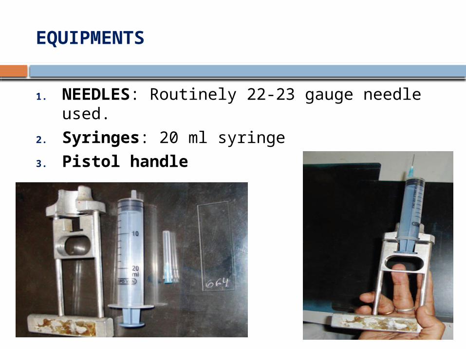
EQUIPMENTS
1. NEEDLES: Routinely 22-23 gauge needle used.
2. Syringes: 20 ml syringe3. Pistol handle

4) Sterile container: Physiological saline or Hank’s balanced solution
5) Slides: clean, dry & free of grease.6) A 0.4 mm haemocytometer coverslip gives
better control over smearing pressure & a more perfect spread
7) Fixatives: 70-90% ethanol, Carnoy’s fixative, 10% buffered formalin, gluteraldehyde used
8) Stains9) Microscopes

FINE NEEDLE ASPIRATION TECHNIQUE
2 techniques:1. FNAC with aspiration2. FNAC without aspiration
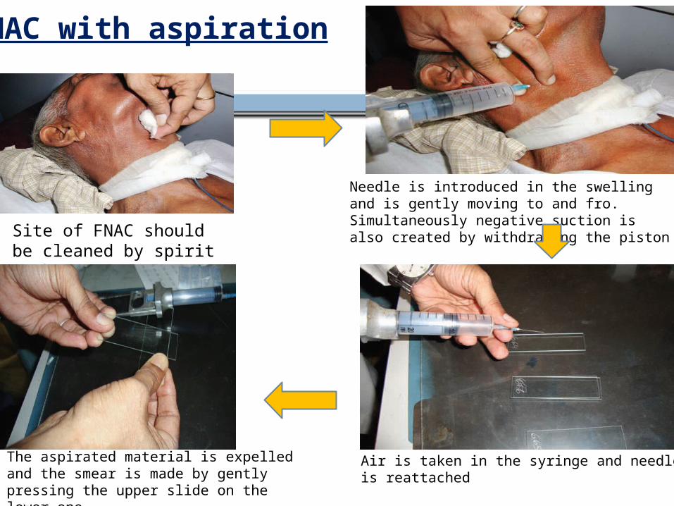
Site of FNAC should be cleaned by spirit swab
Needle is introduced in the swelling and is gently moving to and fro. Simultaneously negative suction is also created by withdrawing the piston
Air is taken in the syringe and needle is reattached
The aspirated material is expelled and the smear is made by gently pressing the upper slide on the lower one
FNAC with aspiration
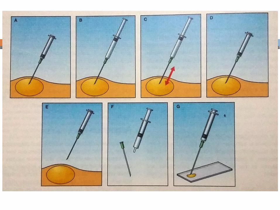

-Introduced by Zajdela in 1987-based on the observation that the capillary pressure in a fine needle is sufficient to keep the detached cell inside the lumen of the needle
FNAC without aspiration
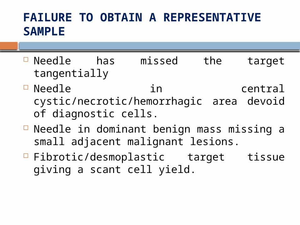
FAILURE TO OBTAIN A REPRESENTATIVE SAMPLE
Needle has missed the target tangentially Needle in central cystic/necrotic/hemorrhagic
area devoid of diagnostic cells. Needle in dominant benign mass missing a
small adjacent malignant lesions. Fibrotic/desmoplastic target tissue giving a
scant cell yield.


PROCESSING THE SAMPLE
Sample expelled on to a clean & dry microscope slide using air in a syringe.
SMEARING
DIRECT
INDIRECT

DIRECT SMEARING
DRYCreamy consistency & consists of numerous cells suspended in a
small amount of tissue fluid
WETSmaller number of cells
suspended in fluid or blood

Indirect smearing: Thin fluid samples are best processed by centrifugation on the cytocentrifuge.
Milipore nucleopore filtration is an alternative
Thinprep technique

FIXATION & STAINING
2 fundamentally different methods of fixation & staining are used in FNAC:
1. Air drying followed by staining with a haematological stain such as MAY GRUNWALD- GIEMSA STAIN , Jenner-Giesma, Diff-Quik
2. Alcohol fixation and staining according to PAP or with H&E.

Papanicolaou stain
Contents: Harris’s hematoxylin Orange G6 EA 50Results: Nuclei- blue/black Cytoplasm (non-keratinizing)- blue/green Keratinizing cells- pink/orange

Romanowsky stain
Contents: Methylene blue/azure B and eosin, dissoved in
acetone-free methanol, include jenner, Giesma, May Grunwald and Leishman stain
Results: Nuclei- purple/blue Cytoplasm- pink/blue Eosinophils- pink/red

Diff-Quik is a commercial Romanowsky stain variant, commonly used in histological staining to rapidly stain and differentiate a variety of smears, commonly blood and non-gynecological smears, including those of fine needle aspirates.

MAY GRUNWALD- GIEMSA STAIN
commonly used staining of blood smearsContents: methylene blue (a basic dye) Azures (also basic dyes) Eosin (an acid dye)
Results: Nuclei of white blood cells and the granules of
basophil granulocytes- blue Red blood cells and eosinophil granules – red cytoplasm of white blood cells - light blue

SPECIAL STAINS
1. PAS or Alcian blue - mucins, glycogen 2. Prussian blue - iron3. Masson-Fontana - melanin4. Congo red - amyloid5. Ziehl-neelson - acid fast bacilli6. Bile pigment- Fauchet’s reagent counterstained
with sirus red.7. Gram, PAS or Gomori’s silver stain for
microorganism

COMPLICATIONS
Usually free of complications Bleedings, hematoma, emphysema (in
lung). Rarely anaphylactic reaction- accidental
rupture of hydrated cyst

FNAC OF SALIVARY GLAND
FNAC of the salivary gland lesions has gained wide clinical recognition.
The incisional biopsy of the salivary gland may cause fistula formation and other complications that can be avoided in FNAC.
For accurate diagnosis of salivary gland lesions, an adequate sample stained by both May Grunwald- Giemsa stain and Papanicolaou’s stain (or H and E) along with detailed clinical history are needed.

Cytology of Normal Salivary Glands
Benign ductal cells: These cells are usually in small clusters or monolayered sheets. The cells are round to oval, with scanty cytoplasm having monomorphic nuclei.
Acinar cells: These are commonly present as small ball like clusters. The individual cells are round with abundant foamy cytoplasm and small round nuclei. The acinar cells may also be present discretely and cells with bare nuclei may be mistaken as lymphocytes.
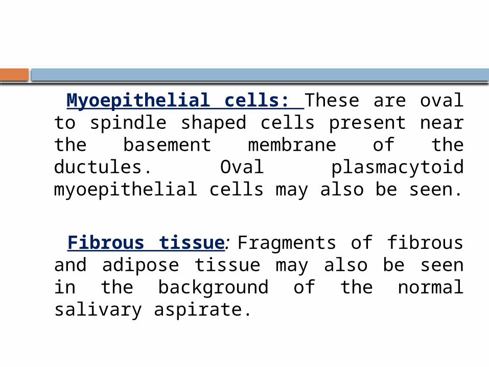
Myoepithelial cells: These are oval to spindle shaped cells present near the basement membrane of the ductules. Oval plasmacytoid myoepithelial cells may also be seen.
Fibrous tissue: Fragments of fibrous and adipose tissue may also be seen in the background of the normal salivary aspirate.
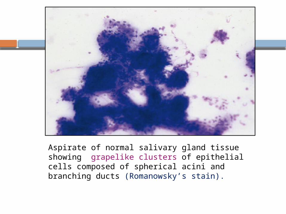
Aspirate of normal salivary gland tissue showing grapelike clusters of epithelial cells composed of spherical acini and branching ducts (Romanowsky’s stain).

Benign acinar cells arranged in ‘‘rosette’’ formation. The cells have abundant foamy vacuolated cytoplasm with indistinct cell borders, and eccentric nuclei (Papanicolaou stain).

Diagnostic approach of salivary gland tumors

Fatty Infiltration Presents as a diffuse enlargement Addition to normal salivary gland elements,
there is significant increase in the amount of adipose tissue situated between ductal and acinar cells
Associated with diabetes, cirrhosis, alcoholism, medications, nutritional deficiencies and hormonal disturbances

Chronic sialadenitis
Commonly results from stones or postsurgical scarring
More frequently seen in the submandibular glandCytology: low cellularity and show predominance of ductal
cells, background has variable number of lymphocytes, occasional plasma cells and neutrophils, and spindle fibroblasts
Squamous and mucinous metaplasia may be focally encountered
Acute sialadenitis: seldom sampled, many neutrophils are admixed with benign salivary gland elements, reactive and reparative changes may be seen
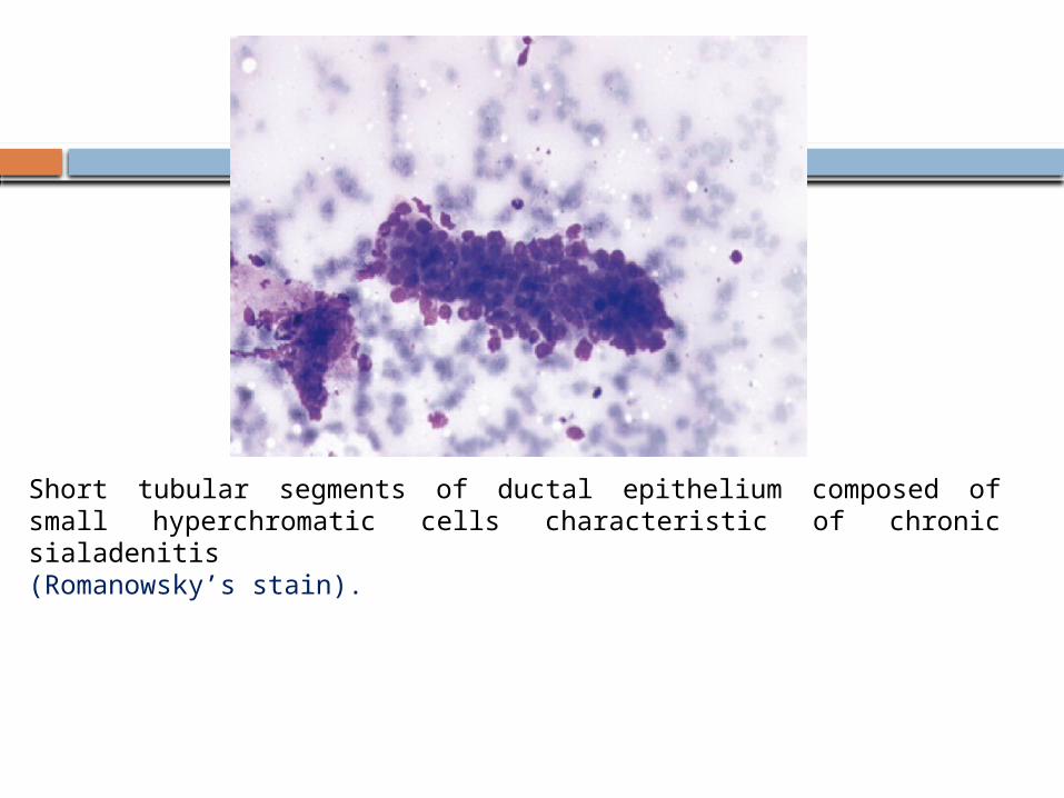
Short tubular segments of ductal epithelium composed of small hyperchromatic cells characteristic of chronic sialadenitis (Romanowsky’s stain).

Benign Lymphoepithelial Cysts of the Parotid Glands
Cytology: Smears - characterized by a mixed lymphoid
infiltrate with a predominance of small mature lymphocytes
Characteristically has a watery proteinaceous background in which the lymphoid elements are distributed
Cuboidal or mucin-containing cells may be found distributed singly in the smear
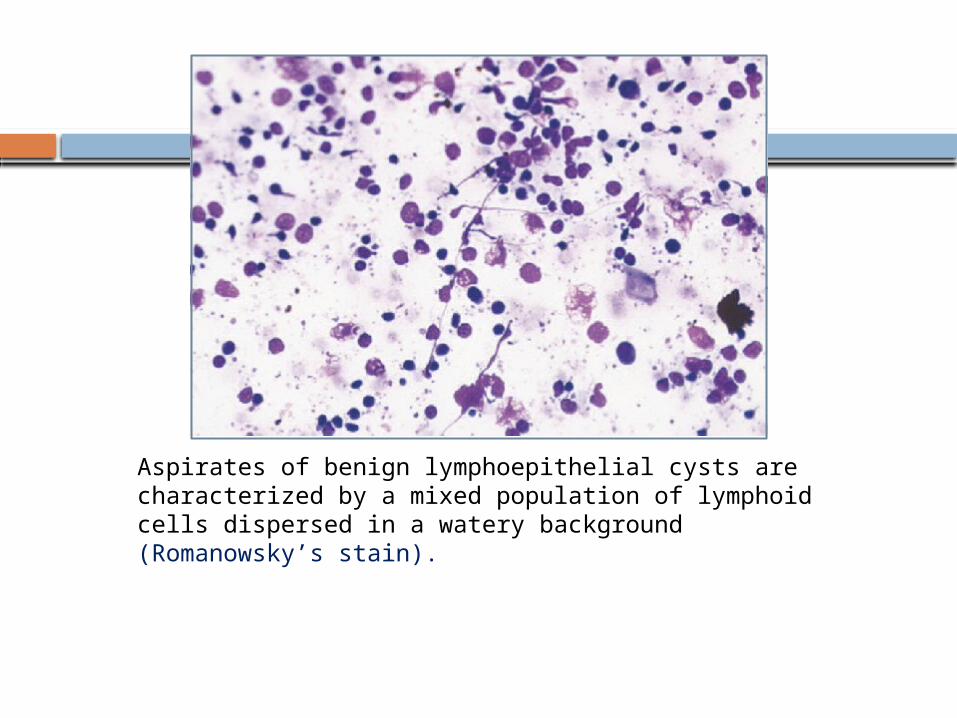
Aspirates of benign lymphoepithelial cysts are characterized by a mixed population of lymphoid cells dispersed in a watery background (Romanowsky’s stain).

Pleomorphic Adenoma
Pleomorphic adenoma (PA) commonly involves the parotid gland (more than 75%).
painless, slow growing, firm to hard swelling of the salivary gland.
Cytology• Pinkish fibrillar chondromyxoid matrix material with
frayed indistinct margins• Clusters of round, ovoid or plasmacytoid epithelial
cells with moderate amount of dense cytoplasm• Clusters and discrete spindle shaped myoepithelial
cells embedded in mesenchymal stroma

Immunochemistry: Epithelial cells are positive for cytokeratin
(CK) Myoepithelial cells are positive for CK and
vimentin (co-expressed), S-100, Glial fibrillary acid protein (GFAP) and Calponin

Long strands of spindle cells embedded in the connective tissue stroma (H & E)
Cellular pleomorphic adenoma showing discrete and cluster of epithelial cells (H & E)
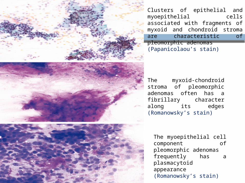
Clusters of epithelial and myoepithelial cells associated with fragments of myxoid and chondroid stroma are characteristic of pleomorphic adenomas (Papanicolaou’s stain)
The myxoid-chondroid stroma of pleomorphic adenomas often has a fibrillary character along its edges (Romanowsky’s stain)
The myoepithelial cell component of pleomorphic adenomasfrequently has a plasmacytoid appearance (Romanowsky’s stain)

Warthin’s Tumor
2nd most common tumor, occurs almost exclusively in parotid gland.
Cytology: The aspirate has a thin watery mucoid
appearance, and consists of a mixed population of lymphocytes, occasional plasma cells, and variable number of oncocytes
mucoid material or greenish-brown dirty fluid Many cohesive sheets of oncocytes Squamous and mucinous metaplasia.

There is admixed population of largeflat sheets of oncocytes and lymphoid cells.
The epithelial cells have abundant dense eosinophilic cytoplasm, well defined cytoplasmic border, enlarged centrally placed nuclei, and prominent nucleoli (Papanicolaou stain)
Aspirates of Warthin tumors are characterized by a mixed population of lymphocytes and clusters of epithelial cells with abundant granular cytoplasm. The cells lie within a dirty proteinaceous background (Romanowsky’s stain).

Basal Cell Adenoma (BCA)
Basal cell adenoma (BCA) is an uncommon salivary gland tumor
The majority of BCA arises in the major salivary gland.
The parotid gland is the predominant site of occurrence and more than 75 percent of BCA arise in parotid gland.

Cytology: Cohesive groups of basaoid cells Peripheral palisading arrangement Round nucleus, bland nuclear chromatin and
scanty cytoplasm Squamous morules Scanty homogeneous acellular stromal
material.

Tightly cohesive clusters of basaloid cells and many background stripped nuclei (DQ stain)
Membranous variant of basal cell adenoma. There is dense hyaline extracellular material surrounding the basaloid cell clusters. (Papanicolaou stain)
Oncocytoma
This is a benign salivary gland neoplasm that predominantly involves parotid gland (about 75%).
The tumor predominantly presents as a painless mobile mass.
Occurs exclusively among elderly people Pain is generally absent.
Cytology: Three-dimensional clusters of oncocytes Polygonal cells with abundant densely granular
eosinophilic cytoplasm Central to eccentric monomorphic round
nucleus.

Predominantly discrete oncocytic cells in FNAC smear (MGG)
Oncocytomas contain epithelial cells with abundant granular cytoplasm identical to those seen in Warthin tumors, but oncocytomas lack the lymphoid component(Romanowsky’s stain).

Myoepithelial Tumors
This tumor comprises only 11.5 percent of all salivary gland neoplasms.
The patient usually presents as a slow growing painless mass in the parotid or minor salivary gland regions.

Cytology:Spindle cell type• Abundant clusters and dissociated spindle cells• Elongated nuclei, fine nuclear chromatin and
inconspicuous nucleoliHyaline myoepithelial cells• Dissociated round to oval cells• Plasmacytoid cells with abundant cytoplasm
and eccentric nucleus.

The hyaline variant of myoepithelioma showing dissociated plasmacytoid cells with abundant cytoplasm and eccentric nucleus (MGG)

Adenoid cystic carcinoma
Adenoid cystic carcinoma (ACC) is a slow growing tumor.
Tendency to recurrence ACC infiltrates local nerves, causes paralysis of
the motor nerves and produces pain in the ear. Most common in parotid

Cytology:• Multiple variable sized globular, spherical or tubular
homogeneous, acellular magenta colored matrix material
• These globules are surrounded by cells• Clusters and dissociated small cells with scanty
cytoplasm• Round monomorphic hyperchromatic nuclei with coarse
chromatin.IHC:High Ki67 index.c-Kit overexpression noted.

Small round cells with scanty cytoplasm along with pinkish hyaline globules (MGG)
Cells arranged around the pinkish globules (MGG)
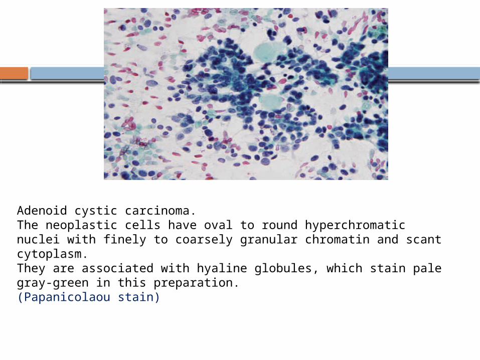
Adenoid cystic carcinoma. The neoplastic cells have oval to round hyperchromatic nuclei with finely to coarsely granular chromatin and scant cytoplasm. They are associated with hyaline globules, which stain pale gray-green in this preparation. (Papanicolaou stain)

Acinic Cell Carcinoma
Malignant epithelial neoplasm of the duct apparatus, but occasional lesions seem to show acinar differentiation
Cytology: low-grade malignancy characterized by sheets of large
cells with abundant cytoplasm The neoplastic cells have foamy/vacuolated cytoplasm
with ill-defined borders, eccentrically placed nuclei, small inconspicuous nucleoli, and lack significant nuclear atypia or pleomorphism
Occasionally lymphocytes and psammoma bodies Clean background.
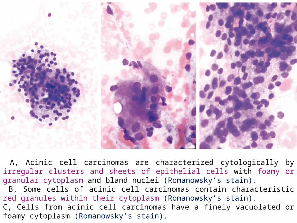
A, Acinic cell carcinomas are characterized cytologically by irregular clusters and sheets of epithelial cells with foamy or granular cytoplasm and bland nuclei (Romanowsky’s stain). B, Some cells of acinic cell carcinomas contain characteristic red granules within their cytoplasm (Romanowsky’s stain).C, Cells from acinic cell carcinomas have a finely vacuolated or foamy cytoplasm (Romanowsky’s stain).

Mucoepidermoid Carcinoma
Mucoepidermoid carcinoma (MEC) originates from the ductal cells of the salivary gland and is commonly located in the parotid gland.
Low-grade MEC is commonly cystic and is cytologically characterized by an admixture of glandular and metaplastic squamous cells.
The background demonstrates mucinous material and debris
The glandular cells may have a ductal appearance (intermediate cells) or may resemble macrophages (mucin-producing cells)

High-grade MEC has a less prominent cystic component and greater degree of atypia, compared with low-grade MEC.
High-grade MEC is cytologically characterized by large pleomorphic cells with predominantly epidermoid or undifferentiated cell features
Glandular cells are rarely seen in high-grade MEC, but their presence in association with squamous cells establishes the diagnosis.

Abundant dissociated squamoid cells with nuclear enlargement and pleomorphism. Occasional mucus secreting cells are also seen (H & E)
High-grade mucoepidermoid carcinoma. The cells are large, pleomorphic and show severe cytologic atypia. The cytoplasm has a dense quality with squamoid features (Papanicolaou stain)

Salivary Duct Carcinoma
Salivary duct carcinoma (SDC) is a rare primary salivary gland malignancy, high and low grades.
High-grade SDC is the most common subtype (>90% of cases) and considered one of the most aggressive salivary gland malignancies.
Moderate to severe nuclear atypia and pleomorphism, presence of associated necrosis in the background constitutes an important clue to the diagnosis of high-grade SDC.
In low-grade SDC, the neoplastic cells have a uniform appearance and show minimal atypia

High-grade salivary duct carcinoma. There are flat sheets of epithelial cells with abundant delicate cytoplasm and moderate nuclear atypia. The nuclei are round to oval in shape and show finely granular chromatin and prominent nucleoli. Abundant necrosis is associated with the malignant cells(Papanicolaou stain)

Epithelial-myoepithelial carcinoma
Epithelialmyoepithelial carcinoma (EMC) is regarded as a low to intermediate-grade malignancy with propensity for local recurrence
FNA is usually of high cellularity and shows two distinct cell populations, ductal and myoepithelial

Epithelial-myoepithelial carcinoma. Dual populations of ductal and myoepithelial cells. The ductal cells are more cohesive, three-dimensional, and show less amount of cytoplasm. The myoepithelial cells are arranged as loosely cohesive groups of large cells with pale vacuolated (clear) cytoplasm and have round-elongated nuclei and small nucleoli (Papanicolaou stain)

Polymorphous Low Grade Adenocarcinoma
Polymorphous low grade adenocarcinoma (PLGA) arises exclusively in the minor salivary glands and 60 percent of the tumor arises from the palate.
The patient usually presents as painless mass in the palate.
Infiltrative growth and perineural invasion seen.

Cytology: Clusters and papillary arrangement of cells Small round to oval cells with scanty to
moderate cytoplasm Round nucleus, fine stippled nuclear chromatin
and inconspicuous nucleoli Magenta colored matrix material.

Cells are in tight cluster with scanty to moderate cytoplasm (H & E)

FNAC OF ORAL CAVITY

Hemangioma
Cytology: Aspirates of hemangiomas are characterized by a
highly bloody background in which are dispersed single, plump, spindle shaped cells and clusters of spindle-shaped to ovoid cells
most aspirates from hemangiomas are of scant cellularity
The aspirated tissue fragments often show a three-dimensional “arcade” architecture with preserved lumen
The nuclei often run in a streaming pattern or may form complex coils or tubular structures

Aspirates from hemangiomas have a bloody background in which are scattered a small number of clusters of spindle cells. These clusters may have a tubular appearance. The nuclei are bland and elongated (Romanowsky’s stain)

Lipoma
Cytology: Aspirates of lipomas grossly yield an oily fluid
that, when smeared, has a glistening appearance. Microscopically lipomas are characterized by
lacelike sheets of adipose tissue. Free fat may be seen in the background and is
especially prominent in air-dried material. The tissue fragments have a three-dimensional
character and are composed of large adipocytes with abundant clear cytoplasm and small round to ovoid nuclei
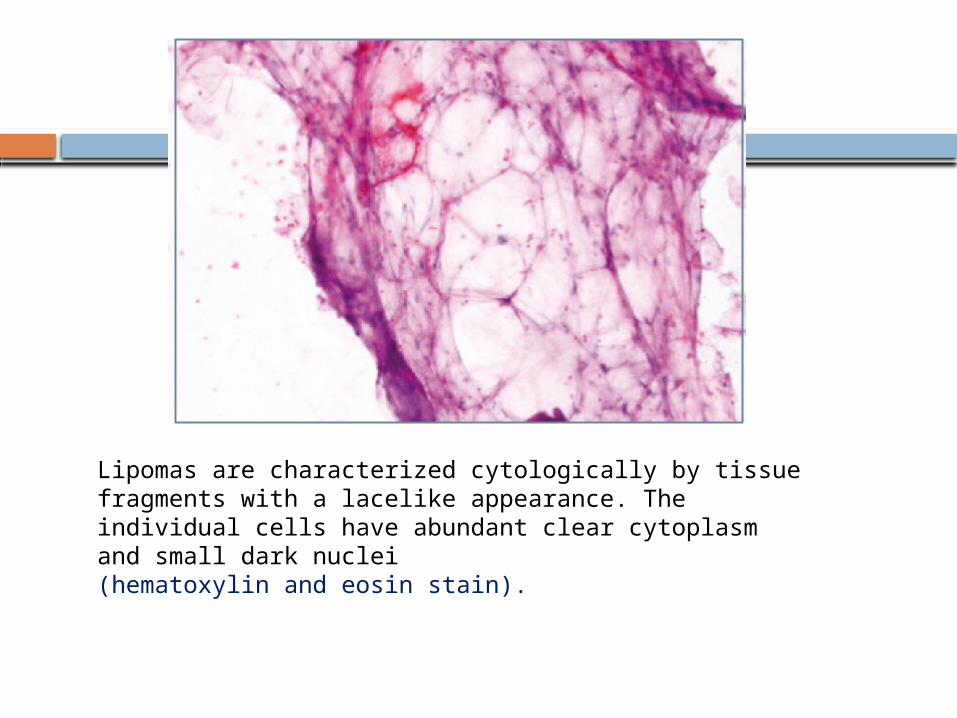
Lipomas are characterized cytologically by tissue fragments with a lacelike appearance. The individual cells have abundant clear cytoplasm and small dark nuclei (hematoxylin and eosin stain).

Granular Cell Tumor
Cytology: cellular with a background of granular debris Cells lie both singly and in syncytial groups. The cytoplasmic borders are indistinct, and the
cytoplasmic membranes frequently rupture, resulting in scattered naked nuclei
Within the syncytial aggregates, the nuclei may form a pseudofollicular pattern or present as flat sheets of cells.

Aspirates from granular cell tumors are characterized by syncytial groups of cells with abundant granular cytoplasm surrounding bland round to oval nuclei. The background of the smear has a dirty granular appearance (Papanicolaou’s stain).
intraoral Cysts and Tumors
Odontogenic cysts, both developmental and inflammatory types, share an essentially identical cytomorphology.
Aspirates contain abundant anucleated squamous cells similar in appearance to superficial and intermediate cells of stratified squamous epithelium
Occasional odontogenic keratocysts will show evidence of mineralization within the squamous cells
Odontogenic Cysts, Including Dentigerous Cysts, Eruption Cysts, Odontogenic Keratocysts, and Periapical Cysts

The cytoplasm of the squamous cells is abundant and brightly eosinophilic on Papanicolaou staining
The cells often have a glassy refractile appearance The nuclei are round or slightly ovoid and centrally
located and possess a granular chromatin Occasional aggregates of keratinized eosinophilic
bodies are seen consistent with squamous pearls Developmental cysts usually contain little
inflammation, whereas inflammatory cysts contain prominent infiltrates of neutrophils and histiocytes.

Ameloblastomas
Cytology: moderately cellular and composed of two types of
epithelial cells. The basaloid or ameloblast-like cells and the squamous
epithelial cells are present in varying proportions Basaloid cells - homogeneous and arranged in tight
clusters with well-defined edges, cells show nuclear palisading
The palisading cells have basophilic cytoplasm with poorly defined cytoplasmic outlines, nuclei are round to oval

Squamous epithelial cells - loosely cohesive and demonstrates a dense glassy cytoplasm.
The aggregates of squamous cells may contain spherical keratinized bodies, round or oval nuclei that are centrally placed, presence of spherical keratinized bodies in the squamous cells is a common feature in ameloblastomas
Foamy macrophages present

Ameloblastoma: Tight cluster of basaloid cells along with loose connective tissue stroma (H & E)
Schwannoma (Neurilemmoma)
Cytology: Generally hypocellular, with the majority of cells
forming small- to moderate-size tissue aggregates
These tissue aggregates have a filamentous appearance and indistinct cytoplasmic membranes
low-power examination- the tissue clusters often have a jigsaw puzzle appearance
High-power examination- reveals these tissue clusters to be composed of spindle-shaped cells with filamentous cytoplasm
Indistinct nuclear palisading is seen, but the cytologic demonstration of Verocay bodies is rare

Aspirates from schwannoma contain tissue aggregates with indistinct cytoplasmic membranes (Romanowsky’s stain). Schwannomas contain spindle cells with filamentous cytoplasm (inset). Nuclei are blunt or pointed with nuclear bends (Romanowsky’s stain).

Calcifying Odontogenic Cysts
Cytology: Smears - moderate cellularity. Cells have a basaloid appearance with scant
cytoplasm. A high proportion of the tissue fragments contain a
population of ghost cells characterized by densely eosinophilic, homogeneous, glassy cytoplasmic structures
Scattered MGCs, calcific debris, and ribbonlike fragments of dense acellular material are scattered in the background visible nuclei

Cementifying Fibroma
Cytology: Smears - cellular and contain single cells or clusters
of ovoid to spindle-shaped fibroblast-like cells, associated with a large number of spherical calcified psammoma bodies
The cytoplasm of the spindle cells is scant with poorly defined cell borders
Presence of numerous spherical calcified psammoma body–like structures admixed with a predominantly fibroblastic population of cells should suggest a diagnosis of benign fibro-osseous lesion

Odontogenic Myxoma
Cytology: Smear- yields abundant glistening viscous material
that is sparsely cellular Romanowsky stained, air-dried preparations show
abundant red-purple extracellular matrix with a granular or fibrillary appearance
Scattered within this matrix are a small number of plump ovoid cells of uniform appearance, have moderate to abundant pale blue cytoplasm that may be finely vacuolated in some cases, nuclei are round to oval with a finely granular chromatin
No epithelial component is identified

Squamous Cell Carcinoma
Most common oral cancerCytology: Divided into keratinizing and nonkeratinizing types Keratinizing squamous cell carcinoma-
predominant single cell presentation, single cells and clusters of neoplastic cells are present.
Keratinized malignant cell: cytoplasm refractile and eosinophilic (PAP, H&E), dense blue (MGG)
Perinuclear halo seen, irregular angular, densely hyperchromatic nuclei.
Squamous pearls are often seen Necrosis

Non keratinising squamous cell carcinoma- Irregular solid cohesive fragments Elongated or spindle shaped nuclei Variable chromatin density in adjacent cells

Well differentiated kertinizing squamous cell carcinoma: Smears are characterized by high cellularity with abundant anucleated and nucleated keratinized squamous cells. (DQ stain)
Poorly differentiated squamous cell carcinoma.Smears show cohesive and dyscohesive group of cells with vesicular nuclei, coarsely granular chromatin, and large prominent nucleoli. No evidence of squamous keratinization is seen inthe specimen (Papanicolaou stain)

Melanoma
Cytology: Smear: highly cellular and characterized by marked
pleomorphism, most cells lie in a dyscohesive pattern Characterized by a mixture of epithelioid cells and smaller
numbers of malignant giant cells containing multiple pleomorphic nuclei.
Background rich in red blood cells, but a prominent tumor diathesis with granular debris is frequent
Romanowsky stain- deep, dark blue to nearly black pigment
Papanicolaou staining- dark brown finely granular pigment.

Melanomas are characterized by dyscohesive spindle or epithelioid cells with prominent nucleoli (Papanicolaou’s stain). Melanin pigment may be seen in melanoma (hematoxylin and eosin stain).

Kaposi Sarcoma
Cytology: Usually hemorrhagic and contain scant tissue
fragments. The individual tissue fragments are of small to
moderate size. The larger tissue aggregates may display ill-
defined vascular spaces and a radial or parallel arrangement of the elongated spindle-shaped cells
Individual cells are represented by naked nuclei with loss of cytoplasm.
Fragments of pink- mauve stroma may be found in the background.

Kaposi sarcoma contains radial arrangements of elongated spindle cells (Romanowsky’s stain)
Conclusion
FNAC is a highly accurate procedure for differentiating benign and malignant lesions.
However, specific cytological diagnosis may be difficult to make in the absence of characteristic architectural patterns.
Diagnosis of aspirates from cystic lesions may be less specific than the solid lesions due to paucity of specific lesional cells in the former and also due to superimposed infection

Reference
Gnepp, diagnostic surgical pathology of the head and neck, 2nd edition.
Surgical Pathology of the Head and Neck, Third Edition, LEON BARNES
Fine needle aspiration cytology, 4TH edition, Svante R Orell.
Fine needle aspiration cytology, Review article, JV LEVER, PA TROTT, AJ WEBBA, J Clin Pathol 1985;38: 1-11.

Thank you