fine structure of a bacterial community associated with - bionames
TRANSCRIPT
Proc. Helminthol. Soc. Wash.54(2), 1987, pp. 212-219
Fine Structure of a Bacterial Community Associated withCyathostomes (Nematoda: Strongylidae) of Zebras
R. C. KRECEK,' R. M. SAYRE,? H. J. ELS,3J. P. VAN NlEKERK,3 AND F. S. MALAN 4
1 Department of Parasitology, Faculty of Veterinary Science, University of Pretoria,P.O. Box 12580, Onderstepoort 0110, Republic of South Africa2 Nematology Laboratory, Agricultural Research Service, USDA,Beltsville, Maryland 207053 Electron Microscope Unit, Medical University of Southern Africa,0204 Medunsa, Republic of South Africa and4 Hoechst Research Farm, P.O. Box 124, 1320 Malelane, Republic of South Africa
ABSTRACT: Microorganisms attached to the posterior and anterior extremities of zebra cyathostomes are studiedby scanning electron (SEM) and transmission electron microscopy (TEM). The predominant constituent of themicrobial community is a filamentous prokaryotic organism, which bears resemblance to Arthromitus Leidy,1849, and is designated Arthromitus-like organism (ALO). ALO is associated with the vulvar and anal openingsof the female's posterior end. The other organisms include those with a filamentous cross wall, a distinct doublecell wall, a blunt end, and spiral shape. These microbes are not observed to cause harm to the cyathostomehost.
KEY WORDS: morphology, SEM, TEM, filamentous prokaryotes, nematodes, equine, strongyles, bacteria,wildlife , cuticle, South Africa.
During recent dry periods in southern Africa,individual Burchell's zebras (Equus burchelli an-tiquorurri) were randomly culled from herds inthe Kruger and Etosha National Parks to reducepopulation pressures. This action provided theopportunity to survey this wild equid for its hel-minth populations.
The large intestine of wild and domestic equidsis the habitat for the majority of more than 109known species of helminth parasites, principallynematodes (Theiler, 1923; Lichtenfels, 1975;Scialdo-Krecek, 1984). Filamentous prokaryoticorganisms were noted attached to the anteriorand posterior extremities, particularly to the vul-var and anal openings of female strongyles, andto the cuticle of both sexes of worms that wererecovered from the large intestines of these ze-bras. During investigations, members of theStrongylidae, the largest family of nematodes inthis equid, were identified and frozen for futurebiochemical studies. Our purposes here are (1)to report on the fine structure, based on scanningand transmission electron microscopy, of bac-terial organisms associated with the cyatho-stomes (Strongylidae: Cyathostominae) from ze-bras, and (2) to present evidence of a microbialcommunity associated with these nematodes.
Materials and Methods
Helminth collection
Five zebras were killed and processed at necropsyaccording to published methods (Malan et al., 198la,b; Scialdo-Krecek, 1984). Live cyathostomes were re-covered from the large intestine at necropsy and main-tained in physiological saline (0.85% NaCl solution) ina water bath at 37°C.
Preparation for electron microscopy
The cyathostomes were examined under a stereo-microscope and those nematodes with filamentous mi-croorganisms attached were identified. Between ex-amination and fixation, the cyathostomes weremaintained in a water bath at 37°C. Those worms in-tended for transmission electron microscopy (TEM)were transferred to 3% glutaraldehyde (GA) in Millo -nig's phosphate buffer at pH 7.2 (room temperature)for 7 days, washed twice for 15 min in Millonig's buffer,and postfixed with 1% OsO4 in 0.15 M sodium caco-dylate buffer. The specimens were washed in 0.15 Msodium cacodylate buffer, dehydrated in 50%, 70%,90%, and 100% (twice) ethanol, cleared in propyleneoxide, and embedded in Polarbed 812 resin. Thin sec-tions through the cyathostome's posterior extremity,including a few mm of the anus and vulva, were cuton a Reichert ultramicrotome, stained with uranyl ace-tate and lead citrate, and photographed in a JEOL 100CX transmission electron microscope at 100 kV.
In preparation for scanning electron microscopy(SEM), the nematodes were fixed in glutaraldehyde as
212
Copyright © 2011, The Helminthological Society of Washington

213
Figures 1-6. Scanning electron micrographs of the filamentous microorganisms associated with zebra cyatho-stomes which resemble Arthromitus Leidy, 1849, and were therefore labeled ALO (Arthromitus-like organism).1. Posterior extremity of female cyathostome showing mass of filaments covering tip of tail, and anal and vulvaropenings. Scale bar 100 j&m. 2. The organisms extrude from the anal opening (arrow). Scale bar 10 ftm. 3. Shortfilaments surround the vulvar opening. Scale bar 10 pm. 4. Fingerlike tips (arrow) of free end of organism suggestsa stage of growth. Scale bar 10 pm. 5. Segmentation evident in what may be older filaments. Scale bar 10 /nm.6. Coccoid bodies (a) on cyathostome cuticle which may give rise to longer segments (b) and eventually filaments(c). Scale bar 10 jim.
above, as well as in 70% ethanol or in 1% picric acidin Karnovsky's, washed in sodium cacodylate buffer,and subsequently dehydrated in 50%, 70%, 90%, and100% (twice) ethanol followed by critical point dryingin CO2. Individual specimens were mounted on stubs,previously coated with a thin carbon layer. This wasfollowed by sputter-coating with an Au-Pd layer. Theywere then viewed in a JEOL 35C scanning electronmicroscope at 8-12 kV.
Results
Filamentous microorganisms were found at-tached to the posterior and anterior extremitiesand along the cuticle of several cyathostomespecies recovered from the large intestines of thezebras. In addition, these organisms were at-tached inside the vulvar and anal openings of
Copyright © 2011, The Helminthological Society of Washington

214 PROCEEDINGS OF THE HELMINTHOLOGICAL SOCIETY
Copyright © 2011, The Helminthological Society of Washington
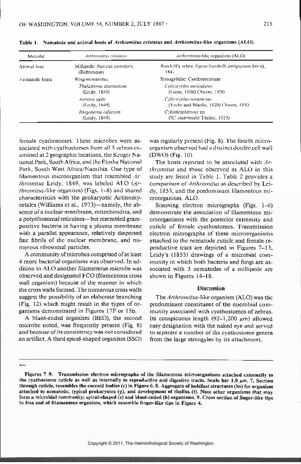
OF WASHINGTON, VOLUME 54, NUMBER 2, JULY 1987 215
Table 1. Nematode and animal hosts of Arthromitus cnstatus and Arthromitus-like organisms (ALO).
Microbe Arthromitus-like organisms (ALO)
Animal host
Nematode hosts
Millipede: Narceus annularis(Rafinesque)
Rhigonematidae:
Thelastoma attenuatuin(Leidy, 1849)
Aorurus agile(Leidy, 1849)
Rhigonema infect um(Leidy, 1849)
Burchell's zebra: Equus burchelli antiquorum Smith,1841
Strongylidae: Cyathostominae
Cylicocyclus auriculatus(Looss, 1900)Chaves, 1930
Cylicocyclus triramosus(Yorke and Macfle, 1920) Chaves, 1930
Cylindropharynx sp.(1C. intermedia Theiler, 1923)
female cyathosomes. These microbes were as-sociated with cyathostomes from all 5 zebras ex-amined at 2 geographic locations, the Kruger Na-tional Park, South Africa, and the Etosha NationalPark, South West Africa/Namibia. One type offilamentous microorganism that resembled Ar-thromitus Leidy, 1849, was labeled ALO (Ar-thromitus-like organism) (Figs. 1-8) and sharedcharacteristics with the prokaryotic Actinomy-cetales (Williams et al., 1973)—namely, the ab-sence of a nuclear membrane, mitochondria, anda polyribosomal reticulum—but resembled gram-positive bacteria in having a plasma membranewith a parallel appearance, relatively dispersedfine fibril s of the nuclear membrane, and nu-merous ribosomal particles.
A community of microbes comprised of at least4 more bacterial organisms was observed. In ad-dition to ALO another filamentous microbe wasobserved and designated FCO (filamentous crosswall organism) because of the manner in whichthe cross walls formed. The numerous cross wallssuggest the possibility of an elaborate branching(Fig. 12) which might result in the types of or-ganisms demonstrated in Figures 17F or 15b.
A blunt-ended organism (BEO), the secondmicrobe noted, was frequently present (Fig. 8)and because of its consistency was not consideredan artifact. A third spiral-shaped organism (SSO)
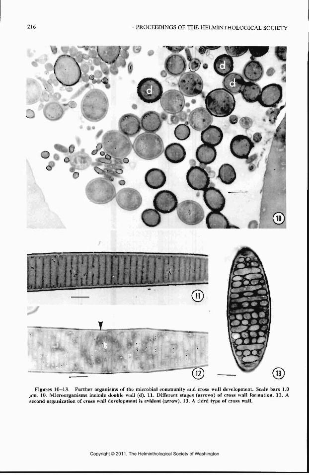
was regularly present (Fig. 8). The fourth micro-organism observed had a distinct double cell wall(DWO) (Fig. 10).
The hosts reported to be associated with Ar-thromitus and those observed as ALO in thisstudy are listed in Table 1. Table 2 provides acomparison of Arthromitus as described by Lei-dy, 1853, and the predominant filamentous mi-croorganism, ALO.
Scanning electron micrographs (Figs. 1-6)demonstrate the association of filamentous mi-croorganisms with the posterior extremity andcuticle of female cyathostomes. Transmissionelectron micrographs of these microorganismsattached to the nematode cuticle and female re-productive tract are depicted in Figures 7-13.Leidy's (1853) drawings of a microbial com-munity in which both bacteria and fungi are as-sociated with 3 nematodes of a millipede areshown in Figures 14-18.
Discussion
The Arthromitus-like organism (ALO) was thepredominant constituent of the microbial com-munity associated with cyathostomes of zebras.Its conspicuous length (92-1,200 ^m) allowedeasy designation with the naked eye and servedto separate a number of the cyathostome generafrom the large strongyles by its attachment.
Figures 7-9. Transmission electron micrographs of the filamentous microorganisms attached externally tothe cyathostome cuticle as well as internally to reproductive and digestive tracts. Scale bar 1.0 jim. 7. Sectionthrough cuticle, resembles the coccoid bodies (c) in Figure 6. 8. Aggregate of holdfast structures (hs) for organismattached to nematode, typical prokaryotes (p), and development of thallus (t). Note other organisms that mayform a microbial community; spiral-shaped (s) and blunt-ended (b) organisms. 9. Cross section of finger-like tipsin free end of filamentous organism, which resemble finger-like tips in Figure 4.
Copyright © 2011, The Helminthological Society of Washington
216 PROCEEDINGS OF THE HELMINTHOLOGICAL SOCIETY
o
.
Figures 10-13. Further organisms of the microbial community and cross wall development. Scale bars 1.0fim. 10. Microorganisms include double wall (d). 11. Different stages (arrows) of cross wall formation. 12. Asecond organization of cross wall development is evident (arrow). 13. A third type of cross wall.
Copyright © 2011, The Helminthological Society of Washington

OF WASHINGTON, VOLUME 54, NUMBER 2, JULY 1987 217
Table 2. A comparison of Arthromltus Leidy, 1849 (Leidy, 1853) with the Arthromitus-like organisms (ALO)of present study.
Arthromitus (after Leidy, 1853) ALO
(1) Thallus—delicate, filamentous, linear, straight or inflected, flexible, colorless,translucent, obtusely rounded at free end.
(2) Pedicle of attachment 1 or more amber-colored round or oval granules or inaggregations of several granules.
(3) Articul i indistinct, but becoming well marked after the development of the in-terior sporular body.
(4) Spore oval, simple, faintly yellowish, translucent, highly refractive, usuallylying oblique and alternating in position in different articuli.
(5) Length 17-2,083 ^m; width 0.15 Mm.(6) Habitat: Mucous membrane of ventriculus and large intestine of Narceus an-
nularis, and also Enterobnnts elegans, Rhigonema infection, Aorurus agile, andThelastoma attenuatiim; from the mucous membrane and its appendages ofthe ventriculus of Passalus cornutus and Polydesmus virginiensis and Eccrinalonga. Usually from lips of anal and generative apertures.
(1) Same, except free end with finger-lik e projection.
(2) Attachment site usually aggregation.
(3) Articuli distinct and indistinct.
(4) Spores not evident.
(5) Length 92-1,200 urn; width 3 /urn.(6) Habitat: Usually of cuticle, anal and
vulvar openings of the posterior ex-tremity of females. Sometimes asso-ciated with anterior extremities andwith males. Only associated with thegenera, Cylicocyclus and Cylindro-pharynx (Strongylidae: Cyathostomi-nae).
Though these filamentous microorganisms bearsimilarities to Arthromitus described by Leidy(1853) (Table 2), they did not exhibit evidenceof endospores, a characteristic of Arthromitus.The possibility exists, however, that our sampleswere observed in a nonendospore-forming phaseof growth. Leidy's (1853) observations took placeover a period of time (i.e., hours or days), where-as our specimens were collected and fixed at onemoment in time.
No correlation between the presence of eggs inthe uteri of female cyathostomes and the occur-rence of ALO was observed. A closer examina-tion may reveal if young females (nongravid) orspent females (beyond egg-laying age) were morefrequently associated with the presence of ALO,or whether ALO affects the production or ex-pulsion of eggs at all. Such an effect is probablyunlikely because the penetration of ALO ap-peared to be superficial and not more than 100-300 urn deep into the reproductive tract.
We feel that the relationship of these micro-organisms to their host is probably of a com-mensal nature and therefore agree with the def-inition proposed by Noble and Noble (1961)—i.e., the association of 2 species in which only 1species may benefit. We can assume 1 of the 2associants in the present study, either the nema-tode or the microbes, is dependent on the other;however, we have no evidence that either coulddefine the nature of the apparent commensalism.Poinar (1979) cites several instances of potentialpathogenic bacteria in nematodes and Andersonet al. (1978), for instance, observed microbes in
association with cuticular lesions. Strongylusedentatus, recovered from horses, belongs to thesame nematode family as the cyathostomes(Lichtenfels, 1975). Anderson et al. (1978) con-sidered the microbes harmful and described 4types of lesions. According to these authors, theassociation of the lesions with the nematode gen-italia suggests that "some lesions of helminthcuticles may be a venereal disease."
The cyathostome species with which ALO areassociated in this study may not be a completelist (Table 1). Those included reflect a randomsampling (400-500 cyathostomes) that was fixedat the zebra postmortem examination. Identifi-cation of further specimens to species level shouldreveal whether host species restriction or prev-alence on a seasonal basis occurs. Cyathostomespecies preference wil l also relate to intestinal sitedistribution since it is a characteristic of thesenematodes (Lichtenfels, 1975). Furthermore, theALO appears to be associated more often withthe posterior and less frequently with the anteriorextremities of the cyathostomes. Closer exami-nation of more specimens wil l reveal whetherother nematode sites are involved.
Leidy (1853) observed a community in whichbacteria and fungi inhabited millipede nema-todes (Figs. 14-18). More extensive microbialcommunities exist consisting of 4-30 protists asin the paunch of wood-eating termites (Marguliset al., 1986). Perhaps the extent of the microbialcommunities in the millipede, termites, and ze-bras is related in some way to the ingestion oforganic matter and the appearance of ALO in 2
Copyright © 2011, The Helminthological Society of Washington

218 PROCEEDINGS OF THE HELMINTHOLOGICAL SOCIETY
16
17
Copyright © 2011, The Helminthological Society of Washington

OF WASHINGTON, VOLUME 54, NUMBER 2, JULY 1987 219
of the 3 hosts. Certainly, given the milieu, itwould be surprising if the nematode's cuticlewould be free of bacteria. It is unlikely that alladherent bacteria are harmful; some may not bepathogenic, and others may benefit the nematode(Sayre and Starr, 1987). Likewise, filamentousbacterial organisms have frequently been re-ported from the gut of mice, dogs, cats, sheep,horses, and pigs (Anderson et al., 1971, 1978;Davis and Savage, 1974; Davis etal., 1976, 1977;Gregory et al., 1985). The present study is thefirst known report of a microbial community be-lieved to be nonpathogenic and associated withstrongyles inside a mammalian gut.
Acknowledgments
We thank T. E. Krecek for his invaluable tech-nical assistance, the National Parks Board (NPB),South Africa, for placing the zebras at our dis-posal, Drs. V. de Vos and L. E. O. Braack of theKruger National Park for their cooperation dur-ing collecting trips, Professor R. W. Lichtwardtfor his helpful comments initially regarding theclassification of these microbes, Patricia A. Pillit tfor her valuable advice regarding the millipedenematodes, and Dr. K. D. Murrell for his helpfulcomments regarding the manuscript.
Literature Cited
Anderson, W. R., P. A. Madden, and M. L. Colglazier.1978. Microbial flora of cuticular lesions onStrongylus edentatus. Proceedings of the Helmin-thological Society of Washington 45:219-225.
, , and F. G. Tromba. 1971. Histopath-ologic and bacteriologic examination of cuticularlesions of Ascaris suum. Journal of Parasitology47:1010-1014.
Davis, C. P., D. Cleven, E. Balish, and C. E. Yale.1977. Bacterial association in the gastrointestinaltract of beagle dogs. Applied and EnvironmentalMicrobiology 34:194-206.
, , J. Brown, and E. Balish. 1976. An-aerobiospirilhim, a new genus of spiral-shapedbacteria. International Journal of Systematic Bac-teriology 26:498-504.
, and D. C. Savage. 1974. Habitat, succession,attachment, and morphology of segmented, fila-
mentous microbes indigenous to the murine gas-trointestinal tract. Infection and Immunity 10:948-956.
Gregory, M. W., R. M. Pittilo, S. J. Ball, and W. M.Hutchison. 1985. Scanning electron microscopyof filamentous organisms associated with coccidialinfections in cats and sheep. Annals of TropicalMedicine and Parasitology 79:473-475.
Leidy, J. 1853. A Flora and Fauna within LivingAnimals. Smithsonian Institution, G. P. Putnamand Company, New York. 67 pp.
Lichtenfels, J. R. 1975. Helminths of domestic equids.Illustrated keys to genera and species with em-phasis on North American forms. Proceedings ofthe Helminthological Society of Washington 42(Special issue): 1-92.
Malan, F. S., R. K. Reinecke, and R. C. Scialdo. 198 la.Recovery of helminths postmortem from equines.I. Parasites in arteries, subperitoneum, liver andlungs. Onderstepoort Journal of Veterinary Re-search 48:141-143.
, , and . 1981b. Recovery of hel-minths postmortem from equines. II . Helminthsand larvae ofGasterophilus in the gastro-intestinaltract and oestrids from the sinuses. OnderstepoortJournal of Veterinary Research 48:145-147.
Margulis, L., D. Chase, and R. Guerrero. 1986. Mi-crobial communities. BioScience 36:160-170.
Noble, E. R., and G. A. Noble. 1961. Parasitology,the Biology of Animal Parasites. Lea and Febiger,Philadelphia, Pennsylvania. 767 pp.
Poinar, G. O., Jr. 1979. Nematodes for BiologicalControl of Insects. CRC Press, Inc., Boca Raton,Florida. 277 pp.
Sayre, R. M., and M. P. Starr. 1987. Bacterial dis-eases and antagonisms of plant-parasitic nema-todes. Pages 000-000 in G. O. Poinar, Jr. and H.Jansson, eds. Nematode Pathology. CRC Press,Inc., Boca Raton, Florida. (In press.)
Scialdo-Krecek, R. C. 1984. The nematode parasitesof Equus zebra hartmannae and Equus burchelliantiquorum from different areas of southern Af-rica. D.Sc. Thesis, University of Pretoria, Pretoria,South Africa. 261 pp.
Theiler, G. 1923. The strongylids and other nema-todes parasitic in the intestinal tract of South Af-rican equines. The Government Printing and Sta-tionery Office, Pretoria. 175 pp.
Williams, S. T., G. P. Sharpies, and R. M. Bradshaw.1973. The fine structure of the Actinomycetales.Pages 113-130 in G. Sykes and F. A. Skinner, eds.Actinomycetales: Characteristics and PracticalImportance. Academic Press, New York.
Figures 14-18. Drawings of a microbial community associated with nematodes from the intestinal tract ofthe millipede Narceus annularis (Rafinesque) according to Leidy (1853). 14. Middle portion of the body ofThelastoma attenuatum Leidy, 1849, with 2 thalli of Enterobryus growing from it. a, Uterus; b, vagina; c, vulva;d, Arthromitus; e, Enterobryus; f, intestine. 15. Posterior extremity of Aorurus agile, a, Rectum; b, anus; c, tuftsof Arthromitus; d, uterus with egg. 16. Lateral view of the posterior extremity of Rhigonema infectum. a, Intestine;b, rectum; c, anus; d, Enterobryus; e, pedicle of attachment of Enterobryus. 17. Posterior extremity of Rhigonemainfectum. a, Intestine; b, rectum; c, young thallus of Enterobryus; d, long pedicle of Enterobryus; e, Arthromituscristatus; f, Cladophytum comatum. 18. Aorurus agile with Enterobryus elegans and Arthromitus cristatus. a,Uterus with eggs; b, anus; c, Arthromitus cristatus; d, Enterobryus elegans.
Copyright © 2011, The Helminthological Society of Washington