first draft report - dwqr...
TRANSCRIPT

FINAL REPORT - ENV 3/04/15.
Stobhill Hospital, 133 Balornock Road, Glasgow G21 3UW.
Typing of Cryptosporidium isolates from
water samples in Scotland (ENV 3/04/05).
Final report to the:
Environment and Rural Affairs
Department,
Agricultural and Biological Research
Group,
SCOTTISH EXECUTIVE.
1
FINAL REPORT - ENV 3/04/15.
Typing of Cryptosporidium isolates from water samples in Scotland
(ENV 3/04/15).
Lead contractor: Dr. H. V. Smith, Scottish Parasite Diagnostic Laboratory, Stobhill
Hospital, 133 Balornock Road, Glasgow, G21 3UW. Telephone: 0141 201 3028; Telefax: 041
201 3029; e-mail: [email protected]
The OBJECTIVE was to gain information on the occurrence of Cryptosporidium spp.
oocysts in raw and treated drinking waters in order to identify the predominant types
existing in water catchment areas and to monitor variations in the oocyst population
distribution over a 1 year period with a view to assisting the assessment of the risk to
human health.
This was accomplished by:
a) confirming the presence of Cryptosporidium species oocysts on microscope slides using
epifluorescence and Nomarski differential interference contrast microscopy.
b) assessing Cryptosporidium species oocyst integrity based on morphology using
Nomarski differential interference contrast microscopy.
c) determining species and / or genotype by established molecular methods in order to
yield identification of distinct Cryptosporidium species, both known and previously
unknown.
This draft final report provides the Scottish Executive (SE) and Scottish Water (SW)
with quality assured information on the occurrence, species and genotype of
Cryptosporidium oocysts in Scottish waters over a 1 year period by analysing oocyst
morphometry and morphology and determining species / genotype of oocysts deposited
on Cryptosporidium Directions (Scottish Water) 2003 microscope slides.
2
FINAL REPORT - ENV 3/04/15.
1. INTRODUCTION
The protozoan parasite, Cryptosporidium, has been implicated in numerous
waterborne and foodborne outbreaks of cryptosporidiosis (Smith and Rose 1990, 1998;
Girdwood and Smith, 1999; Fayer et al. 2000; Slifko et al. 2000). Cryptosporidium has a
complex life cycle, involving both asexual and sexual reproductive cycles, which is
completed within an individual host, and transmission is via an environmentally robust oocyst
excreted in the faeces of the infected host. Currently, there is debate concerning the number of
species within the genus Cryptosporidium. Sixteen species of Cryptosporidium are valid and
which might occur in our environment. There are: Cryptosporidium hominis described
originally in humans, C. parvum, in man and numerous other mammals, C. andersoni and C.
bovis in cattle, C. muris in mice, C. felis in cats, C. suis in pigs, C. wrairi in guinea pigs, C.
canis in dogs C. meleagridis in turkeys, C. baileyi in chickens, C. galli in birds, C.
saurophilum in lizards, C. serpentis in snakes, C. scophthalmi and C. molnari in fish. In
addition, there are a further 40 (or more) Cryptosporidium genotypes, which differ
significantly in their molecular signatures but, as yet, have not been ascribed species status
(Smith et al., 2007; Tables 1a and b).
Genetic analyses reveal that at least seven species (C. hominis, C. parvum, C.
meleagridis, C. felis, C. canis, C. suis, and C. muris) and two genotypes (monkey and
cervine) of Cryptosporidium are associated with human disease (Caccio et al., 2005, Smith et
al., 2006) but C. parvum and C. hominis remain the most common species infecting humans.
Species of Cryptosporidium reported to have crossed host-specificity barriers and detected in
human stools are C. meleagridis, C. felis, C. muris, C. suis and C. canis and Cryptosporidium
cervine and monkey genotypes (Table 1a).
Table 1a. Differences in host range between species within the genus Cryptosporidium
Cryptosporidium
species
Dimensions of
oocysts (µm)
Primary host Human host range
Immuno(competent)
Immuno(compromised)
C. parvum 4.5 x 5.5 mammals Competent & compromised
C. hominis 4.5 x 5.5 humans Competent & compromised
C. muris 5.6 x 7.4 mammals Competent
C. felis 4.5 x 5.0 felids Competent & compromised
C. meleagridis 4.5-4.0 x 4.6-5.2 Turkeys, humans Competent & compromised
3
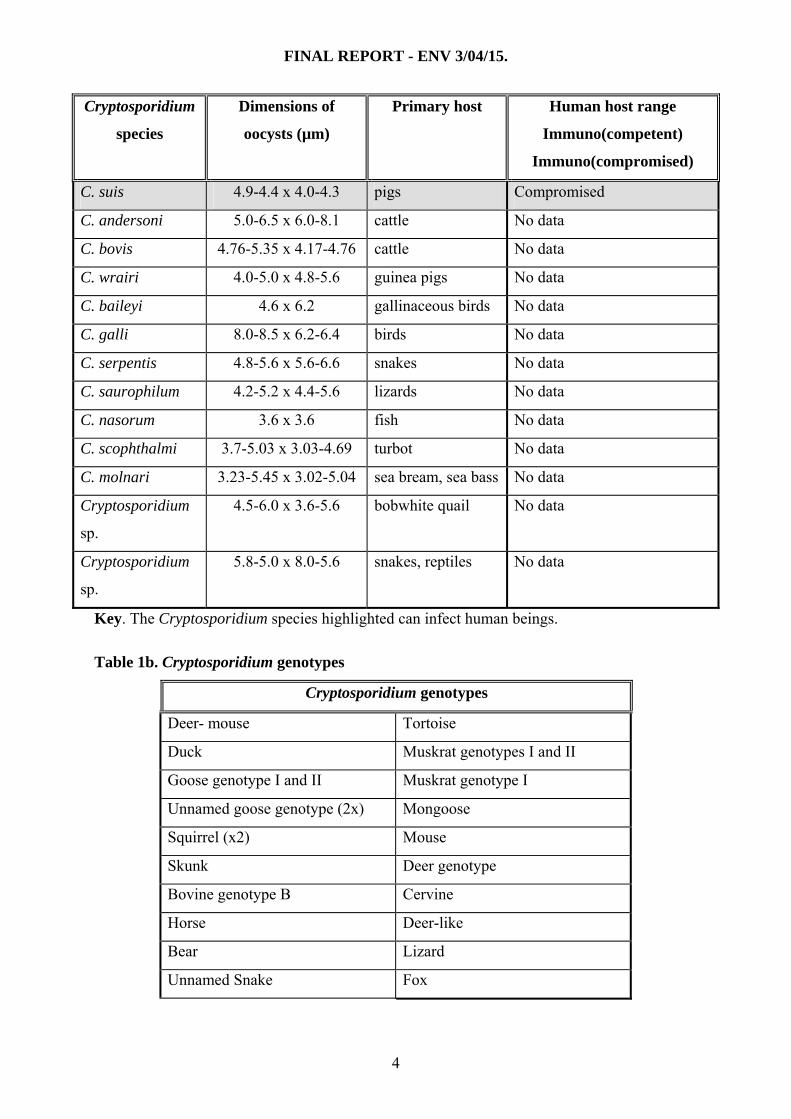
FINAL REPORT - ENV 3/04/15.
Cryptosporidium
species
Dimensions of
oocysts (µm)
Primary host Human host range
Immuno(competent)
Immuno(compromised)
C. suis 4.9-4.4 x 4.0-4.3 pigs Compromised
C. andersoni 5.0-6.5 x 6.0-8.1 cattle No data
C. bovis 4.76-5.35 x 4.17-4.76 cattle No data
C. wrairi 4.0-5.0 x 4.8-5.6 guinea pigs No data
C. baileyi 4.6 x 6.2 gallinaceous birds No data
C. galli 8.0-8.5 x 6.2-6.4 birds No data
C. serpentis 4.8-5.6 x 5.6-6.6 snakes No data
C. saurophilum 4.2-5.2 x 4.4-5.6 lizards No data
C. nasorum 3.6 x 3.6 fish No data
C. scophthalmi 3.7-5.03 x 3.03-4.69 turbot No data
C. molnari 3.23-5.45 x 3.02-5.04 sea bream, sea bass No data
Cryptosporidium
sp.
4.5-6.0 x 3.6-5.6 bobwhite quail No data
Cryptosporidium
sp.
5.8-5.0 x 8.0-5.6 snakes, reptiles No data
Key. The Cryptosporidium species highlighted can infect human beings.
Table 1b. Cryptosporidium genotypes
Cryptosporidium genotypes
Deer- mouse Tortoise
Duck Muskrat genotypes I and II
Goose genotype I and II Muskrat genotype I
Unnamed goose genotype (2x) Mongoose
Squirrel (x2) Mouse
Skunk Deer genotype
Bovine genotype B Cervine
Horse Deer-like
Bear Lizard
Unnamed Snake Fox
4
FINAL REPORT - ENV 3/04/15.
Cryptosporidium genotypes
Unnamed Snake (W11) Woodcock
Pig genotype II ferret
Most human volunteer infectivity studies have used C. parvum oocyst isolates. Of 29
healthy human volunteers, with no evidence of previous Cryptosporidium infection, 20%
became infected following an oral dose of 30 C. parvum (Iowa isolate, bovine) oocysts
(DuPont, et al., 1995). A dose of 300 oocysts caused infection in 88%, and 1000 oocysts
produced infection in 100% of volunteers tested. The median infective dose was calculated to
be 132 oocysts. Of the volunteers who excreted oocysts, 39% developed diarrhoea and one
other enteric symptom. Those with diarrhoea excreted more oocysts than those without
diarrhoea, and were more likely to excrete oocysts on consecutive days (Chappell, et al.,
1996). Previous exposure confers some protection against reinfection. A 14 fold increase in
ID50 occurred in volunteers with pre-existing anti-C. parvum serum IgG (Chappell et al.,
1999).
Similar results were obtained with one recent human volunteer infectivity study using
C. hominis. Twenty one adult healthy volunteers were challenged with 10-500 oocysts (isolate
TU502). Of these, 16 individuals (76.2%) had evidence of infection. The ID50 was estimated
as 10-83 oocysts using clinical and microbiological definitions of infection, respectively.
Diarrhea occurred in 40% of subjects receiving 10 oocysts with a stepwise increase to 75% in
those receiving 500 oocysts. Most infected persons elicited a serum IgG immune response
(Chappell et al., 2006).
The infectivity of different C. parvum isolates can vary in healthy human adult
volunteers. Isolates differed in their ID50, in their attack rate, and in the duration of diarrhoea
they induced (Okhuysen et al., 1999). The median infectious dose is 9 oocysts for the TAMU
(equine) isolate, 132 oocysts for the Iowa isolate and 1042 oocysts for the UCP (bovine)
isolate of C. parvum (Okhuysen et al., 1999).
Transmission occurs via any route by which material contaminated with viable
oocysts excreted by infected hosts can reach the intestine of a susceptible host. Person to
person transmission, via the faecal-oral route, is a major route and has been documented
between family / household members, health workers and their patients, and children in day-
care centres (probably due to the lower standards of personal hygiene exhibited by pre-school
children) and other institutions. Cryptosporidium oocysts are frequent contaminants of water,
5
FINAL REPORT - ENV 3/04/15.
with contributions from infected human and non-human hosts, livestock and agricultural
practices and infected feral and transport hosts (Smith and Rose, 1990, 1998; Smith et al.,
1995; Smith and Lloyd, 1997). The most important route of environmental transmission is
through the contamination of water by oocysts (Robertson et al., 1994; Lisle and Rose, 1995;
Smith et al., 1995; Rose et al., 1997; Smith and Rose, 1998). Waterborne cryptosporidiosis,
associated with community water systems, has been reported primarily from North America
and Europe (Smith et al., 1995; Smith and Rose, 1998; Kourenti et al., 2006).
In order to safeguard public water supplies from oocyst contamination, various
recommendations have been made and the sampling and monitoring of water and
environmental samples for oocysts has become a concern of increasing importance for the
water industry and other interested bodies. Water microbiologists and epidemiologists require
knowledge on the source and level of contamination, the viability of the organisms, the
relationship to indicator organisms, and the reservoirs of infection, while engineers and utility
operators require knowledge on (oo)cyst removal and inactivation by treatment processes.
Regulators of drinking and wastewater programmes require to know where and when these
organisms occur in water, the suitability and availability of monitoring methods, and whether
treatment requirements should be standardised.
Following a large outbreak of cryptosporidiosis in the Torbay area of Devon, where
drinking water was strongly implicated, the case brought against the water company, of
supplying water unfit for human consumption, was rejected by the Court on the grounds that
epidemiological evidence was not admissible. In England and Wales, the Government
promulgated Regulations to ensure that drinking water was treated to adequately remove
Cryptosporidium spp. In England and Wales, the Government promulgated Regulations to
ensure that drinking water was treated to adequately remove Cryptosporidium spp. Water
undertakers are required to determine whether there is a significant risk from
Cryptosporidium oocysts in water supplied from waterworks and to comply with the
requirement for treating the water intended to be supplied. A treatment standard was set,
based on a standard method that sampled at least 40 litres per hour of treated water, as a
continuous sample, over a 24 hour period, and to implement the regulations, Standard
Operating Protocols for monitoring Cryptosporidium oocysts in water supplies were identified
for analysing samples. An average of less than 1 oocyst in 10 litres of the final water sampled
over the 24 hour period was required as evidence of effective water treatment.
6

FINAL REPORT - ENV 3/04/15.
In order to permit the use of analytical evidence in a Court of Law, strict rules for all aspects
of sampling and analysis were laid down and an information letter, since updated, identified
the Protocol containing Standard Operating Protocols (SOPs) for monitoring Cryptosporidium
oocysts in water supplies (Anon., 2005). Identification and enumeration of oocysts using
modifications of Smith et al. (1989) and Grimason et al., (1994) is performed on air dried
oocysts, methanol fixed onto glass microscope slides and stained with a DWI approved
commercially available monoclonal antibody that recognises exposed epitopes on oocysts
walls (FITC-C-mAb) and the nuclear fluorogen 4'6-diamidino-2-phenyl indole (DAPI). Slides
are viewed under the appropriate filters of an epifluorescence microscope and oocysts
identified, measured and enumerated.
The same SOPs are used in Scotland [The Cryptosporidium (Scottish Water)
Directions 2003]
http://www.scotland.gov.uk/Publications/2004/01/18727/31490. The Cryptosporidium
(Scottish Water) Directions 2003, which came into force on 01/01/2004, requires Scottish
Water to implement the recommendations contained in the Third Report of the 'Group of
Experts on Cryptosporidium in Water Supplies' (Anon. 1998), and sets out a framework for
assessing the risk of Cryptosporidium in public water supplies in Scotland. It requires Scottish
Water to assign a score to each of their supplies depending on the assessed risk of
Cryptosporidium contamination of the catchment, and, for those high- risk supplies, requires
continuous monitoring for Cryptosporidium oocysts. The revised ‘Directions’ provide for
more widespread testing for Cryptosporidium to provide data about background levels in
water supplies. A provision for Cryptosporidium sampling at all water treatment works was
put in place between January and June 2004, and from June 2004, every supply in Scotland
will be tested at least once a month with the frequency of testing being based on the assessed
risk and the flow through the works. These revised ‘Directions’ presented the ideal
opportunity to investigate the Cryptosporidium species / genotypes present in Scottish raw
and final waters.
All objects which fulfil the definition of a C. parvum oocyst must be included in the
count. Objects which are less than 4 x 4μm or greater than 6 x 6μm, should be noted and
included with the final report, but not in the count. The species of Cryptosporidium cannot be
determined by viewing these slides because the dimensions of oocysts from species which are
infectious to humans and those which are not can overlap (Table 1). Molecular methods offer
the solution to this problem, and methods based on the polymerase chain reaction (PCR) are
7
FINAL REPORT - ENV 3/04/15.
available for genotyping and speciating Cryptosporidium, primarily from stool samples where
oocyst density, and hence extractable DNA, is high.
Oocysts occur at low densities in water (Smith and Rose, 1990, 1998; Smith et al.,
1995) and methods which can genotype small numbers of organisms reliably and
reproducibly from water concentrates are required to determine which species occur, and with
what frequency, in water. DNA extraction is at the centre of efficient PCR amplification and
the detection of small numbers of oocysts by molecular methods. A standard, maximised
method for DNA extraction from C. parvum oocysts is essential both for detecting small
numbers of oocysts and for evaluating the sensitivity of detection by PCR using different
primers. Disruption of the robust oocyst wall is a prerequisite to the release of sporozoite
nuclei and effective DNA extraction, while the liberation of DNA from bound protein, is
essential both for efficient primer annealing and successful PCR amplification.
Environmental contamination with oocysts of Cryptosporidium species that are not
infectious to susceptible human hosts contributes to the difficulties in assessing the risk to
public health from waterborne oocysts. The extent of the occurrence of species other than C.
parvum in the environment is only now being addressed. Xiao et al. (2001b) reported the
analysis of 29 storm water samples in the USA, which revealed the presence of
Cryptosporidium spp. in 27 of them, mainly wildlife Cryptosporidium genotypes. The most
common genotypes / species found in surface waters were C. parvum, C. hominis and C.
andersoni, with C. andersoni reported to be the most commonly found in wastewater (8
samples). However, restriction fragment length polymorphism (RFLP) patterns indicated
mixed populations and sequence analysis of the amplicons indicated that only 4 genotypes
had 100% homology with previously known sequences. A more recent study on storm waters
reported that 94.4% (n = 107) of samples analysed from 3 watersheds in New York were
linked to animal sources in a total of 22 Cryptosporidium species and genotypes identified.
However, only 11 of these identified species/genotypes could be attributed to known species /
groups of animals: Cryptosporidium opossum I, cervine, muskrat I and II, deer, snake and
skunk genotypes and C. baileyi, C. parvum and C. hominis species (Jiang et al., 2005).
Environmental matrices contain many inhibitory substances in varying quantities,
which will decrease the sensitivity of detection. This demands more effective methods both
for neutralising inhibitory effects and extracting nucleic acids. IMS can reduce the inhibitors
of PCR (e.g. clays, pH, humic and fulvic acids, polysaccharides and other organic
compounds, salts and heavy metals, etc.) as well as other substances that co-purify with
nucleic acids, and which are found frequently in water concentrates. Cryptosporidium positive
8

FINAL REPORT - ENV 3/04/15.
slides contain oocysts stained with antibodies labelled with FITC-C-mAb and DAPI, which
could reduce or inhibit PCR amplification. Our data indicate that DNA extraction in our lysis
buffer system does not reduce PCR amplification. Currently, for many PCR assays, there is a
distinct difference between laboratory and field outcomes.
In 2005, the Drinking Water Quality Regulator for Scotland and Scottish Water
awarded a 1 year research contract to SPDL to genotype Cryptosporidium oocysts detected in
the Scottish Water (SW) Routine Cryptosporidium Monitoring Programme.
2. MATERIALS AND METHODS
2.1 Cryptosporidium positive water monitoring slides
SW selected Cryptosporidium positive slides to be sent to SPDL by courier. These
consisted of all final water samples and selected raw water samples. Approximately 20 slides
were received and analysed weekly. Each sample was allocated an unique SPDL number
which was used throughout the process of work (sample, lysate test tubes, PCR1 and PCR2
test tubes).
2.1.1 Microscopic examination of slides
Each microscope slide received from SW had been stained with FITC-C-mAb and
DAPI. Slides were re-examined at SPDL according to the DWI SOP for ‘Monitoring of
Cryptosporidium oocysts in Treated Water Supplies to Satisfy Water Supply (Water Quality)
(Amendment) Regulations 1999, SI No. 1524 Part 2’. An object, located under the x20
objective, which fitted the initial criteria attributed to a Cryptosporidium oocyst, was
examined further using the x40 and x100 oil immersion objectives. DAPI intercalates with the
nuclei of the Cryptosporidium oocysts sporozoites within viable and non-viable oocysts. The
presence (and where possible the number) of DAPI stained sporozoite nuclei in each oocyst
was determined. Nomarski differential interference contrast (DIC) optics were used to
determine the internal morphology of oocysts.
2.1.2 Microscope optics
An Olympus BH-2 epifluorescence microscope equipped with Nomarski DIC optics
was used to view the prepared slides. Epifluorescence microscopy using ultra-violet (UV)
excitation (excitation 355 nm, emission 450 nm) was used to determine the presence of the
DAPI stained sporozoite nuclei. A blue filter block (excitation 490 nm; emission 510 nm) was
9

FINAL REPORT - ENV 3/04/15.
used to visualise FITC-C-mAb emissions. Nomarski DIC optics was used to determine
internal morphology.
2.2 DNA extraction from slides
DNA extraction from slides was performed according to Nichols et al. (2004). The
methodology consists in the following sequential steps: i) removal of oocysts from slides by
scraping the slide surface, ii) preparation of oocyst lysate i.e. liberation of sporozoite DNA in
suspension by freezing and thawing oocysts, iii) digestion of proteins closely associated with
the DNA with proteinase K (pK) and iv) inactivation of pK by heat treatment followed by
high speed centrifugation to remove particulate material from the lysate. Lysates were stored
at -20°C until used.
2.2.1 Protocol for removing oocysts from slides (slide scraping)
All slides were processed to completion, individually. Slides were placed on absorbent
tissue and a cotton swab moistened in nail varnish remover was applied onto the nail varnish
to soften it. The perimeter of the coverslip was swabbed with the impregnated swab to soften
the nail varnish, and the opposite end of the swab was used to scrape the nail varnish from the
coverslip / slide interface onto the absorbent tissue. A clean scalpel blade was positioned
between a corner of the coverslip and the slide surface and the coverslip was gently levered
off the slide. The opposite end of the coverslip was held gently to avoid sideways movement
of the coverslip or slide. Once lifted, the coverslip was inverted and placed onto the absorbent
tissue. The Teflon®-coated area of the Regulatory slide surrounding the well was dried with a
small piece of folded absorbent tissue, then 10µl of lysis buffer (LB; 50 mM Tris-HCl pH 8.0,
1 mM EDTA pH 8.0, 0.5% SDS) were pipetted onto the well of the slide. The entire surface
of the well was scraped with a sterile 10 µl bacteriological inoculation loop (Nunc, UK), and
once scraped, the loop was placed on a support so that it did not rest on a contaminated
surface.
Residual LB was aspirated by tilting a slide to an angle of about 45°C from the
horizontal towards the operator, and aspirating the fluid which collected at the bottom of the
well by placing the tip of a P20 Gilson pipette fitted with a filter tipped pipette tip close to,
but not touching, the fluid.
The LB, containing the scraped sample, was pipetted into an appropriately labelled 1.5
ml screw cap microcentrifuge tube. A further 10 µl aliquot of fresh LB was deposited onto the
sample well using a clean pipette tip, and the sample scraped using the same inoculation loop.
10

FINAL REPORT - ENV 3/04/15.
Once scraped, the loop was placed on a support so that it did not rest on a contaminated
surface. All liquid was removed from the well as described above, then the slide was rotated
through 180º and the slide scraping steps were repeated, twice again. The final volume of the
sample amounted to ∼40µl. The loop was snapped, carefully, by pressing it against the inner
wall of the microcentrifuge tube and the rim and left in the tube, which was capped. Swabs,
gloves and absorbent tissues were disposed of immediately after each sample was removed
from a slide. Once scraped, the slide was retained and re-examined by epifluorescence
microscopy to determine the efficiency of the removal procedure.
2.2.2 Preparation of oocyst lysate by freezing and thawing
DNA extraction was conducted in a designated area. Sample tubes were placed in a
polystyrene rack, which was used to support the tubes during freeze-thawing. The polystyrene
rack was floated on the liquid nitrogen (LN) for 1 min, fully immersing the tubes, then the
rack was transferred to a 65°C water bath for 1 min. This freeze / thawing cycle was repeated
15 times, and every 5 cycles, tube contents were gently mixed by rocking the rack. Each
sample was centrifuged at 14,000 g for 10 sec to ensure that all the sample lysate was
deposited at the base of the tube. Lysate was transferred into a clean tube containing 1.6 µL of
pK at 5 mg mL-1 using a pipette fitted with a filter tipped pipette tip and incubated at 55°C for
3 h in a water bath. Following incubation, capped tubes were centrifuged at 14,000 g for 10
sec to ensure that all the sample lysate was deposited at the base of the tube. Samples were
incubated at 90°C for 20 min in a water bath to denature pK, chilled on ice for ∼1 min, then
centrifuged at 14,000 × g, for 5 min at room temperature. All supernatant (approximately 30
μL) was transferred to a clean, labelled tube and stored at -20°C until used.
2.3 DNA PCR amplification
2.3.1. PCR assays
Two nested PCR assays that target the 18S rRNA gene were used:
Locus 1: The 18S rRNA gene fragment of Xiao et al. (1999, 2001). This PCR-RFLP assay
will determine most currently recognised Cryptosporidium species genotypes. The initial
RFLP analysis of this locus requires two separate enzymatic digestion of the PCR product
with the restriction endonucleases AseI and SspI.
11
FINAL REPORT - ENV 3/04/15.
Locus 2: The 18S rRNA gene fragment of Johnson et al. (1995). This PCR-RFLP assay uses
two published primers sequences in a nested assay incorporating the published CPB-
DIAGR/F primers amplicon. Both outer primers amplify all major species and are
Cryptosporidium specific. The initial RFLP analysis of this locus requires a one-tube
simultaneous enzymatic digestion with two restriction endonucleases AseI and DraI.
2.3.2 PCR reactions
PCR reactions were set up in a designated laboratory in a UV pre-sterilised hood.
Each reaction was performed in either 50 or 100 μl containing pre-mixed reagents at final
concentrations of 200 μM of each of the four dNTP’s; BSA at 400 µg ml-1; MgCl2 at
concentrations varying from 2.0 to 6 mM, depending on the PCR assay; 2.5U of Taq
polymerase (ABgene, UK); Tween 20 (2% concentration) and primers (MWG Biotech. UK
Ltd., Milton Keynes, UK), at the concentration specified for each assay in 1x PCR buffer IV
(ABgene, UK). Two μl of DNA template (lysate defrosted at room temperature, mixed by
vortexing for 10 sec and pulsed at 14,000 x g for 10 sec in a microcentrifuge) were used for
first round amplification. Three negative controls were set up for each PCR run: one using the
water designated for preparing the megamix (performed in the laboratory designated for pre-
PCR manipulations), one using LB set up before dispensing the test samples and one set up
after all the test samples for an individual PCR run were dispensed. One positive control of a
known DNA concentration, which was appropriate to each PCR run, was set up as the last
sample. Secondary PCRs were set up by transferring 2 µl of primary PCR to 100 µl total
reaction volume following published protocols. PCR amplifications were performed in Perkin
Elmer thermocyclers model 9600 following published amplification protocols.
2.3.3 Analysis of PCR product by gel electrophoresis
PCR product was visualised by gel electrophoresis in 1.4% standard agarose gels,
stained with ethidium bromide on a UV Transiluminator (UVT-20M/V, Herolab; UV
emission = 302 nm). Gels were photographed using the Gel Doc 2000 system (Bio-Rad, UK),
equipped with QuantityOne software for gel documentation.
2.4 Species identification by PCR-RFLP
2.4.1 Enzymatic digestion of PCR products
12
FINAL REPORT - ENV 3/04/15.
Restriction enzymes DraI, SspI and DdeI (Invitrogen, UK) and AseI and MboII
(NewEngland Biolabs, UK) were used according to the manufacturer’s instructions. Twenty
microlitres of PCR product were digested with 20U of each enzyme in a total volume of 50 μl
in the appropriate buffer provided by the manufacturer. Digestions were completed at 37°C
for 1 – 2 h and the digested products were resolved in 2% standard agarose gels. When
simultaneous digestion with DraI and AseI was performed, NE buffer 3 (NewEngland
Biolabs, UK) was used. This provides 100% efficiency of digestion with AseI and
approximately 85% with DraI. Gel image documentation was as described in section 2.3.3.
2.4.2 Interpretation of RFLP patterns and sequential digestions
Locus 1. Interpretation of RFLP profiles obtained by separate digestion of amplicons
with restriction enzymes AseI and SspI was as published by Xiao et al. (1999). The enzymes
DdeI and MboII were used, respectively, to differentiate between C. muris and C. andersoni
and between C. parvum, C. bovis and the deer-like genotype (Feng et al., 2006).
Locus 2. Interpretation of RFLP profiles obtained by simultaneous digestion of CPB-
DIAG amplicons with restriction enzymes AseI / DraI obtained was as published by Nichols
et al. (2003). The enzymes DdeI and MboII were used, respectively, to differentiate between
C. muris and C. andersoni and between C. parvum, C. bovis and the deer-like genotype (Feng
et al., 2006). The enzyme SpeI was used to differentiate between C. meleagridis / ferret /
mouse genotypes from the cervine genotype. The group C. meleagridis / ferret / mouse /
cervine genotypes have identical patterns on digestion with AseI / DraI , however, only the
cervine genotype possess sites for the restriction enzyme SpeI.
2.5 Amplicon sequencing
Sequence analysis of amplicons obtained with locus 2 was performed on selected
samples, initially, to confirm the species or genotypes identified by RFLP and later to attempt
to elucidate dubious results and new RFLP patterns that may represent unknown genotypes.
Only samples containing a single species or genotype, as determined by RFLP analysis, were
selected for sequencing. Sequencing was performed in an automated DNA sequencer, the
Licor L4200-L2 (MWG-Biotech, Milton Keynes, UK) using the dye terminator technology
with the forward primer (CPB-DIAGF) tagged with the 700-nm infrared dye and reverse
primer (CPB-DIAGR) tagged with the 800-nm infrared dye (MWG-Biotech, Milton Keynes
UK). Purification of amplicons for sequencing and gel sequencing runs was as previously
described (Clarke et al., 2001).
13
FINAL REPORT - ENV 3/04/15.
2.6 Creation of the database
During the initial stages of the project a database was created using the software
programme Microsoft Excel. A copy of the completed database is attached to this report.
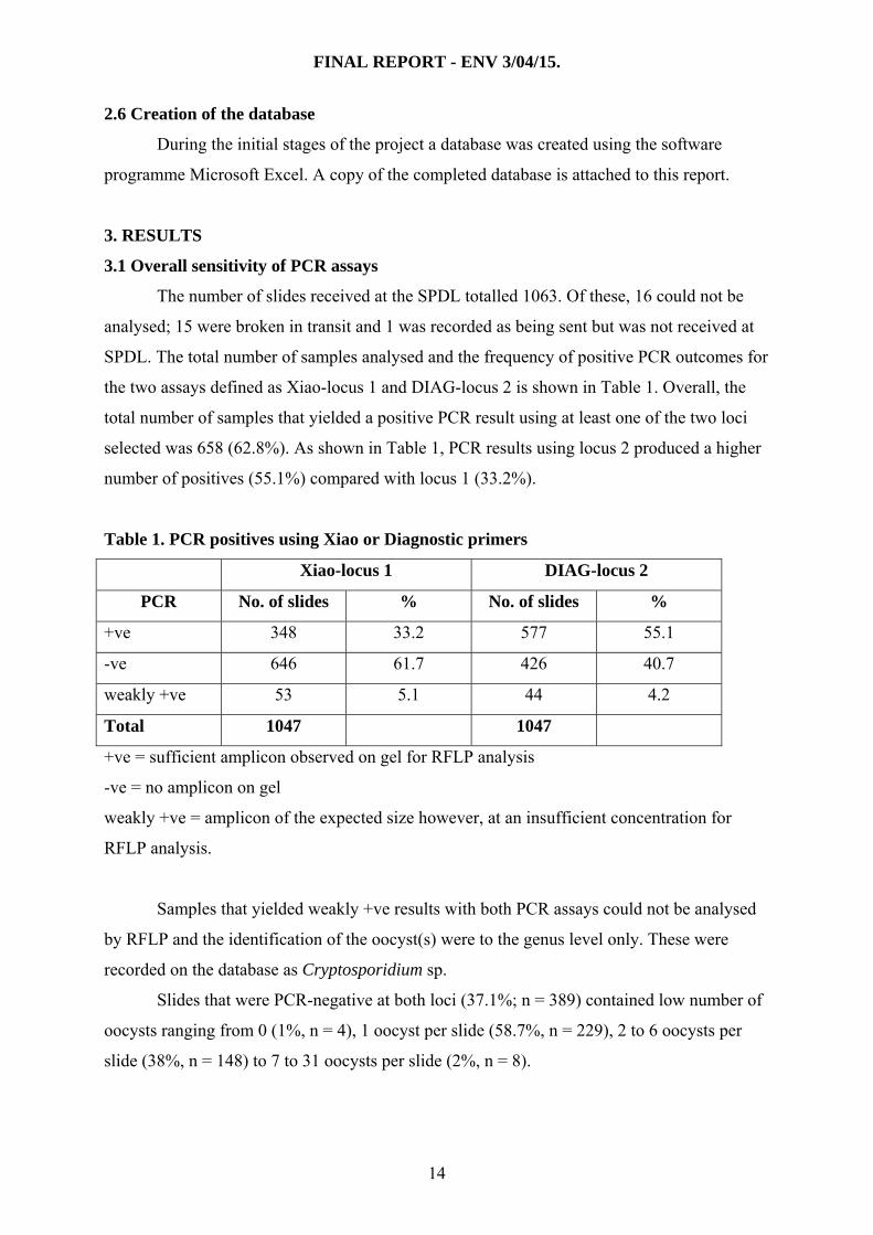
3. RESULTS
3.1 Overall sensitivity of PCR assays
The number of slides received at the SPDL totalled 1063. Of these, 16 could not be
analysed; 15 were broken in transit and 1 was recorded as being sent but was not received at
SPDL. The total number of samples analysed and the frequency of positive PCR outcomes for
the two assays defined as Xiao-locus 1 and DIAG-locus 2 is shown in Table 1. Overall, the
total number of samples that yielded a positive PCR result using at least one of the two loci
selected was 658 (62.8%). As shown in Table 1, PCR results using locus 2 produced a higher
number of positives (55.1%) compared with locus 1 (33.2%).
Table 1. PCR positives using Xiao or Diagnostic primers
Xiao-locus 1 DIAG-locus 2
PCR No. of slides % No. of slides %
+ve 348 33.2 577 55.1
-ve 646 61.7 426 40.7
weakly +ve 53 5.1 44 4.2
Total 1047 1047
+ve = sufficient amplicon observed on gel for RFLP analysis
-ve = no amplicon on gel
weakly +ve = amplicon of the expected size however, at an insufficient concentration for
RFLP analysis.
Samples that yielded weakly +ve results with both PCR assays could not be analysed
by RFLP and the identification of the oocyst(s) were to the genus level only. These were
recorded on the database as Cryptosporidium sp.
Slides that were PCR-negative at both loci (37.1%; n = 389) contained low number of
oocysts ranging from 0 (1%, n = 4), 1 oocyst per slide (58.7%, n = 229), 2 to 6 oocysts per
slide (38%, n = 148) to 7 to 31 oocysts per slide (2%, n = 8).
14

FINAL REPORT - ENV 3/04/15.
3.2 Analysis of raw and final waters and sensitivity of the PCR assays
Of the total 1047 water samples analysed 456 were from raw water sources (43.5 %)
and 591 were from final water sources (56.4 %). Table 2 shows the distribution of the PCR
results at both loci in raw and final water types.
Table 2. Number of positives in raw and final samples using Diagnostic or Xiao primers
PCR Raw Final Results DIAG Xiao DIAG Xiao
+ve 281 178 296 170 -ve 160 250 266 396
weakly +ve 15 28 29 25 Total no. of
slides
456
591 +ve = sufficient amplicon observed on gel for RFLP analysis
-ve = no amplicons on gel
weakly +ve = amplicon of the expected size however, at insufficient concentration for RFLP
analysis. When a sample yielded weakly +ve results with both PCR assays no RFLP could be
performed and the identification of the oocyst(s) was to the genus level only. This was
recorded on the database as Cryptosporidium sp.
The PCR results in Table 2 indicate that the Xiao –locus 1 assay successfully
amplified 39% and 28.8% of raw and final waters, respectively. The DIAG-locus 2 assay
amplified a larger number of samples, 61.6% and 51.1%, in raw and final waters,
respectively.
3.3 Distribution of oocyst numbers on slides
On receipt at SPDL, slides were re-examined and oocysts enumerated. The number of
nuclei per oocyst enumerated by DAPI staining was identified whenever possible and the
results noted in the database. The presence of oocysts larger than 4-5 μm was noted as “large
oocysts” whenever they were observed. Table 3 shows the distribution of slides according to
the number of oocysts they contained [(as enumerated at SW and including Cryptosporidium
like bodies (clb) which will include oocysts that were >4-5 μm].
From five slides that contained no observable oocysts, one resulted in positive PCR
amplification with both assays. Most slides (63.6%, n = 666) contained 1 - 2 oocysts. The
Xiao-locus 1 and DIAG-locus 2 PCRs amplified 27.8% and 46.8% of slides containing 1-2
oocysts, respectively.
15

FINAL REPORT - ENV 3/04/15.
Table 3. PCR positives using the Diagnostic or Xiao primers based upon the number of
oocysts.
DIAG PCR Xiao PCR
No. of
oocysts
on slides
(SW)
No. of
samples
Frequency
%
–ve +ve
(weakly
+ve)
-ve +ve
(weakly +ve)
none 5 0.5 4 1 4 1
1 463 44.2 249 194 (20) 327 118 (18)
2 203 19.4 76 118 (9) 126 67 (10)
3 111 10.6 42 68 (1) 64 36 (11)
4 68 6.5 26 39 (3) 37 29 (2)
5 36 3.4 6 29 (1) 17 17 (2)
6 37 3.5 13 23 (1) 23 12 (2)
7 15 1.4 1 13 (1) 5 9 (1)
8 12 1.1 3 7 (2) 6 4 (2)
9-18 41 3.9 5 32 (3) 25 14 (2)
20-49 26 2.5 1 23 (2) 9 16 (1)
51-100 15 1.4 0 15 3 12 (1)
101-397 13 1.2 0 13 1 11 (1)
860 1 0.09 - 1 - 1
4256 1 0.09 - 1 - 1
Total no.
of samples
1047
Slides containing >200 oocysts were final waters from the following sites: ARDGOUR_FNC
(219 oocysts, 30/08/05); WATERSTEIN_FNC (219 oocysts, week 28/08/05-01/09/05);
ARDGOUR_FNC (257 oocysts, week 27/10/05-02/11/05); ARDGOUR_FNC (295 oocysts,
13/10/05-20/10/05); ARDGOUR_FNC (358 oocysts, week 02/09/05-08/09/05 );
ARDGOUR_FNC (346 oocysts, 02/09/05-08/09/05), Skye (397 oocysts, from 23/08/05);
WATERSTEIN_FNC (4256 oocysts, week 15/09/05- 21/09/05); WATERSTEIN_FNC (860
oocysts, week 15/09/05- 21/09/05).
16

FINAL REPORT - ENV 3/04/15.
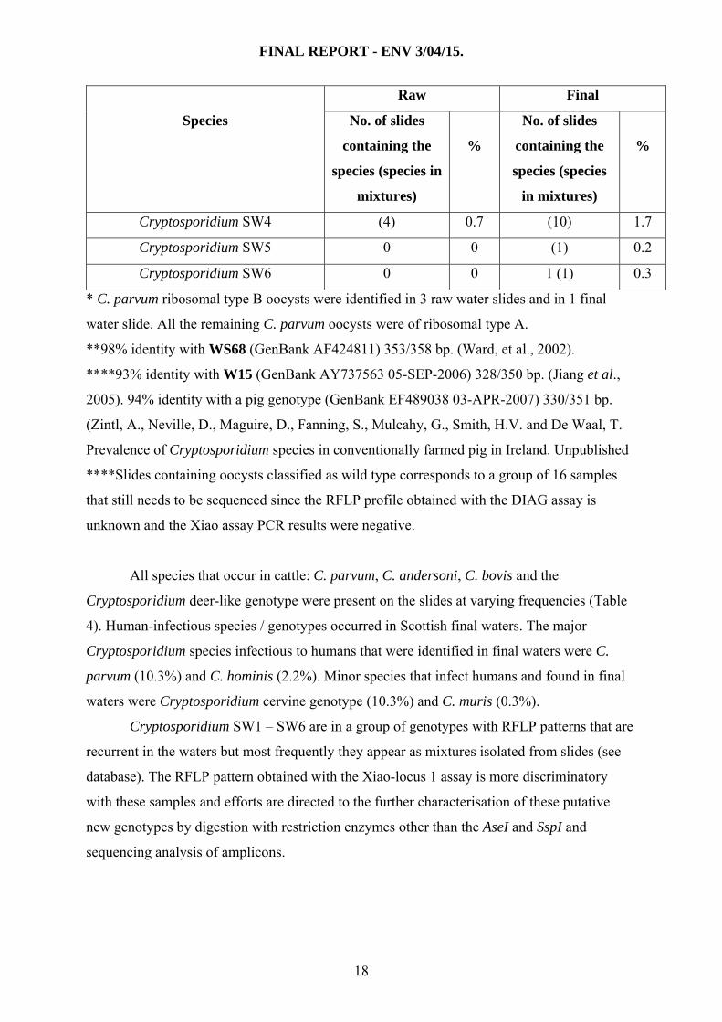
3.4 Identification and distribution of Cryptosporidium species / genotypes in raw and
final waters
Table 4 shows the frequency of species and genotypes present in raw and final waters.
The most frequently observed species were C. andersoni, C. parvum and the Cryptosporidium
cervine genotype in both raw and final waters. Overall, oocysts from 601 slides could be
genotyped (excludes 389 PCR negatives, 16 not available and 41 still to be sequenced).
Table 4. Frequency (%) of Cryptosporidium species / genotypes present in raw and final
water slides (n = 601) present either as single or mixed species.
Raw Final
Species No. of slides
containing the
species (species in
mixtures)
%
No. of slides
containing the
species (species
in mixtures)
%
C. andersoni 114 (21) 22.5 24 (12) 6.0
C. baileyi 8 (7) 2.5 12 (6) 3.0
C. parvum * 48 (21) 11.5 25 (37) 10.3
C. bovis 2 0.3 5 0.8
C. hominis 7 (5) 2.0 7 (6) 2.2
C. muris 2 (2) 0.7 2 0.3
Cryptosporidium muskrat genotype I 1 0.2 1 (2) 0.5
Cryptosporidium muskrat genotype II 2 0.3 9 (9) 3.0
Cryptosporidium cervine genotype 27 4.5 49 (13) 10.3
Cryptosporidium opossum genotype I 1 0.2 1 (2) 0.5
Cryptosporidium sp. 13 2.1 41 6.8
Cryptosporidium deer-like genotype 0 0 3 0.5
Similar to WS68** 1 0.2 0 0
Similar to W15 storm waters*** 0 0 1 0.2
Mixture of species 52 8.6 100 16.6
Wild type**** 9 1.5 11 (20) 5.1
Cryptosporidium SW1 2 (10) 2.0 10 (45) 9.1
Cryptosporidium SW2 1 (10) 1.8 6 (27) 5.5
Cryptosporidium SW3 1 0.2 (2) 0.3
17
FINAL REPORT - ENV 3/04/15.
Raw Final
Species No. of slides
containing the
species (species in
mixtures)
%
No. of slides
containing the
species (species
in mixtures)
%
Cryptosporidium SW4 (4) 0.7 (10) 1.7
Cryptosporidium SW5 0 0 (1) 0.2
Cryptosporidium SW6 0 0 1 (1) 0.3
* C. parvum ribosomal type B oocysts were identified in 3 raw water slides and in 1 final
water slide. All the remaining C. parvum oocysts were of ribosomal type A.
**98% identity with WS68 (GenBank AF424811) 353/358 bp. (Ward, et al., 2002).
****93% identity with W15 (GenBank AY737563 05-SEP-2006) 328/350 bp. (Jiang et al.,
2005). 94% identity with a pig genotype (GenBank EF489038 03-APR-2007) 330/351 bp.
(Zintl, A., Neville, D., Maguire, D., Fanning, S., Mulcahy, G., Smith, H.V. and De Waal, T.
Prevalence of Cryptosporidium species in conventionally farmed pig in Ireland. Unpublished
****Slides containing oocysts classified as wild type corresponds to a group of 16 samples
that still needs to be sequenced since the RFLP profile obtained with the DIAG assay is
unknown and the Xiao assay PCR results were negative.
All species that occur in cattle: C. parvum, C. andersoni, C. bovis and the
Cryptosporidium deer-like genotype were present on the slides at varying frequencies (Table
4). Human-infectious species / genotypes occurred in Scottish final waters. The major
Cryptosporidium species infectious to humans that were identified in final waters were C.
parvum (10.3%) and C. hominis (2.2%). Minor species that infect humans and found in final
waters were Cryptosporidium cervine genotype (10.3%) and C. muris (0.3%).
Cryptosporidium SW1 – SW6 are in a group of genotypes with RFLP patterns that are
recurrent in the waters but most frequently they appear as mixtures isolated from slides (see
database). The RFLP pattern obtained with the Xiao-locus 1 assay is more discriminatory
with these samples and efforts are directed to the further characterisation of these putative
new genotypes by digestion with restriction enzymes other than the AseI and SspI and
sequencing analysis of amplicons.
18
FINAL REPORT - ENV 3/04/15.
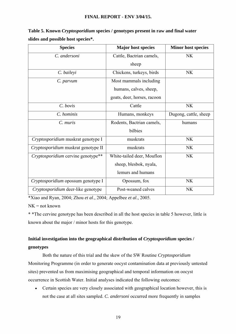
Table 5. Known Cryptosporidium species / genotypes present in raw and final water
slides and possible host species*.
Species Major host species Minor host species
C. andersoni Cattle, Bactrian camels,
sheep
NK
C. baileyi Chickens, turkeys, birds NK
C. parvum Most mammals including
humans, calves, sheep,
goats, deer, horses, racoon
C. bovis Cattle NK
C. hominis Humans, monkeys Dugong, cattle, sheep
C. muris Rodents, Bactrian camels,
bilbies
humans
Cryptosporidium muskrat genotype I muskrats NK
Cryptosporidium muskrat genotype II muskrats NK
Cryptosporidium cervine genotype** White-tailed deer, Mouflon
sheep, blesbok, nyala,
lemurs and humans
NK
Cryptosporidium opossum genotype I Opossum, fox NK
Cryptosporidium deer-like genotype Post-weaned calves NK
*Xiao and Ryan, 2004; Zhou et al., 2004; Appelbee et al., 2005.
NK = not known
* *The cervine genotype has been described in all the host species in table 5 however, little is
known about the major / minor hosts for this genotype.
Initial investigation into the geographical distribution of Cryptosporidium species /
genotypes
Both the nature of this trial and the skew of the SW Routine Cryptosporidium
Monitoring Programme (in order to generate oocyst contamination data at previously untested
sites) prevented us from maximising geographical and temporal information on oocyst
occurrence in Scottish Water. Initial analyses indicated the following outcomes:
• Certain species are very closely associated with geographical location however, this is
not the case at all sites sampled. C. andersoni occurred more frequently in samples
19

FINAL REPORT - ENV 3/04/15.
from the north east and south central Scotland. There were more mixtures of species /
genotypes in samples from the north of Scotland.
C. parvum occurred more frequently between July and September, C. baileyi was
mostly present during July to December and C. andersoni peaked in the winter months
before declining in number.
4. CONCLUSIONS
1. With few notable exceptions, Cryptosporidium oocysts occur in low abundance in both
raw and final water samples in the samples analysed.
2. PCR inhibitors prevented PCR amplification in some oocyst positive samples. One oocyst
negative sample produced an amplicon using both assays, probably because of the
presence of naked DNA in the sample.
3. We compared assay sensitivity in raw and final water concentrates at two 18S rRNA loci.
the Diag – locus 2 assay was more sensitive than the Xiao – locus 1 assay in both raw and
final water concentrates (39% and 28.8% of raw and final waters, and 61.6% and 51.1%
of raw and final waters, respectively).
4. The predominant species identified in raw and final waters were C. andersoni, C. parvum
and the Cryptosporidium cervine genotype. C. parvum and the Cryptosporidium cervine
genotype are infectious to humans.
5. We identified 6 putative new Cryptosporidium species / genotypes (SW1 - SW6) which
we are characterising further.
6. Certain geographical locations were very closely associated with certain species.
7. There were some relatedness between season and species.
8. This is the first study conduced to determine the species / genotype of Cryptosporidium in
Scottish waters and the outcomes highlight the importance of an effective
multidisciplinary approach to the study.
5. REFERENCES
Anonymous. (1998). Cryptosporidium in water supplies. Third Report of the Group of
Experts; Chairman, Professor Ian Bouchier. Department of the Environment, Transport and
the Regions, Department of Health. London, ISBN 1 85112 131 5. HMSO. 171pp.
20
FINAL REPORT - ENV 3/04/15.
Anonymous. (2005). DWI Information letter 2005. The Water Supply (Water Quality)
(Amendment) Regulations 2000, SI No. 3184 England and 2001, SI No. 3911 (W.323) Wales:
Cryptosporidium in Water Supplies: Laboratory and Analytical Procedures. Part 2, June 2005.
Protocol containing Standard Operating Protocols (SOPs) for the monitoring of
Cryptosporidium oocysts in water supplies. UK Drinking Water Inspectorate. [Online]
www.dwi.detr.gov.uk
Cacciò, S.M., Thompson, R.C.A., McLauchlin, J., Smith, H.V., 2005. Unravelling
Cryptosporidium and Giardia epidemiology. Trends in Parasitology, 21, 430-437.
Chappell, C.L., Okhuysen, P.C., Sterling, C.R. and DuPont, H.L. (1996). Cryptosporidium
parvum: intensity of infection and oocyst excretion patterns in healthy volunteers. Journal of
Infectious Diseases 173, 232-236.
Chappell, C.L., Okhuysen, P.C., Sterling, C.R., Wang, C., Jakubowski, W. and DuPont, H.L.
(1999). Infectivity of Cryptosporidium parvum in healthy adults with pre-existing anti-
C.parvum serum immunoglobulin G. American Journal of Tropical Medicine and Hygiene
60, 157-164.
Chappell, C.L., Okhuysen, P.C., Langer-Curry, R., Widmer, G., Akiyoshi, D.E. Tanriverdi, S.
and Tzipori, S. (2006). Cryptosporidium hominis: experimental challenge of healthy adults.
American Journal of Tropical Medicine and Hygiene, 75, 851-857.
Clarke, S.C., Diggle, M.A., and Edwards, G.F.S. (2001). Semiautomation of multilocus
sequence typing for the characterisation of clinical isolates of Neisseria meningitides. Journal
of Clinical Microbiology, 39, 3066-3071.
DuPont, H.L., Chappell, C.L, Sterling, C.R., Okhuysen, P.C., Rose, J.B. and Jakubowski, W.
(1995). ‘The infectivity of Cryptosporidium parvum in health volunteers’. New England
Journal of Medicine 332, 855-859.
Fayer, R., Morgan, U. and Upton, S.J. (2000). Epidemiology of Cryptosporidium:
transmission, detection and identification. International Journal of Parasitology 30, 1305-
1322.
Feng, Y., Ortega, Y., He, G., Das, P., Xu, M., Zhang, X., Fayer, R., Gatei, W., Cama, V., and
Xiao, L. (2006). Wide geographic distribution of Cryptosporidium bovis and the deer-like
genotypes in bovines. Veterinary Parasitology, 144, 1-9.
Girdwood, R.W.A and Smith, H.V. (1999). Giardia. In: Encyclopaedia of Food Microbiology
(eds. R. Robinson, C. Batt & P. Patel) Academic Press, London and New York. pp. 946-954.
21
FINAL REPORT - ENV 3/04/15.
Grimason, A.M., Smith, H.V., Parker, J.F.W., Bukhari, Z., Campbell, A.T. and Robertson,
L.J. (1994). Application of DAPI and immunofluorescence for enhanced identification of
Cryptosporidium spp. oocysts in water samples. Water Research. 28, 733-736.
Jiang, J., Alderisio, K.A., and Xiao, L. (2005). Distribution of Cryptosporidium genotypes in
storm event water samples from three watersheds in New York. Applied and Environmental
Microbiology 71, 4446-4454.
Johnson, D.W., Pieniazek, N.J., Griffin, D.W., Misener, L. and Rose, J.B. (1995).
Development of a PCR protocol for sensitive detection of Cryptosporidium in water samples.
Applied and Environmental Microbiology 61, 3849-3855.
Kourenti, K., Karanis, P. and Smith, H.V. (2006). Waterborne transmission of protozoan
parasites: a worldwide review of outbreaks and lessons learnt. Journal of Water and Health.
5, 1-38.
Lisle, J. T. and Rose, J.B. (1995). Cryptosporidium contamination of water in the USA and UK:
a mini review. Journal of Water SRT Aqua 44, 103-117.
Nichols, R.A.B., Campbell, B.M. and Smith, H.V. (2003). Identification of Cryptosporidium
spp. oocysts in UK noncarbonated natural mineral waters and drinking waters using a
modified nested PCR-RFLP assay. Applied and Environmental Microbiology 69, 4183-4189.
Nichols R.A.B. and Smith, H.V. (2004). Optimisation of DNA extraction and molecular
detection of Cryptosporidium parvum oocysts in natural mineral water sources. Journal of
Food Protection. 67, 524-532.
Okhuysen, P.C., Chappell, C.L., Crab, J.H., Sterling S.R., Du Pont, H.L., 1999. Virulence of
three distinct Cryptosporidium parvum isolates for healthy adults. Journal of Infectious
Diseases 180, 1275-1281.
Robertson, L.J., Smith, H.V. and Ongerth, J.E. (1994). Cryptosporidium and
cryptosporidiosis. Part 3: Development of water treatment technologies to remove and
inactivate oocysts. Microbiology Europe 2, 18-26.
Rose, J.B., Lisle, J.T. and LeChevallier, M. (1997). Waterborne cryptosporidiosis, Incidence,
outbreaks and treatment strategies. In Cryptosporidium and cryptosporidiosis. (ed. Fayer, R.),
Chapter 4. pp. 95-111. CRC Press, Boca Raton, Florida.
Slifco, T.R. Smith, H.V. and Rose, J.B. (2000). Emerging parasite zoonoses associated with
food and water. International Journal for Parasitology 30, 1379-1393.
Smith, H.V., Parker, J.F.W., Girdwood, R.W.A., Gilmour, R. A., Smith, P. G., Morris, G. P.,
Grimason, A.M. and Jackson, M.J. (1989). A modified method for the detection of
22
FINAL REPORT - ENV 3/04/15.
Cryptosporidium spp. oocysts in water-related samples. Communicable Diseases Scotland
89/15, 7-13.
Smith, H.V. and Rose, J.B. (1990). Waterborne cryptosporidiosis. Parasitology Today 6, 8-
12.
Smith, H.V., Robertson, L.J. and Ongerth. J.E. (1995). Cryptosporidiosis and giardiasis: the
impact of waterborne transmission. Journal of Water SRT - Aqua 44(6): 258-274.
Smith, H.V. and Rose, J.B. (1998).Waterborne cryptosporidiosis: current status. Parasitology
Today 14, 14-22.
Smith, H.V. and Lloyd, A. (1997). Protozoan parasites in drinking water: a UK perspective.
New World Water 1, 109-116.
Smith, H.V., Cacciò, S.M., Tait, A., McLauchlin, J. and Thompson, R.C.A. (2006). Tools for
investigating the abiotic transmission of Cryptosporidium and Giardia infections in humans.
Trends in Parasitology 22, 160-166.
Smith, H.V. Cacciò, S.M., Cook, N., Nichols, R.A.B. and Tait, A. (2007). Cryptosporidium
and Giardia as foodborne zoonoses. Veterinary Parasitology In press.
Ward, P.J., Deplazes, P., Regli, W., Rinder, H. and Mathis, A. (2002). Detection of eight
Cryptosporidium genotypes in surface and waste waters in Europe. Parasitology, 124, 359-
368.
Xiao L., Morgan, U.M., Limor, J., Escalante L., Arrowwood, M., Shulaw, W., Thompson,
R.C.A., Fayer R and A. A. Lal. (1999). Genetic diversity within Cryptosporidium parvum and
related Cryptosporidium species. Applied and Environmental Microbiology 65, 3386-3391.
Xiao, L., Sing, A., Limor, J., Grazyck, T., Gradus, S. and Lal, A.A. (2001). Molecular
characterisation of Cryptosporidium oocysts in samples of raw surface water and wastewater.
Applied and Environmental Microbiology 67, 1097-1101.
23