flávio humberto torres dias feio de azevedo the role of...
TRANSCRIPT

Escola de Ciências
Flávio Humberto Torres Dias Feio de Azevedo
The role of MAPK signalling pathways in acetic acid-induced cell death of Saccharomyces cerevisiae
Tese de Mestrado Mestrado em Genética Molecular
Trabalho efectuado sob a orientação de:
Doutora Susana Alexandra Rodrigues Chaves
Professor Doutor Björn Fredrik Johansson
Novembro, 2011
Universidade do Minho

DECLARAÇÃO
Nome: Flávio Humberto Torres Dias Feio de Azevedo
Endereço electrónico: [email protected]
Número do Bilhete de Identidade: 11234559
Título da tese: The role of MAPK signalling pathways in acetic acid-induced cell death of
Saccharomyces cerevisiae
Orientador(es): Doutora Susana Alexandra Rodrigues Chaves
Professor Doutor Björn Fredrik Johansson
Ano de conclusão: 2011
Designação do Mestrado: Mestrado em Genética Molecular
É AUTORIZADA A REPRODUÇÃO PARCIAL DESTA TESE/TRABALHO APENAS PARA EFEITOS DE
INVESTIGAÇÃO, MEDIANTE DECLARAÇÃO ESCRITA DO INTERESSADO, QUE A TAL SE
COMPROMETE.
Universidade do Minho, ___/___/______
Assinatura: ________________________________________________
iii
Agradecimentos
Agradeço à minha família pelo incondicional Amor e apoio ao longo dos anos.
Agradeço aos meus orientadores, Drª. Susana Alexandra Rodrigues Chaves e Prof. Drº Björn Fredrik Johansson, por todo o seu empenho e conhecimentos investidos na concretização deste trabalho.
Agradeço à Prof. Drª Manuela Côrte-Real e à Prof. Drª Maria João Sousa pelo acolhimento no seio do seu grupo de investigação.
Agradeço aos meus colegas de laboratório (Andreia Afonso, Andreia Pacheco, Dário Trindade, Dulce Cunha, Gabriela Ribeiro, Helena Pereira, João Lopes, Marlene Sousa, Rui Silva, Sara Alves, Vitória Maciel, António Rego, Jorge Rodrigues, Rita Cunha, Ana Marta e Marco Cruz) por toda a ajuda e companheirismo, que fizeram com que as longas horas de trabalho se tornassem menos penosas.
Agradeço à Joana Tulha a ajuda dada na formatação da tese.
Agradeço a todo o pessoal técnico e auxiliar do departamento de Biologia pelo contributo na realização deste trabalho.
Este trabalho foi desenvolvido e financiado pelo projecto FCT, (PTDC/BIA-BCM/69448/2006.
...feci quod potui, faciant meliora potentes
...firmum in vita nihil

iv
Summary
Mitogenic Activated Protein Kinase (MAPK) cascades are important signalling
pathways that allow yeast cells to swiftly adapt to changing environmental conditions.
Previous studies suggested that the High Osmolarity Glycerol (HOG) MAPK pathway
and ceramide production are involved in acetic-acid induced apoptosis in yeast.
Evidence that changes in the levels of endogenous ceramides can affect yeast cell fate
has also been put forth. However, knowledge on the molecular basis of acetic acid
induced cell death and ceramide-induced cell changes, as well as the signalling
pathways involved in these processes, is still lacking.
In this work, we tried to elucidate the role of MAPK signalling pathways in the cell
response to ceramide and in acetic acid-induced cell death. Cells treated with the soluble
ceramide analog N-acetyl-D-sphingosine (C2-ceramide) maintained plasma membrane
integrity and did not produce reactive oxygen species detectable by DHE staining. A
subpopulation of cells died when exposed to C2-ceramide but the surviving part
eventually grew, so no reduction in plate counts was observed. Preliminary results
indicated that deletion mutants in MAPK components showed smaller sensitive sub-
populations than the reference strain and were therefore deemed to be more resistant to
C2-ceramide, namely wsc3∆. The MAPK mutants wsc2∆, wsc3∆, ste20∆, slt2∆ and
mkk1∆/mkk2∆ were significantly more resistant to acetic acid than the reference strain.
These mutants also displayed a lower percentage of cells with increased accumulation
of ROS and a higher percentage of cells with preserved plasma membrane integrity than
the wild type strain. The WSC2 and WSC3 genes encode sensors in the cell wall
integrity MAPK pathway, suggesting a role for this signalling pathway in both ceramide
and acetic acid induced cell death. The link between acetic acid-induced cell death and
cell wall integrity is not known, but lipid peroxidation by acetic acid may be a plausible
candidate.
v
Sumário
As cascatas “Mitogenic Activated Protein Kinase” (MAPK) são vias de sinalização
importantes que permitem que as células de levedura se adaptem rapidamente às
mudanças das condições ambientais. Estudos anteriores sugeriram que a via “High
Osmolarity Glycerol” (HOG) MAPK e a produção de ceramida estão envolvidas na
morte celular apoptótica de leveduras induzida por ácido acético. Há também evidências
que alterações nos níveis de ceramidas endógenas podem afectar o destino de células de
levedura. No entanto, ainda não há um conhecimento exacto sobre a base molecular da
morte celular induzida por ácido acético, sobre as mudanças induzidas ao nível celular
pela ceramida, bem como as vias de sinalização envolvidas nestes processos. Neste
trabalho, procurou-se elucidar o papel das vias de sinalização MAPK na resposta celular
à ceramida e na morte celular induzida por ácido acético. Células tratadas com o
análogo solúvel da ceramida N-acetil-D-esfingosina (C2-ceramida) mantiveram a
integridade da membrana plasmática e não produziram espécies reactivas de oxigénio
detectadas através da incubação com DHE. Numa cultura exposta a C2-ceramida, uma
subpopulação das células morreu, enquanto outra parte da cultura cresceu, não se
observando assim na placa uma redução de Cfus em relação ao tempo zero. Alguns
mutantes deletados em componentes das vias MAPK mostraram subpopulações menos
sensíveis do que a estirpe selvagem e assim consideraram-se como sendo estirpes mais
resistentes à C2-ceramida, nomeadamente o mutante wsc3∆. Os mutantes wsc2∆,
wsc3∆, ste20∆, slt2∆ e mkk1∆/mkk2∆ foram significativamente mais resistentes ao
ácido acético do que a estirpe selvagem. Esses mutantes mostraram também uma menor
percentagem de células exibindo uma acumulação de ROS e uma maior percentual de
células com integridade da membrana plasmática preservada do que a estirpe selvagem.
Os genes WSC2 e WSC3 codificam sensores membranares na via MAPK da integridade
da parede, sugerindo um papel para esta via de sinalização tanto na morte celular
induzida por ácido acético como nas alterações induzidas por ceramida. Não é
conhecida a ligação entre morte celular induzida por ácido acético, mas a peroxidação
lipídica pode ser uma hipótese provável.
vi
Table of Contents
Agradecimentos ………………………………………………………………………………………..
Summary………………………………………………………………………………………………..
Sumário ………………………………………………………………………………………………..
Table of contents ……………………………………………………………………………….……...
Abbreviations……………………………………………………………………………………….......
1. Introduction ………………………………………………………………………………….…...
1 - 1 Cell death …………….……………………………………………………………….……
1 - 1.1 Yeast apoptosis…………… …………………………..………………………………
1 - 2 Ceramide signalling in Saccharomyces
cerevisiae…….……………………………………..
1 - 3 MAP kinase pathways in the yeast Saccharomyces cerevisiae………………………….….
1 - 3.1 The pheromone/mating response pathway.……………………………………………
1 - 3.2 Invasive growth/pseudohyphal development pathway………………………….…….
1 - 3.3 Cell wall integrity pathway ….……………………………….………………………
1 - 3.4 High Osmolarity Glycerol (HOG) pathway…………………………………………...
2. Objectives and research plan….………………………………………………………………….
3. Materials and Methods………………………………………………………………………........
3 - 1 Strains ………………………...…………………………………………………………….
3 - 2 Growth conditions and treatments…………………………...……………………………...
3 - 3 Flow cytometry ……………………………………...…………………………..………….
4. Results ……………………………………………………..………….……………………….
4 - 1 Characterization of C2-ceramide-induced cell death……………………………………..
4 - 2 Construction of deletion mutants in components of the MAPK pathways.……...……….
4 - 3 Deletion mutants in components of the MAPK pathways are resistant to C2-ceramide-
induced cell death………………………………...………………………...……………….
4 - 4 C2-ceramide does not lead to an increase in Reactive Oxygen Species or loss of
membrane integrity………………………………………………………………………….
4 - 5 Components of the MAPK pathways modulate acetic acid-induced cell death…………..
5. Discussion …………..……………………………………………………………………………
6. References ………………………………………………………………………………………
7. Attachments ………..……………………………………………………………………………
iii
iv
v
vi
vii
2
2
3
5
7
8
9
10
12
16
20
20
21
22
24
24
26
26
27
28
33
39
47
vii
Abbreviations
MAPK Mitogenic Activated Protein Kinase
MAPKK MAP kinase kinase
MAPKKK MAP kinase kinase kinase
MAPKKKK MAP kinase kinase kinase kinase
HOG high osmolarity glycerol
CWI Cell wall integrity
GPCR G protein-coupled receptor
PAK p21-activated kinase
MCA1 metacaspase 1
AIF apoptosis-inducing factor
MOMP mitochondrial outer membrane permeabilization
cyt c cytochrome c
AAC ADP/ATP carrier proteins
LCB long-chain base
C2-ceramide N-acetyl-D-sphyngosine (C2-ceramide
H2O2 hydrogen peroxide
ROS reactive oxygen species
PI propidium iodide
DHE dihydroethidium
c.f.u. colony forming units
∆Ψm mitochondrial membrane potential

Chapter 1 INTRODUCTION
This chapter contains sections from the publications:
C2-phytoceramide sensitizes S. cerevisiae cells to osmotic stress. Pacheco A, Azevedo F, Chaves SR,
Côrte-Real M, Sousa MJ (in preparation)
Vacuole-mitochondrial cross-talk during apoptosis in yeast: a model for understanding lysosome-
mitochondria-mediated apoptosis in mammals. Sousa MJ, Azevedo F, Pedras A, Marques C, Coutinho
OP, Preto A, Gerós H, Chaves SR, Côrte-Real M. Biochem Soc Trans. 2011 Oct; 39(5):1533-7.

Introduction
2
1. Introduction
1.1 Cell death
Cell death plays an important role in the maintenance of tissue homeostasis, and can be
defined as an irreversible loss of plasma membrane integrity [1]. Two forms of cell
death have been described more frequently: apoptosis and necrosis.
Necrosis is a type of cell death defined by morphological features, such as increasing
the volume of the cytoplasm, plasma membrane rupture, swelling of cytoplasmic
organelles (mitochondria, endoplasmic reticulum, etc..) and a moderate chromatin
condensation [1]. It is traditionally defined as a form of cell death that is uncontrolled,
chaotic, and accidental, due to exposure to extreme environmental conditions [2].
Recently, this assumption has been challenged and in some instances necrosis is now
seen as a regulated, programmed process [2-4].
Apoptosis is a mechanism that deals with the homeostatic removal of cells that have
been mutated, infected or are simply superfluous in multicellular organisms. Several
diseases can be linked to poor regulation of apoptosis, including AIDS, cancer, and
neurodegenerative disorders. Apoptosis is a mechanism of programmed cell death
accompanied by morphological characteristics such as cell rounding, reduction in
cellular volume, condensation and fragmentation of chromatin, and the formation of cell
fragments (apoptotic bodies) [1]. The process of apoptosis ensures that cells are quickly
removed without rupture of the plasma membrane, thus preventing inflammation of the
environment. Since deregulation of apoptosis is associated with severe human
pathologies, the identification of components of the different apoptotic pathways and
the understanding of mechanisms underlying their regulation is critical for the
development of new strategies for prevention and treatment against those diseases.
Different biological models, including Caenorhabditis elegans and Drosophila
melanogaster, were use as core models for cell death research. Yeast was not used in
early apoptosis studies, since it was considered that there was no advantage for a
unicellular organism to commit suicide. After the complete sequencing of the
Saccharomyces cerevisiae genome, an exhaustive search for homologies in databases
failed to uncover potential regulators of apoptosis, reinforcing the idea that this
mechanism was not present in this yeast. However, although yeast is a unicellular
organism, apoptosis could have an evolutionary advantage at the colony level. In the

Introduction
3
wild, yeasts are multicellular colonies and not individuals, in which apoptosis may be a
mechanism that saves and releases nutrients to the healthier cells. Thus, apoptosis acts
as a mechanism of self-preservation of the colony as a whole [5]. Indeed an apoptotic
pathway in yeast was later found in yeast as described in more detail in the next section.
This knowledge, associated with the several attributes of yeast such as its rapid growth,
its simple and inexpensive culturing, ease and safety in handling, and especially its well
defined genetic system and easy genetic manipulation make this unicellular eukaryote
an attractive model system for cell death research.
1.1.1 Yeast apoptosis
The first indication that there is a basic apoptotic mechanism in yeast came from the
finding that a mutation in the S. cerevisiae gene CDC48 results in cell death with
characteristics of apoptosis such as DNA fragmentation, chromatin condensation, and
exposure of phosphatidylserine on the outer plasma membrane [6]. Later, it was
described that an apoptotic cell death can also be induced by H2O2 [7, 8], acetic acid [8,
9], hyperosmotic shock [8], exposure to UV radiation [10], viral "killer" toxins [11],
pheromones [12], chronological [13, 14] and replicative aging [15], and in colonies on
solid media [16]. In virtually all these stimuli, the type of cell death observed is
dependent on the intensity of the applied stress, i.e., low or moderate doses lead to death
by apoptosis, while high doses cause a cell death such as necrosis [7-11]. Afterwards,
several genes involved in yeast apoptotic cell death were identified. The yeast protein
Yor197w, with structural homology with mammalian caspases, was called yeast
metacaspase 1 (MCA1) [17]. Key elements of apoptosis in mammalian cells, such as
HtrA2/Omi, EndoG, and the apoptosis-inducing factor (AIF), are also present in the
yeast genome [18]. The release of cytochrome c (cyt c) from mitochondria, another
landmark of human apoptosis, also occurs in S. cerevisiae [19]. Moreover, several
studies have determined that the expression of pro-apoptotic proteins in yeast can cause
cell death, such as expression of human Bax [20, 21]. Co-expression of the anti-
apoptotic proteins Bcl-2 or Bcl-xL in yeast cells can rescue the lethality caused by
expression of Bax, indicating that at least some features of the regulation of mammalian
apoptotic proteins are conserved in yeast [20].

Introduction
4
Fig. 1. Proteins and pathways in yeast apoptosis (adapted from [22])
In yeast, acetic acid-induced apoptosis is among the best-characterized apoptotic
pathways, and, like in mammalian cells, mitochondria play a key role in this process.
Indeed, different alterations in mitochondrial structure and function occurring during
acetic acid-induced apoptosis have been identified. These changes include reduction in
cristae number and mitochondrial swelling [23], a transient mitochondrial hyper-
followed by depolarization, production of reactive oxygen species, decrease in
cytochrome oxidase activity and mitochondrial outer membrane permeabilization
(MOMP), with concomitant release of cyt c and yeast Aifp [19, 24]. Yeast orthologs of
some of the mammalian permeability transition pore components were found to be
involved in MOMP and cyt c release. While deletion of POR1 (yeast voltage dependent
anion channel) enhances apoptosis triggered by acetic acid, CPR3 (mitochondrial
cyclophilin) deletion has no effect. In contrast, absence of ADP/ATP carrier proteins
(AAC), yeast orthologs of the adenine nucleotide transporter, protects cells exposed to
acetic acid and impairs MOMP and cyt c release [25]. Mitochondrial proteins involved
in fission/fusion, namely, Fis1p, Dnm1p, Mdv1p [26] have also been implicated in the
execution of the yeast apoptotic program induced by acetic acid. Despite the large
number of proteins shown to be involved in acetic acid-induced cell death, the upstream
events and pathways that signal acetic acid-induced apoptosis, as well apoptosis
induced by other stimuli, remain to be identified.

Introduction
5
1.2 Ceramide signalling in Saccharomyces cerevisiae
Sphingolipids are the main lipid component in eukaryotes plasma membranes. The
components of all sphingolipids are a long-chain base (LCB), a fatty acid, and a polar
head group. The shingolipid ceramide acts as an important second messanger in
mammalian cells. Indeed, rapid and transient changes in sphingolipid metabolism are
associated with a wide range of cellular processes such as cell growth, differentiation,
apoptosis, stress responses, and senescence. Ceramide and sphingosine levels increase
in response to stress and in apoptosis induced by several stimuli such as FAS activation
[27] and anticancer drugs [28]. Ceramides regulate mammalian apoptosis by
transcriptional-dependent and -independent mechanisms. The transcriptional-
independent mechanism of ceramide-mediated apoptosis involves generation of reactive
oxygen species and release of mitochondrial intermembrane space proteins, with a pro-
apoptotic role and is inhibited by anti-apoptotic members of the Bcl-2 family [29]. The
former involves transcriptional activation of different signalling cascades including the
c-Jun-N-terminal Kinase (JNK) pathway, also called the Stress Activated Protein
Kinase (SAPK) pathway, which requires Ras and Rac1 [30]. Other intracellular targets
of ceramide include kinase suppressor of Ras, Raf1, PKC and the ceramide-activated
protein phosphatases PP1 and PP2A. Ceramide can also promote the clustering of death
receptors and interferes with the relay of PI3K signals by activating protein
phosphatases, such as ceramide-activated protein phosphatase (CAPP) [31].
As mentioned above, the yeast S. cerevisiae has been extensively used in the elucidation
of numerous cellular and molecular processes shared across species, such as cell cycle
control and apoptosis [32]. Several studies indicate that the ceramide pathway is a
ubiquitous and conserved signalling system from yeast to human [29]. Similarly to
mammalian cells, yeast ceramide levels increase in response to diverse stress treatments
[33] and different studies showed that perturbations in sphingolipid metabolism
determine cell fate. Indeed, expression of mammalian sphingomyelin synthase (SMS1)
suppresses Bax-mediated yeast cell death and confers increased resistance to different
apoptosis inducers [34]. These results suggest that SMS1, which uses ceramide to
synthesize sphingomyelin, protects cells against death by counteracting stress-induced
accumulation of the pro-apoptotic ceramide. Very recently, it has also been reported that
exogenous N-acetyl-D-sphyngosine (C2-ceramide) can trigger a mitochondria-mediated

Introduction
6
cell death process [35]. On the other hand, a different study showed that overexpression
of YDC1, coding for a dihydroceramidase which hydrolyzes ceramide, results in
reduced chronological lifespan and increased apoptotic cell death associated with
fragmentation and dysfunction of mitochondria and vacuoles. These effects were
reverted by exogenous addition of ceramide to YDC1-overexpressing cultures [36],
suggesting ceramide can also have a protective effect in the cell. Yet another study
showed that a yeast mutant deficient in Isc1p, a member of the family of the neutral
sphingomyelinases, displays increased apoptotic cell death in response to hydrogen
peroxide and during chronological aging [37]. More recently, a lipidomic approach
revealed that these phenotypes were associated with increased levels of dihydro-C26-
ceramide and phyto-C26-ceramide, again suggesting that ceramide signalling is indeed
involved in yeast programmed cell death [38]. Exposure to C2-ceramide, a synthetic
cell-permeable ceramide analog, specifically inhibits proliferation of Saccharomyces
cerevisiae cultures in a dose-dependent manner and increases a serine/threonine
phosphatase activity in cell crude extracts with biochemical characteristics similar to
those of mammalian ceramide-activated protein phosphatase (CAPP) [39]. Yeast CAPP,
composed of one catalytic subunit, Sit4p, and two regulatory subunits, Tpd3 and Cdc55,
was subsequently found to mediate the proliferation arrest induced by C2-ceramide by
blocking cells at the G1 phase of the cell cycle [40]. More recently, it was found that
deletion of SIT4 reverts the premature ageing and hydrogen peroxide sensitivity of
isc1∆ cells and eliminates the respiratory defects and catalase A deficiency exhibited by
this mutant [38]. Therefore, activation of Sit4p is involved in the mitochondrial
dysfunction induced by increased levels of dihydro-C26-ceramide and phyto-C26-
ceramide and leads to the shortened chronological lifespan and oxidative stress
sensitivity of the isc1∆ mutant. These results provided the first glimpse of how changes
in the levels of endogenous ceramides can affect cell fate in yeast. However, knowledge
on the molecular basis of ceramide-induced cell changes, as well as of the role of
signalling pathways in this process, is still lacking.
Introduction
7
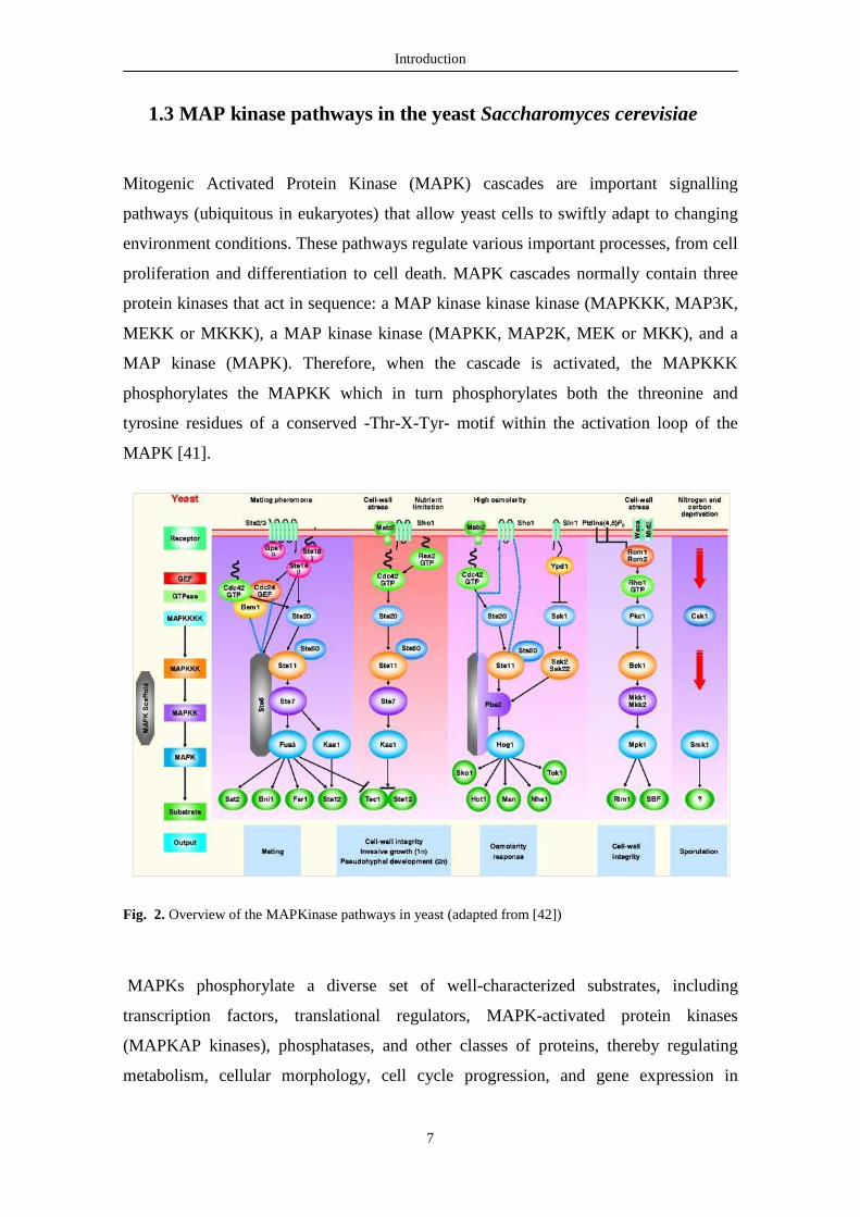
1.3 MAP kinase pathways in the yeast Saccharomyces cerevisiae
Mitogenic Activated Protein Kinase (MAPK) cascades are important signalling
pathways (ubiquitous in eukaryotes) that allow yeast cells to swiftly adapt to changing
environment conditions. These pathways regulate various important processes, from cell
proliferation and differentiation to cell death. MAPK cascades normally contain three
protein kinases that act in sequence: a MAP kinase kinase kinase (MAPKKK, MAP3K,
MEKK or MKKK), a MAP kinase kinase (MAPKK, MAP2K, MEK or MKK), and a
MAP kinase (MAPK). Therefore, when the cascade is activated, the MAPKKK
phosphorylates the MAPKK which in turn phosphorylates both the threonine and
tyrosine residues of a conserved -Thr-X-Tyr- motif within the activation loop of the
MAPK [41].
Fig. 2. Overview of the MAPKinase pathways in yeast (adapted from [42])
MAPKs phosphorylate a diverse set of well-characterized substrates, including
transcription factors, translational regulators, MAPK-activated protein kinases
(MAPKAP kinases), phosphatases, and other classes of proteins, thereby regulating
metabolism, cellular morphology, cell cycle progression, and gene expression in
Introduction
8
response to a variety of extracellular stresses and molecular signals [43]. The specificity
of the MAPK pathways is regulated at several levels, including kinase-kinase and
kinase-substrate interactions, colocalization of kinases by scaffold proteins, and
inhibition of cross-talk/output by the MAPKs themselves [44]. S. cerevisiae contains
five MAPKs, Fus3p, Kss1p, Hog1p, Slt2/Mpk1p and Smk1p, on five functionally
distinct cascades, associated with the pheromone-mating response, invasive
growth/pseudohyphal development, high osmolarity, cell wall integrity, and sporulation,
respectively [45]. The five MAP kinases are controlled by four MAPKKs, Ste7p
(regulating Fus3p and Kss1p), Pbs2p and the redundant pair Mkk1p/Mkk2p, and by
four MAPKKKs, Ste11p, the redundant pair Skk2p/Skk22p and Bck1p. The specificity
of signal transduction is guaranteed by scaffold proteins [46], Ste5p for the pheromone-
mating response pathway, and Pbs2p for the HOG pathway.
1.3.1 The pheromone/mating response pathway
S. cerevisiae can exist as either diploid or haploid cells. Haploids can be of two mating
types, MATa or MATα. Cells of opposing mating types can mate, by undergoing cellular
and nuclear fusion, originating a diploid cell MATa/MATα. This process is initiated by
the release of mating pheromones a-factor and α-factor, from respectively MATa and
MATα cells, which act on cells of the opposing mating type. The a-factor interacts with
MATα cells by binding to the G protein-coupled receptor (GPCR) Ste3p; in turn the α-
factor interacts with MATa cells by binding to GPCR Ste2/3p [47, 48]. Binding of the
mating pheromones activates the plasma membrane GPCRs, Ste2p and Ste3p, which in
turn induces the dissociation of the heterotrimeric G protein Gpa1p-Ste14p-Ste18p, in
Gα (Gpa1p) and the Gβγ complex (Ste14p-Ste18p) [49]. The Gβγ complex then
activates the downstream proteins Ste20p and Ste5p. It is important to point out that
Ste20p, a member of the p21-activated kinase (PAK) family of protein kinases,
activates three of the yeast MAPK pathways, namely pheromone-mating response,
invasive growth/pseudohyphal development, and high osmolarity. In these pathways,
Ste20p is activated by the Small rho-like GTPase Cdc42p, and activated Ste20p is
responsible for the phosphorylation and activation of Ste11p. Cdc4p2 localizes to the
plasma membrane, and Ste20p is brought to its vicinity via its binding to Bem1p, an
adaptor protein [50]. Likewise, Set11p is recruited to the same vicinity as activated
Introduction
9
Ste20p because it interacts with the adaptor protein Ste50p, which in turn can associate
with Cdc42p, thus connecting Ste11p to Ste20p at the plasma membrane [51, 52].
Activated Ste11p phosphorylates and activates the MAPKK Ste7p, which in turn
phosphorylates and activates the MAPK Fus3p. The signal transduction along the
pathway is mediated by the scaffold protein Ste5p, which binds both the Gβγ complex
and all three component kinases of the pheromone-mating response MAPK cascade
Ste11p, Ste7p, Fus3p [53, 54].
The cellular responses to pheromones and the subsequent actions elicited by the MAPK
Fus3p include cell cycle arrest in G1, polarized cell growth to form cellular projections
(“shmoos”) towards a mating partner, expression of proteins required for cell adhesion,
cell fusion (plasmogamy), and nuclear fusion (karyogamy).
1.3.2 Invasive growth/pseudohyphal development pathway
In environments were nutrients become limiting, yeast cells will undergo morphological
changes, becoming more elongated, and proliferate in a unipolar pattern. Cells growing
under those conditions exhibit increased cell-cell adhesion, cell-substratum adhesion,
and increased capacity to penetrate the substratum [43]. These sets of changes induced
by nutrient limitation are named invasive growth in haploids (elicited by glucose
limitation) and pseudohyphal development in diploids (elicited by nitrogen limitation)
[55]. The switch from a normal yeast state to an invasive growth/pseudohyphal
development involves a signalling pathway very similar to the pheromone-mating
response pathway [56]. This pathway is comprised of a MAPK cascade that mediates
signal transduction from two GTP binding proteins; active Ras2p (yeast homologous of
mammalian H-Ras), which mediates activation of Cdc42p [56, 57]. In this pathway,
several transmembrane proteins are necessary to elicit filamentous growth: Sho1p,
Msb2p, Mep2p and Gprp [58]. Sho1p can form a complex with Msb2p, and absence of
either protein blocks activation of the MAPK Kss1p and prevents filamentous growth
[58]. As in the pheromone-mating response pathway, Cdc42p is required for the
function of the PAK Ste20p. The subsequent MAPkinase module is basically the same,
except the scaffold protein Ste5p from the pheromone-mating response pathway is
absent.

Introduction
10
1.3.3. Cell wall integrity pathway
The yeast cell wall is a strong and rigid barrier that protects cells from extreme changes
in the environment. The yeast cell wall has four major functions: stabilization of
internal osmotic conditions, protection against physical stress, maintenance of cell
shape, which is a precondition for morphogenesis, and its function as a scaffold for
proteins [59]. It consists of an inner layer of load-bearing polysaccharides (glucan
polymers and chitin), acting as a scaffold for a protective outer layer of mannoproteins
that extend into the medium [59]. The inner layer is composed mainly (80 to 90%) of
β1,3-glucan chains with some β1,6-linked glucan branches. Polymers of β1,6-glucan
chains make up most of the remainder of the inner layer (8 to 18%), with chitin chains
representing the smallest fraction (1 to 2%) [59]. The Cell Wall Integrity (CWI)
signalling pathway is the predominant signalling pathway employed by yeast S.
cerevisiae to maintain cell wall integrity during growth and development and to cope
with cell wall stresses caused by external stimulus.
Fig. 3. Yeast cell wall (adapted from [103] )
The CWI pathway is regulated by the cell cycle and is activated by several cell wall
stresses caused by external stimuli (heat stress, hypotonic stress, mating pheromone,
cell wall stressing agents, oxidative stress, actin cytoskeleton depolarization, etc). This
MAP kinase cascade consists of five cell surface sensors, Wsc1p (aka Slg1, Hcs77),
Introduction
11
Mid2p, Mtl1p, Wsc2p and Wsc3p, coupled to the GTP-binding protein Rho1p [60],
which activates an array of effectors: Pkc1p, an essential activator (MPKKKK)
homologous to the mammalian PKC isoforms, the MAPKKK Bck1p, the redundant pair
of MAPKK Mkk1/2p and the MAPK Slt2p also known as Mpk1p [61].
Pkc1p was the first component of this pathway identified. pkc1∆ mutants show a growth
defect that can be rescued by an osmoregulator (1 M sorbitol) at 30ºC or lower
temperatures [62]. Mutants in components of the MAP kinase downstream of Pkc1p
showed a similar phenotype to pkc1∆ mutants, though less severe. For instance, the
growth defect of a bck1∆ mutant strain was rescued by 1 M sorbitol even a 37ºC,
suggesting that Pkc1p regulates others targets beside the MAP kinase cascade [63].
Roh1p, a G-protein homologous to mammalian RhoA [64], is considered the prime
regulator of CWI signalling. In S. cerevisiae, there are four additional related GTP-
binding proteins: Rho2p (53% identity to Rho1), Cdc42p (50%), Rho3p (44%), and
Rho4p (38%) [65]. Rho proteins alternate between the active GTP-bound state and the
inactive GDP-bound state. They are regulated both by GTPase-activating proteins
(GAPs) and guanosine nucleotide exchange factors (GEFs), acting in opposition. Rho1p
is regulated by four GAPs - Bem2p, Sac7p, Bag7p and Lrg1p - and is stimulated
through the action of the Rom1p and Rom2p GEFs [65, 66]. Four effectors of Rho1p
have been described: the Pkc1p protein kinase, the 1,3-β-glucan synthase (GS), the
Bni1p and Bnr1p formin proteins, and the Skn7p transcription factor (activated in the
Sln1p branch of the HOG pathway) [67].

Introduction
12
Fig. 4. Cell wall integrity (CWI) signalling pathway (adapted from [61]).
The MAPKinase cascade of the CWI pathway mediates transcriptional responses by
way of two regulators, the SBF (Swi4/Swi6p) complex and Rlm1p, which are targets of
phosphorylation by the MAPK Slt2/Mpk1p [61]. The SBF complex is composed of the
DNA-binding subunit Swi4p and the regulatory subunit Swi6p [68]. This complex is
vital in the G1 phase of the cell cycle and stimulates the periodic expression of the
cyclin genes CLN1, CLN2, PCL1, and PCL2, as well as a large number of genes
required for bud emergence, including genes encoding enzymes required for cell wall
metabolism [68].
1.3.4 High Osmolarity Glycerol (HOG) pathway
In the natural habitat of yeast cells, a dramatic increase of the external osmolarity
(hyper-osmotic shock) is a typical scenario. A common solution shared by different
types of cells is accumulating solutes to balance the internal cellular osmotic pressure
with the external environment. In the case of yeast cells, they increase the synthesis of
glycerol in a mechanism referred to as the High Osmolarity Glycerol response. The
HOG pathway contains two transmembrane proteins, Sho1p and Sln1p (an osmosensor)
and two different inputs can lead to activation of Hog1p.
Under hypo-osmotic conditions, Sln1p is constitutively active and catalyzes
autophosphorylation. Subsequently, Sln1p phosphorylates an intermediate protein, the
Introduction
13
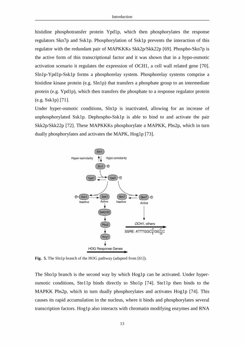
histidine phosphotransfer protein Ypd1p, which then phosphorylates the response
regulators Skn7p and Ssk1p. Phosphorylation of Ssk1p prevents the interaction of this
regulator with the redundant pair of MAPKKKs Skk2p/Skk22p [69]. Phospho-Skn7p is
the active form of this transcriptional factor and it was shown that in a hypo-osmotic
activation scenario it regulates the expression of OCH1, a cell wall related gene [70].
Sln1p-Ypd1p-Ssk1p forms a phosphorelay system. Phosphorelay systems comprise a
histidine kinase protein (e.g. Sln1p) that transfers a phosphate group to an intermediate
protein (e.g. Ypd1p), which then transfers the phosphate to a response regulator protein
(e.g. Ssk1p) [71].
Under hyper-osmotic conditions, Sln1p is inactivated, allowing for an increase of
unphosphorylated Ssk1p. Dephospho-Ssk1p is able to bind to and activate the pair
Skk2p/Skk22p [72]. These MAPKKKs phosphorylate a MAPKK, Pbs2p, which in turn
dually phosphorylates and activates the MAPK, Hog1p [73].
Fig. 5. The Sln1p branch of the HOG pathway (adapted from [61]).
The Sho1p branch is the second way by which Hog1p can be activated. Under hyper-
osmotic conditions, Ste11p binds directly to Sho1p [74]. Ste11p then binds to the
MAPKK Pbs2p, which in turn dually phosphorylates and activates Hog1p [74]. This
causes its rapid accumulation in the nucleus, where it binds and phosphorylates several
transcription factors. Hog1p also interacts with chromatin modifying enzymes and RNA
Introduction
14
polymerase II, and affects the expression of hundreds of genes in response to
hyperosmotic shock [75].
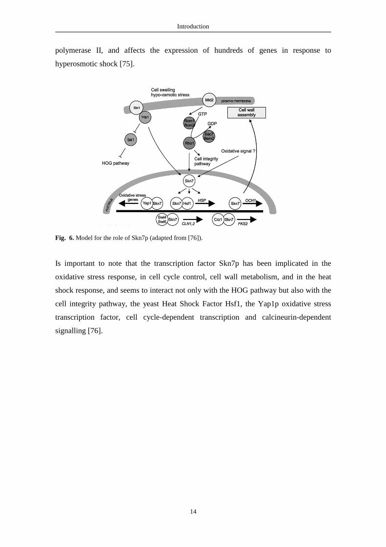
Fig. 6. Model for the role of Skn7p (adapted from [76]).
Is important to note that the transcription factor Skn7p has been implicated in the
oxidative stress response, in cell cycle control, cell wall metabolism, and in the heat
shock response, and seems to interact not only with the HOG pathway but also with the
cell integrity pathway, the yeast Heat Shock Factor Hsf1, the Yap1p oxidative stress
transcription factor, cell cycle-dependent transcription and calcineurin-dependent
signalling [76].

Chapter 2 OBJECTIVES AND RESEARCH PLAN

Objectives and Research plan
16
2. Objectives and research plan
Expression of mammalian sphingomyelin synthase (SMS1) suppresses Bax-mediated
cell death in yeast and confers increased resistance to different apoptosis inducers [34].
These results suggest that SMS1, using ceramide to synthesize sphingomyelin, protects
against death by counteracting stress-induced accumulation of the proapoptotic
ceramide. Data from our lab (unpublished) show that mutants in Isc1p, the yeast
ortholog of mammalian neutral sphingomyelinase (N-SMase) are more resistant to
acetic acid-induced cell death. These data suggest acid-induced apoptosis involves a
ceramide-mediated pathway. However, it is not know if ceramide induces cell death in
yeast, or if the regulatory mechanisms might involve conserved pathways such as
mitogen activated protein kinase (MAPK) signalling. The involvement of these
signalling pathways in acetic acid-induced apoptosis is also not known. We thus set out
to characterize the effect of ceramide on yeast cell death, as well as the potential
involvement of MAPK signalling pathways in this process and in acetic acid-induced
apoptosis.
A) Elucidation of signalling pathways in ceramide-induced apoptosis
Rationale:
Ceramide, which is generated intracellularly in response to different stress stimuli acts
as a second messenger in the initiation phase of apoptosis and appears to regulate
apoptosis through two different mechanisms. One involves transcriptional activation of
different signalling cascades, including the JNK pathway, and the other occurs via
alteration of mitochondrial function. Yet, the molecular mechanisms underlying
ceramide pro-apoptotic activity, including its targets and the signalling systems with
which it interacts, are for the most part incomplete.
Objective:
This work aimed to identify points of crosstalk between ceramide and MAPK signalling
pathways. The identification of these targets provides means to further characterize the
ceramide-induced death pathway in yeast and will give information on new putative
Objectives and Research plan
17
targets in mammalian cells. It could also provide an association, at the signalling level,
between yeast and mammalian apoptosis. Moreover, this data could also offer a link
between yeast stress signalling networks and mammalian apoptosis, which would
facilitate further research on apoptosis.
Research plan:
1) Characterization of ceramide-induced death in the Saccharomyces cerevisiae wild
type strain W303
2) Construction of MAPK knock-out mutants in the W303 strain background
3) Characterization of ceramide-induced death in the mutant strains constructed in (2)
B) Elucidation of signalling pathways in acetic acid-induced apoptosis
Rationale:
In yeast, acetic acid-induced apoptosis is one of the best characterized yeast apoptotic
pathways [9, 77]. It has been shown that expression of Bcl-xL decreased acetic acid
induced apoptosis, which correlated with phosphorylation of Bcl-xL [77]. Pre-treatment
of yeast cells expressing Bcl-xL with JNK inhibitor II renders the cells resistant to
acetic acid-induced apoptosis and eliminates the Bcl-xL phosphorylated form. These
results suggest there is a yeast pathway homologue to the mammalian SAPK/JNK,
responsible for acid-induced Bcl-xL phosphorylation. The mammalian protein kinases
p38 and SAPK/JNK have been shown to be structurally and functionally homologous to
Hog1, the MAPK from High Osmolarity Glycerol (HOG) pathway of yeast [78].
Therefore, it is expected that they share similar regulatory mechanisms [79]. Recent
data also suggest that acetic acid activates the HOG pathway [80], and also leads to
phosphorylation of Slt2p, a MAPKK from the PKC cascade. These results suggest an
intricate relation between PKC and HOG signalling in acetic acid-induced apoptosis.
Still, the involvement of other MAPK pathways cannot be discarded.

Objectives and Research plan
18
Objective:
This work aimed to characterize the involvement of MAPK signalling pathways in acetic
acid-induced cell death. Our working hypothesis was that acetic acid-induced apoptosis
involves ceramide production, and it was expected that at least the MAPK mutants
identified in (A) would exhibit an identical phenotype regarding their response to acetic
acid apoptosis-inducing concentrations. Since sensors for acetic acid had not been
identified so far, possible candidate plasma membrane proteins were studied in order to
try to identify other putative upstream effectors of acetic acid-induced cell death. We
expected to ascertain whether acetic acid directly, and/or through changes in
sphingolipids, regulates proliferation and apoptosis through opposite survival and death
signalling pathways, or whether the stress-activated MAP kinases, necessary for growth
in the presence of acetic acid in yeast, were also involved in cell death. In addition, we
anticipated unraveling any possible connections between the two processes.
Research plan:
1) Screen the yeast strains disrupted in genes coding for components of the MAPK
pathways that have been constructed in (A) for increased pro- or anti-apoptotic activity
by assessing decreased or increased sensitivity to acetic acid-induced cell death.
2) Test the strains presenting altered phenotypes for apoptotic markers, to determine at
what level the mutations are interfering with the cell death process.

Chapter 3 MATERIALS AND METHODS

Materials and Methods
20
3. Materials and methods
3.1 Strains
The yeast S. cerevisiae strain W303-1A (MATa, ura3-52, trp1∆ 2, leu2-3,112, his3-11,
ade2-1, can1-100) was used throughout this work as the wild type strain. Several
mutant strains deleted in key components of the MAPK pathways were constructed by
replacing the respective genes in the W303-1A strain with a kanMX4 disruption
cassette (Table I). Each disruption cassette was amplified by PCR, from genomic DNA
purified from the respective Euroscarf deletion strain, using the flanking primers A and
D and the primers Kanrev and Kanfwd that anneal in the kanMX4 cassette, as described
in the Saccharomyces Genome Deletion Project database (Fig. 7A) [81, 82, 104].
Cassettes were transformed into W303-1A, and transformants selected on YEPD plates
containing 200 µg/ml geneticin (Sigma). Correct integration of the cassette was
confirmed by PCR using primers α and δ that anneal upstream and downstream of the
insertion and the primers KanB and KanC, that anneal in the kanMX4 cassette,
respectively (Fig. 7B).
Fig. 7. Strategy used for the deletion of yeast ORFs (A) and confirmation of the correct integration of the
deletion module (B) (adapted from [104]).
(B) (A)

Materials and Methods
21
3.2 Growth conditions and treatments
Cells were maintained in rich medium (YPD) (1% yeast extract, 2% glucose, 2% bacto-
peptone, 2% agar) and grown in synthetic complete medium (SC-Gal) (0.67% Bacto-
yeast nitrogen base w/o amino acids (Difco), 2% galactose and 0.2% Dropout mix).
For ceramide treatment, yeast cells were grown as described above until exponential
phase (OD600nm – 0.5-0.6), harvested by centrifugation and suspended in fresh SC-Gal
medium with 40 µM of N-acetylsphingosine (C2-ceramide, Sigma) dissolved in DMSO,
or the equivalent volume of solvent (DMSO 0.1%, v/v) and incubated for 240 minutes
at 30ºC with agitation (200 revolutions/min (rpm). Samples were taken at different time
points, diluted to 10-4 in 1:10 serial dilutions in deionised sterilized water, and 40 µL
drops were spotted on YPD agar plates in replicates of seven. Colony forming units
(c.f.u.) were counted after 48 h incubation at 30ºC. Cell viability was calculated as
percentage of cfus normalized to the OD600 of the culture in relation to time zero. Cell
viability for C2- ceramide treated cells was then normalized to the cell viability of
DMSO-treated cells (see: Attachment II).
For acetic acid treatment, yeast cells were grown overnight in liquid SC-Gal until
exponential growth-phase (OD600nm – 0.5-0.6) at 30ºC with agitation (200 rpm). Cells
were harvested by centrifugation and suspended in fresh SC-Gal medium (pH 3) with
120 mM acetic acid, and incubated for 200 minutes at 30ºC in 50 mL Erlenmeyer flasks
with an air:liquid ratio of 5:1 in a mechanical shaker at 200 rpm. Samples were taken at
different time points, diluted to 10-4 in 1:10 serial dilutions in deionised sterilized water,
and 40 µL drops were spotted on YPD agar plates in replicates of seven. Colony
forming units (c.f.u.) were counted after 48 h incubation at 30ºC. Cell viability was
calculated as percentage of cfus in relation to time zero.
Materials and Methods
22
3.3 Flow cytometry
During acetic acid and C2-ceramide treatments, samples were also taken to assess loss
of plasma membrane integrity and production of reactive oxygen species (ROS) by flow
cytometric analysis in an EPICS® XL™ (Beckman COULTER®) flow cytometer
equipped with an argon-ion laser emitting a 488-nm beam at 15mW. Cells were
collected by centrifugation, washed in deionised water, suspended in phosphate buffered
saline (PBS) and stained with 1 µg/mL propidium iodide (PI, Sigma) or 2 µM/mL
dihydroethidium (DHE, Sigma) for 30 min at room temperature, in the dark.
Monoparametric detection of PI fluorescence was performed using FL-3 (488/620 nm)
and detection of DHE was performed using FL-4 (488/675 nm).

Chapter 4 RESULTS
Results
24
4. Results
Characterization of C2-ceramide induced cell death
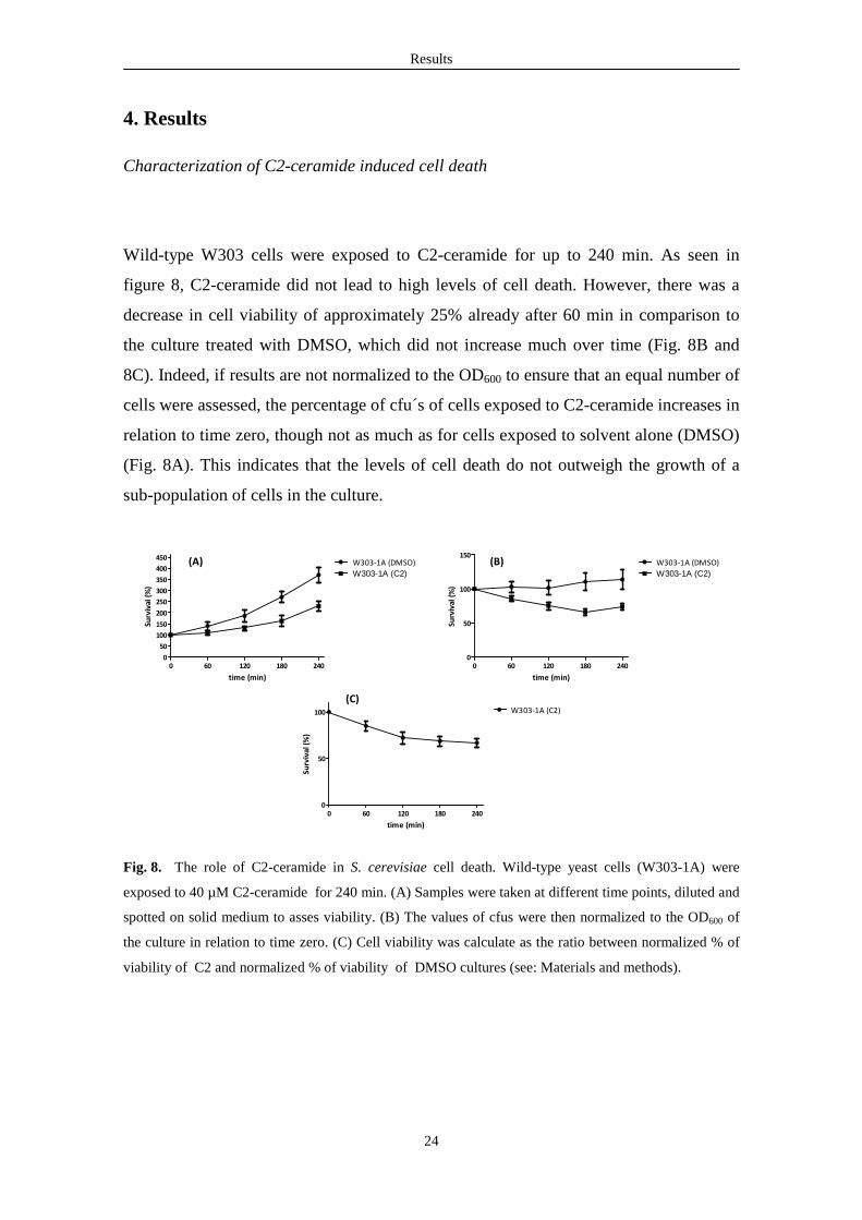
Wild-type W303 cells were exposed to C2-ceramide for up to 240 min. As seen in
figure 8, C2-ceramide did not lead to high levels of cell death. However, there was a
decrease in cell viability of approximately 25% already after 60 min in comparison to
the culture treated with DMSO, which did not increase much over time (Fig. 8B and
8C). Indeed, if results are not normalized to the OD600 to ensure that an equal number of
cells were assessed, the percentage of cfu´s of cells exposed to C2-ceramide increases in
relation to time zero, though not as much as for cells exposed to solvent alone (DMSO)
(Fig. 8A). This indicates that the levels of cell death do not outweigh the growth of a
sub-population of cells in the culture.
0 60 120 180 2400
50
100
150
200
250
300
350
400
450W303-1A (DMSO)
W303-1A (C2)
time (min)
Surv
ival
(%
)
0 60 120 180 2400
50
100
150W303-1A (DMSO)
W303-1A (C2)
time (min)
Surv
ival
(%
)
0 60 120 180 2400
50
100 W303-1A (C2)
time (min)
Surv
ival
(%
)
Fig. 8. The role of C2-ceramide in S. cerevisiae cell death. Wild-type yeast cells (W303-1A) were
exposed to 40 µM C2-ceramide for 240 min. (A) Samples were taken at different time points, diluted and
spotted on solid medium to asses viability. (B) The values of cfus were then normalized to the OD600 of
the culture in relation to time zero. (C) Cell viability was calculate as the ratio between normalized % of
viability of C2 and normalized % of viability of DMSO cultures (see: Materials and methods).
(A) (B)
(C)
Results
25
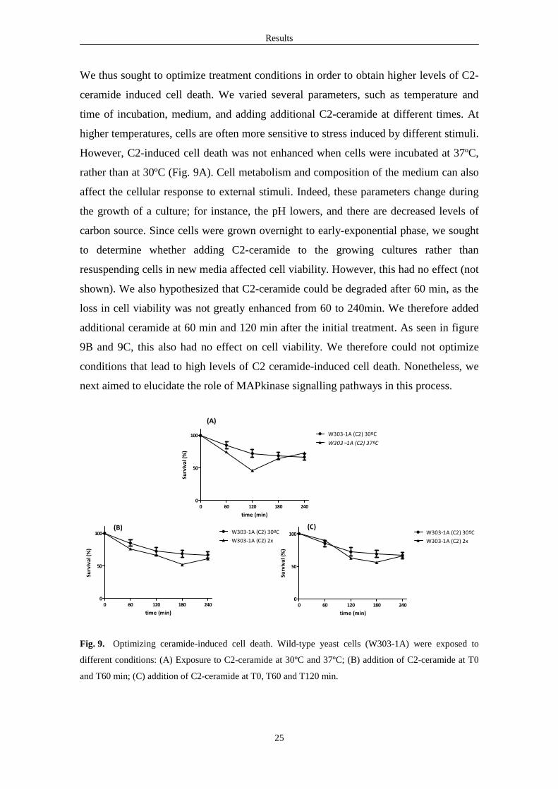
We thus sought to optimize treatment conditions in order to obtain higher levels of C2-
ceramide induced cell death. We varied several parameters, such as temperature and
time of incubation, medium, and adding additional C2-ceramide at different times. At
higher temperatures, cells are often more sensitive to stress induced by different stimuli.
However, C2-induced cell death was not enhanced when cells were incubated at 37ºC,
rather than at 30ºC (Fig. 9A). Cell metabolism and composition of the medium can also
affect the cellular response to external stimuli. Indeed, these parameters change during
the growth of a culture; for instance, the pH lowers, and there are decreased levels of
carbon source. Since cells were grown overnight to early-exponential phase, we sought
to determine whether adding C2-ceramide to the growing cultures rather than
resuspending cells in new media affected cell viability. However, this had no effect (not
shown). We also hypothesized that C2-ceramide could be degraded after 60 min, as the
loss in cell viability was not greatly enhanced from 60 to 240min. We therefore added
additional ceramide at 60 min and 120 min after the initial treatment. As seen in figure
9B and 9C, this also had no effect on cell viability. We therefore could not optimize
conditions that lead to high levels of C2 ceramide-induced cell death. Nonetheless, we
next aimed to elucidate the role of MAPkinase signalling pathways in this process.
Fig. 9. Optimizing ceramide-induced cell death. Wild-type yeast cells (W303-1A) were exposed to
different conditions: (A) Exposure to C2-ceramide at 30ºC and 37ºC; (B) addition of C2-ceramide at T0
and T60 min; (C) addition of C2-ceramide at T0, T60 and T120 min.
0 60 120 180 2400
50
100 W303-1A (C2) 30ºC
W303−1A (C2) 37ºC
time (min)
Surv
ival
(%
)
0 60 120 180 2400
50
100 W303-1A (C2) 30ºC
W303-1A (C2) 2x
time (min)
Surv
ival
(%
)
0 60 120 180 2400
50
100 W303-1A (C2) 30ºC
W303-1A (C2) 2x
time (min)
Surv
ival
(%
)
(A)
(B) (C)
Results
26
Construction of deletion mutants in components of the MAPK pathways
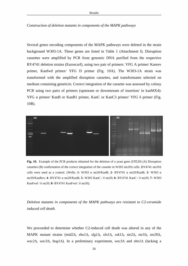
Several genes encoding components of the MAPK pathways were deleted in the strain
background W303-1A. These genes are listed in Table 1 (Attachment I). Disruption
cassettes were amplified by PCR from genomic DNA purified from the respective
BY4741 deletion strains (Euroscarf), using two pair of primers: YFG A primer/ Kanrev
primer, Kanfwd primer/ YFG D primer (Fig. 10A). The W303-1A strain was
transformed with the amplified disruption cassettes, and transformants selected on
medium containing geneticin. Correct integration of the cassette was assessed by colony
PCR using two pairs of primers (upstream or downstream of insertion/ in kanMX4):
YFG α primer/ KanB or KanB1 primer, KanC or KanC3 primer/ YFG δ primer (Fig.
10B).
Fig. 10. Example of the PCR products obtained for the deletion of a yeast gene (STE20) (A) Disruption
cassettes (B) confirmation of the correct integration of the cassette in W303 ste20∆ cells. BY4741 ste20∆
cells were used as a control. (Wells: 1- W303 α ste20/KanB; 2- BY4741 α ste20/KanB; 3- W303 α
ste20/KanRev; 4- BY4741 α ste20/KanB; 5- W303 KanC / δ ste20; 6- BY4741 KanC / δ ste20; 7- W303
KanFwd / δ ste20; 8- BY4741 KanFwd / δ ste20).
Deletion mutants in components of the MAPK pathways are resistant to C2-ceramide
induced cell death.
We proceeded to determine whether C2-induced cell death was altered in any of the
MAPK mutant strains (mid2∆, sho1∆, slg1∆, sln1∆, ssk1∆, ste2∆, ste3∆, ste20∆,
wsc2∆, wsc3∆, hog1∆). In a preliminary experiment, wsc3∆ and sho1∆ (lacking a
(A) (B)
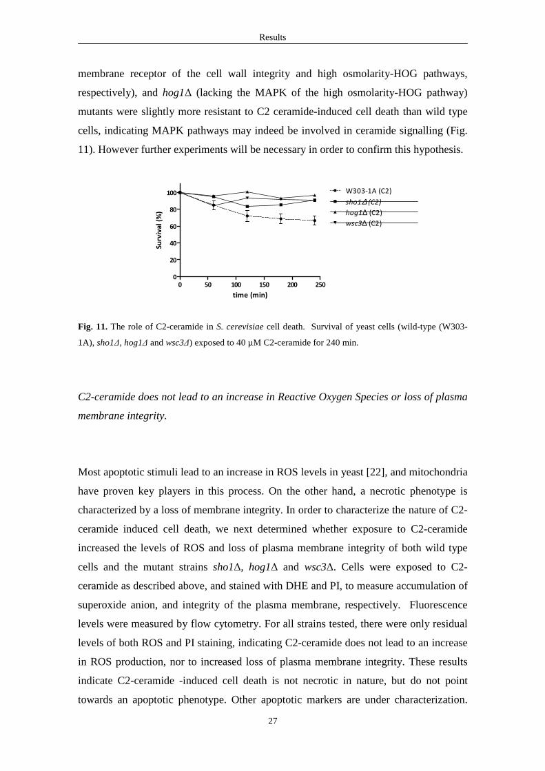
Results
27
membrane receptor of the cell wall integrity and high osmolarity-HOG pathways,
respectively), and hog1∆ (lacking the MAPK of the high osmolarity-HOG pathway)
mutants were slightly more resistant to C2 ceramide-induced cell death than wild type
cells, indicating MAPK pathways may indeed be involved in ceramide signalling (Fig.
11). However further experiments will be necessary in order to confirm this hypothesis.
0 50 100 150 200 2500
20
40
60
80
100 W303-1A (C2)
sho1∆ (C2)
hog1∆ (C2)
wsc3∆ (C2)
time (min)
Surv
ival
(%
)
Fig. 11. The role of C2-ceramide in S. cerevisiae cell death. Survival of yeast cells (wild-type (W303-
1A), sho1∆, hog1∆ and wsc3∆) exposed to 40 µM C2-ceramide for 240 min.
C2-ceramide does not lead to an increase in Reactive Oxygen Species or loss of plasma
membrane integrity.
Most apoptotic stimuli lead to an increase in ROS levels in yeast [22], and mitochondria
have proven key players in this process. On the other hand, a necrotic phenotype is
characterized by a loss of membrane integrity. In order to characterize the nature of C2-
ceramide induced cell death, we next determined whether exposure to C2-ceramide
increased the levels of ROS and loss of plasma membrane integrity of both wild type
cells and the mutant strains sho1∆, hog1∆ and wsc3∆. Cells were exposed to C2-
ceramide as described above, and stained with DHE and PI, to measure accumulation of
superoxide anion, and integrity of the plasma membrane, respectively. Fluorescence
levels were measured by flow cytometry. For all strains tested, there were only residual
levels of both ROS and PI staining, indicating C2-ceramide does not lead to an increase
in ROS production, nor to increased loss of plasma membrane integrity. These results
indicate C2-ceramide -induced cell death is not necrotic in nature, but do not point
towards an apoptotic phenotype. Other apoptotic markers are under characterization.
Results
28
Since C2-ceramide is not the equivalent of yeast endogenous ceramides, it is possible
we cannot mimic the role of increased ceramide levels in yeast by adding exogenous
C2-ceramide. As there is evidence that ceramide levels play an important role in acetic
acid-induced cell death, we next aimed to further characterize the signalling pathways
involved in the apoptotic phenotype triggered by acetic acid (another master thesis from
our lab).
Components of the MAPK pathways modulate acetic acid-induced cell death.
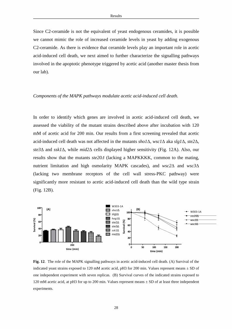
In order to identify which genes are involved in acetic acid-induced cell death, we
assessed the viability of the mutant strains described above after incubation with 120
mM of acetic acid for 200 min. Our results from a first screening revealed that acetic
acid-induced cell death was not affected in the mutants sho1∆, wsc1∆ aka slg1∆, ste2∆,
ste3∆ and ssk1∆, while mid2∆ cells displayed higher sensitivity (Fig. 12A). Also, our
results show that the mutants ste20∆ (lacking a MAPKKKK, common to the mating,
nutrient limitation and high osmolarity MAPK cascades), and wsc2∆ and wsc3∆
(lacking two membrane receptors of the cell wall stress-PKC pathway) were
significantly more resistant to acetic acid-induced cell death than the wild type strain
(Fig. 12B).
Fig. 12. The role of the MAPK signalling pathways in acetic acid-induced cell death. (A) Survival of the
indicated yeast strains exposed to 120 mM acetic acid, pH3 for 200 min. Values represent means ± SD of
one independent experiment with seven replicas. (B) Survival curves of the indicated strains exposed to
120 mM acetic acid, at pH3 for up to 200 min. Values represent means ± SD of at least three independent
experiments.
2000
20
40
60
80
100W303-1A
sho1∆
ssk1∆
hog1∆ste2∆ste3∆
slg1∆
mid2∆
time (min)
Surv
ival
(%
)
0 50 100 150 2000
20
40
60
80
100 W303-1A
ste20∆wsc2∆wsc3∆
time (min)
Surv
ival
(%
)
(A) (B)
Results
29
These observations indicate that MAPK signalling pathways are involved in acetic acid-
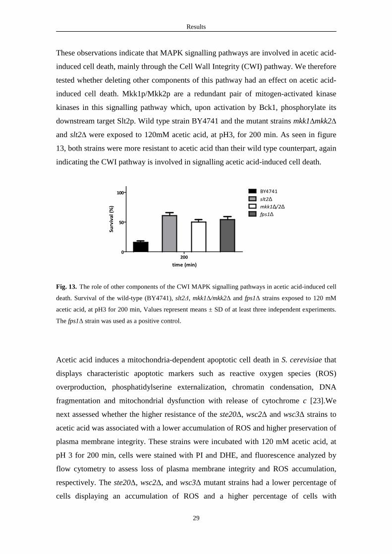
induced cell death, mainly through the Cell Wall Integrity (CWI) pathway. We therefore
tested whether deleting other components of this pathway had an effect on acetic acid-
induced cell death. Mkk1p/Mkk2p are a redundant pair of mitogen-activated kinase
kinases in this signalling pathway which, upon activation by Bck1, phosphorylate its
downstream target Slt2p. Wild type strain BY4741 and the mutant strains mkk1∆mkk2∆
and slt2∆ were exposed to 120mM acetic acid, at pH3, for 200 min. As seen in figure
13, both strains were more resistant to acetic acid than their wild type counterpart, again
indicating the CWI pathway is involved in signalling acetic acid-induced cell death.
2000
50
100 BY4741
slt2∆mkk1∆/2∆fps1∆
time (min)
Surv
ival
(%
)
Fig. 13. The role of other components of the CWI MAPK signalling pathways in acetic acid-induced cell
death. Survival of the wild-type (BY4741), slt2∆, mkk1∆/mkk2∆ and fps1∆ strains exposed to 120 mM
acetic acid, at pH3 for 200 min, Values represent means ± SD of at least three independent experiments.
The fps1∆ strain was used as a positive control.
Acetic acid induces a mitochondria-dependent apoptotic cell death in S. cerevisiae that
displays characteristic apoptotic markers such as reactive oxygen species (ROS)
overproduction, phosphatidylserine externalization, chromatin condensation, DNA
fragmentation and mitochondrial dysfunction with release of cytochrome c [23].We
next assessed whether the higher resistance of the ste20∆, wsc2∆ and wsc3∆ strains to
acetic acid was associated with a lower accumulation of ROS and higher preservation of
plasma membrane integrity. These strains were incubated with 120 mM acetic acid, at
pH 3 for 200 min, cells were stained with PI and DHE, and fluorescence analyzed by
flow cytometry to assess loss of plasma membrane integrity and ROS accumulation,
respectively. The ste20∆, wsc2∆, and wsc3∆ mutant strains had a lower percentage of
cells displaying an accumulation of ROS and a higher percentage of cells with

Results
30
preserved plasma membrane integrity than the wild type strain (Fig. 14). This is
consistent with a role for Wsc2p and Wsc3p as sensor transducers in acetic acid-induced
apoptosis signalling. Ste20p is involved in signal transduction of a number of pathways,
so it remains to be determined whether its role in acetic acid-induced apoptosis is
related to MAPK signalling and/or to other pathways.
Fig. 14. Percentage of cells displaying PI internalization (A) and intracellular ROS levels (B) assessed by
flow cytometry after treatment with 120 mM acetic acid, at pH3, for 200 min. Values are represented as
means ± SD of at least three independent experiments with at least 20000 cells counted in each time
point. Asterisks represent significance from control by one-way ANOVA test: *** p < 0.001.
The histone H2B S10 is involved in acetic acid-induced cell death
It has previously been reported that histone H2B is phosphorylated at S10 after
exposure to apoptotic stimuli such as hydrogen peroxide, acetic acid, and α-factor, and
inhibiting this phosphorylation event renders cells more resistant to these stimuli [83,
84]. The authors also showed that this phosphorylation is mediated by the Ste20p kinase
in vivo and in vitro [83, 84]. This raised the hypothesis that the resistant phenotype
observed in ste20∆ cells was simply due to a defect in phosphorylation of the histone
H2B S10, and not to its role in the MAPK signalling pathways. This seems to be the
case for H2O2-induced apoptosis, as the strains ste20∆, H2B S10A (a strain with non-
phosphorylatable H2B S10) and the double mutant H2B S10A ste20∆ were equally
more resistant to H2O2-induced cell death than wild type cells. To test this hypothesis,
we compared the viability of these strains (Wt, ste20∆, H2B S10A, H2B S10A ste20∆)
after exposure to acetic acid. Unexpectedly, in our preliminary results the strain H2B
W303-1A ste20∆∆∆∆ wsc2∆∆∆∆ wsc3∆∆∆∆0
20
40
60
80
PI
(%) *** *** ***
W303-1A ste20∆∆∆∆ wsc2∆∆∆∆ wsc3∆∆∆∆0
20
40
60
80
*** ******
DH
E (%
)
(A) (B)

Results
31
S10A had increased levels of plasma membrane integrity after exposure to acetic acid
than strains ste20∆ or even H2B S10A ste20∆ (Fig. 15). This strain was constructed in
this study, using the reagents provided by Dr Allis as described in Materials and
Methods. We are currently deleting STE20 in this strain background in order to exclude
any alterations during the construction of this strain are responsible for the observed
phenotype.
Fig. 15. Percentage of cells displaying PI internalization assessed by flow cytometry after treatment with
120 mM acetic acid, at pH3, for 200 min.
40
50
60
70
80
Wt ste20∆ H2B S10AH2B S10A ste20∆
PI (
%)

Chapter 5 DISCUSSION

Discussion
33
5. Discussion
In this work, we investigated the possible involvement of the different MAPK pathways
in S. cerevisiae ceramide and acetic acid-induced cell death. We intended to confirm
whether acetic acid directly and/or through changes in sphingolipids regulates
proliferation and apoptosis through opposite survival and death signalling pathways,
and whether the stress-activated MAP kinases, involved in acetic acid resistance in
yeast, are also involved in cell death. We also aimed to establish a link between the two
processes. We started by determining whether exposure of the W303-1A strain to C2-
ceramide resulted in cell death. We found that cells exposed to C2-ceramide showed a
decrease in viability when compared with cells exposed to the solvent DMSO.
However, cultures continued to grow in the presence of C2-ceramide. This seems to
indicate that only a small fraction of the culture dies, whereas a large fraction of the
cells survives and grows. Further optimization of the treatment conditions (temperature,
ceramide concentration) did not increase C2-ceramide-induced cell death. Therefore, we
next proceeded to determine if any of the mutant strains constructed showed altered
phenotypes after incubation with C2-ceramide. Of all the mutants analyzed, wsc3∆,
sho1∆ and hog1∆ were slightly more resistant to C2 ceramide-induced cell death than
wild type cells, indicating that signalling C2-ceramide-induced cell death may involve
MAPK pathways. However, these results are very preliminary and warrant further
confirmation. In the wild type strain or in these mutants, C2-ceramide did not induce
loss of plasma membrane integrity, which would be indicative of necrosis, or an
increase in intercellular ROS levels, which would suggest a mitochondria-mediated
process. Therefore, the nature of C2-ceramide-induced death is still uncharacterized.
The fact that C2-ceramide led to only a small reduction on cell viability further
hampered our studies. It is known that sphingolipids are generated in response to heat
stress in both mammals and yeasts [33, 85, 86]. It was also shown that C2-ceramide
mimics the effects elicited by endogenously generated ceramide in the response of
mammalian cells to different death stimuli [87]. However, it is possible that C2-
ceramide is not the best molecule to use while studying the role of sphingolipid
signalling in yeast apoptosis. Sphingolipids contain a long-chain base (LCBs in yeast
are dihydrosphingosine (DHS) and phytosphingosine (PHS)), a fatty acid and a polar
head group. LCBs are the best-characterized heat-induced sphingolipid signalling

Discussion
34
molecules in yeast, whereas in mammals it is ceramide that signals cells to undergo
apoptosis during severe heat stress and other stresses [33, 85, 86]. LCBs regulate
various signalling pathways in S. cerevisiae, such as the Target Of Rampamycin
Complex 2 (TORC2) and calcineurin, which in turn regulate ceramide synthase activity
in response to heat and other stresses; PKC1 is one of the downstream partners [86, 88].
It would be more appropriate to use the LCBs dihydrosphingosine and
phytosphingosine to better define the cell death process mediated by sphingolipids.
However, these molecules are not easily synthesized and produced in soluble form to
allow their use in the proposed studies.
Regarding the characterization of the involvement of MAP kinase pathways in cell
death induced by acetic acid, our results from a first screening revealed that acetic acid-
induced cell death was not affected in the mutants sho1∆, wsc1∆ aka slg1∆, ste2∆,
ste3∆ and ssk1∆, while mid2∆ cells displayed higher sensitivity. In contrast, wsc2∆,
wsc3∆ and ste20∆ strains, as well as hog1∆ cells, were significantly more resistant to
acetic acid-induced cell death. These results are discussed bellow.
Mid2p is a plasma membrane sensor of the CWI pathway. The mid2∆ strain showed
increased sensitivity to acetic acid-induced cell death. It has previously been shown that
low extracellular pH conditions activate the CWI pathway via the cell sensor Mid2p
[89]. In addition, Mid2p likely activates the transcription factor Skn7p [90], which has
been associated with the control of oxidative stress response, among other roles [91-93].
Accordingly, deletion of SKN7 sensitizes cells to oxidative stress [91-93]. Absence of
Mid2p could result in lower activity of Skn7p, although Skn7p can function
independently of Mid2p. It would therefore be interesting to determine whether skn7∆
cells are also more sensitive to acetic acid-induced cell death.
It has previously been shown that hog1∆ cells are more sensitive to growth on acetic
acid-containing plates. In contrast, deletion of the aquaglyceroporin Fps1p, which
mediates uptake of acetic acid, increases resistance to acetic acid of cells grown under
the same conditions. The same authors showed that Hog1p phosphorylates Fps1p,
targeting it for degradation. The phenotype of the hog1∆fps1∆ mutant was
indistinguishable from that of the fps1∆ strain, indicating Hog1p contributes to
resistance of cells to chronic exposure to acetic acid through its role in lowering the
levels of Fps1p, thus reducing the accumulation of intracellular acetic acid [94].
Unexpectedly, we found that the hog1∆ strain was more resistant to acetic acid than the

Discussion
35
wild type strain. This may be explained by the different strain background used.
However, it is also possible that deleting HOG1 can provide enough energy to increase
the capacity to repair damage in the short term. In oxidative stress, e.g. after exposure
to acetic acid or H2O2, there is an increase in the levels of reactive oxygen species
(ROS). ROS can cause oxidative damage in proteins, lipids and mainly in DNA, thus
compromising cell viability. Cells have several defense mechanisms and invest energy
to cope with the consequences of cellular damage, and express protective proteins or
metabolites such as DNA damage repair enzymes, SOD, or catalase. Though a previous
study showed that exposure to acetic acid did not alter catalase or superoxide dismutase
activity in wild-type cells, acetic acid-induced cell death decreased in cells
overexpressing catalase T and increased when Cu,Zn superoxide dismutase is
overexpressed, suggesting that hydrogen peroxide contributes to acetic acid-induced cell
death [95].
Osmotic stress leads to the production of glycerol, which is needed for osmotic
adjustment. However, this requires a great deal of resources and energy. It has been
shown that Hog1p plays a role in establishing the metabolic conditions for elevated
glycerol production, as well as ensure there are adequate levels of enzymes necessary
for glycerol production [96]. Therefore, deleting HOG1 could prevent production of
glycerol, thus increasing the available energy, which could then be used in recovering
from damage. In the short term (up to 200 min, the length of our assay), reducing the
resources channeled to produce glycerol could make resources available to the repair of
damage. However, Hog1p would be required in the long run, thus rendering cells more
sensitive to osmotic stress. Further studies will be required to test this hypothesis.
Ste20p is a MAPKKKK in the pheromone-mating response, invasive growth/
pseudohyphal development and HOG pathways, and is therefore not specific to one
particular pathway. It is however interesting to note that it does not play a role in the
CWI pathway. It is likely that the resistant phenotype observed in ste20∆ cells was
simply due to a defect in phosphorylation of the histone H2B S10, and not to its role in
the MAPK signalling pathways, which would indicate that acetic acid specifically
induces the CWI pathway. This hypothesis is currently under investigation.
Wsc2p and Wsc3p function as plasma membranes receptors of the CWI pathway. Our
initial results therefore suggested that this pathway is involved in signalling acetic acid-
induced apoptosis, where Wsc2p and Wsc3p could function as sensors. We therefore
tested other mutants lacking the MAPK and MAPKK of the CWI pathway, slt2∆ and
Discussion
36
mkk1∆/mkk2∆, in which phosphorylation of downstream kinases is absent. These
mutants were also more resistant to acetic acid-induced cell death. Our data therefore
indicate that the CWI pathway is involved in signalling acetic acid- induced cell death,
as blocking signal transduction in this pathway renders cells more resistant to acetic
acid. The mutants slt2∆, mkk1∆/mkk2∆, and bck1∆ are more sensitive to a variety of
stimuli [76], as they have altered composition of the cell wall [76]. However, they are
more resistant to oxidative stress. It would be interesting to further analyze which are
the effectors of Stl2p, and the final targets of acetic acid-induced signalling. Slt2p (aka
Mpk1p) targets the transcription factor complex SBF (Swi4p/Swi6p) and Rlm1p. The
SBF complex is composed of the DNA-binding subunit Swi4p and the regulatory
subunit Swi6p. In cell wall stresses, Fks2p (Catalytic subunit of 1,3-beta-glucan
synthase) is induced, which requires the SBF complex. Indeed, activated
(phosphorylated) Slt2p was detected in a complex with Swi4p and Swi6p at the FKS2
promoter [97]. However, when cells are challenged with oxidative stress, Swi6p can
also be directly oxidized in a specific cysteine residue (Cys404), resulting in a G1 delay
while cells repair cellular damage. Swi6p can therefore function as an oxidative stress
sensor for the regulation of the cell cycle, independently of Swi4p [98, 99]. It has been
described that swi6∆ cells are more sensitive to chronic exposure to acetic acid, though
its role has not been defined. It would be interesting to determine whether Swi4p,
Swi6p, and Fks2p play a role in acetic acid-induced apoptosis, and whether it differs
from that in chronic exposure to acetic acid. In a strain lacking Slt2p or lacking the
ability to phosphorylate Slt2p (mkk1∆/mkk2∆), Swi6p could be more available to suffer
direct oxidation, and thus could better control damage repair. This phenomenon, of
enhanced damage repair by the greater availability of Swi6p maybe transient, because in
the long term cell wall damage induced by acetic acid can be deleterious in cells lacking
Slt2p (which by itself is very sensitive to various stresses), even though genes
responsible for cell wall assembly can be controlled by other effectors. The need to fully
block signal transduction in the CWI pathway to render a resistant phenotype to acetic
acid should be tested further through studies in a bck1∆ strain.
The Hog1p pathway seems to be important for resistance to acetic acid at low pH. In
accordance, a sub-lethal growth inhibitory concentration of acetic acid was shown to
promote the phosphorylation of Hog1p and Slt2p [80]. However, from the 101 viable
kinase mutants of the Euroscarf collection, only hog1∆, pbs2∆, ssk1∆ and ctk2∆
exhibited deficient growth in the presence of acetic acid. Activation of Hog1p by acetic
Discussion
37
acid was shown to depend on the presence of SSK1 and PBS2, but not of SHO1 or
STE11. In the same screening, loss of the cell integrity MAP kinase (Slt2p/Mpk1p) was
found to slightly increase acetate resistance. In what concerns the known plasma
membrane sensors of MAPK pathways, acetate-induced Hog1p activation appears to
involve the Sln1p, whereas Slt2p activation was dependent on Wsc1p [100]. These data
together with our results indicate that not all stress-activated MAP kinases involved in
acetic acid resistance in yeast are also involved in acetic acid induced apoptosis.
The wsc3∆ mutant displayed a resistance phenotype to both ceramide and acetic-acid
induced cell death. The higher survival of wsc3∆, deficient in a sensor in the cell wall
integrity MAPK pathway, suggests a role for this signalling pathway in both ceramide
and acetic acid induced cell death. The link between acetic acid-induced cell death and
cell wall integrity is not known, but lipid peroxidation by acetic acid may be a plausible
candidate. Indeed, it has been shown that that linoleic acid hydroperoxide (LoaOOH), a
product of radical attack on an unsaturated lipid, activates the CWI MAPK Slt2p
(Mkk1p) [101].
Finally, in our study, exposure to acetic acid resulted in a high percentage of cells
displaying loss of plasma membrane integrity, as well as high ROS production. This is
in disagreement with an apoptotic phenotype but may be indicative of secondary
necrosis following an initial apoptotic process. These markers should therefore be
monitored at earlier time points under our experimental conditions. Other apoptotic
markers should also be assessed in the future. However, there is growing evidence that
necrosis is a controlled process. It has been described that necrotic programmed cell
death in yeast is accompanied by typical morphological and cell functional changes.
These include plasma membrane rupture, mitochondrial outer membrane
permeabilization (MOMP), dissipation of mitochondrial potential, ATP depletion,
overproduction of reactive oxygen species (ROS), and nuclear release of high mobility
group box-1 (HMGB1) protein. In the future, it will be important to monitor necrotic
markers such as the nuclear release of yeast HMGB1 (Nhp6Ap) [102]. Interestingly,
Hsp90p has been functionally associated with regulation of necrosis and MAPKs such
as Slt2p may be targets of Hsp90p, indicating MAPK pathways may also be involved in
programmed necrosis [102].

Chapter 6 REFERENCES
References
39
6 - References
1. Kroemer, G., W.S. El-Deiry, P. Golstein, M.E. Peter, D. Vaux, P. Vandenabeele, B. Zhivotovsky, M.V. Blagosklonny, W. Malorni, R.A. Knight, M. Piacentini, S. Nagata, and G. Melino (2005). Classification of cell death: recommendations of the Nomenclature Committee on Cell Death. Cell Death Differ. 12 Suppl 2: 1463-7.
2. Syntichaki, P. and N. Tavernarakis (2002). Death by necrosis. Uncontrollable catastrophe, or is
there order behind the chaos? EMBO Rep. 3(7): 604-9. 3. Edinger, A.L. and C.B. Thompson (2004). Death by design: apoptosis, necrosis and autophagy.
Curr Opin Cell Biol. 16(6): 663-9.
4. Golstein, P. and G. Kroemer (2007). Cell death by necrosis: towards a molecular definition. Trends Biochem Sci. 32(1): 37-43.
5. Gourlay, C.W., W. Du, and K.R. Ayscough (2006). Apoptosis in yeast--mechanisms and benefits
to a unicellular organism. Mol Microbiol. 62(6): 1515-21. 6. Madeo, F., E. Frohlich, and K.U. Frohlich (1997). A yeast mutant showing diagnostic markers of
early and late apoptosis. J Cell Biol. 139(3): 729-34. 7. Madeo, F., E. Frohlich, M. Ligr, M. Grey, S.J. Sigrist, D.H. Wolf, and K.U. Frohlich (1999). Oxygen
stress: a regulator of apoptosis in yeast. J Cell Biol. 145(4): 757-67. 8. Ribeiro, G.F., M. Corte-Real, and B. Johansson (2006). Characterization of DNA damage in yeast
apoptosis induced by hydrogen peroxide, acetic acid, and hyperosmotic shock. Mol Biol Cell. 17(10): 4584-91.
9. Ludovico, P., M.J. Sousa, M.T. Silva, C. Leao, and M. Corte-Real (2001). Saccharomyces
cerevisiae commits to a programmed cell death process in response to acetic acid. Microbiology. 147(Pt 9): 2409-15.
10. Del Carratore, R., C. Della Croce, M. Simili, E. Taccini, M. Scavuzzo, and S. Sbrana (2002). Cell
cycle and morphological alterations as indicative of apoptosis promoted by UV irradiation in S. cerevisiae. Mutat Res. 513(1-2): 183-91.
11. Reiter, J., E. Herker, F. Madeo, and M.J. Schmitt (2005). Viral killer toxins induce caspase-
mediated apoptosis in yeast. J Cell Biol. 168(3): 353-8. 12. Severin, F.F. and A.A. Hyman (2002). Pheromone induces programmed cell death in S.
cerevisiae. Curr Biol. 12(7): R233-5. 13. Fabrizio, P., L. Battistella, R. Vardavas, C. Gattazzo, L.L. Liou, A. Diaspro, J.W. Dossen, E.B. Gralla,
and V.D. Longo (2004). Superoxide is a mediator of an altruistic aging program in Saccharomyces cerevisiae. J Cell Biol. 166(7): 1055-67.
14. Herker, E., H. Jungwirth, K.A. Lehmann, C. Maldener, K.U. Frohlich, S. Wissing, S. Buttner, M.
Fehr, S. Sigrist, and F. Madeo (2004). Chronological aging leads to apoptosis in yeast. J Cell Biol. 164(4): 501-7.
15. Laun, P., A. Pichova, F. Madeo, J. Fuchs, A. Ellinger, S. Kohlwein, I. Dawes, K.U. Frohlich, and M.
Breitenbach (2001). Aged mother cells of Saccharomyces cerevisiae show markers of oxidative stress and apoptosis. Mol Microbiol. 39(5): 1166-73.

References
40
16. Vachova, L. and Z. Palkova (2005). Physiological regulation of yeast cell death in multicellular colonies is triggered by ammonia. J Cell Biol. 169(5): 711-7.
17. Madeo, F., E. Herker, C. Maldener, S. Wissing, S. Lachelt, M. Herlan, M. Fehr, K. Lauber, S.J.
Sigrist, S. Wesselborg, and K.U. Frohlich (2002). A caspase-related protease regulates apoptosis in yeast. Mol Cell. 9(4): 911-7.
18. Ludovico, P., F. Madeo, and M. Silva (2005). Yeast programmed cell death: an intricate puzzle.
IUBMB Life. 57(3): 129-35. 19. Ludovico, P., F. Rodrigues, A. Almeida, M.T. Silva, A. Barrientos, and M. Corte-Real (2002).
Cytochrome c release and mitochondria involvement in programmed cell death induced by acetic acid in Saccharomyces cerevisiae. Mol Biol Cell. 13(8): 2598-606.
20. Ligr, M., F. Madeo, E. Frohlich, W. Hilt, K.U. Frohlich, and D.H. Wolf (1998). Mammalian Bax
triggers apoptotic changes in yeast. FEBS Lett. 438(1-2): 61-5. 21. Priault, M., N. Camougrand, B. Chaudhuri, J. Schaeffer, and S. Manon (1999). Comparison of the
effects of bax-expression in yeast under fermentative and respiratory conditions: investigation of the role of adenine nucleotides carrier and cytochrome c. FEBS Lett. 456(2): 232-8.
22. Carmona-Gutierrez, D., T. Eisenberg, S. Buttner, C. Meisinger, G. Kroemer, and F. Madeo (2010).
Apoptosis in yeast: triggers, pathways, subroutines. Cell Death Differ. 17(5): 763-73. 23. Pereira, C., R.D. Silva, L. Saraiva, B. Johansson, M.J. Sousa, and M. Corte-Real (2008).
Mitochondria-dependent apoptosis in yeast. Biochim Biophys Acta. 1783(7): 1286-302. 24. Wissing, S., P. Ludovico, E. Herker, S. Buttner, S.M. Engelhardt, T. Decker, A. Link, A. Proksch, F.
Rodrigues, M. Corte-Real, K.U. Frohlich, J. Manns, C. Cande, S.J. Sigrist, G. Kroemer, and F. Madeo (2004). An AIF orthologue regulates apoptosis in yeast. J Cell Biol. 166(7): 969-74.
25. Pereira, C., N. Camougrand, S. Manon, M.J. Sousa, and M. Corte-Real (2007). ADP/ATP carrier is
required for mitochondrial outer membrane permeabilization and cytochrome c release in yeast apoptosis. Mol Microbiol. 66(3): 571-82.
26. Fannjiang, Y., W.C. Cheng, S.J. Lee, B. Qi, J. Pevsner, J.M. McCaffery, R.B. Hill, G. Basanez, and
J.M. Hardwick (2004). Mitochondrial fission proteins regulate programmed cell death in yeast. Genes Dev. 18(22): 2785-97.
27. Tepper, A.D., J.G. Cock, E. de Vries, J. Borst, and W.J. van Blitterswijk (1997). CD95/Fas-induced
ceramide formation proceeds with slow kinetics and is not blocked by caspase-3/CPP32 inhibition. J Biol Chem. 272(39): 24308-12.
28. Bose, R., M. Verheij, A. Haimovitz-Friedman, K. Scotto, Z. Fuks, and R. Kolesnick (1995).
Ceramide synthase mediates daunorubicin-induced apoptosis: an alternative mechanism for generating death signals. Cell. 82(3): 405-14.
29. Mathias, S., L.A. Pena, and R.N. Kolesnick (1998). Signal transduction of stress via ceramide.
Biochem J. 335 ( Pt 3): 465-80. 30. Brenner, B., U. Koppenhoefer, C. Weinstock, O. Linderkamp, F. Lang, and E. Gulbins (1997). Fas-
or ceramide-induced apoptosis is mediated by a Rac1-regulated activation of Jun N-terminal kinase/p38 kinases and GADD153. J Biol Chem. 272(35): 22173-81.
31. Wymann, M.P. and R. Schneiter (2008). Lipid signalling in disease. Nat Rev Mol Cell Biol. 9(2):
162-76.
References
41
32. Karathia, H., E. Vilaprinyo, A. Sorribas, and R. Alves (2011). Saccharomyces cerevisiae as a model organism: a comparative study. PLoS One. 6(2): e16015.
33. Dickson, R.C., C. Sumanasekera, and R.L. Lester (2006). Functions and metabolism of
sphingolipids in Saccharomyces cerevisiae. Prog Lipid Res. 45(6): 447-65. 34. Yang, Z., C. Khoury, G. Jean-Baptiste, and M.T. Greenwood (2006). Identification of mouse
sphingomyelin synthase 1 as a suppressor of Bax-mediated cell death in yeast. FEMS Yeast Res. 6(5): 751-62.
35. Carmona-Gutierrez, D., A. Reisenbichler, P. Heimbucher, M.A. Bauer, R.J. Braun, C. Ruckenstuhl,
S. Buttner, T. Eisenberg, P. Rockenfeller, K.U. Frohlich, G. Kroemer, and F. Madeo (2011). Ceramide triggers metacaspase-independent mitochondrial cell death in yeast. Cell Cycle. 10(22).
36. Aerts, A.M., P. Zabrocki, I.E. Francois, D. Carmona-Gutierrez, G. Govaert, C. Mao, B. Smets, F.
Madeo, J. Winderickx, B.P. Cammue, and K. Thevissen (2008). Ydc1p ceramidase triggers organelle fragmentation, apoptosis and accelerated ageing in yeast. Cell Mol Life Sci. 65(12): 1933-42.
37. Almeida, T., M. Marques, D. Mojzita, M.A. Amorim, R.D. Silva, B. Almeida, P. Rodrigues, P.
Ludovico, S. Hohmann, P. Moradas-Ferreira, M. Corte-Real, and V. Costa (2008). Isc1p plays a key role in hydrogen peroxide resistance and chronological lifespan through modulation of iron levels and apoptosis. Mol Biol Cell. 19(3): 865-76.
38. Barbosa, A.D., H. Osorio, K.J. Sims, T. Almeida, M. Alves, J. Bielawski, M.A. Amorim, P. Moradas-
Ferreira, Y.A. Hannun, and V. Costa (2011). Role for Sit4p-dependent mitochondrial dysfunction in mediating the shortened chronological lifespan and oxidative stress sensitivity of Isc1p-deficient cells. Mol Microbiol. 81(2): 515-27.
39. Fishbein, J.D., R.T. Dobrowsky, A. Bielawska, S. Garrett, and Y.A. Hannun (1993). Ceramide-
mediated growth inhibition and CAPP are conserved in Saccharomyces cerevisiae. J Biol Chem. 268(13): 9255-61.
40. Nickels, J.T. and J.R. Broach (1996). A ceramide-activated protein phosphatase mediates
ceramide-induced G1 arrest of Saccharomyces cerevisiae. Genes Dev. 10(4): 382-94. 41. Marshall, C.J. (1994). MAP kinase kinase kinase, MAP kinase kinase and MAP kinase. Curr Opin
Genet Dev. 4(1): 82-9. 42. Qi, M. and E.A. Elion (2005). MAP kinase pathways. J Cell Sci. 118(Pt 16): 3569-72. 43. Chen, R.E. and J. Thorner (2007). Function and regulation in MAPK signalling pathways: lessons
learned from the yeast Saccharomyces cerevisiae. Biochim Biophys Acta. 1773(8): 1311-40. 44. Burack, W.R. and A.S. Shaw (2000). Signal transduction: hanging on a scaffold. Curr Opin Cell
Biol. 12(2): 211-6. 45. Hunter, T. and G.D. Plowman (1997). The protein kinases of budding yeast: six score and more.
Trends Biochem Sci. 22(1): 18-22. 46. Pawson, T. and J.D. Scott (1997). Signalling through scaffold, anchoring, and adaptor proteins.
Science. 278(5346): 2075-80. 47. Burkholder, A.C. and L.H. Hartwell (1985). The yeast alpha-factor receptor: structural properties
deduced from the sequence of the STE2 gene. Nucleic Acids Res. 13(23): 8463-75.
References
42
48. Chen, L. and N.G. Davis (2000). Recycling of the yeast a-factor receptor. J Cell Biol. 151(3): 731-8.
49. Gustin, M.C., J. Albertyn, M. Alexander, and K. Davenport (1998). MAP kinase pathways in the
yeast Saccharomyces cerevisiae. Microbiol Mol Biol Rev. 62(4): 1264-300. 50. Winters, M.J. and P.M. Pryciak (2005). Interaction with the SH3 domain protein Bem1 regulates
signalling by the Saccharomyces cerevisiae p21-activated kinase Ste20. Mol Cell Biol. 25(6): 2177-90.
51. Tatebayashi, K., K. Yamamoto, K. Tanaka, T. Tomida, T. Maruoka, E. Kasukawa, and H. Saito
(2006). Adaptor functions of Cdc42, Ste50, and Sho1 in the yeast osmoregulatory HOG MAPK pathway. EMBO J. 25(13): 3033-44.
52. Wu, C., E. Leberer, D.Y. Thomas, and M. Whiteway (1999). Functional characterization of the
interaction of Ste50p with Ste11p MAPKKK in Saccharomyces cerevisiae. Mol Biol Cell. 10(7): 2425-40.
53. Whiteway, M.S., C. Wu, T. Leeuw, K. Clark, A. Fourest-Lieuvin, D.Y. Thomas, and E. Leberer
(1995). Association of the yeast pheromone response G protein beta gamma subunits with the MAP kinase scaffold Ste5p. Science. 269(5230): 1572-5.
54. Choi, K.Y., B. Satterberg, D.M. Lyons, and E.A. Elion (1994). Ste5 tethers multiple protein kinases
in the MAP kinase cascade required for mating in S. cerevisiae. Cell. 78(3): 499-512. 55. Truckses, D.M., L.S. Garrenton, and J. Thorner (2004). Jekyll and Hyde in the microbial world.
Science. 306(5701): 1509-11. 56. Roberts, R.L. and G.R. Fink (1994). Elements of a single MAP kinase cascade in Saccharomyces
cerevisiae mediate two developmental programs in the same cell type: mating and invasive growth. Genes Dev. 8(24): 2974-85.
57. Mosch, H.U., R.L. Roberts, and G.R. Fink (1996). Ras2 signals via the Cdc42/Ste20/mitogen-
activated protein kinase module to induce filamentous growth in Saccharomyces cerevisiae. Proc Natl Acad Sci U S A. 93(11): 5352-6.
58. Cullen, P.J., W. Sabbagh, Jr., E. Graham, M.M. Irick, E.K. van Olden, C. Neal, J. Delrow, L.
Bardwell, and G.F. Sprague, Jr. (2004). A signalling mucin at the head of the Cdc42- and MAPK-dependent filamentous growth pathway in yeast. Genes Dev. 18(14): 1695-708.
59. Klis, F.M., A. Boorsma, and P.W. De Groot (2006). Cell wall construction in Saccharomyces
cerevisiae. Yeast. 23(3): 185-202. 60. Rodicio, R. and J.J. Heinisch (2010). Together we are strong--cell wall integrity sensors in yeasts.
Yeast. 27(8): 531-40. 61. Levin, D.E. (2005). Cell wall integrity signalling in Saccharomyces cerevisiae. Microbiol Mol Biol
Rev. 69(2): 262-91. 62. Levin, D.E. and E. Bartlett-Heubusch (1992). Mutants in the S. cerevisiae PKC1 gene display a
cell cycle-specific osmotic stability defect. J Cell Biol. 116(5): 1221-9. 63. Lee, K.S. and D.E. Levin (1992). Dominant mutations in a gene encoding a putative protein
kinase (BCK1) bypass the requirement for a Saccharomyces cerevisiae protein kinase C homolog. Mol Cell Biol. 12(1): 172-82.
References
43
64. Qadota, H., Y. Anraku, D. Botstein, and Y. Ohya (1994). Conditional lethality of a yeast strain expressing human RHOA in place of RHO1. Proc Natl Acad Sci U S A. 91(20): 9317-21.
65. Dohlman, H.G. and J.W. Thorner (2001). Regulation of G protein-initiated signal transduction in
yeast: paradigms and principles. Annu Rev Biochem. 70: 703-54. 66. Roumanie, O., C. Weinachter, I. Larrieu, M. Crouzet, and F. Doignon (2001). Functional
characterization of the Bag7, Lrg1 and Rgd2 RhoGAP proteins from Saccharomyces cerevisiae. FEBS Lett. 506(2): 149-56.
67. Cabib, E., J. Drgonova, and T. Drgon (1998). Role of small G proteins in yeast cell polarization
and wall biosynthesis. Annu Rev Biochem. 67: 307-33. 68. Iyer, V.R., C.E. Horak, C.S. Scafe, D. Botstein, M. Snyder, and P.O. Brown (2001). Genomic
binding sites of the yeast cell-cycle transcription factors SBF and MBF. Nature. 409(6819): 533-8.
69. Posas, F., S.M. Wurgler-Murphy, T. Maeda, E.A. Witten, T.C. Thai, and H. Saito (1996). Yeast
HOG1 MAP kinase cascade is regulated by a multistep phosphorelay mechanism in the SLN1-YPD1-SSK1 "two-component" osmosensor. Cell. 86(6): 865-75.
70. Li, S., S. Dean, Z. Li, J. Horecka, R.J. Deschenes, and J.S. Fassler (2002). The eukaryotic two-
component histidine kinase Sln1p regulates OCH1 via the transcription factor, Skn7p. Mol Biol Cell. 13(2): 412-24.
71. West, A.H. and A.M. Stock (2001). Histidine kinases and response regulator proteins in two-
component signalling systems. Trends Biochem Sci. 26(6): 369-76. 72. Posas, F. and H. Saito (1998). Activation of the yeast SSK2 MAP kinase kinase kinase by the SSK1
two-component response regulator. EMBO J. 17(5): 1385-94. 73. O'Rourke, S.M. and I. Herskowitz (2004). Unique and redundant roles for HOG MAPK pathway
components as revealed by whole-genome expression analysis. Mol Biol Cell. 15(2): 532-42. 74. Zarrinpar, A., R.P. Bhattacharyya, M.P. Nittler, and W.A. Lim (2004). Sho1 and Pbs2 act as
coscaffolds linking components in the yeast high osmolarity MAP kinase pathway. Mol Cell. 14(6): 825-32.
75. de Nadal, E., P.M. Alepuz, and F. Posas (2002). Dealing with osmostress through MAP kinase
activation. EMBO Rep. 3(8): 735-40. 76. Hohmann, S. (2002). Osmotic stress signalling and osmoadaptation in yeasts. Microbiol Mol Biol
Rev. 66(2): 300-72. 77. Saraiva, L., R.D. Silva, G. Pereira, J. Goncalves, and M. Corte-Real (2006). Specific modulation of
apoptosis and Bcl-xL phosphorylation in yeast by distinct mammalian protein kinase C isoforms. J Cell Sci. 119(Pt 15): 3171-81.
78. Kultz, D., A. Garcia-Perez, J.D. Ferraris, and M.B. Burg (1997). Distinct regulation of
osmoprotective genes in yeast and mammals. Aldose reductase osmotic response element is induced independent of p38 and stress-activated protein kinase/Jun N-terminal kinase in rabbit kidney cells. J Biol Chem. 272(20): 13165-70.
79. Takekawa, M., T. Maeda, and H. Saito (1998). Protein phosphatase 2Calpha inhibits the human
stress-responsive p38 and JNK MAPK pathways. EMBO J. 17(16): 4744-52.
References
44
80. Mollapour, M. and P.W. Piper (2006). Hog1p mitogen-activated protein kinase determines acetic acid resistance in Saccharomyces cerevisiae. FEMS Yeast Res. 6(8): 1274-80.
81. Baudin, A., O. Ozier-Kalogeropoulos, A. Denouel, F. Lacroute, and C. Cullin (1993). A simple and
efficient method for direct gene deletion in Saccharomyces cerevisiae. Nucleic Acids Res. 21(14): 3329-30.
82. Wach, A., A. Brachat, R. Pohlmann, and P. Philippsen (1994). New heterologous modules for
classical or PCR-based gene disruptions in Saccharomyces cerevisiae. Yeast. 10(13): 1793-808. 83. Ahn, S.H., W.L. Cheung, J.Y. Hsu, R.L. Diaz, M.M. Smith, and C.D. Allis (2005). Sterile 20 kinase
phosphorylates histone H2B at serine 10 during hydrogen peroxide-induced apoptosis in S. cerevisiae. Cell. 120(1): 25-36.
84. Ahn, S.H., R.L. Diaz, M. Grunstein, and C.D. Allis (2006). Histone H2B deacetylation at lysine 11
is required for yeast apoptosis induced by phosphorylation of H2B at serine 10. Mol Cell. 24(2): 211-20.
85. Jenkins, G.M. (2003). The emerging role for sphingolipids in the eukaryotic heat shock
response. Cell Mol Life Sci. 60(4): 701-10. 86. Dickson, R.C. (2008). Thematic review series: sphingolipids. New insights into sphingolipid
metabolism and function in budding yeast. J Lipid Res. 49(5): 909-21. 87. Guenther, G.G., E.R. Peralta, K.R. Rosales, S.Y. Wong, L.J. Siskind, and A.L. Edinger (2008).
Ceramide starves cells to death by downregulating nutrient transporter proteins. Proc Natl Acad Sci U S A. 105(45): 17402-7.
88. Aronova, S., K. Wedaman, P.A. Aronov, K. Fontes, K. Ramos, B.D. Hammock, and T. Powers
(2008). Regulation of ceramide biosynthesis by TOR complex 2. Cell Metab. 7(2): 148-58. 89. Claret, S., X. Gatti, F. Doignon, D. Thoraval, and M. Crouzet (2005). The Rgd1p Rho GTPase-
activating protein and the Mid2p cell wall sensor are required at low pH for protein kinase C pathway activation and cell survival in Saccharomyces cerevisiae. Eukaryot Cell. 4(8): 1375-86.
90. Ketela, T., R. Green, and H. Bussey (1999). Saccharomyces cerevisiae mid2p is a potential cell
wall stress sensor and upstream activator of the PKC1-MPK1 cell integrity pathway. J Bacteriol. 181(11): 3330-40.
91. Morgan, B.A., G.R. Banks, W.M. Toone, D. Raitt, S. Kuge, and L.H. Johnston (1997). The Skn7
response regulator controls gene expression in the oxidative stress response of the budding yeast Saccharomyces cerevisiae. EMBO J. 16(5): 1035-44.
92. Raitt, D.C., A.L. Johnson, A.M. Erkine, K. Makino, B. Morgan, D.S. Gross, and L.H. Johnston
(2000). The Skn7 response regulator of Saccharomyces cerevisiae interacts with Hsf1 in vivo and is required for the induction of heat shock genes by oxidative stress. Mol Biol Cell. 11(7): 2335-47.
93. Lee, J., C. Godon, G. Lagniel, D. Spector, J. Garin, J. Labarre, and M.B. Toledano (1999). Yap1 and
Skn7 control two specialized oxidative stress response regulons in yeast. J Biol Chem. 274(23): 16040-6.
94. Mollapour, M. and P.W. Piper (2007). Hog1 mitogen-activated protein kinase phosphorylation
targets the yeast Fps1 aquaglyceroporin for endocytosis, thereby rendering cells resistant to acetic acid. Mol Cell Biol. 27(18): 6446-56.
References
45
95. Guaragnella, N., L. Antonacci, S. Giannattasio, E. Marra, and S. Passarella (2008). Catalase T and Cu,Zn-superoxide dismutase in the acetic acid-induced programmed cell death in Saccharomyces cerevisiae. FEBS Lett. 582(2): 210-4.
96. Westfall, P.J., J.C. Patterson, R.E. Chen, and J. Thorner (2008). Stress resistance and signal
fidelity independent of nuclear MAPK function. Proc Natl Acad Sci U S A. 105(34): 12212-7. 97. Kim, K.Y., A.W. Truman, and D.E. Levin (2008). Yeast Mpk1 mitogen-activated protein kinase
activates transcription through Swi4/Swi6 by a noncatalytic mechanism that requires upstream signal. Mol Cell Biol. 28(8): 2579-89.
98. Chiu, J., C.M. Tactacan, S.X. Tan, R.C. Lin, M.A. Wouters, and I.W. Dawes (2011). Cell cycle
sensing of oxidative stress in Saccharomyces cerevisiae by oxidation of a specific cysteine residue in the transcription factor Swi6p. J Biol Chem. 286(7): 5204-14.
99. Fong, C.S., M.D. Temple, N. Alic, J. Chiu, M. Durchdewald, G.W. Thorpe, V.J. Higgins, and I.W.
Dawes (2008). Oxidant-induced cell-cycle delay in Saccharomyces cerevisiae: the involvement of the SWI6 transcription factor. FEMS Yeast Res. 8(3): 386-99.
100. Mollapour, M., A. Shepherd, and P.W. Piper (2009). Presence of the Fps1p aquaglyceroporin
channel is essential for Hog1p activation, but suppresses Slt2(Mpk1)p activation, with acetic acid stress of yeast. Microbiology. 155(Pt 10): 3304-11.
101. Alic, N., V.J. Higgins, A. Pichova, M. Breitenbach, and I.W. Dawes (2003). Lipid hydroperoxides
activate the mitogen-activated protein kinase Mpk1p in Saccharomyces cerevisiae. J Biol Chem. 278(43): 41849-55.
102. Eisenberg, T., D. Carmona-Gutierrez, S. Buttner, N. Tavernarakis, and F. Madeo (2010). Necrosis
in yeast. Apoptosis. 15(3): 257-68.
103. http://www.electroextraction.org/project/
104. http://www-sequence.stanford.edu/group/yeast_deletion_project/deletions3.html

Chapter 7 ATTACHMENTS
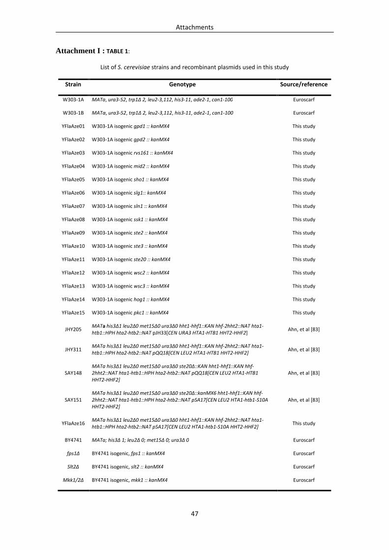
Attachments
47
Attachment I : TABLE 1:
List of S. cerevisiae strains and recombinant plasmids used in this study
Strain Genotype Source/reference
W303-1A MATa, ura3-52, trp1Δ 2, leu2-3,112, his3-11, ade2-1, can1-100 Euroscarf
W303-1B MATα, ura3-52, trp1Δ 2, leu2-3,112, his3-11, ade2-1, can1-100 Euroscarf
YFlaAze01 W303-1A isogenic gpd1 :: kanMX4 This study
YFlaAze02 W303-1A isogenic gpd2 :: kanMX4 This study
YFlaAze03 W303-1A isogenic rvs161 :: kanMX4 This study
YFlaAze04 W303-1A isogenic mid2 :: kanMX4 This study
YFlaAze05 W303-1A isogenic sho1 :: kanMX4 This study
YFlaAze06 W303-1A isogenic slg1:: kanMX4 This study
YFlaAze07 W303-1A isogenic sln1 :: kanMX4 This study
YFlaAze08 W303-1A isogenic ssk1 :: kanMX4 This study
YFlaAze09 W303-1A isogenic ste2 :: kanMX4 This study
YFlaAze10 W303-1A isogenic ste3 :: kanMX4 This study
YFlaAze11 W303-1A isogenic ste20 :: kanMX4 This study
YFlaAze12 W303-1A isogenic wsc2 :: kanMX4 This study
YFlaAze13 W303-1A isogenic wsc3 :: kanMX4 This study
YFlaAze14 W303-1A isogenic hog1 :: kanMX4 This study
YFlaAze15 W303-1A isogenic pkc1 :: kanMX4 This study
JHY205 MATa his3Δ1 leu2Δ0 met15Δ0 ura3Δ0 hht1-hhf1::KAN hhf-2hht2::NAT hta1-
htb1::HPH hta2-htb2::NAT pJH33[CEN URA3 HTA1-HTB1 HHT2-HHF2] Ahn, et al [83]
JHY311 MATa his3Δ1 leu2Δ0 met15Δ0 ura3Δ0 hht1-hhf1::KAN hhf-2hht2::NAT hta1-
htb1::HPH hta2-htb2::NAT pQQ18[CEN LEU2 HTA1-HTB1 HHT2-HHF2] Ahn, et al [83]
SAY148 MATa his3Δ1 leu2Δ0 met15Δ0 ura3Δ0 ste20Δ::KAN hht1-hhf1::KAN hhf-
2hht2::NAT hta1-htb1::HPH hta2-htb2::NAT pQQ18[CEN LEU2 HTA1-HTB1
HHT2-HHF2]
Ahn, et al [83]
SAY151 MATa his3Δ1 leu2Δ0 met15Δ0 ura3Δ0 ste20Δ::kanMX6 hht1-hhf1::KAN hhf-
2hht2::NAT hta1-htb1::HPH hta2-htb2::NAT pSA17[CEN LEU2 HTA1-htb1-S10A
HHT2-HHF2]
Ahn, et al [83]
YFlaAze16 MATa his3Δ1 leu2Δ0 met15Δ0 ura3Δ0 hht1-hhf1::KAN hhf-2hht2::NAT hta1-
htb1::HPH hta2-htb2::NAT pSA17[CEN LEU2 HTA1-htb1-S10A HHT2-HHF2] This study
BY4741 MATa; his3Δ 1; leu2Δ 0; met15Δ 0; ura3Δ 0 Euroscarf
fps1Δ BY4741 isogenic, fps1 :: kanMX4 Euroscarf
Slt2Δ BY4741 isogenic, slt2 :: kanMX4 Euroscarf
Mkk1/2Δ BY4741 isogenic, mkk1 :: kanMX4 Euroscarf

Attachments
48
Attachment II
Equations
100exp
% ×=ectedcfus
measuredcfusviability (1)
0
0expTOD
TODTcfusectedcfus n×
= (2)
%100×DMSO
ceramide (3)