floral structure and dynamics of nectar production...
TRANSCRIPT

FLORAL STRUCTURE AND DYNAMICS OF NECTAR PRODUCTION INECHINACEA PALLIDA VAR. ANGUSTIFOLIA (ASTERACEAE)
Tyler J. Wist and Arthur R. Davis1
Department of Biology, University of Saskatchewan, 112 Science Place, Saskatoon, Saskatchewan S7N 5E2, Canada
The reproductive structure of the disk florets of Echinacea pallida var. angustifolia (Asteraceae) in relation toinsect pollination was investigated using light, fluorescence, and scanning electron microscopy. The study ofthis self-incompatible species emphasized pollen production, pollen-stigma interactions, transmitting tissue,and vasculature within the style. Nectary structure and nectar production dynamics were also examined.Produced in the fused anther tubes, the trinucleate pollen with yellow pollenkitt was plentiful per floret,yielding a pollen : ovule ratio of 24,130. Encircling the style base at the ovary summit, the floral nectary pos-sessed modified stomata whose pores, as well as nonstomatal gaps in the epidermis, provided apoplasticpathways for nectar escape and reabsorption. Phloem alone supplied the gland interior, the sieve element–companion cell complexes reaching up to the nectary epidermis. Nectar was hexose dominant, its volume andnectar-sugar quantity per floret peaking on the afternoon of the first day of anthesis until the morning of thesecond day. Nectar production only occurred in half of the florets for 3 d, rarely for 5 d. Potential honeyproduction from fields of this species was estimated at 2.1–11.9 kg/ha.
Keywords: floral nectar, nectary, pollen-stigma interactions, pollination, style.
Introduction
Based on molecular data, Echinacea angustifolia has recentlybeen reclassified as a subspecies of Echinacea pallida (Binns et al.2000). Echinacea pallida var. angustifolia (narrow-leaved purpleconeflower) is a native species on the North American prairies,where it is also grown as a specialty crop on a limited acreageto supply the demand for herbal preparations from its variousorgans. The primary commodity of E. pallida var. angustifoliais its thick tap root, but secondary products include aerial stemsand achenes that may be brought to market during the yearsbefore root harvest. The species is self incompatible (McGregor1968) and hence reliant on cross-pollination for fruit set.
The inflorescences of several Echinacea spp. were studiedin detail and included measurements of many floral organs ofE. pallida var. angustifolia (McGregor 1968). Disk florets im-mediately inward from the capitulum’s showy pink ray florets(fig. 1A) open first and are protandrous (Wagenius 2004). How-ever, during the pistillate phase of the disk florets, when mostself pollen has been gathered by insects and stigmas are re-ceptive to outcross pollen, nectar is the primary reward offeredto foraging insects. No studies of nectar production through-out flowering exist for this species, yet such an investiga-tion is essential to understanding the attractiveness of theself-incompatible disk florets to insect foragers.
Although inflorescences are visited by honey bees (Apismellifera L.) for nectar and pollen, there are no ratings cur-rently available for Echinacea spp. as honey plants (Crane et al.1984). Introducing honeybee colonies to fields of E. pallidavar. angustifolia could fulfill the crop’s cross-pollination re-quirements and provide a prolonged source of floral nectar
for honey production. The potential therefore exists to develophoney as a product from Echinacea fields while providing anadequate supply of bees as cross-pollinators to ensure the pro-duction of high-quality achenes.
Accordingly, the objectives of this study of E. pallida var.angustifolia are fourfold. For the first time, (1) structural repro-ductive features of the self-incompatible disk florets as theyrelate to insect pollination are examined; (2) the interaction ofpollen grains with the stigma and pollen-tube growth throughthe style are investigated; (3) the production of nectar by diskflorets is followed from anthesis to senescence, in order to un-derstand periodicity of disk-floret attractiveness to insects; and(4) the potential for honey production is estimated.
Material and Methods
Plants and Field Sites
Mature plants of Echinacea pallida var. angustifolia (DC)Cronq. were sampled at three field sites in the summers of2004 and 2005. Chief Whitecap Park of the Meewasin ValleyAuthority is located near the South Saskatchewan River onthe northeastern side of Saskatoon. This field site, once a cul-tivated plot of this species, still contained thousands of plantsin a nonmanaged setting.
The second site was an organic farm located 7 km west ofSaskatoon on Valley Road, on the west bank of the SouthSaskatchewan River near Poplar Bluffs Canoe Launch. In 2004,the plants were in their fifth year of growth, and many hadattained a bushy habit with numerous inflorescences per plant.
Eight plants of E. pallida var. angustifolia were transplantedfrom the first site into a garden plot north of the W. P. ThompsonBuilding, University of Saskatchewan, in spring 2003. In 2004and 2005, the number of E. pallida var. angustifolia plants at
1 Author for correspondence; e-mail: [email protected].
Manuscript received August 2007; revised manuscript received November 2007.
708
Int. J. Plant Sci. 169(6):708–722. 2008.
� 2008 by The University of Chicago. All rights reserved.
1058-5893/2008/16906-0002$15.00 DOI: 10.1086/533602

this third field site increased naturally. Voucher plants wereplaced in the W. P. Fraser Herbarium (SASK). For compara-tive purposes, some observations were also made from diskflorets of Echinacea purpurea L. (Moench) growing on a green-house bench (Wist and Davis 2006).
Disk-Floret Structure
SEM. For morphological study of overall structure, disk flo-rets of the mature bud, the dehiscent staminate (SP), the first-day pistillate (PP1) stage, and the senescent (PP3) stage (fig.
1A, 1C) were harvested from E. pallida var. angustifolia plantsat the first and third field sites and processed for SEM. Inflo-rescences were bagged before the first whorl of disk floretsreached anthesis in order to prevent potential insect damageto the florets. Florets were removed from inflorescences be-fore fixation in 2% glutaraldehyde (GA) in 25 mM Na phos-phate buffer, pH 6.8, for 0.5 h. After rinsing three times withphosphate buffer, tissues were postfixed for 2 h in 1% OsO4
in buffer. Three rinses each of buffer and distilled water werefollowed by dehydration in a graded series of acetone. Tissueswere critical-point dried with liquid CO2 (Polaron Instruments,
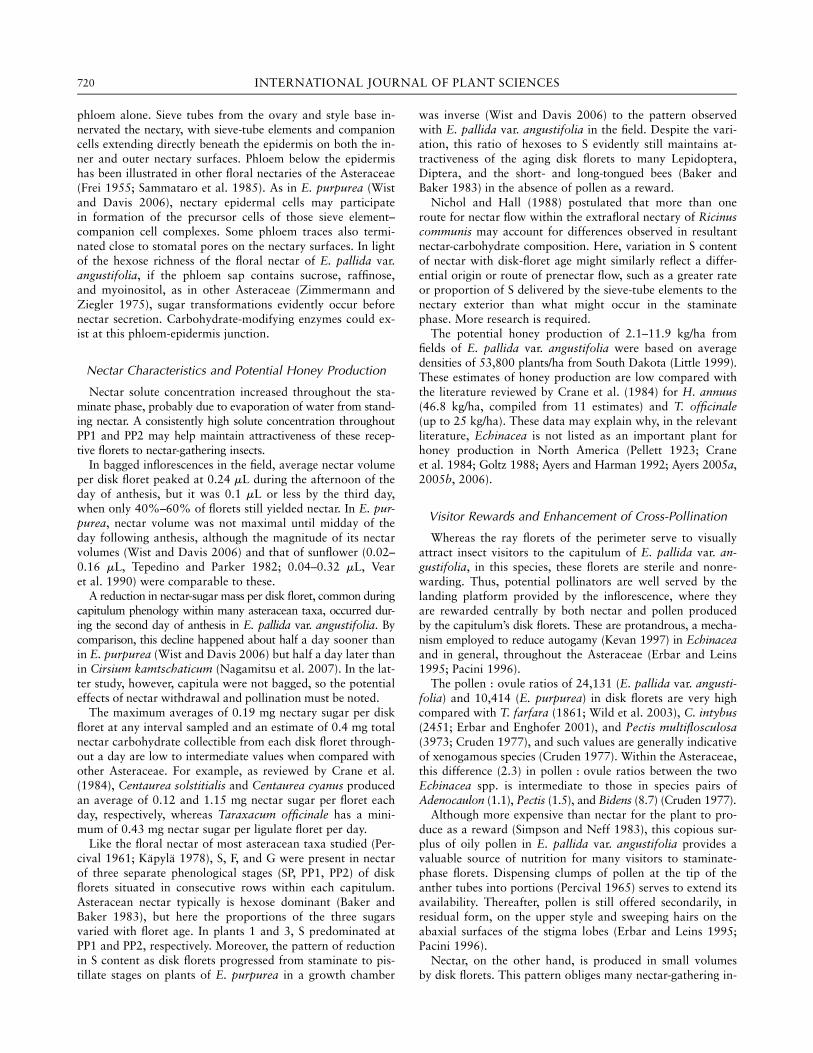
Fig. 1 A, Inflorescence of Echinacea pallida var. angustifolia visited by bee flies (Systoechus vulgaris), showing disk florets of the staminate
phase (SP); first day (PP1), second day (PP2), and third day (PP3) of the pistillate phase; ray florets (rf ); and paleae (pa). B, Disk floret dissected toshow the adnate filament (f ), anthers (an), calyx (ca), nectary (n), ovary (oy), stigma lobes (sg), style (st), and tubular corolla (ct). C, Disk florets
from five phases: mature bud, staminate phase (SP); first day (PP1), second day (PP2), and third day (PP3) of the pistillate phase; anther (an);
ovary (oy); stigma lobes (sg); style (st).
709WIST & DAVIS—FLORAL STRUCTURE IN ECHINACEA
Watford), mounted on aluminum stubs with two-sided tape,coated with gold (Edwards S150B Sputter Coater, Wilmington,MA), observed with a Philips SEM 505 (Eindhoven, Nether-lands) at 30 kV, and photographed with Polaroid 665 positive/negative film. Negatives and positives were scanned (Epson 3200Photo, Epson CX3810, Toronto, Ontario), and images were la-beled and arranged using Adobe Photoshop 7.0.
For detailed studies of nectary morphology, nectaries fromdisk florets in four distinct phenological stages (mature bud,SP, PP1, and senescent PP3; fig. 1C) were removed from ma-ture inflorescences of two plants and processed for SEM. Insome samples, exterior floral organs were first removed to ex-pose the nectary, which remained on the inferior ovary (fig.1B). Nectary dimensions and the number and developmentalstage of the modified stomata on the nectary surface werecompared between floral stages.
LM. Disk florets of two distinct developmental stages (latestaminate phase SP 24 h after anthesis, when the pollen massremained on the prereceptive stigma lobes of the pollen pre-senter, and early PP2 48 h postanthesis, with stigma lobes fullybifurcated and receptive) were harvested from the same inflo-rescence and dissected to the nectary atop a cross-sectionedovary. Tissues were fixed in 1.5% GA in 25 mM Na phos-phate buffer, pH 6.8, for 0.5 h at room temperature beforetransferring to 3% GA in buffer for 2 h. On ice, samples wererinsed with buffer for 1–12 h, postfixed overnight with 1%OsO4, then rinsed with distilled water before dehydration inan ethanol series. A gradual substitution of ethanol by propyl-ene oxide preceded sample infiltration and embedding inAraldite 502 resin at 60�C for 24 h. Semithin sections (2 mm)of floral nectaries were then cut on a Reichert OMU3 (Tucson,AZ) ultramicrotome, floated on distilled water, briefly flamedto heat-fix them to microscope slides, stained with toluidineblue (O’Brien and McCully 1981), rinsed of excess stain, andthen mounted in immersion oil under coverslips. Sections wereexamined with a Zeiss Universal (Thornwood, NY) microscope,photographed with Fujifilm Superia Iso 100 film, and arrangedusing Adobe Photoshop 7.0. Fresh tissues, including the floralnectary, were also examined after staining with I2KI for starch(Johansen 1940) and pollen grains stained with acetocarmine(Belling 1921).
Fluorescence microscopy. The occurrence of phloem in flo-ral organs such as the nectary and style was investigated us-ing epifluorescence optics. Intact florets above the ovary werestained overnight at room temperature in 0.1% aniline bluein 0.1 M K3PO4, which binds to callose (b-1,3-glucan) of sieve-tube elements (Martin 1959). The whole mount was thenslightly compressed with a coverslip on a microscope slide.
Determination of pollen quantity per floret. For E. purpurea,the average number of pollen grains per disk floret was deter-mined by counting the total grains from all five anthers withinone indehiscent but otherwise mature anther tube from eachof five plants. Four additional disk florets from the same capit-ulum of one of the same plants were randomly selected andtheir pollen contents counted. The entire anther tube was dis-sected from a disk floret before dehiscence, ensuring that allgrains produced were still present, and then split open with arazor blade to release pollen into a drop of water on a micro-scope slide. The opened anther tube was transferred throughfive subsequent water droplets until all pollen was removed
from anthers and suspended in water. Coverslips were placedover the droplets and sealed with nail polish to prevent evapo-ration. All grains from each anther tube were counted at 3100using a compound microscope (Olympus, Center Valley, PA).
For E. pallida var. angustifolia, this same method was em-ployed for the first disk floret sampled. However, owing tothe larger quantity of pollen produced in this species, maturebuds with their corolla tips closed were subsampled for theirpollen as follows. Anthers were carefully stripped longitudinallyfrom their fused tubes and transferred to an Eppendorf cen-trifuge tube (1.5 mL) containing 750 mL distilled water. Pollenwas dislodged and suspended by vortexing (Mini Vortexer,VWR, West Chester, PA) for 2 min at top speed. After 1 h, eachcentrifuge tube was vortexed again for 1 min to eliminate pollenclumping, providing an even distribution of solitary grains forcounting when 75 mL of the suspension was applied by pipetteto a microscope slide. One more disk floret from each of threeplants, plus five florets randomly chosen from the same row ofthe capitulum of another plant, were also subsampled.
Nectar dynamics throughout floret phenology. To estimatethe quantity of nectar sugar per stage of floret phenology en-countered by potential pollinators on the capitulum of E. pallidavar. angustifolia, inflorescences were bagged (2.5 mesh/mm) toexclude nectar loss to insects. Sampling of nectar was attemptedfrom disk florets during three intervals (morning: 0900–1100;midday: 1101–1400; afternoon: 1401–1900) for each phenologi-cal stage (SP, PP1–PP4). Stage PP4 is a floret like PP2 in figure 1Cbut not cross-pollinated and hence not yet senescent, by thefourth day of its pistillate phase. Disk florets were sampled fornectar in the field but not within 24 h of precipitation, to avoidany nectar dilution by rain. An individual floret was sampledonly once, rather than on repeated occasions, and a range of5–15 disk florets per phenological stage was assayed per inflo-rescence. Nectar was harvested from four inflorescences of dif-ferent plants at each of sites 1 and 2 in 2004. The majority ofnectar sampling, however, was conducted on 12 plants and 14inflorescences at site 3 in 2004 and 2005.
Nectar volume. A 0.2-mL microcapillary tube (DrummondMicrocaps) of common bore was inserted to the corolla base,and the volume of withdrawn nectar was calculated from theheight of the nectar column.
Nectar solute concentration. Following volume measurement,nectar was expelled onto the prismatic surface of a handheldrefractometer (0%–50%, 40%–85%; Bellingham and Stanley,Tunbridge Wells, Kent) modified by the manufacturer for smallquantities of fluid. Approximately 90% of samples yielded areading. These nectar solute concentrations based on mass(NCM) were corrected to 20.0�C (manufacturer’s manual).
Nectar-sugar quantity per disk floret. Corrected nectarsolute concentrations (%NCM) were converted to microgramssolute per microliter nectar (NCV) using the equation of Burquezand Corbet (1991). The nectar-sugar quantity per disk floretwas estimated as the product of nectar volume and NCV.
Nectar-carbohydrate composition. For three plants, nectarwas collected separately for each of three phenological stages(SP, PP1, PP2) by pooling nectar from several florets per whorlinto a 1-mL microcapillary before expulsion onto a filter-paperwick (McKenna and Thomson 1988). Nectar volumes in PP3and PP4 stage florets were insufficient for carbohydrate analysis.Sampling from these three stages of florets was achievable from
710 INTERNATIONAL JOURNAL OF PLANT SCIENCES
consecutive whorls within the same inflorescence, thereby al-lowing comparison of nectar-carbohydrate composition withina genotype. Air-dried wicks were stored individually in labeledenvelopes at room temperature. Wicks were later eluted indi-vidually in 2 mL Eppendorf tubes containing 150–1000 mL ofpure distilled water, depending on the dilution rate required toallow carbohydrate peaks to fall within the range of standardcurves (r ¼ 0:99 for each carbohydrate) created for each majornectar sugar (glucose, fructose, sucrose; 5–200 ppm). After filter-ing, 50 mL of each sample were analyzed in duplicate using a Wa-ters HPLC system, as described in Davis et al. (1998).
Results
Inflorescences and Ray Florets
Typically, in its first year of growth, each plant of Echina-cea pallida var. angustifolia begins as a basal rosette of leavesthat bolts in late spring or early summer to produce a singlecapitulum. However, some plants do not produce an inflores-cence in their first year of growth. By the third through fifthyears of growth, the plant often takes on a bushy appearanceand can produce multiple inflorescences from shoots derivedfrom lateral rhizomes. The root and rhizome are perennialand increase in size during each growing season, sending outnew shoots and leaves each spring.
The first inflorescence to reach anthesis was borne termi-nally on the central stalk (peduncle). Other inflorescencesmay develop from axillary buds on the shoot, depending onplant age. Surrounding the inflorescence were green, imbri-cate, involucral bracts covered in trichomes visible beneaththe ray florets (fig. 1A). The cone-shaped capitulum entersanthesis with maturation of the outer single whorl of sterile,ligulate (ray) florets, which surround multiple whorls of diskflorets (fig. 1A). Corollas of ray florets comprised three petalsin the ligule, lacked ‘‘nectar-guide’’ patterns, and ranged incolor from purplish pink to deep purple; most drooped whenmature. Ray florets were sterile, lacking an androecium anda full gynoecium, except for an ovary. In rare cases, ray flo-rets possessed a stigma and style, but it was not determinedwhether these florets produced viable achenes. In all otherray florets, the ovary wall of the achene hardened but failedto yield a viable fruit due to the incomplete gynoecium.
Phenology of Disk Floret
After expansion and pigmentation of the corolla of the rayflorets (fig. 1A) that coincided with lateral expansion of theinflorescence, the protandrous disk florets developed centrip-etally and sequentially, in whorls. The mean number of diskflorets per capitulum was 202 6 3:7 (SE; n ¼ 280 plantssampled at the three field sites over two seasons). One whorlof mature buds reached anthesis per day by entering into thestaminate stage (SP) upon anther dehiscence (fig. 1A, 1C).Anthers fused along their length protruded beyond the corollatube in the early morning of the staminate phase, and a yellowpollen mass (fig. 1C, SP) typically was presented by 0900, de-pending on climate. Pollen release often coincided with inci-dence of the morning sun on the anthers; pollen was notpresented during rainy periods. On the second day of flower-
ing, the elongating style extended through the anther tubeand pollen mass before the two stigma lobes reflexed; thisinitial pistillate phase was designated PP1 (fig. 1A, 1C). StagePP2 (fig. 1C) marked the second day of stigmatic receptivity.Disk florets remained in the receptive pistillate stage for upto 8 d (e.g., PP2 in fig. 1C), until anthesis was complete, oruntil cross-pollination occurred, at which point the stigma lobesand style shrivelled at senescence and withdrew into the co-rolla tube (senescent PP3 in fig. 1C).
Perianth Morphology of Disk Florets
Each disk floret of E. pallida var. angustifolia was subtendedby a bract (palea) (fig. 1A) that gave the capitulum’s centeran echinate appearance. Each disk floret was perfect and epig-ynous (fig. 1B), with the inferior ovary embedded in the cone-shaped inflorescence.
Sepals of the fused calyx (pappus) were highly reduced and ex-isted only as a fringe of green tissue at the ovary summit, sur-rounding the corolla base (fig. 1B, 1C; fig. 2E, 2F). The pappushad unevenly serrate margins (fig. 2F) with a prominent exten-sion on the side adjacent to the palea (fig. 2E, 2F, bottom).
Corolla color varied from green at anthesis (fig. 1C, left)to reddish purple in the pistillate phase (fig. 1C, right). Thecampanulate corolla of five petals (fig. 2B, 2D) initially had anarrow base (figs. 1C, left, 2E) that gradually enlarged to abulge (fig. 1C, right), thus serving as a nectar reservoir. Atthe corolla apex, petal lobes were distinct (fig. 1C; 2B, 2D).Patches of short, unicellular trichomes were often found onthe lobe margins (fig. 2A). However, uniseriate trichomes oc-curred infrequently and at random intervals on the outer co-rolla surface (fig. 2B, 2D).
Androecium of Disk Florets
The androecium was composed of five synantherous sta-mens. The stamens were epipetalous, their filaments adnateto the petals basally (figs. 1B, 2E, 4A). Anthers were coales-cent and fused into a tube (fig. 2D), the filaments remainingfree (fig. 2E). Each mature anther contained two elongatedpollen sacs (microsporangia) that released pollen grains byinward longitudinal dehiscence up to the bases of the anther’sapical extensions (fig. 2C). Pollen aggregation within pollensacs was evident when anther walls were removed, leavingthe grains molded into the shape of the pollen sacs (fig. 2G).Pollen presentation was secondary, with the microgameto-phytes released inside the tube being swept out and presentedon the top of the anther tube (SP in fig. 1C) by the extrusionof the stigma (fig. 2D) and contraction of the filaments (PP1in fig. 1C), which was evident when gentle pressure was ap-plied to filaments with a glass micropipette. Each morningbetween sunrise and 0900, a new whorl of disk florets openedinto the indehiscent staminate stage (SPi). The florets releasedtheir pollen from the top of the tube between 0900 and 1100,weather permitting, as they entered the dehiscent staminatephase (SPd). By 2130, florets had passed through the staminatephase and entered the receptive pistillate phase (PP1) charac-terized by reflexed stigma lobes.
Pollen grains of E. pallida var. angustifolia were yellow atdehiscence (fig. 1A–1C) and had a tendency to clump togetherdue to the presence of yellow droplets of oily pollenkitt ad-
711WIST & DAVIS—FLORAL STRUCTURE IN ECHINACEA
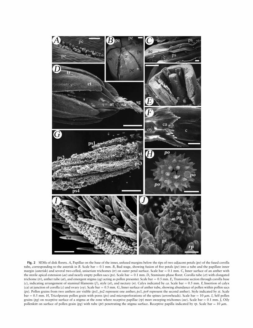
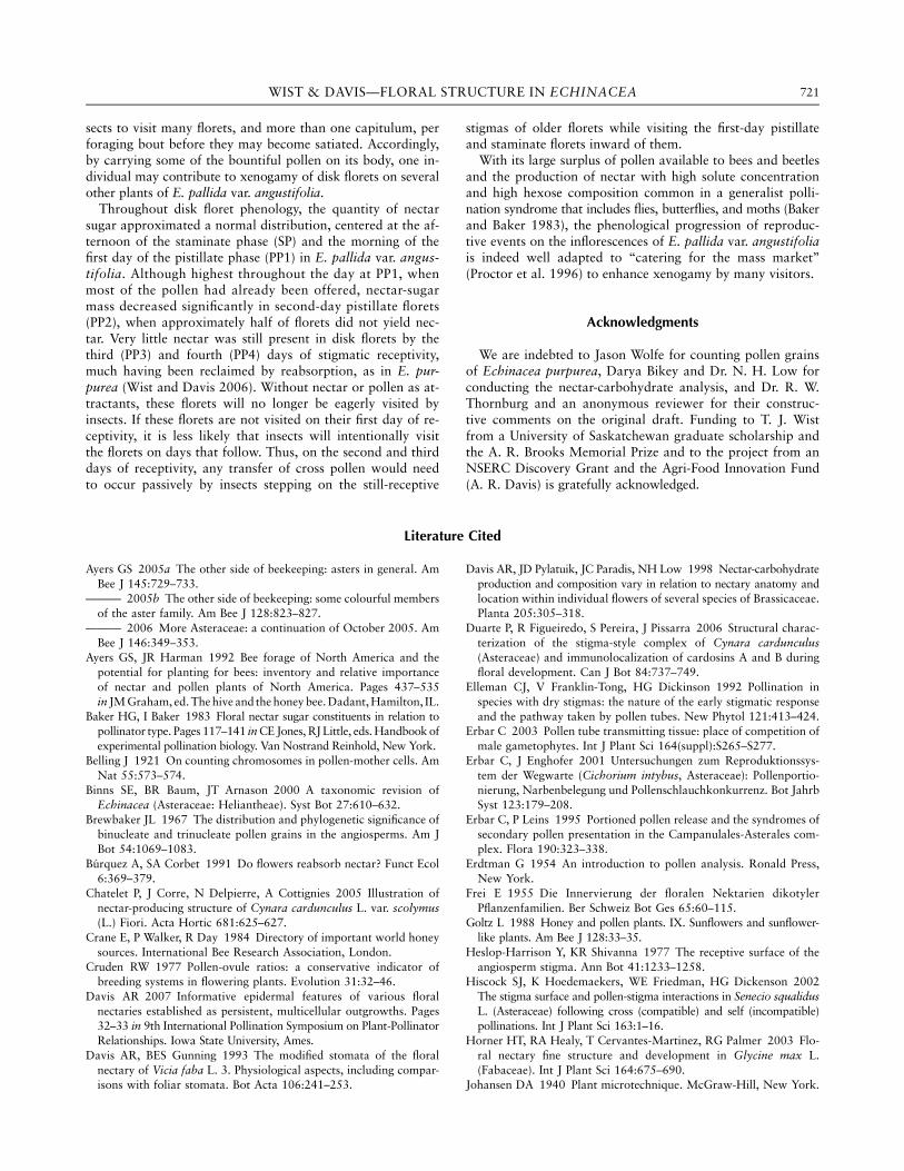
Fig. 2 SEMs of disk florets. A, Papillae on the base of the inner, unfused margins below the tips of two adjacent petals (pe) of the fused corolla
tube, corresponding to the asterisk in B. Scale bar ¼ 0:1 mm. B, Bud stage, showing fusion of five petals (pe) into a tube and the papillate inner
margin (asterisk) and several two-celled, uniseriate trichomes (tr) on outer petal surface. Scale bar ¼ 0:1 mm. C, Inner surface of an anther withthe sterile apical extension (ae) and nearly empty pollen sacs (ps). Scale bar ¼ 0:1 mm. D, Staminate-phase floret. Corolla tube (ct) with elongated
trichome (tr), anther tube (at), and emergent stigma (sg) acting as pollen presenter. Scale bar ¼ 0:5 mm. E, Transverse section through corolla base
(c), indicating arrangement of staminal filaments (f ), style (st), and nectary (n). Calyx indicated by ca. Scale bar ¼ 0:5 mm. F, Insertion of calyx
(ca) at junction of corolla (c) and ovary (oy). Scale bar ¼ 0:5 mm. G, Inner surface of anther tube, showing abundance of pollen within pollen sacs(ps). Pollen grains from two anthers are visible (ps1, ps2 represent one anther; ps3, ps4 represent the second anther). Style indicated by st. Scale
bar ¼ 0:5 mm. H, Tricolporate pollen grain with pores (po) and microperforations of the spines (arrowheads). Scale bar ¼ 10 mm. I, Self pollen
grains (pg) on receptive surface of a stigma at the zone where receptive papillae (rp) meet sweeping trichomes (sw). Scale bar ¼ 0:1 mm. J, Oilypollenkitt on surface of pollen grain (pg) with tube (pt) penetrating the stigma surface. Receptive papilla indicated by rp. Scale bar ¼ 10 mm.
hering to the grain exterior (figs. 2J, 3A). Grains were echi-nate with microperforated spines and tricolporate with threetypically prominent apertures (fig. 2H).
Pollen grains of E. pallida var. angustifolia and also Echi-nacea purpurea (Wist 2005) were trinucleate at the time ofanther dehiscence (fig. 3A). One round tube (vegetative cell)nucleus and two laminar sperm cells—often parallel to eachother—were visible within the cytoplasm.
In E. purpurea, the average quantity of pollen grains fromfive indehiscent anther tubes randomly selected within onecapitulum was 9296 6 666 (SE). Total pollen from single diskflorets from four other plants ranged from 7464 to 14,231grains. Thus, with a solitary ovule available per floret (fig. 4A),the pollen : ovule ratio was 10;414 6 2239 (SE; n ¼ 5 plants).
In E. pallida var. angustifolia, five disk florets within thesame row of an inflorescence averaged 26;580 6 591 (SE)pollen grains. From the other four plants sampled, total pol-len per disk floret varied from 17,910 to 29,464 grains, yield-ing a pollen : ovule ratio of 24;131 6 4770 (SE; n ¼ 5).
Gynoecium of Disk Florets
The gynoecium consisted of one bilobed stigma, one style,and one inferior ovary (fig. 1B). Within the unilocular ovarywas one ovule (fig. 4A). The stigma lobes were equal in length(fig. 1B) and had long sweeping trichomes (fig. 2D, 2I; fig. 4I)on the nonreceptive abaxial surface that transported pollengrains from the interior of the anther tube and presentedthem above the anther tips (SP in fig. 1C). The two receptivesurfaces of the stigma abutted and covered each other at thistime, preventing the majority of self pollen from contactingthem. Separation of the stigmatic lobes (fig. 1B; PP1 in fig.1C) signified onset of the pistillate phase. The receptive adax-ial surface of the stigma was covered with papillae (fig. 2I,2J; fig. 4I) that lacked any exudate (i.e., a dry-type stigma).If the stigma was not cross-pollinated after several days of re-ceptivity, the stigma lobes reflexed further to potentiallybring their receptive adaxial surfaces into contact with selfpollen grains remaining on the anthers.
One vascular bundle containing both xylem and phloementered each stigmatic lobe (fig. 4I) after traveling up eitherside of the style (fig. 3B) from its base (fig. 3D, 3E), often withseveral isolated sieve tubes found several cell layers awayfrom the phloem of the two wide vascular bundles (fig. 3B, 3D).These isolated phloem traces were also found in E. purpurea (fig.3C). In the center of the style was the transmitting tract forconducting pollen tubes to the ovule (fig. 3C, 3D). The centerof the transmitting tract was hollow near the base of the stylein E. pallida var. angustifolia (fig. 3D; fig. 4G, 4H, 4J) butlacked this hollow center in the majority of the upper portionof the style in both E. pallida var. angustifolia and E. purpurea(fig. 3C). A dense-staining intercellular matrix occurred amongcells of the transmitting tract (fig. 3C, 3D). The transmittingtract extended into each stigma lobe and was found directlybeneath the papillae of the receptive surface, where it eventu-ally declined to one cell layer at the stigma lobe tip from a denselayer 4–5 cells in thickness in the rest of the lobe (fig. 4I).Each compatible pollen grain on the receptive surface couldproduce a single pollen tube (fig. 2J) that passed between thepapillae and penetrated the transmitting tissue beneath.
Nectary Anatomy
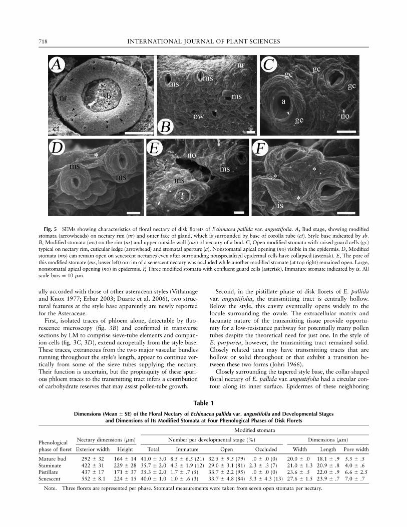
The yellowish nectary of the disk florets takes its final cup-shaped and pentamerous form (fig. 5A) from pressure exertedon its inner surface during its formation by the style base andthe bases of the five fused petals from outside it. Nectary diam-eter increased throughout the disk-floret phenology by meansof senescent florets that are slightly less than twice the width ofmature buds (table 1). Nectary height was less variable acrossstages, with mean sizes ranging from 164 to 229 mm (table 1).
Modified stomata occurred on the nectary surface and werethe most likely route for nectar escape. However, nonstomatalopenings that may also serve as sites of nectar escape wereobserved in the epidermis (fig. 5C, 5E). Most stomata occurredsingly, but there were infrequent exceptions (figs. 3F, 5F) whereguard mother cells of adjacent stomata may have arisen fromthe same cell division. Both guard cells and nonspecializedepidermal cells had a ridged appearance owing to the cuticularridges that on the guard cells ran circumferentially along eachside of the pore (fig. 5C, 5D, 5F). Nectaries had an average of35–41 modified stomata (table 1), with the majority located onthe upper rim (fig. 3E; fig. 4D, 4E, 4G; fig. 5A) but with severaloccurring on the lower nectary walls (figs. 3E, 5B). Nectaries ofmature buds had a greater proportion of immature stomata(21%; table 1), where the outer cuticle spanning the circum-ferential ledges had not yet torn to reveal the stomatal porebeneath, than those of the three older floret stages. In prese-cretory mature buds, the majority of modified stomata wereopen, however, and remained so throughout the stages of floretdevelopment until some (13%, table 1) were occluded (fig.5E, bottom), sometimes by underlying parenchyma cells (fig.5F). Most modified stomata remained open during the senes-cent phase (84%, table 1) even when adjacent epidermal cellshad already collapsed (fig. 5D), and pore width remained large(table 1). Substomatal chambers beneath the modified stomatacould be large (fig. 4F) or small (fig. 4D, 4E, bottom left).
Modified stomata contained more starch (fig. 3E, 3F; fig.4E, 4G) than both nonspecialized epidermal cells and nectaryparenchyma cells (fig. 3F; fig. 4F, 4G). Within the nectary,parenchyma cells were the dominant cell type and were smallbut variable in size (fig. 4F), ranging from ;10 to 20 mm.
The vasculature of the nectary consisted of phloem alonewith no xylem progressing beyond the style or ovary into thenectary. Nectary phloem had two origins: vertical tracesthrough phloem sieve tubes from the ovary (fig. 4B–4D) andhorizontally oriented traces arising from phloem at the ex-treme base of the style (fig. 4H, 4J). Within the nectary, sievetubes could branch and extend horizontally (fig. 3F, 3G), thencontinue vertically until they ended near or, in infrequent in-stances, at (fig. 3H) the pores of modified stomata. Sieve tubeswere found adjacent to the outer epidermis of the nectary (fig.4B, 4D), and both sieve elements and companion cells were indirect contact with epidermal cells (fig. 4F, 4H, 4K).
Dynamics of Nectar Secretion
Plants of E. pallida var. angustifolia from the three fieldsites were sampled for nectar, and these data were combinedbecause the patterns were similar. Figure 6 illustrates the changesin mean nectar volume and nectar-sugar quantity per diskfloret spanning the commencement of nectar accumulation
713WIST & DAVIS—FLORAL STRUCTURE IN ECHINACEA
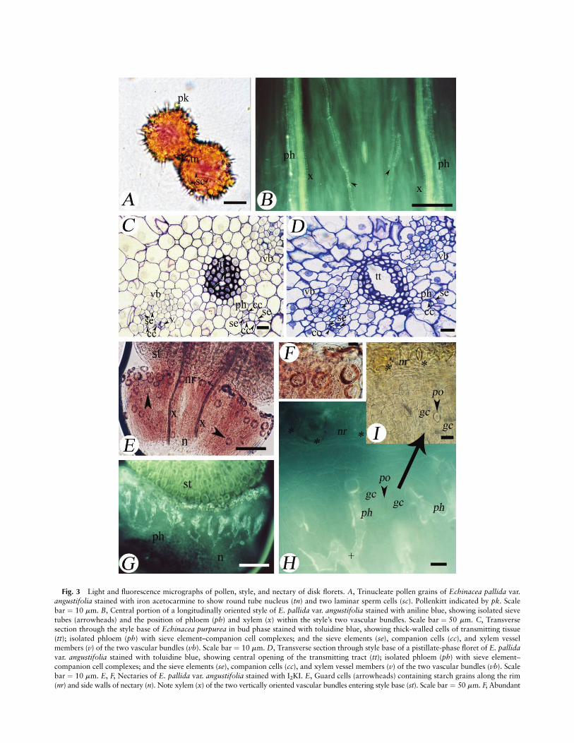
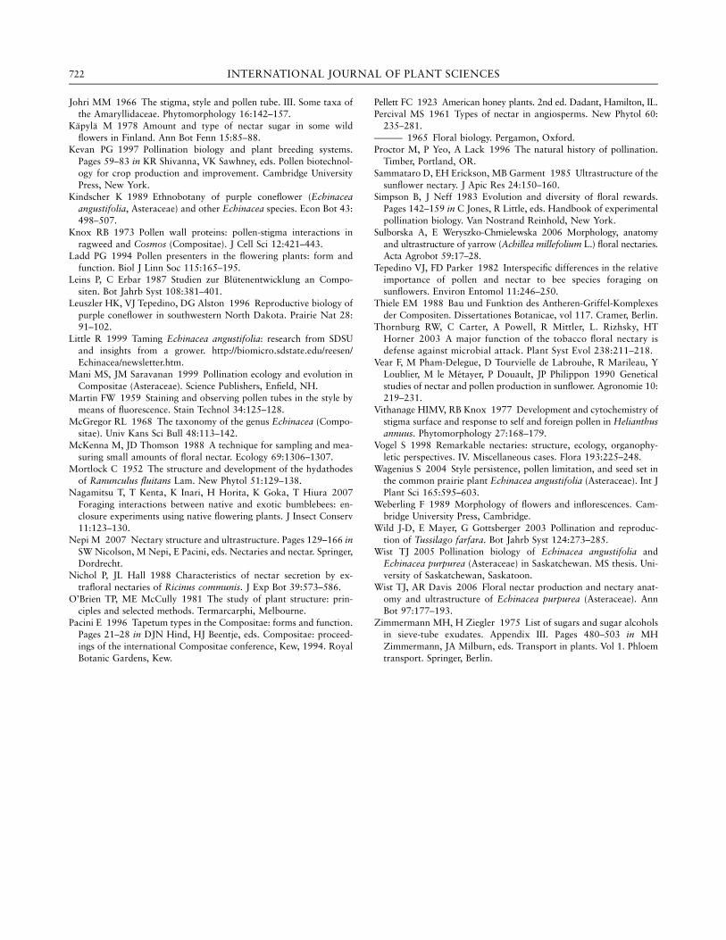
Fig. 3 Light and fluorescence micrographs of pollen, style, and nectary of disk florets. A, Trinucleate pollen grains of Echinacea pallida var.
angustifolia stained with iron acetocarmine to show round tube nucleus (tn) and two laminar sperm cells (sc). Pollenkitt indicated by pk. Scale
bar ¼ 10 mm. B, Central portion of a longitudinally oriented style of E. pallida var. angustifolia stained with aniline blue, showing isolated sieve
tubes (arrowheads) and the position of phloem (ph) and xylem (x) within the style’s two vascular bundles. Scale bar ¼ 50 mm. C, Transversesection through the style base of Echinacea purpurea in bud phase stained with toluidine blue, showing thick-walled cells of transmitting tissue
(tt); isolated phloem (ph) with sieve element–companion cell complexes; and the sieve elements (se), companion cells (cc), and xylem vessel
members (v) of the two vascular bundles (vb). Scale bar ¼ 10 mm. D, Transverse section through style base of a pistillate-phase floret of E. pallidavar. angustifolia stained with toluidine blue, showing central opening of the transmitting tract (tt); isolated phloem (ph) with sieve element–companion cell complexes; and the sieve elements (se), companion cells (cc), and xylem vessel members (v) of the two vascular bundles (vb). Scale
bar ¼ 10 mm. E, F, Nectaries of E. pallida var. angustifolia stained with I2KI. E, Guard cells (arrowheads) containing starch grains along the rim
(nr) and side walls of nectary (n). Note xylem (x) of the two vertically oriented vascular bundles entering style base (st). Scale bar ¼ 50 mm. F, Abundant
(morning of the staminate phase SP) to the cessation of nec-tar production by the end of the third day of the pistillatephase (PP3), 4 d postanthesis.
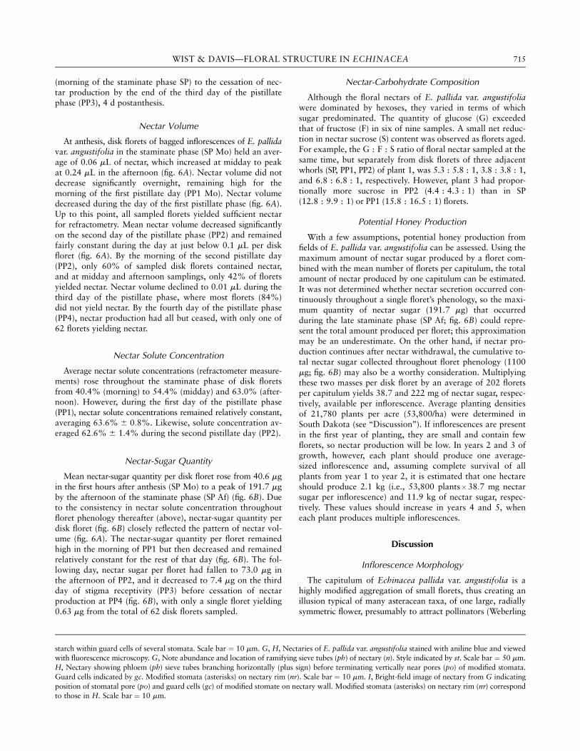
Nectar Volume
At anthesis, disk florets of bagged inflorescences of E. pallidavar. angustifolia in the staminate phase (SP Mo) held an aver-age of 0.06 mL of nectar, which increased at midday to peakat 0.24 mL in the afternoon (fig. 6A). Nectar volume did notdecrease significantly overnight, remaining high for themorning of the first pistillate day (PP1 Mo). Nectar volumedecreased during the day of the first pistillate phase (fig. 6A).Up to this point, all sampled florets yielded sufficient nectarfor refractometry. Mean nectar volume decreased significantlyon the second day of the pistillate phase (PP2) and remainedfairly constant during the day at just below 0.1 mL per diskfloret (fig. 6A). By the morning of the second pistillate day(PP2), only 60% of sampled disk florets contained nectar,and at midday and afternoon samplings, only 42% of floretsyielded nectar. Nectar volume declined to 0.01 mL during thethird day of the pistillate phase, where most florets (84%)did not yield nectar. By the fourth day of the pistillate phase(PP4), nectar production had all but ceased, with only one of62 florets yielding nectar.
Nectar Solute Concentration
Average nectar solute concentrations (refractometer measure-ments) rose throughout the staminate phase of disk floretsfrom 40.4% (morning) to 54.4% (midday) and 63.0% (after-noon). However, during the first day of the pistillate phase(PP1), nectar solute concentrations remained relatively constant,averaging 63:6% 6 0:8%. Likewise, solute concentration av-eraged 62:6% 6 1:4% during the second pistillate day (PP2).
Nectar-Sugar Quantity
Mean nectar-sugar quantity per disk floret rose from 40.6 mgin the first hours after anthesis (SP Mo) to a peak of 191.7 mgby the afternoon of the staminate phase (SP Af) (fig. 6B). Dueto the consistency in nectar solute concentration throughoutfloret phenology thereafter (above), nectar-sugar quantity perdisk floret (fig. 6B) closely reflected the pattern of nectar vol-ume (fig. 6A). The nectar-sugar quantity per floret remainedhigh in the morning of PP1 but then decreased and remainedrelatively constant for the rest of that day (fig. 6B). The fol-lowing day, nectar sugar per floret had fallen to 73.0 mg inthe afternoon of PP2, and it decreased to 7.4 mg on the thirdday of stigma receptivity (PP3) before cessation of nectarproduction at PP4 (fig. 6B), with only a single floret yielding0.63 mg from the total of 62 disk florets sampled.
Nectar-Carbohydrate Composition
Although the floral nectars of E. pallida var. angustifoliawere dominated by hexoses, they varied in terms of whichsugar predominated. The quantity of glucose (G) exceededthat of fructose (F) in six of nine samples. A small net reduc-tion in nectar sucrose (S) content was observed as florets aged.For example, the G : F : S ratio of floral nectar sampled at thesame time, but separately from disk florets of three adjacentwhorls (SP, PP1, PP2) of plant 1, was 5.3 : 5.8 : 1, 3.8 : 3.8 : 1,and 6.8 : 6.8 : 1, respectively. However, plant 3 had propor-tionally more sucrose in PP2 (4.4 : 4.3 : 1) than in SP(12.8 : 9.9 : 1) or PP1 (15.8 : 16.5 : 1) florets.
Potential Honey Production
With a few assumptions, potential honey production fromfields of E. pallida var. angustifolia can be assessed. Using themaximum amount of nectar sugar produced by a floret com-bined with the mean number of florets per capitulum, the totalamount of nectar produced by one capitulum can be estimated.It was not determined whether nectar secretion occurred con-tinuously throughout a single floret’s phenology, so the maxi-mum quantity of nectar sugar (191.7 mg) that occurredduring the late staminate phase (SP Af; fig. 6B) could repre-sent the total amount produced per floret; this approximationmay be an underestimate. On the other hand, if nectar pro-duction continues after nectar withdrawal, the cumulative to-tal nectar sugar collected throughout floret phenology (1100mg; fig. 6B) may also be a worthy consideration. Multiplyingthese two masses per disk floret by an average of 202 floretsper capitulum yields 38.7 and 222 mg of nectar sugar, respec-tively, available per inflorescence. Average planting densitiesof 21,780 plants per acre (53,800/ha) were determined inSouth Dakota (see ‘‘Discussion’’). If inflorescences are presentin the first year of planting, they are small and contain fewflorets, so nectar production will be low. In years 2 and 3 ofgrowth, however, each plant should produce one average-sized inflorescence and, assuming complete survival of allplants from year 1 to year 2, it is estimated that one hectareshould produce 2.1 kg (i.e., 53,800 plants 3 38:7 mg nectarsugar per inflorescence) and 11.9 kg of nectar sugar, respec-tively. These values should increase in years 4 and 5, wheneach plant produces multiple inflorescences.
Discussion
Inflorescence Morphology
The capitulum of Echinacea pallida var. angustifolia is ahighly modified aggregation of small florets, thus creating anillusion typical of many asteracean taxa, of one large, radiallysymmetric flower, presumably to attract pollinators (Weberling
starch within guard cells of several stomata. Scale bar ¼ 10 mm. G, H, Nectaries of E. pallida var. angustifolia stained with aniline blue and viewedwith fluorescence microscopy. G, Note abundance and location of ramifying sieve tubes (ph) of nectary (n). Style indicated by st. Scale bar ¼ 50 mm.
H, Nectary showing phloem (ph) sieve tubes branching horizontally (plus sign) before terminating vertically near pores (po) of modified stomata.
Guard cells indicated by gc. Modified stomata (asterisks) on nectary rim (nr). Scale bar ¼ 10 mm. I, Bright-field image of nectary from G indicating
position of stomatal pore (po) and guard cells (gc) of modified stomate on nectary wall. Modified stomata (asterisks) on nectary rim (nr) correspondto those in H. Scale bar ¼ 10 mm.
715WIST & DAVIS—FLORAL STRUCTURE IN ECHINACEA

Fig. 4 Light micrographs of disk florets of Echinacea pallida var. angustifolia. A–D, Longitudinal sections through a staminate-phase floret.E–K, Cross sections through style base and nectary of a pistillate-stage floret. A, Tangential section showing nectary (n) below globular base of
style (st), and ovule (ov) within inferior ovary (oy). Staminal filaments (f ), corolla tube (ct), and reduced calyx (ca) also evident. Scale bar ¼ 100 mm.
B, Vascular trace extending from ovary base toward bases of corolla tube (ct) and nectary (n) with xylem vessels (x) and phloem sieve tubes
(arrowheads) evident in longitudinal section. Phloem (ph) sieve elements and companion cells occur in cross section. C, Higher magnification ofB showing the longitudinal vessels of xylem (x) and cross-sectional phloem (ph) in the ovary. D, Portion of nectary disk enclosed by base of
corolla tube (ct) with phloem extending as a sieve tube through ovary (oy) and presumably connecting to phloem below nectary epidermis
(arrowheads). Style indicated by st. E, Nectary rim with guard cells (gc) of modified stomata cut in various planes to show amyloplasts (s)within guard cells, substomatal chambers (sc), and undulating surface of style base (st). Stomatal aperture indicated by a. F, Large substomatalchamber (sc) continuous with aperture of a modified stomate. Note narrow interface between style base (st) and inner epidermis of nectary (ep)
and presence of a phloem sieve element (se) and companion cell (cc) adjacent to inner epidermis. Guard cells indicated by gc. G, Oblique
section of nectary showing intact guard cells (gc) amid deteriorating cells along rim (nr). At right (asterisk), cells of nectary base are contiguouswith cells of style base (st). Transmitting tract indicated by tt. Note concave nature of exposed stylar epidermal cells (arrowhead), giving an

1989; Proctor et al. 1996). The common name, purple cone-flower, derives from the raised base of the inflorescence thatbears the disk florets. The outer purple, petal-like ray floretsare the first to reach anthesis and attract insects visually, butin this species, they are functionally sterile, lacking an androe-cium, entire gynoecium, or nectary. Each disk floret is sub-tended by a modified bract (palea) protruding beyond thecorolla, and this stiff, spiny structure led to the generic namefrom the Greek echinos, a hedgehog (Kindscher 1989).
Structure and Phenology of Disk Florets
The membranous, scalelike pappus is the reduced form of thefree, upper limbs of the sepals and apparently serves no func-tional purpose to disk florets of E. pallida var. angustifolia otherthan the calyx base fusing around the ovary, thus forming partof the indehiscent, mature fruit, as in several other asteraceantaxa (Mani and Saravanan 1999). Without a hairlike pappusspecialized for wind dispersal, such as in dandelion Taraxacumofficinale, primary dispersal of the Echinacea achenes is reduced.
The fused corolla tubes of E. pallida var. angustifolia flo-rets did not differ from the descriptions of McGregor (1968),who reported their lengths as 6–8 mm. The corolla’s bellshape protects the androecium and upper gynoecium prean-thesis, and its basal expansion postanthesis serves as a nectarreservoir. Filament bases also attach at the corolla base, com-mon in the Asteraceae (Leins and Erbar 1987). At the stamentips, the sterile anther apices may function in pollen portion-ing (Thiele 1988) and for protection of pollen shed withinthe anther tube from environmental damage, e.g., by exclud-ing rain during the period when the style does not yet presentpollen. At maturity, each bisporangiate anther of the tubecontained microgametophytes that fell within the range (19–26 mm) reported for E. pallida var. angustifolia by McGregor(1968), ranking them small to intermediate within Asteraceae(Erdtman 1954). Pollen grains were echinate, a common fea-ture of the Tubuliflorae (Erdtman 1954). The spines plus oilypollenkitt probably assist dispersal by helping grains adhereto body hairs of inflorescence-visiting insects. Before antherdehiscence, mature pollen grains of E. pallida var. angustifo-lia and Echinacea purpurea (Wist 2005) already containeda vegetative (tube) nucleus plus two sperm cells, evidentlythe first record of trinucleate pollen in Echinacea, typical ofAsteraceae (Brewbaker 1967).
The gynoecium of each epigynous disk floret consisted ofan inferior, unilocular ovary containing a single ovule. Atopthe ovary, the style led to a bilobed stigma, conventional inAsteraceae (Mani and Saravanan 1999).
Stigmas of 17 asteracean genera were described as ‘‘dry,’’a feature common in taxa that employ a sporophytic self-
incompatibility system to prevent self pollination (Heslop-Harrison and Shivanna 1977). The stigma of E. pallida var.angustifolia bifurcates into two lobes that reflex when recep-tive and may become moist, according to Leuszler et al. (1996).No evidence of a ‘‘wet’’ stigma was found in our observationsof E. pallida var. angustifolia, but the stigmas of other Aster-aceae may produce a small exudate (Knox 1973; Vithanageand Knox 1977; Elleman et al. 1992; Hiscock et al. 2002),and so technically are not dry (Elleman et al. 1992) but semi-dry (Hiscock et al. 2002). Stigmas of Echinacea may also sharethis semidry condition.
Stigmas of E. pallida var. angustifolia served a dual func-tion during floret phenology. Initially, in the staminate phase,the unreceptive stigma with its adpressed lobes acted as a sec-ondary pollen presenter (see Pacini 1996). The abaxial, non-receptive surface of the stigma bore elongated trichomes thatswept out pollen grains from the anther tube for presentationin the staminate phase, as the style lengthened and the stami-nal filaments contracted. Although pollen-catching hairs areabsent on the stigma of some Asteraceae (Knox 1973), thistype of active pollen presentation is typical (Ladd 1994; Pacini1996). In addition, the stigma lobes reflexed during the diskfloret’s pistillate phase to expose their adaxial, receptive sur-faces of unicellular papillae. Xenogamous pollen grains trans-ferred by pollinators adhered to the surface of these papillae,where germination of pollen tubes initiated from pores onthe grain surface. The single, elongating pollen tube from acompatible grain penetrated the stigma surface extracellularly,between receptive papillae, presumably where the cuticle isabsent or thin (Elleman et al. 1992; Hiscock et al. 2002). Stig-mas that remained free from cross-pollination for several dayscurled downward and contacted self pollen that may yet clingresidually to the style and/or anther tips (fig. 2C). However,the effectiveness of this final, autogamous self-pollination pro-cess is unknown. The pistillate phase in E. pallida var. angus-tifolia eventually ends with floret senescence after 8–10 d,when the style shrivels and retracts into the corolla tube (fig.1C; Wagenius 2004).
The style interior served as the conduit for pollen-tubegrowth of compatible grains from the stigma to the inferiorovary. A central, clearly delimited core of transmitting tractoccurred between the style’s two vascular bundles, extendingupward into each stigmatic lobe. Transmitting tissue also oc-curs below the stigmatic papillae in Cichorium intybus (Erbar2003), Cynara cardunculus (Duarte et al. 2006), and Helianthusannuus (Vithanage and Knox 1977). Passage of pollen tubesoccurred extracellularly within the matrix surrounding thetransmitting-tract cells, which in transverse section were round-ish and small in diameter. Although these characteristics ofthe pollen-tube pathway in E. pallida var. angustifolia gener-
irregular surface. H, Horizontal phloem trace with sieve element (se) and companion cell (cc) extending externally from one of the two regular
vascular bundles (vb) and traversing stylar tissue (st) toward nectary (n). Transmitting tract indicated by tt. Note phloem cells adjacent to
epidermis and amid parenchyma cells of nectary (small arrowheads). I, Longitudinal section through tip of stigma lobe with a 1–4-celled layer
of transmitting tract (tt) directly beneath papillae (rp) of receptive surface. Sweeping trichomes (sw) of pollen presenter and vessels of xylemtrace (x). J, Horizontal sieve tube element (se) and companion cell (cc) supplying nectary and originating from phloem (arrowhead) external to
vascular bundles (vb) of style. K, Higher magnification of outer epidermis of H. Sieve elements (se) and companion cells (cc) of vascular tissue
next to outer epidermis (ep) of nectary. Other sieve element–companion cell complexes (arrowheads) evident amid nectary parenchyma. Scale
bars for B–K ¼ 10 mm.
717WIST & DAVIS—FLORAL STRUCTURE IN ECHINACEA
ally accorded with those of other asteracean styles (Vithanageand Knox 1977; Erbar 2003; Duarte et al. 2006), two struc-tural features at the style base apparently are newly reportedfor the Asteraceae.
First, isolated traces of phloem alone, detectable by fluo-rescence microscopy (fig. 3B) and confirmed in transversesections by LM to comprise sieve-tube elements and compan-ion cells (fig. 3C, 3D), extend acropetally from the style base.These traces, extraneous from the two major vascular bundlesrunning throughout the style’s length, appear to continue ver-tically from some of the sieve tubes supplying the nectary.Their function is uncertain, but the propinquity of these spuri-ous phloem traces to the transmitting tract infers a contributionof carbohydrate reserves that may assist pollen-tube growth.
Second, in the pistillate phase of disk florets of E. pallidavar. angustifolia, the transmitting tract is centrally hollow.Below the style, this cavity eventually opens widely to thelocule surrounding the ovule. The extracellular matrix andlacunate nature of the transmitting tissue provide opportu-nity for a low-resistance pathway for potentially many pollentubes despite the theoretical need for just one. In the style ofE. purpurea, however, the transmitting tract remained solid.Closely related taxa may have transmitting tracts that arehollow or solid throughout or that exhibit a transition be-tween these two forms (Johri 1966).
Closely surrounding the tapered style base, the collar-shapedfloral nectary of E. pallida var. angustifolia had a circular con-tour along its inner surface. Epidermes of these neighboring
Table 1
Dimensions (Mean 6 SE) of the Floral Nectary of Echinacea pallida var. angustifolia and Developmental Stagesand Dimensions of Its Modified Stomata at Four Phenological Phases of Disk Florets
Modified stomata
Nectary dimensions (mm) Number per developmental stage (%) Dimensions (mm)Phenological
phase of floret Exterior width Height Total Immature Open Occluded Width Length Pore width
Mature bud 292 6 32 164 6 14 41.0 6 3.0 8.5 6 6.5 (21) 32.5 6 9.5 (79) .0 6 .0 (0) 20.0 6 .0 18.1 6 .9 5.5 6 .5
Staminate 422 6 31 229 6 28 35.7 6 2.0 4.3 6 1.9 (12) 29.0 6 3.1 (81) 2.3 6 .3 (7) 21.0 6 1.3 20.9 6 .8 4.0 6 .6Pistillate 437 6 17 171 6 37 35.3 6 2.0 1.7 6 .7 (5) 33.7 6 2.2 (95) .0 6 .0 (0) 23.6 6 .5 22.0 6 .9 6.6 6 2.5
Senescent 552 6 8.1 224 6 15 40.0 6 1.0 1.0 6 .6 (3) 33.7 6 4.8 (84) 5.3 6 4.3 (13) 27.6 6 1.5 23.9 6 .7 7.0 6 .7
Note. Three florets are represented per phase. Stomatal measurements were taken from seven open stomata per nectary.
Fig. 5 SEMs showing characteristics of floral nectary of disk florets of Echinacea pallida var. angustifolia. A, Bud stage, showing modified
stomata (arrowheads) on nectary rim (nr) and outer face of gland, which is surrounded by base of corolla tube (ct). Style base indicated by sb.B, Modified stomata (ms) on the rim (nr) and upper outside wall (ow) of nectary of a bud. C, Open modified stomata with raised guard cells (gc)
typical on nectary rim, cuticular ledge (arrowhead) and stomatal aperture (a). Nonstomatal apical opening (no) visible in the epidermis. D, Modified
stomata (ms) can remain open on senescent nectaries even after surrounding nonspecialized epidermal cells have collapsed (asterisk). E, The pore of
this modified stomate (ms, lower left) on rim of a senescent nectary was occluded while another modified stomate (at top right) remained open. Large,nonstomatal apical opening (no) in epidermis. F, Three modified stomata with confluent guard cells (asterisk). Immature stomate indicated by is. All
scale bars ¼ 10 mm.
718 INTERNATIONAL JOURNAL OF PLANT SCIENCES
floral organs showed clear signs of pressure exerted at this in-terface (fig. 4F, 4G). Consequently, the basal style surface wasirregular, the outer periclinal walls of its epidermal cells oftenbeing depressed (fig. 4D). Similarly, to its exterior, the nectarywas molded closely by the five petals of the corolla tube, yieldinga pentagonal form as in E. purpurea (Wist and Davis 2006)and Achillea millefolium (Sulborska and Weryszko-Chmielewska2006).
Overall, many other structural features were shared be-tween the floral nectaries of E. purpurea (Wist and Davis2006) and E. pallida var. angustifolia, though with quantita-tive differences. The latter was about 100 mm wider and 60mm taller, and the average number of stomata (38) on its sur-face was about 10 higher. At any phenological stage, the flo-ral nectary of each species could possess immature open andoccluded stomatal pores (table 1). Pore occlusion is common(Thornburg et al. 2003; Wist and Davis 2006; Davis 2007;Nepi 2007), though the nature of the occluding material inthis species is unknown. Open stomata surveyed on nectariesof E. pallida var. angustifolia had wider apertures, and sub-stomatal chambers within the same gland could range greatly
in magnitude, the largest eliminating contact with the guardcells above. The evidence from these two studies of Echina-cea does not lend itself to a regulatory role of nectar flow bythe nectary stomata (Wist and Davis 2006), though pore-widthclosure noted in some asteracean nectaries has suggested reg-ulatory capabilities of the stomata (Sammataro et al. 1985;Chatelet et al. 2005). Guard cells on the nectary of E. pallidavar. angustifolia remained turgid and contained an abundanceof starch, despite initial collapse of the postsecretory gland,like the demise of the annular nectary of Glycine max (Horneret al. 2003).
Apical openings near stomata on the rim occurred as gaps inthe nectary epidermis. Like stomatal pores, these openings forma continuum with intercellular spaces and so provide an apo-plastic pathway for nectar escape from the gland. Other floralnectaries bearing stomata (Davis and Gunning 1993; Vogel1998; Davis 2007) and certain hydathodes (Mortlock 1952)possess similar gaps.
Like ;32% of the Asteraceae studied to date (referencesin Wist and Davis 2006), the floral nectary of E. pallida var.angustifolia received a direct vascular supply consisting of
Fig. 6 Nectar characteristics of disk florets of Echinacea pallida var. angustifolia. A, Nectar volume (mean 6 SE). B, Nectar-sugar quantity
(mean 6 SE). Morning (Mo), midday (Md), afternoon (Af ). Staminate phase (SP); first day (PP1), second day (PP2), third day (PP3), and fourthday (PP4) of the pistillate phase. The numbers in parentheses represent, per floret stage and time of day, the total number of disk florets sampled
(A) and all florets sampled minus those that yielded nectar but in insufficient quantities to have their nectar-solute concentrations register by
refractometry (B).
719WIST & DAVIS—FLORAL STRUCTURE IN ECHINACEA
phloem alone. Sieve tubes from the ovary and style base in-nervated the nectary, with sieve-tube elements and companioncells extending directly beneath the epidermis on both the in-ner and outer nectary surfaces. Phloem below the epidermishas been illustrated in other floral nectaries of the Asteraceae(Frei 1955; Sammataro et al. 1985). As in E. purpurea (Wistand Davis 2006), nectary epidermal cells may participatein formation of the precursor cells of those sieve element–companion cell complexes. Some phloem traces also termi-nated close to stomatal pores on the nectary surfaces. In lightof the hexose richness of the floral nectar of E. pallida var.angustifolia, if the phloem sap contains sucrose, raffinose,and myoinositol, as in other Asteraceae (Zimmermann andZiegler 1975), sugar transformations evidently occur beforenectar secretion. Carbohydrate-modifying enzymes could ex-ist at this phloem-epidermis junction.
Nectar Characteristics and Potential Honey Production
Nectar solute concentration increased throughout the sta-minate phase, probably due to evaporation of water from stand-ing nectar. A consistently high solute concentration throughoutPP1 and PP2 may help maintain attractiveness of these recep-tive florets to nectar-gathering insects.
In bagged inflorescences in the field, average nectar volumeper disk floret peaked at 0.24 mL during the afternoon of theday of anthesis, but it was 0.1 mL or less by the third day,when only 40%–60% of florets still yielded nectar. In E. pur-purea, nectar volume was not maximal until midday of theday following anthesis, although the magnitude of its nectarvolumes (Wist and Davis 2006) and that of sunflower (0.02–0.16 mL, Tepedino and Parker 1982; 0.04–0.32 mL, Vearet al. 1990) were comparable to these.
A reduction in nectar-sugar mass per disk floret, common duringcapitulum phenology within many asteracean taxa, occurred dur-ing the second day of anthesis in E. pallida var. angustifolia. Bycomparison, this decline happened about half a day sooner thanin E. purpurea (Wist and Davis 2006) but half a day later thanin Cirsium kamtschaticum (Nagamitsu et al. 2007). In the lat-ter study, however, capitula were not bagged, so the potentialeffects of nectar withdrawal and pollination must be noted.
The maximum averages of 0.19 mg nectary sugar per diskfloret at any interval sampled and an estimate of 0.4 mg totalnectar carbohydrate collectible from each disk floret through-out a day are low to intermediate values when compared withother Asteraceae. For example, as reviewed by Crane et al.(1984), Centaurea solstitialis and Centaurea cyanus producedan average of 0.12 and 1.15 mg nectar sugar per floret eachday, respectively, whereas Taraxacum officinale has a mini-mum of 0.43 mg nectar sugar per ligulate floret per day.
Like the floral nectar of most asteracean taxa studied (Per-cival 1961; Kapyla 1978), S, F, and G were present in nectarof three separate phenological stages (SP, PP1, PP2) of diskflorets situated in consecutive rows within each capitulum.Asteracean nectar typically is hexose dominant (Baker andBaker 1983), but here the proportions of the three sugarsvaried with floret age. In plants 1 and 3, S predominated atPP1 and PP2, respectively. Moreover, the pattern of reductionin S content as disk florets progressed from staminate to pis-tillate stages on plants of E. purpurea in a growth chamber
was inverse (Wist and Davis 2006) to the pattern observedwith E. pallida var. angustifolia in the field. Despite the vari-ation, this ratio of hexoses to S evidently still maintains at-tractiveness of the aging disk florets to many Lepidoptera,Diptera, and the short- and long-tongued bees (Baker andBaker 1983) in the absence of pollen as a reward.
Nichol and Hall (1988) postulated that more than oneroute for nectar flow within the extrafloral nectary of Ricinuscommunis may account for differences observed in resultantnectar-carbohydrate composition. Here, variation in S contentof nectar with disk-floret age might similarly reflect a differ-ential origin or route of prenectar flow, such as a greater rateor proportion of S delivered by the sieve-tube elements to thenectary exterior than what might occur in the staminatephase. More research is required.
The potential honey production of 2.1–11.9 kg/ha fromfields of E. pallida var. angustifolia were based on averagedensities of 53,800 plants/ha from South Dakota (Little 1999).These estimates of honey production are low compared withthe literature reviewed by Crane et al. (1984) for H. annuus(46.8 kg/ha, compiled from 11 estimates) and T. officinale(up to 25 kg/ha). These data may explain why, in the relevantliterature, Echinacea is not listed as an important plant forhoney production in North America (Pellett 1923; Craneet al. 1984; Goltz 1988; Ayers and Harman 1992; Ayers 2005a,2005b, 2006).
Visitor Rewards and Enhancement of Cross-Pollination
Whereas the ray florets of the perimeter serve to visuallyattract insect visitors to the capitulum of E. pallida var. an-gustifolia, in this species, these florets are sterile and nonre-warding. Thus, potential pollinators are well served by thelanding platform provided by the inflorescence, where theyare rewarded centrally by both nectar and pollen producedby the capitulum’s disk florets. These are protandrous, a mecha-nism employed to reduce autogamy (Kevan 1997) in Echinaceaand in general, throughout the Asteraceae (Erbar and Leins1995; Pacini 1996).
The pollen : ovule ratios of 24,131 (E. pallida var. angusti-folia) and 10,414 (E. purpurea) in disk florets are very highcompared with T. farfara (1861; Wild et al. 2003), C. intybus(2451; Erbar and Enghofer 2001), and Pectis multiflosculosa(3973; Cruden 1977), and such values are generally indicativeof xenogamous species (Cruden 1977). Within the Asteraceae,this difference (2.3) in pollen : ovule ratios between the twoEchinacea spp. is intermediate to those in species pairs ofAdenocaulon (1.1), Pectis (1.5), and Bidens (8.7) (Cruden 1977).
Although more expensive than nectar for the plant to pro-duce as a reward (Simpson and Neff 1983), this copious sur-plus of oily pollen in E. pallida var. angustifolia provides avaluable source of nutrition for many visitors to staminate-phase florets. Dispensing clumps of pollen at the tip of theanther tubes into portions (Percival 1965) serves to extend itsavailability. Thereafter, pollen is still offered secondarily, inresidual form, on the upper style and sweeping hairs on theabaxial surfaces of the stigma lobes (Erbar and Leins 1995;Pacini 1996).
Nectar, on the other hand, is produced in small volumesby disk florets. This pattern obliges many nectar-gathering in-
720 INTERNATIONAL JOURNAL OF PLANT SCIENCES
sects to visit many florets, and more than one capitulum, perforaging bout before they may become satiated. Accordingly,by carrying some of the bountiful pollen on its body, one in-dividual may contribute to xenogamy of disk florets on severalother plants of E. pallida var. angustifolia.
Throughout disk floret phenology, the quantity of nectarsugar approximated a normal distribution, centered at the af-ternoon of the staminate phase (SP) and the morning of thefirst day of the pistillate phase (PP1) in E. pallida var. angus-tifolia. Although highest throughout the day at PP1, whenmost of the pollen had already been offered, nectar-sugarmass decreased significantly in second-day pistillate florets(PP2), when approximately half of florets did not yield nec-tar. Very little nectar was still present in disk florets by thethird (PP3) and fourth (PP4) days of stigmatic receptivity,much having been reclaimed by reabsorption, as in E. pur-purea (Wist and Davis 2006). Without nectar or pollen as at-tractants, these florets will no longer be eagerly visited byinsects. If these florets are not visited on their first day of re-ceptivity, it is less likely that insects will intentionally visitthe florets on days that follow. Thus, on the second and thirddays of receptivity, any transfer of cross pollen would needto occur passively by insects stepping on the still-receptive
stigmas of older florets while visiting the first-day pistillateand staminate florets inward of them.
With its large surplus of pollen available to bees and beetlesand the production of nectar with high solute concentrationand high hexose composition common in a generalist polli-nation syndrome that includes flies, butterflies, and moths (Bakerand Baker 1983), the phenological progression of reproduc-tive events on the inflorescences of E. pallida var. angustifoliais indeed well adapted to ‘‘catering for the mass market’’(Proctor et al. 1996) to enhance xenogamy by many visitors.
Acknowledgments
We are indebted to Jason Wolfe for counting pollen grainsof Echinacea purpurea, Darya Bikey and Dr. N. H. Low forconducting the nectar-carbohydrate analysis, and Dr. R. W.Thornburg and an anonymous reviewer for their construc-tive comments on the original draft. Funding to T. J. Wistfrom a University of Saskatchewan graduate scholarship andthe A. R. Brooks Memorial Prize and to the project from anNSERC Discovery Grant and the Agri-Food Innovation Fund(A. R. Davis) is gratefully acknowledged.
Literature Cited
Ayers GS 2005a The other side of beekeeping: asters in general. Am
Bee J 145:729–733.——— 2005b The other side of beekeeping: some colourful members
of the aster family. Am Bee J 128:823–827.
——— 2006 More Asteraceae: a continuation of October 2005. Am
Bee J 146:349–353.Ayers GS, JR Harman 1992 Bee forage of North America and the
potential for planting for bees: inventory and relative importance
of nectar and pollen plants of North America. Pages 437–535
in JM Graham, ed. The hive and the honey bee. Dadant, Hamilton, IL.Baker HG, I Baker 1983 Floral nectar sugar constituents in relation to
pollinator type. Pages 117–141 in CE Jones, RJ Little, eds. Handbook of
experimental pollination biology. Van Nostrand Reinhold, New York.Belling J 1921 On counting chromosomes in pollen-mother cells. Am
Nat 55:573–574.
Binns SE, BR Baum, JT Arnason 2000 A taxonomic revision of
Echinacea (Asteraceae: Heliantheae). Syst Bot 27:610–632.Brewbaker JL 1967 The distribution and phylogenetic significance of
binucleate and trinucleate pollen grains in the angiosperms. Am J
Bot 54:1069–1083.
Burquez A, SA Corbet 1991 Do flowers reabsorb nectar? Funct Ecol6:369–379.
Chatelet P, J Corre, N Delpierre, A Cottignies 2005 Illustration of
nectar-producing structure of Cynara cardunculus L. var. scolymus(L.) Fiori. Acta Hortic 681:625–627.
Crane E, P Walker, R Day 1984 Directory of important world honey
sources. International Bee Research Association, London.
Cruden RW 1977 Pollen-ovule ratios: a conservative indicator ofbreeding systems in flowering plants. Evolution 31:32–46.
Davis AR 2007 Informative epidermal features of various floral
nectaries established as persistent, multicellular outgrowths. Pages
32–33 in 9th International Pollination Symposium on Plant-PollinatorRelationships. Iowa State University, Ames.
Davis AR, BES Gunning 1993 The modified stomata of the floral
nectary of Vicia faba L. 3. Physiological aspects, including compar-isons with foliar stomata. Bot Acta 106:241–253.
Davis AR, JD Pylatuik, JC Paradis, NH Low 1998 Nectar-carbohydrate
production and composition vary in relation to nectary anatomy andlocation within individual flowers of several species of Brassicaceae.
Planta 205:305–318.
Duarte P, R Figueiredo, S Pereira, J Pissarra 2006 Structural charac-
terization of the stigma-style complex of Cynara cardunculus(Asteraceae) and immunolocalization of cardosins A and B during
floral development. Can J Bot 84:737–749.
Elleman CJ, V Franklin-Tong, HG Dickinson 1992 Pollination in
species with dry stigmas: the nature of the early stigmatic responseand the pathway taken by pollen tubes. New Phytol 121:413–424.
Erbar C 2003 Pollen tube transmitting tissue: place of competition of
male gametophytes. Int J Plant Sci 164(suppl):S265–S277.Erbar C, J Enghofer 2001 Untersuchungen zum Reproduktionssys-
tem der Wegwarte (Cichorium intybus, Asteraceae): Pollenportio-
nierung, Narbenbelegung und Pollenschlauchkonkurrenz. Bot Jahrb
Syst 123:179–208.Erbar C, P Leins 1995 Portioned pollen release and the syndromes of
secondary pollen presentation in the Campanulales-Asterales com-
plex. Flora 190:323–338.
Erdtman G 1954 An introduction to pollen analysis. Ronald Press,New York.
Frei E 1955 Die Innervierung der floralen Nektarien dikotyler
Pflanzenfamilien. Ber Schweiz Bot Ges 65:60–115.Goltz L 1988 Honey and pollen plants. IX. Sunflowers and sunflower-
like plants. Am Bee J 128:33–35.
Heslop-Harrison Y, KR Shivanna 1977 The receptive surface of the
angiosperm stigma. Ann Bot 41:1233–1258.Hiscock SJ, K Hoedemaekers, WE Friedman, HG Dickenson 2002
The stigma surface and pollen-stigma interactions in Senecio squalidusL. (Asteraceae) following cross (compatible) and self (incompatible)
pollinations. Int J Plant Sci 163:1–16.Horner HT, RA Healy, T Cervantes-Martinez, RG Palmer 2003 Flo-
ral nectary fine structure and development in Glycine max L.
(Fabaceae). Int J Plant Sci 164:675–690.Johansen DA 1940 Plant microtechnique. McGraw-Hill, New York.
721WIST & DAVIS—FLORAL STRUCTURE IN ECHINACEA
Johri MM 1966 The stigma, style and pollen tube. III. Some taxa ofthe Amaryllidaceae. Phytomorphology 16:142–157.
Kapyla M 1978 Amount and type of nectar sugar in some wild
flowers in Finland. Ann Bot Fenn 15:85–88.Kevan PG 1997 Pollination biology and plant breeding systems.
Pages 59–83 in KR Shivanna, VK Sawhney, eds. Pollen biotechnol-
ogy for crop production and improvement. Cambridge University
Press, New York.Kindscher K 1989 Ethnobotany of purple coneflower (Echinacea
angustifolia, Asteraceae) and other Echinacea species. Econ Bot 43:
498–507.
Knox RB 1973 Pollen wall proteins: pollen-stigma interactions inragweed and Cosmos (Compositae). J Cell Sci 12:421–443.
Ladd PG 1994 Pollen presenters in the flowering plants: form and
function. Biol J Linn Soc 115:165–195.Leins P, C Erbar 1987 Studien zur Blutenentwicklung an Compo-
siten. Bot Jahrb Syst 108:381–401.
Leuszler HK, VJ Tepedino, DG Alston 1996 Reproductive biology of
purple coneflower in southwestern North Dakota. Prairie Nat 28:91–102.
Little R 1999 Taming Echinacea angustifolia: research from SDSU
and insights from a grower. http://biomicro.sdstate.edu/reesen/
Echinacea/newsletter.htm.Mani MS, JM Saravanan 1999 Pollination ecology and evolution in
Compositae (Asteraceae). Science Publishers, Enfield, NH.
Martin FW 1959 Staining and observing pollen tubes in the style bymeans of fluorescence. Stain Technol 34:125–128.
McGregor RL 1968 The taxonomy of the genus Echinacea (Compo-
sitae). Univ Kans Sci Bull 48:113–142.
McKenna M, JD Thomson 1988 A technique for sampling and mea-suring small amounts of floral nectar. Ecology 69:1306–1307.
Mortlock C 1952 The structure and development of the hydathodes
of Ranunculus fluitans Lam. New Phytol 51:129–138.
Nagamitsu T, T Kenta, K Inari, H Horita, K Goka, T Hiura 2007Foraging interactions between native and exotic bumblebees: en-
closure experiments using native flowering plants. J Insect Conserv
11:123–130.
Nepi M 2007 Nectary structure and ultrastructure. Pages 129–166 inSW Nicolson, M Nepi, E Pacini, eds. Nectaries and nectar. Springer,
Dordrecht.
Nichol P, JL Hall 1988 Characteristics of nectar secretion by ex-trafloral nectaries of Ricinus communis. J Exp Bot 39:573–586.
O’Brien TP, ME McCully 1981 The study of plant structure: prin-
ciples and selected methods. Termarcarphi, Melbourne.
Pacini E 1996 Tapetum types in the Compositae: forms and function.Pages 21–28 in DJN Hind, HJ Beentje, eds. Compositae: proceed-
ings of the international Compositae conference, Kew, 1994. Royal
Botanic Gardens, Kew.
Pellett FC 1923 American honey plants. 2nd ed. Dadant, Hamilton, IL.Percival MS 1961 Types of nectar in angiosperms. New Phytol 60:
235–281.
——— 1965 Floral biology. Pergamon, Oxford.Proctor M, P Yeo, A Lack 1996 The natural history of pollination.
Timber, Portland, OR.
Sammataro D, EH Erickson, MB Garment 1985 Ultrastructure of the
sunflower nectary. J Apic Res 24:150–160.Simpson B, J Neff 1983 Evolution and diversity of floral rewards.
Pages 142–159 in C Jones, R Little, eds. Handbook of experimental
pollination biology. Van Nostrand Reinhold, New York.
Sulborska A, E Weryszko-Chmielewska 2006 Morphology, anatomyand ultrastructure of yarrow (Achillea millefolium L.) floral nectaries.
Acta Agrobot 59:17–28.
Tepedino VJ, FD Parker 1982 Interspecific differences in the relativeimportance of pollen and nectar to bee species foraging on
sunflowers. Environ Entomol 11:246–250.
Thiele EM 1988 Bau und Funktion des Antheren-Griffel-Komplexes
der Compositen. Dissertationes Botanicae, vol 117. Cramer, Berlin.Thornburg RW, C Carter, A Powell, R Mittler, L. Rizhsky, HT
Horner 2003 A major function of the tobacco floral nectary is
defense against microbial attack. Plant Syst Evol 238:211–218.
Vear F, M Pham-Delegue, D Tourvielle de Labrouhe, R Marileau, YLoublier, M le Metayer, P Douault, JP Philippon 1990 Genetical
studies of nectar and pollen production in sunflower. Agronomie 10:
219–231.Vithanage HIMV, RB Knox 1977 Development and cytochemistry of
stigma surface and response to self and foreign pollen in Helianthusannuus. Phytomorphology 27:168–179.
Vogel S 1998 Remarkable nectaries: structure, ecology, organophy-letic perspectives. IV. Miscellaneous cases. Flora 193:225–248.
Wagenius S 2004 Style persistence, pollen limitation, and seed set in
the common prairie plant Echinacea angustifolia (Asteraceae). Int J
Plant Sci 165:595–603.Weberling F 1989 Morphology of flowers and inflorescences. Cam-
bridge University Press, Cambridge.
Wild J-D, E Mayer, G Gottsberger 2003 Pollination and reproduc-
tion of Tussilago farfara. Bot Jahrb Syst 124:273–285.Wist TJ 2005 Pollination biology of Echinacea angustifolia and
Echinacea purpurea (Asteraceae) in Saskatchewan. MS thesis. Uni-
versity of Saskatchewan, Saskatoon.Wist TJ, AR Davis 2006 Floral nectar production and nectary anat-
omy and ultrastructure of Echinacea purpurea (Asteraceae). Ann
Bot 97:177–193.
Zimmermann MH, H Ziegler 1975 List of sugars and sugar alcoholsin sieve-tube exudates. Appendix III. Pages 480–503 in MH
Zimmermann, JA Milburn, eds. Transport in plants. Vol 1. Phloem
transport. Springer, Berlin.
722 INTERNATIONAL JOURNAL OF PLANT SCIENCES