fluid dynamics simulations to understand bav aortopathy
TRANSCRIPT

Department of Cardiac Surgery
Fluid Dynamics Simulations to Understand BAV AortopathyMechanisms and RisksBicuspid Aortic Valve, Numerical Interpretation of the Aortic Root Hemodynamics
International Meeting on Aortic Disease 2018, Liege, Belgium
13.09.2018
Luca Koechlin, Denis Berdajs
Departement of Cardiac Surgery, University Hospital Basel, Switzerland

Disclosures
None
Luca Koechlin, Denis Berdajs; [email protected], Departement of Cardiac Surgery, University Hospital Basel

Key points
Introduction
Anatomical background, definition of the Aortic root 3-D geometry
Material and methods
Results impact of the BAV morphology on the local aortic root hemodynamics
impact of aortic valve pathology on the local hemodynamic profile in the
ascending aorta, aortic arch and thoracic aorta
ConclusionLuca Koechlin, Denis Berdajs; [email protected], Departement of Cardiac Surgery, University Hospital Basel

Introduction
Bicuspid aortic valve (BAV) occurs in about 0.5% to 1.5% of
population
BAV is associated to the elevated morbidity and mortality related to
the
aortic valve dysfunction and
arteriopathy (aneurysm formation and/or dissection)
Luca Koechlin, Denis Berdajs; [email protected], Departement of Cardiac Surgery, University Hospital Basel

Introduction
Evidence in recent literature regarding the impact of the BAV
morphology on the local aortic root hemodynamics being
responsible for development of the BAV dysfunction is restricted
The predictive factors associated with acute aortic syndrome are
well defined, however, the exact mechanism resulting in wall
disruption is not known
Luca Koechlin, Denis Berdajs; [email protected], Departement of Cardiac Surgery, University Hospital Basel

Introduction
Elevated pressure, low shear stress and turbulent flow pattern have
been associated with type A and B aortic dissection
Low shear stress and high pressure are predictive factors for vascular
wall degeneration and sclerosis
Luca Koechlin, Denis Berdajs; [email protected], Departement of Cardiac Surgery, University Hospital Basel

Introduction
Aim was to evaluate the impact of aortic valve pathology (BAV with
insufficiency or stenosis) on the local hemodynamic profile in the
ascending aorta, aortic arch and thoracic aorta
For this purpose, based on experimental data obtained fluid dynamic
model of BAV with aortic valve insufficency and stenosis were
developed
Luca Koechlin, Denis Berdajs; [email protected], Departement of Cardiac Surgery, University Hospital Basel
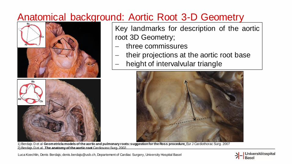
Anatomical background: Aortic Root 3-D Geometry
Luca Koechlin, Denis Berdajs; [email protected], Departement of Cardiac Surgery, University Hospital Basel
1) Berdajs D et al Geometricla models of the aortic and pulmonary roots: suggestionfor the Ross procedure Eur J Cardiothorac Surg. 2007
2) Berdajs D et al The anatomy of the aortic root Cardiovasc Surg. 2002.
Key landmarks for description of the aortic
root 3D Geometry;
three commissures
their projections at the aortic root base
height of intervalvular triangle

Anatomical background: Aortic Root 3-D Geometry
Luca Koechlin, Denis Berdajs; [email protected], Departement of Cardiac Surgery, University Hospital Basel
1) Berdajs D et all. Geometricla models of the aortic and pulmonary roots: suggestion for the Ross procedure Eur J Cardiothorac Surg. 2007 Jan;31(1):31-5
2) Berdajs D et all The anatomy of the aortic root Cardiovasc Surg. 2002 Aug;10(4):320-7.
Connecting the determined 6
landmarks results a three
sided prism
The three sided prism describes the
aortic root natural asymmetry.
This asymmetry may be defined with one
single parameter; by Aortic Root Vector.

Methods
Under experimental conditions
Type I BAV with valve
insufficiency (n=10 animals) and
valve stenosis (n=10 animals)
was created
The 4D aortic root deformation
was registered by implantation of
6 micro-sonometric (n=6) high-
resolution (200 Hz) crystals in
each aortic root modality
Luca Koechlin, Denis Berdajs; [email protected], Departement of Cardiac Surgery, University Hospital Basel
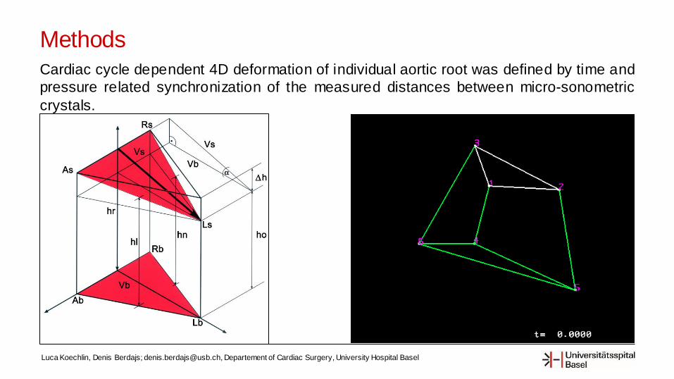
MethodsCardiac cycle dependent 4D deformation of individual aortic root was defined by time and
pressure related synchronization of the measured distances between micro-sonometric
crystals.
Luca Koechlin, Denis Berdajs; [email protected], Departement of Cardiac Surgery, University Hospital Basel

Methods
Computed fluid dynamic models (CFD), based on experimental
data, for the bicuspid aortic valve with insufficiency and stenosis were
established in order to evaluate
local pressure and shear stress profile at the surface of the
individual aortic root elements
4-D pressure and flow related computed fluid dynamic simulation of
thoracic aorta in BAV with aortic valve stenosis and insufficiency was
performed in order to simulate
pressure, velocity and shear stress profiles from the aortic root
up to the descending thoracic aorta.
Luca Koechlin, Denis Berdajs; [email protected], Departement of Cardiac Surgery, University Hospital Basel

Results: BAV with aortic valve insufficiency
Luca Koechlin, Denis Berdajs; [email protected], Departement of Cardiac Surgery, University Hospital Basel
Pressure Profile in BAV with valve insufficiency:
Moderate to low tangential pressure (40-65mmHg)
was present at:
• leaflets,
• coaptations,
• intervalvular triangles,
• three commissures
Almost during the whole period of cardiac cycle
At peak ejection moderately elevated pressure (50-
80mmHg) was registered at mentioned elements
Shear stress Profile in BAV with valve
insufficiency:
Low shear stress (0-0.5Pa) was found at all
components of the aortic root over whole cardiac
cycle
Pressure Shear stress
Pressure/Shear stress Profile of BAV with insufficiency at peak ejection

Results: BAV with aortic valve stenosis
Luca Koechlin, Denis Berdajs; [email protected], Departement of Cardiac Surgery, University Hospital Basel
Pressure Profile in BAV with valve stenosis:
Low tangential pressure (0-30mmHg) was present at
• leaflets,
• cooaptations,
• intervalvular triangles,
• three commissures
Almost during the whole period of cardiac cycle
At peak ejection moderately elevated pressure was
registered (50-80mmHg)
Shear stress Profile in BAV with valve stenosis:
Low shear stress was present (0-0.5Pa) at mentioned
components almost during the whole period of cardiac cycle
At peak ejection :
Low shear stress (0-0.5Pa), was registered
• at inferior 1/3 of the leaflets and triangles
High shear stress was registered
• superior 2/3 of leaflets
• commissures and cooaptations (0.8-1.5Pa),
• leaflets fusions sites (>2Pa)
Pressure/Shear stress Profile of BAV with stenosis at peak ejection
Pressure Shear stress

Results: BAV with aortic valve insufficiency (BAV)
Luca Koechlin, Denis Berdajs; [email protected], Departement of Cardiac Surgery, University Hospital Basel
In aortic valve insufficiency,
low shear stresses, with large
blood flow velocity oscillations,
were found
• at the ascending aorta,
• at the lesser curvature of
the aortic arch,
• in front of cervical vessels
and• at aortic isthm
Shear stress Pressure Flow profile

Results: BAV with aortic valve stenosis
Luca Koechlin, Denis Berdajs; [email protected], Departement of Cardiac Surgery, University Hospital Basel
In aortic valve stenosis
high shear stress with
elevated pressure were found
• at the sinotubularjunction,
• at the ascending aorta
• at the ostium of both
cervical arteries
Shear stress Pressure Flow profile

Conclusion
Luca Koechlin, Denis Berdajs; [email protected], Departement of Cardiac Surgery, University Hospital Basel
In BAV with stenosis during the
ejection period of cardiac cycle the
leaflets are exposed to moderately
elevated pressure and high shear stress.
This especially at the coaptation surface.
In contrast in BAV with insufficiency at
ejection phase the moderately elevated
pressure was combined with low shear
stress.

Conclusion
Luca Koechlin, Denis Berdajs; [email protected], Departement of Cardiac Surgery, University Hospital Basel
In real-time pressure-flow numerical
simulation of BAV with valve insufficiency
the low shear stresses and
turbulent/oscillation flow regions were
documented at traditional levels of entry tears in aortic dissection type A and B.
In comparison, in in vivo simulation of BAV
with valve stenosis, high shear stress with
elevated pressure at the ascending aorta and aortic arch may be identified as
contributing elements for vessel dilatation,
aneurysm formation and direct intimal tear
typically for type A aortic dissection.

Conclusion
Luca Koechlin, Denis Berdajs; [email protected], Departement of Cardiac Surgery, University Hospital Basel
According to the present results one can conclude that bicuspid stenotic aortic
valve is hemodynamically less favorable situation as compared to the BAV with
insufficiency.
The elevated pressure conjoined with elevated shear stress in stenotic BAV may in long term promote degenerative processes of the leaflets and consequently
the failure of the valve function.
Further predictive models for type A and B aortic dissection can be
developed in the future.