fmri: biological basis and experiment design intro history basic mechanism neurohemodynamic coupling
Post on 20-Dec-2015
215 views
TRANSCRIPT
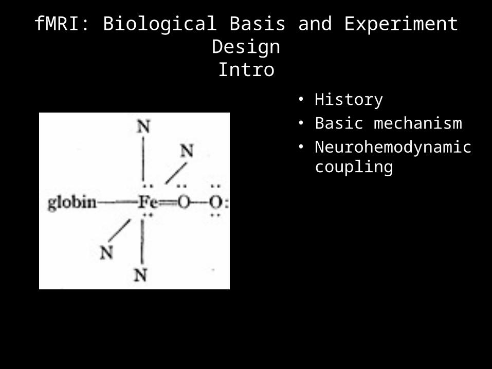
fMRI: Biological Basis and Experiment DesignIntro
• History• Basic mechanism• Neurohemodynamic
coupling
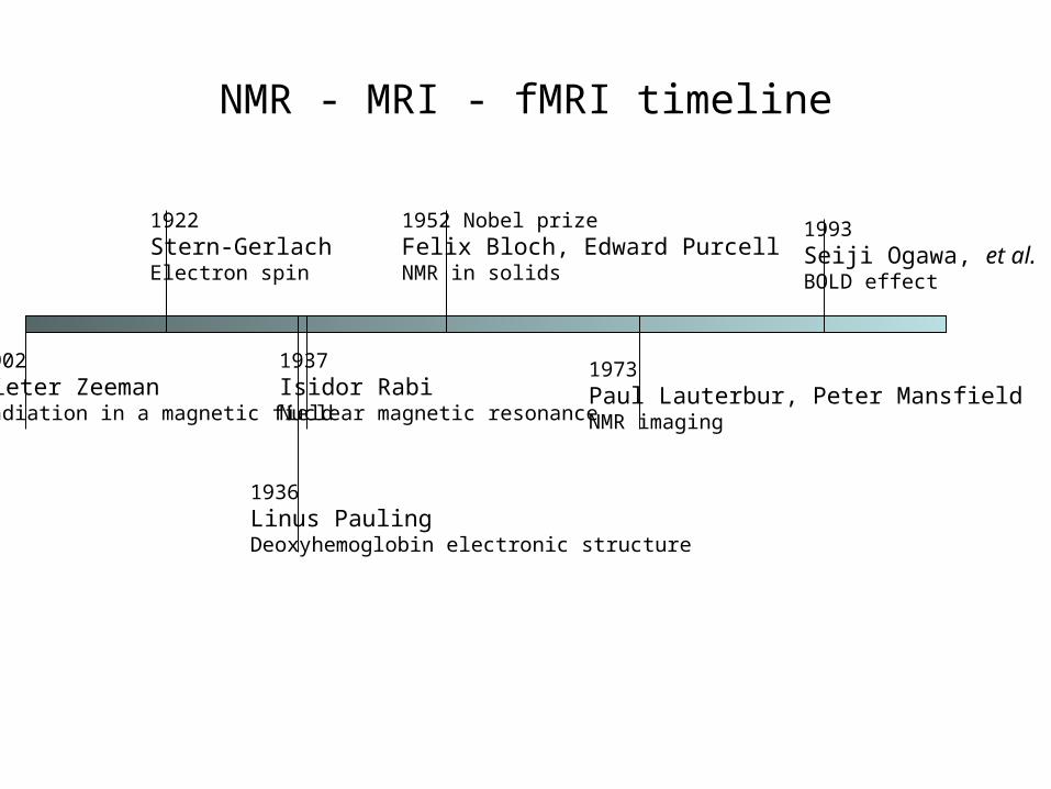
NMR - MRI - fMRI timeline
1922Stern-GerlachElectron spin
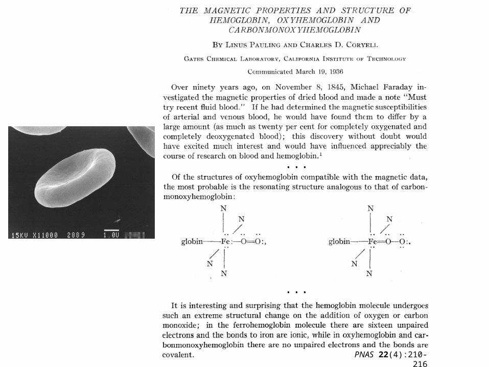
1936Linus PaulingDeoxyhemoglobin electronic structure
1937Isidor RabiNuclear magnetic resonance
1952 Nobel prizeFelix Bloch, Edward PurcellNMR in solids
1973Paul Lauterbur, Peter MansfieldNMR imaging
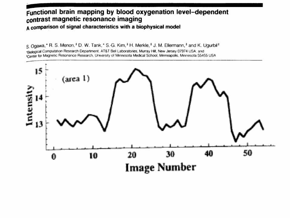
1993Seiji Ogawa, et al.BOLD effect
1902Pieter ZeemanRadiation in a magnetic field
...
...
PNAS 22(4):210-216
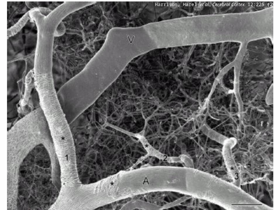
Harrison, Harel et al., Cerebral Cortex 12:225 (2002)
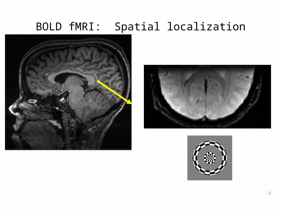
BOLD fMRI: Spatial localization
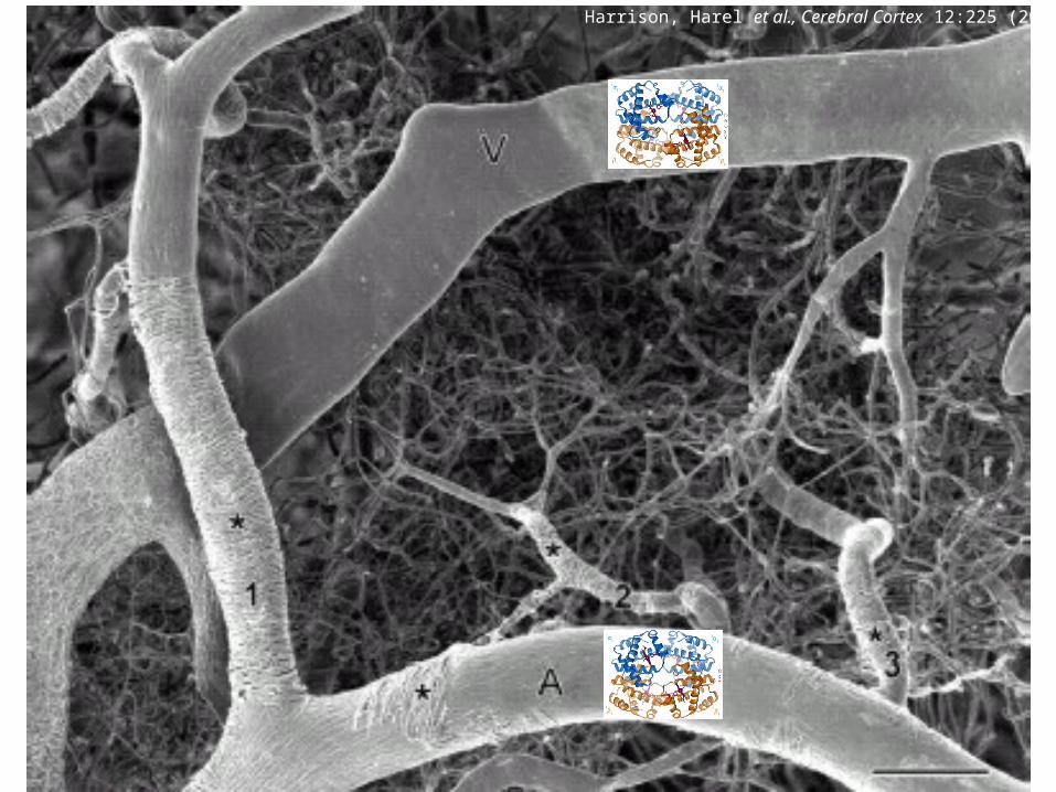
Harrison, Harel et al., Cerebral Cortex 12:225 (2002)
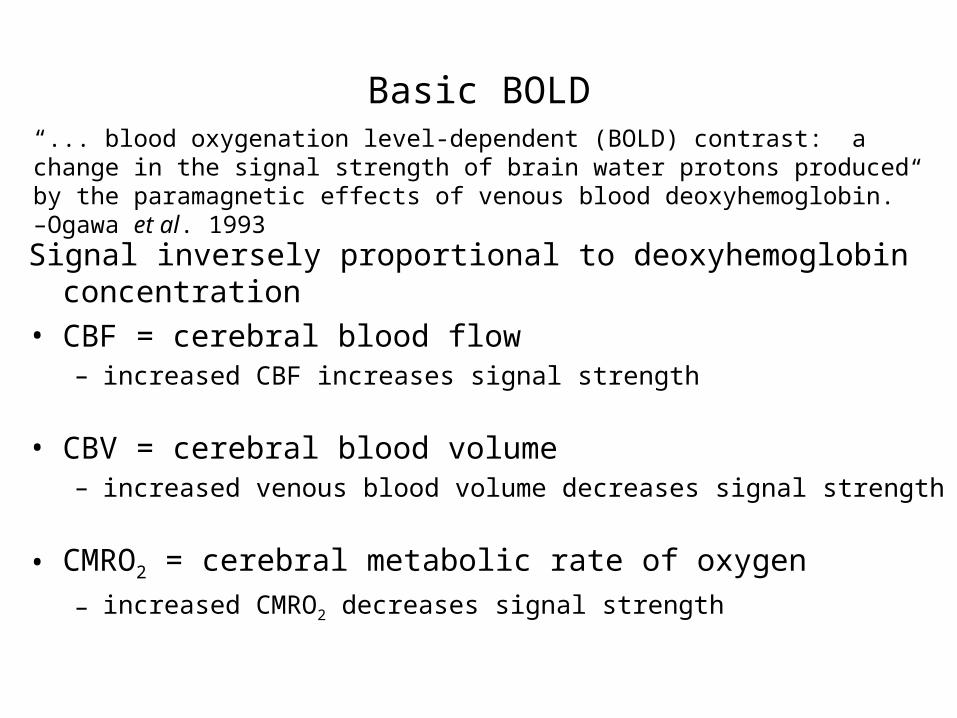
Basic BOLD
Signal inversely proportional to deoxyhemoglobin concentration• CBF = cerebral blood flow
– increased CBF increases signal strength
• CBV = cerebral blood volume– increased venous blood volume decreases signal strength
• CMRO2 = cerebral metabolic rate of oxygen
– increased CMRO2 decreases signal strength
“... blood oxygenation level-dependent (BOLD) contrast: a change in the signal strength of brain water protons produced by the paramagnetic effects of venous blood deoxyhemoglobin.” –Ogawa et al. 1993

And here we start speaking in vague generalities.
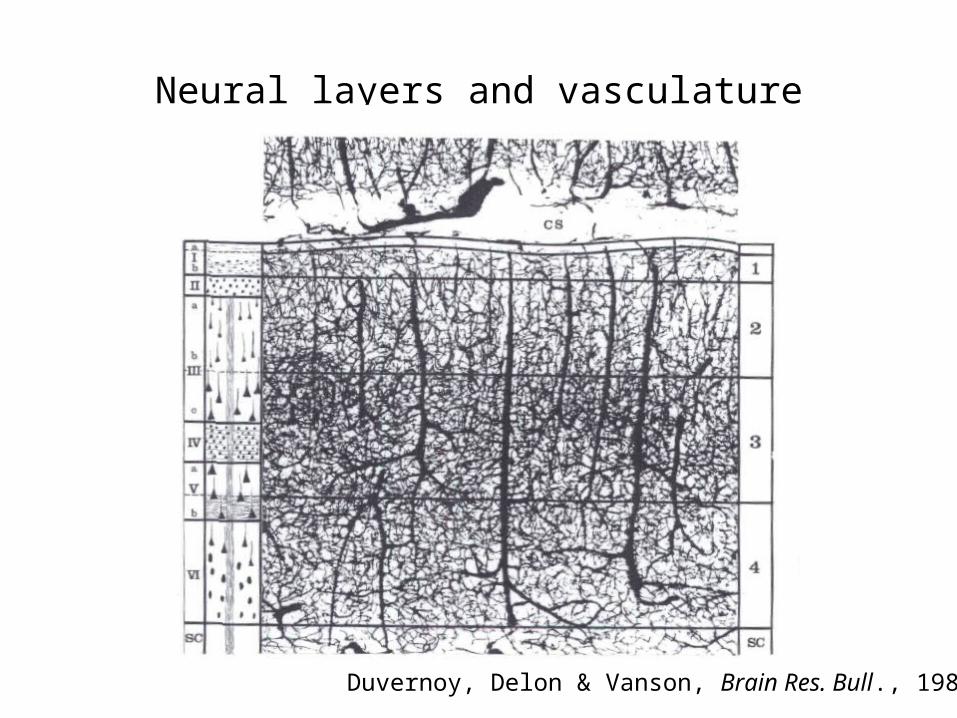
Neural layers and vasculature
Duvernoy, Delon & Vanson, Brain Res. Bull., 1981
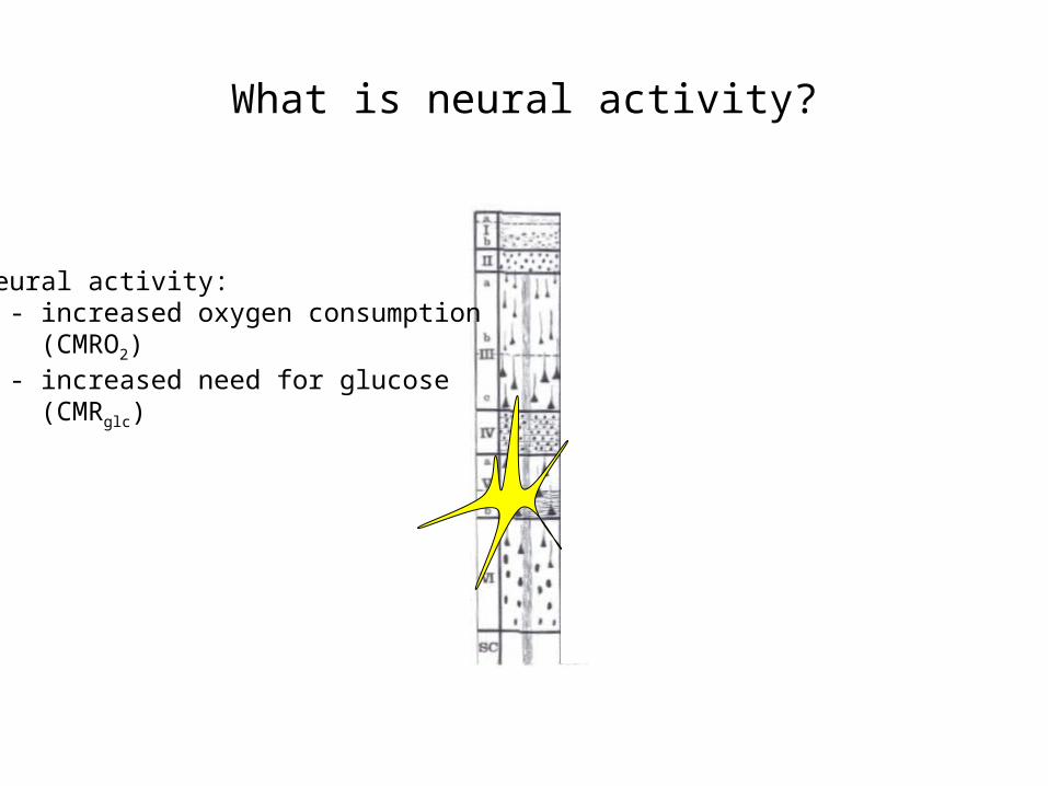
What is neural activity?
Neural activity: - increased oxygen consumption (CMRO2) - increased need for glucose (CMRglc)
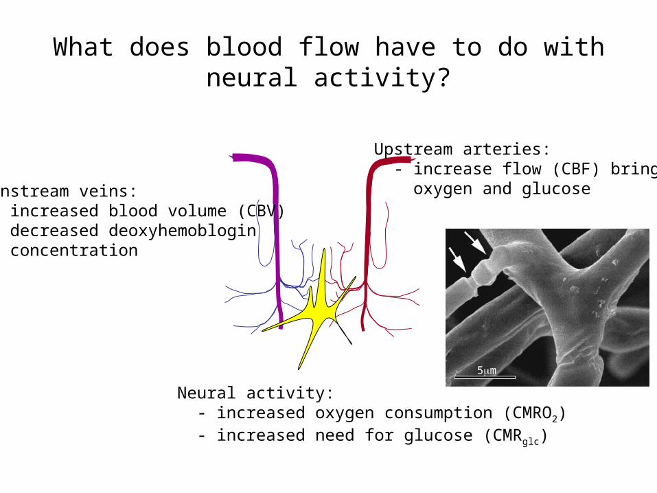
What does blood flow have to do with neural activity?
5m
Neural activity: - increased oxygen consumption (CMRO2) - increased need for glucose (CMRglc)
Upstream arteries: - increase flow (CBF) brings oxygen and glucose Downstream veins:
- increased blood volume (CBV) - decreased deoxyhemoblogin concentration