fmri: biological basis and experiment design lecture 3 cell metabolism vascular architecture blood...
Post on 20-Dec-2015
221 views
TRANSCRIPT
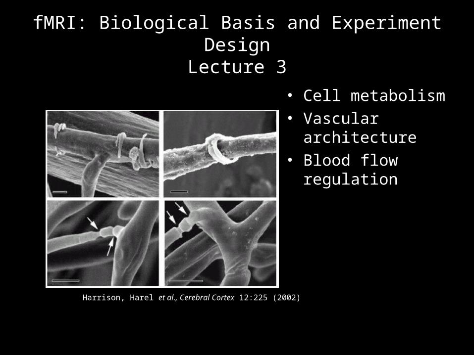
fMRI: Biological Basis and Experiment DesignLecture 3
• Cell metabolism• Vascular architecture• Blood flow regulation
Harrison, Harel et al., Cerebral Cortex 12:225 (2002)
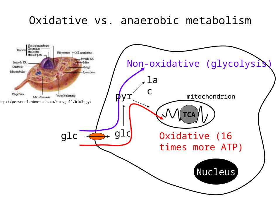
Oxidative vs. anaerobic metabolism
http://personal.nbnet.nb.ca/trevgall/biology/
Non-oxidative (glycolysis)
TCA
Nucleus
mitochondrion
Oxidative (16 times more ATP)
glc glc
pyr
lac
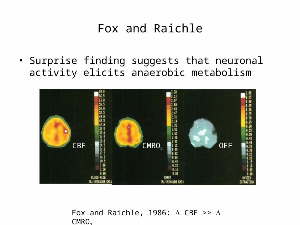
Fox and Raichle
• Surprise finding suggests that neuronal activity elicits anaerobic metabolism
Fox and Raichle, 1986: CBF >> CMRO2
CBF CMRO2 OEF
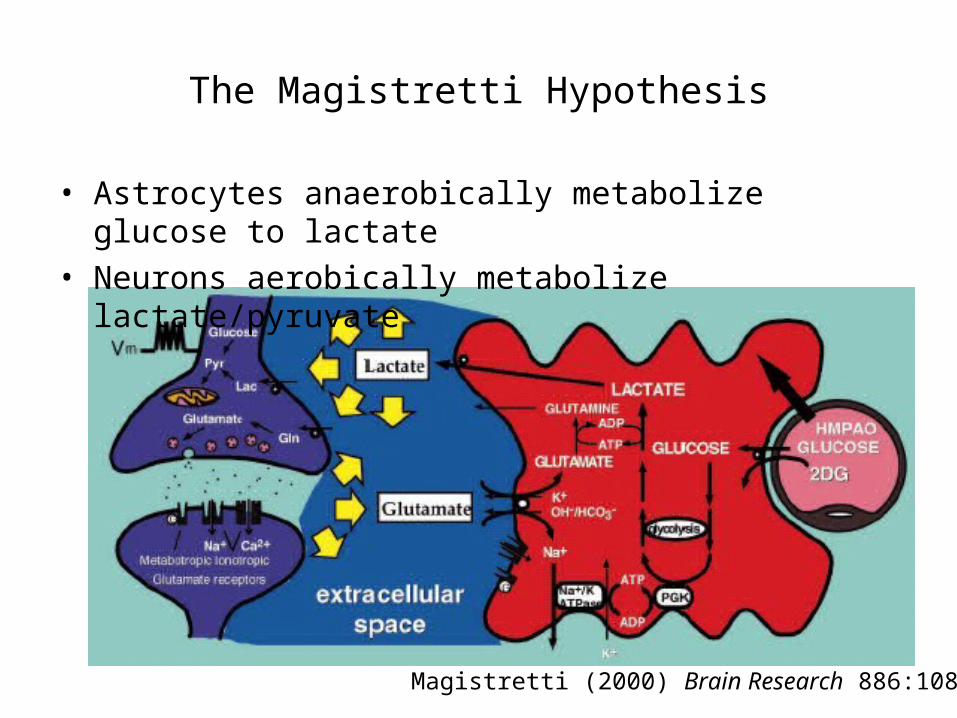
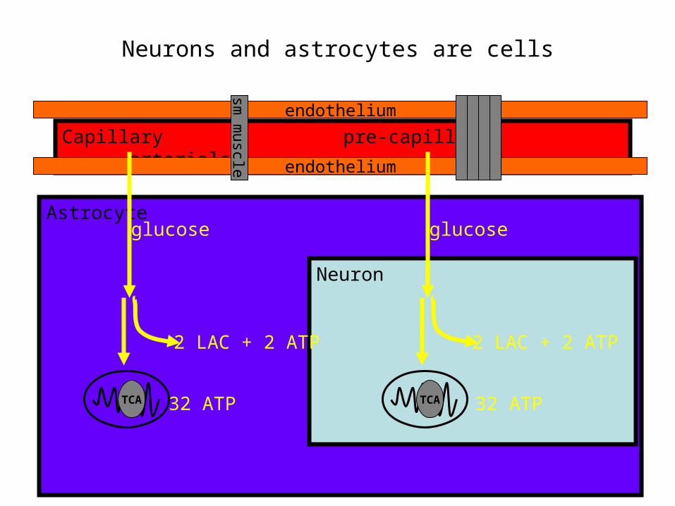
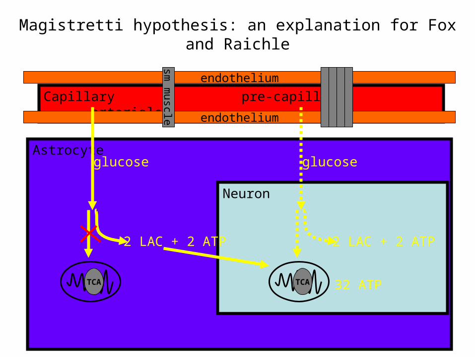
The Magistretti Hypothesis
• Astrocytes anaerobically metabolize glucose to lactate• Neurons aerobically metabolize lactate/pyruvate
Magistretti (2000) Brain Research 886:108
Capillary pre-capillary arteriole endothelium
endothelium
sm muscle
Neurons and astrocytes are cells
Astrocyte
Neuron
2 LAC + 2 ATP
TCA
glucose
2 LAC + 2 ATP
TCA
glucose
32 ATP 32 ATP
Capillary pre-capillary arteriole endothelium
endothelium
sm muscle
Magistretti hypothesis: an explanation for Fox and Raichle
Astrocyte
Neuron
2 LAC + 2 ATP
TCA
glucose
2 LAC + 2 ATP
TCA
glucose
32 ATP
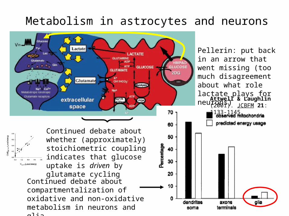
Metabolism in astrocytes and neurons
Pellerin: put back in an arrow that went missing (too much disagreement about what role lactate plays for neurons)Attwell & Laughlin
(2001). JCBFM 21: 1133-1145.
Continued debate about whether (approximately) stoichiometric coupling indicates that glucose uptake is driven by glutamate cycling
Continued debate about compartmentalization of oxidative and non-oxidative metabolism in neurons and glia
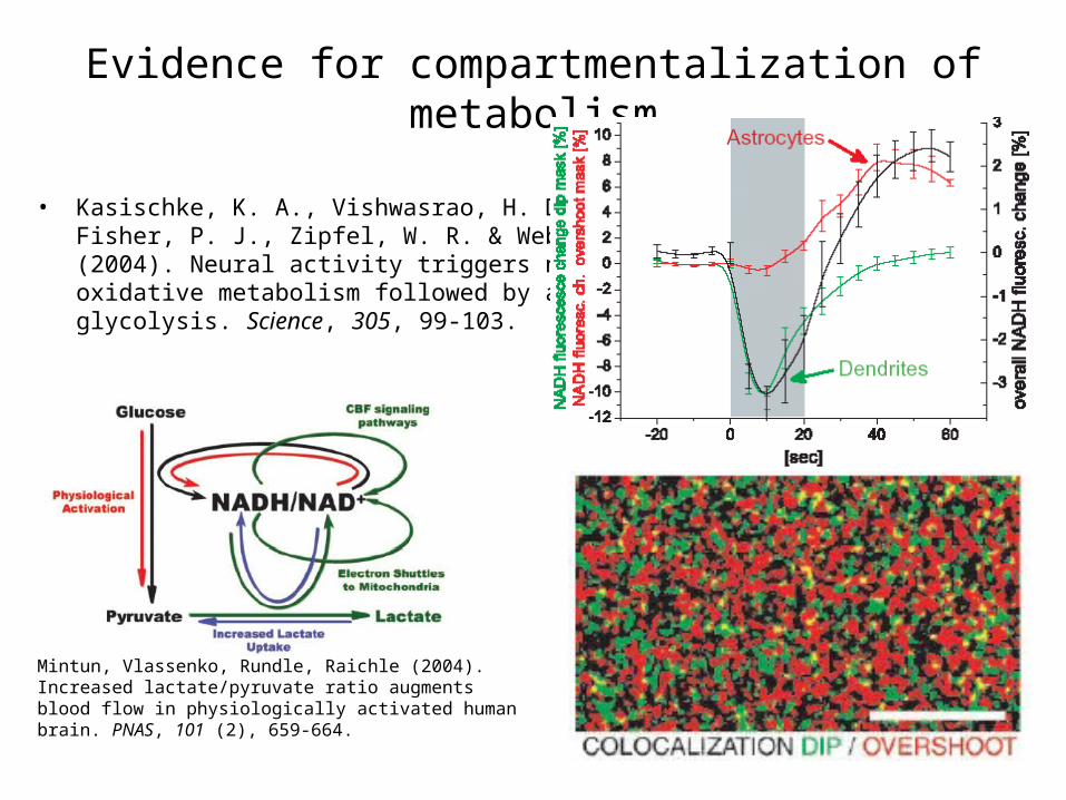
Evidence for compartmentalization of metabolism
• Kasischke, K. A., Vishwasrao, H. D., Fisher, P. J., Zipfel, W. R. & Webb, W. W. (2004). Neural activity triggers neuronal oxidative metabolism followed by astrocytic glycolysis. Science, 305, 99-103.
Mintun, Vlassenko, Rundle, Raichle (2004). Increased lactate/pyruvate ratio augments blood flow in physiologically activated human brain. PNAS, 101 (2), 659-664.
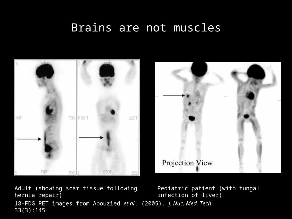
Brains are not muscles
Pediatric patient (with fungal infection of liver)Adult (showing scar tissue following hernia repair)
18-FDG PET images from Abouzied et al. (2005). J. Nuc. Med. Tech. 33(3):145
Capillary pre-capillary arteriole endothelium
endothelium
sm muscle
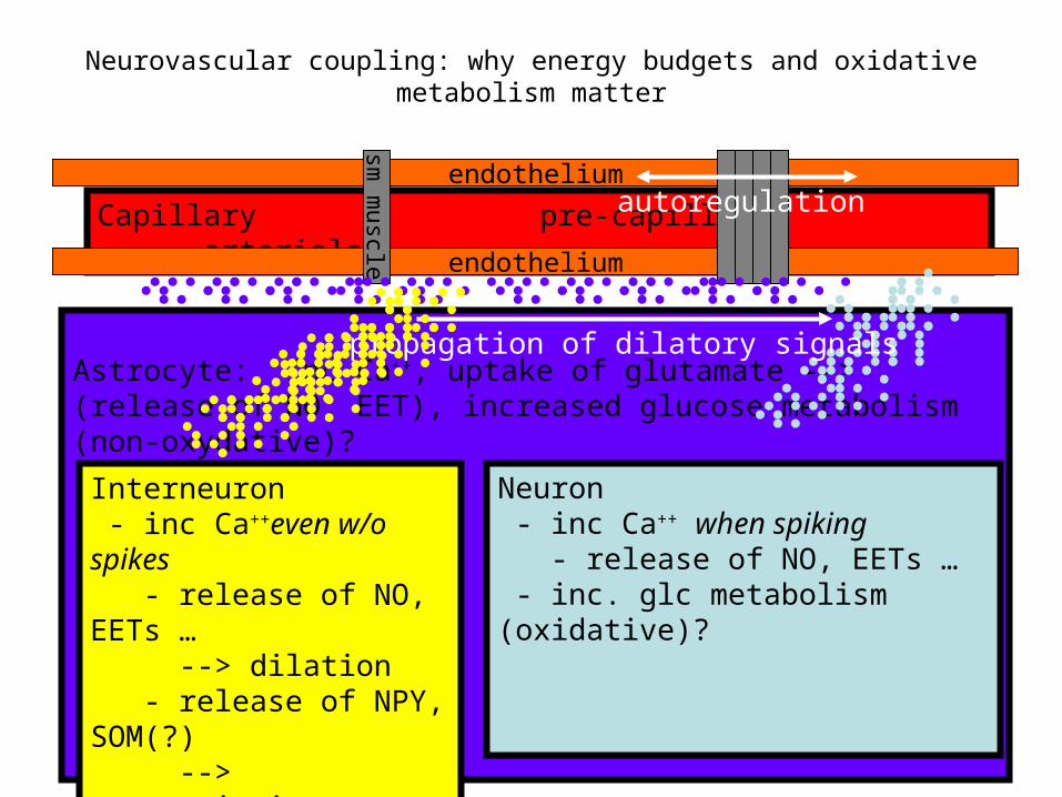
Neurovascular coupling: why energy budgets and oxidative metabolism matter
Astrocyte: Inc Ca++, uptake of glutamate --> (release of NO, EET), increased glucose metabolism (non-oxydative)?
Interneuron - inc Ca++even w/o spikes - release of NO, EETs … --> dilation - release of NPY, SOM(?) --> contstriction - inc. glc metabolism?
Neuron - inc Ca++ when spiking - release of NO, EETs … - inc. glc metabolism (oxidative)?
propagation of dilatory signals
autoregulation
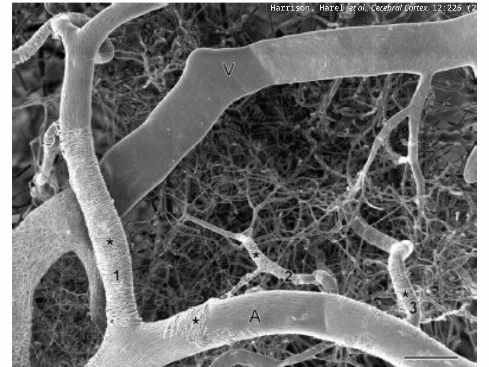
Harrison, Harel et al., Cerebral Cortex 12:225 (2002)
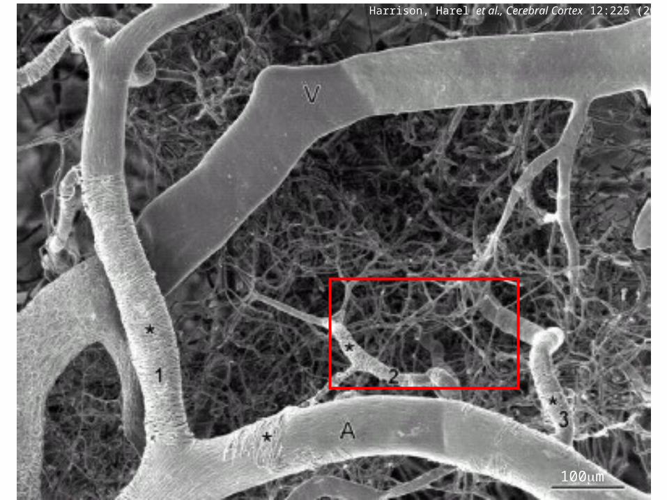
Harrison, Harel et al., Cerebral Cortex 12:225 (2002)
100m
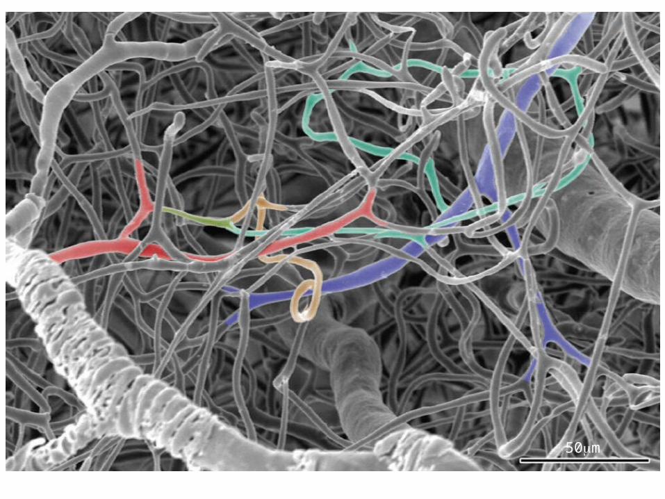
50m
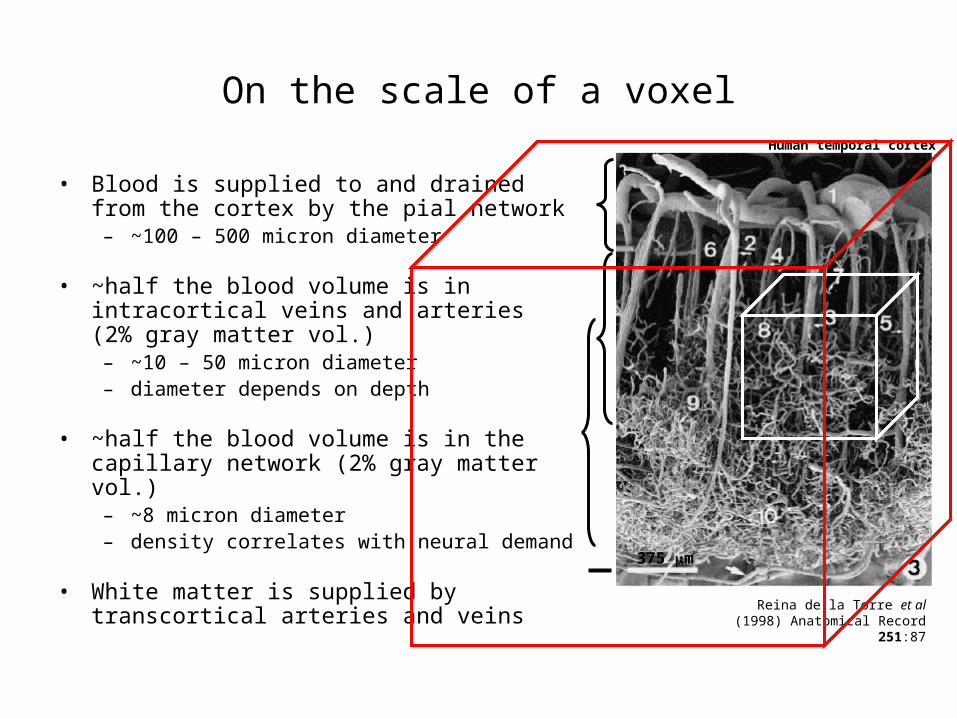
On the scale of a voxel
• Blood is supplied to and drained from the cortex by the pial network
– ~100 – 500 micron diameter
• ~half the blood volume is in intracortical veins and arteries (2% gray matter vol.)
– ~10 – 50 micron diameter– diameter depends on depth
• ~half the blood volume is in the capillary network (2% gray matter vol.)
– ~8 micron diameter– density correlates with neural demand
• White matter is supplied by transcortical arteries and veins
Human temporal cortex
Reina de la Torre et al (1998) Anatomical Record
251:87
375 m
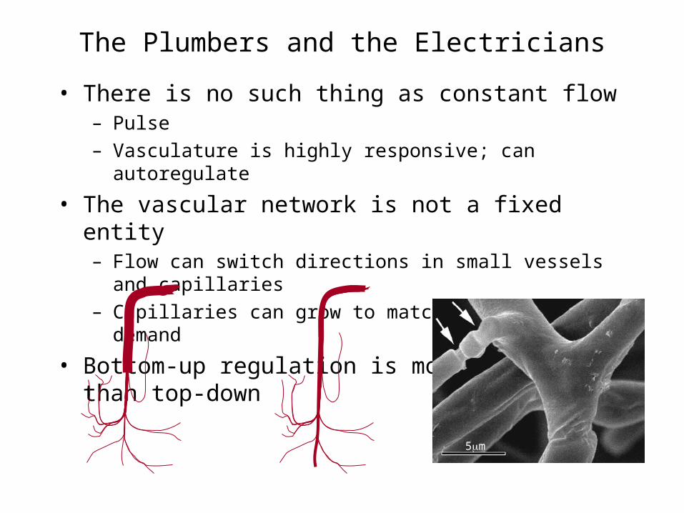
The Plumbers and the Electricians
• There is no such thing as constant flow– Pulse– Vasculature is highly responsive; can autoregulate
• The vascular network is not a fixed entity– Flow can switch directions in small vessels and capillaries– Capillaries can grow to match metabolic demand
• Bottom-up regulation is more practical than top-down
5m
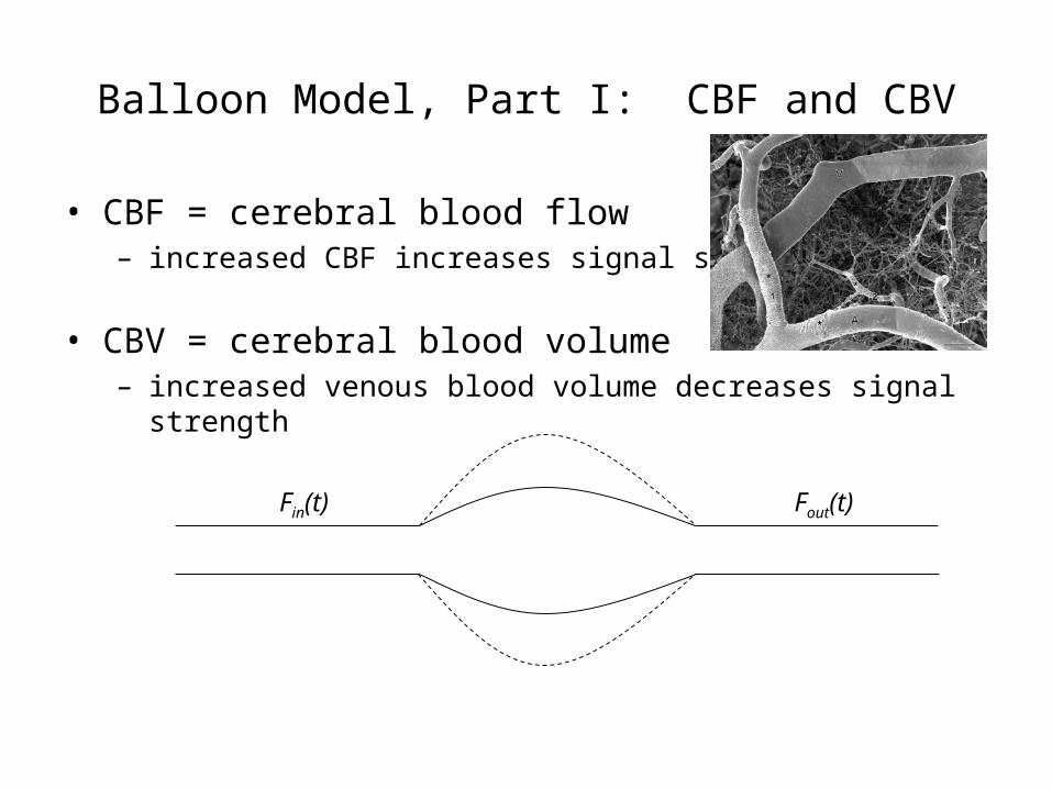
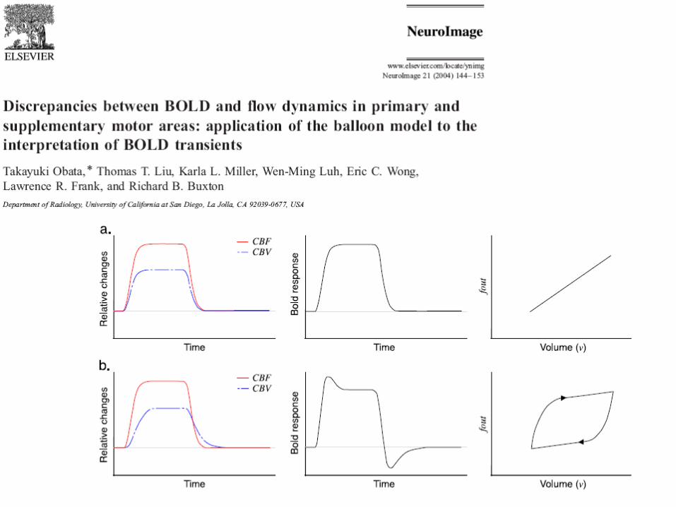
Balloon Model, Part I: CBF and CBV
• CBF = cerebral blood flow– increased CBF increases signal strength
• CBV = cerebral blood volume– increased venous blood volume decreases signal strength
Fout(t)Fin(t)
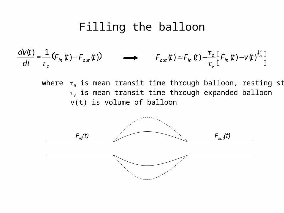
Filling the balloon
Fout(t)Fin(t)
( ))()(1)(
0
tFtFdt
tdvoutin −=
τ ⎥⎦⎤
⎢⎣⎡ −−≅ α
ττ 1
)()()()( tvtFtFtF inv
oinout
where τ0 is mean transit time through balloon, resting stateτv is mean transit time through expanded balloonv(t) is volume of balloon