folding of an intrinsically disordered protein by ...ibmmsrvlakitu.unibe.ch/altmann/bah et al....
TRANSCRIPT

LETTERdoi:10.1038/nature13999
Folding of an intrinsically disordered protein byphosphorylation as a regulatory switchAlaji Bah1,2, Robert M. Vernon1,2, Zeba Siddiqui1, Mickael Krzeminski1,2, Ranjith Muhandiram2,3, Charlie Zhao1,Nahum Sonenberg4, Lewis E. Kay1,2,3,5 & Julie D. Forman-Kay1,2
Intrinsically disordered proteins play important roles in cell signalling,transcription, translation and cell cycle regulation1,2. Although theylack stable tertiary structure, many intrinsically disordered proteinsundergo disorder-to-order transitions upon binding to partners3,4.Similarly, several folded proteins use regulated order-to-disordertransitions to mediate biological function5,6. In principle, the functionof intrinsically disordered proteins may be controlled by post-trans-lational modifications that lead to structural changes such as folding,although this has not been observed. Here we show that multisitephosphorylation induces folding of the intrinsically disordered 4E-BP2, the major neural isoform of the family of three mammalianproteins that bind eIF4E and suppress cap-dependent translation ini-tiation. In its non-phosphorylated state, 4E-BP2 interacts tightly witheIF4E using both a canonical YXXXXLW motif (starting at Y54) thatundergoes a disorder-to-helix transition upon binding and a dyna-mic secondary binding site7–11. We demonstrate that phosphorylationat T37 and T46 induces folding of residues P18–R62 of 4E-BP2 into afour-stranded b-domain that sequesters the helical YXXXXLW motifinto a partly buried b-strand, blocking its accessibility to eIF4E. Thefolded state of pT37pT46 4E-BP2 is weakly stable, decreasing affinityby 100-fold and leading to an order-to-disorder transition upon bind-ing to eIF4E, whereas fully phosphorylated 4E-BP2 is more stable,decreasing affinity by a factor of approximately 4,000. These resultshighlight stabilization of a phosphorylation-induced fold as the essen-tial mechanism for phospho-regulation of the 4E-BP:eIF4E inter-action and exemplify a new mode of biological regulation mediatedby intrinsically disordered proteins.
Proteins exist in a continuum of states, from ensembles of intercon-verting conformers for intrinsically disordered proteins (IDPs) to stablestructures for folded proteins2,4,12,13. Although many undergo subtle con-formational rearrangements in response to biological signals, someundergo larger changes: that is, disorder-to-order or order-to-disordertransitions4 that can be mediated by interactions with ligands. Post-translational modifications (PTMs) such as phosphorylation can alsopotentially induce such transitions. While phosphorylation can convertthe folded oligomerization domain of nucleophosmin into a disorderedmonomer14,15, PTMs have been generally known only to stabilize ordestabilize individual secondary structural elements in IDPs16–18. Thus,PTM-mediated folding of a protein domain, as shown here for the phos-phorylation-induced folding of 4E-BP2, is a novel regulatory mechanismfor IDPs.
Here we study the phospho-regulation of the 4E-BP:eIF4E interaction19.The 4E-BPs compete with eIF4G for eIF4E binding to prevent cap-dependent translation initiation by using similar canonical eIF4E-bindingYXXXXLW motifs20,21. Non-phosphorylated or minimally phosphory-lated 4E-BPs interact tightly with eIF4E, while the binding of highly phos-phorylated 4E-BPs is much weaker and can be outcompeted by eIF4G.T37 and T46 are known to be phosphorylated first, followed by T70and S65 (ref. 22). Interestingly, in their free states, highly phosphorylated
4E-BPs are very stable while non- or minimally phosphorylated 4E-BPs are targeted for degradation with ubiquitination at K57 (within theYXXXXLW motif) by the KLHL25-CUL3 E3 ligase23. However, there isno consensus on how phosphorylation regulates binding to eIF4E oraffects the stability of 4E-BPs. Phosphorylation may result in electro-static repulsion with the negative surface of eIF4E21 or S65 phosphoryla-tion could inhibit binding by destabilizing the YXXXXLW motif helicalstructure24. The inability of glutamic acid phospho-mimetics to weaken4E-BP:eIF4E binding has also suggested that additional PTMs may berequired25.
To address the phospho-regulatory mechanism, we have used NMR,isothermal titration calorimetry (ITC) and mutagenesis. Figure 1 andExtended Data Fig. 1 show structural and dynamic properties of wild-type (WT) 4E-BP2 uniformly phosphorylated at T37, T46, S65, T70 andS83 (Fig. 1a). IDPs such as non-phosphorylated 4E-BP2 have intensepeaks with narrow 1HN chemical shift dispersion26. Unlike other IDPsfor which phosphorylation only causes downfield chemical shifts ofphosphorylated residues27, 4E-BP2 phosphorylation induces widespreaddownfield and upfield chemical shifts for residues spanning T19–R62,suggesting folding upon phosphorylation (Fig. 1b). Notably, G39 andG48, the first glycines in the six-residue repeat sequences (TTPGGT)containing two of the five phosphorylation sites, show dramatic down-field 1HN chemical shift changes (Fig. 1b, inset). Peaks for the rest ofthe protein are intense with narrow 1HN chemical shift dispersion, indi-cating that these residues remain disordered. Like its non-phosphorylatedstate11, phosphorylated 4E-BP2 exchanges between major and minorconformers, probably from cis–trans isomerization of the multipleprolines and between unfolded and partly folded states for the foldedregion (see below). Lower [1H]–15N nuclear Overhauser effect (NOE)values are expected for IDPs because of their rapid motions26,28. Elevated[1H]–15N NOEs for most residues from T19–R62 (Fig. 1c), along withthe large increase in chemical shift dispersion and the high amide protontemperature coefficients indicative of intramolecular hydrogen bonding29
(Extended Data Fig. 1d), provide strong evidence that this region foldsupon phosphorylation, while the rest of the protein remains disordered.
We made alanine mutations to mimic in vivo phosphorylation states of4E-BP2 (ref. 22) (Extended Data Fig. 2). No significant change in globaldispersion was observed for 4E-BP2 phosphorylated only at S65/T70/S83 (Extended Data Fig. 2a), demonstrating that it remains disordered,while phosphorylating T37 and T46 (pT37pT46) induces a 4E-BP2 foldidentical to phosphorylated wild type (pWT) (Extended Data Fig. 2b).Interestingly, when phosphorylated individually, pT37 or pT46 result ina partly folded state, with some chemical shift changes indicative of orderedstructure (pT37) and the presence of one b-turn leading to one char-acteristic downfield shifted glycine peak (G39 for pT37, G48 for pT46;Extended Data Fig. 2c, d). Thus, phosphorylation of both T37 and T46 isnecessary and sufficient for phosphorylation-induced folding of 4E-BP2.
We determined the structure of P18–R62 using CS-Rosetta30 includ-ing initially only chemical shifts, and subsequently adding NOEs (see
1Molecular Structure and Function Program, Hospital for Sick Children, Toronto, Ontario M5G 0A4, Canada. 2Department of Biochemistry, University of Toronto, Toronto, Ontario M5S 1A8, Canada.3Department of Molecular Genetics, University of Toronto, Toronto, Ontario M5S 1A8, Canada. 4Department of Biochemistry and Goodman Cancer Research Centre, McGill University, Montreal, QuebecH3G 1Y6, Canada. 5Department of Chemistry, University of Toronto, Toronto, Ontario M5S 3H6, Canada.
0 0 M O N T H 2 0 1 4 | V O L 0 0 0 | N A T U R E | 1
Macmillan Publishers Limited. All rights reserved©2014

Methods, Extended Data Figs 3–6 and Extended Data Table 1a, b). Thedomain is a four-stranded b-fold (Fig. 2), with b1(T19–I24), b2(C35–pT37), b3(L42–T45), b4(R51–R56) and a 310 helix (A27–Q29). pT37and pT46 are central to a network of hydrogen bonds that stabilize tightb-turns composed of the identical pTPGGT motifs connecting strandsb2 with b3 (pT37–T41) and b3 with b4 (pT46–T50). The hydrogenbonding correlates well with 1HN temperature coefficients (ExtendedData Fig. 1d). G39 and G48 amide protons hydrogen bond to T37 and
T46 phosphate groups, respectively, explaining their unexpectedly largedownfield chemical shifts (Fig. 1a, inset, and Extended Data Fig. 2b). TheYXXXXLw eIF4E-binding motif forms part of strandb4 with Y54 largelyburied within a hydrophobic cluster also involving V22, I24, L30, P31,Y34 and I52 (Fig. 2a). The phosphorylation-induced structure provides amechanism by which phosphorylation reduces eIF4E binding by seques-tering the helical YXXXXLw motif into a b-strand. Its burial stabilizesthe b-fold; phosphorylated Y54A/L59A [p(Y54A/L59A)] retains the b-turns, evident from the characteristic downfield 1HN shifts of G39 andG48, but no longer folds (Fig. 3a). A stable fold sequestering YXXXXLwcould also block ubiquitination of K57, thereby preventing degradationin vivo23.
ITC (Extended Data Table 2 and Extended Data Fig. 7) and NMR datawere obtained on 4E-BP2 variants to probe the role of electrostatics infolding and reducing eIF4E affinity. Unlike pT37pT46, neither T37D/T46D nor T37E/T46E (phospho-mimics) showed evidence of folding(Extended Data Fig. 2e, f). In contrast to pT37pT46, which has reducedaffinity (dissociation constant (Kd) 5 267 6 32 nM), T37D/T46D andT37E/T46E bind tightly to eIF4E (Kd 5 3.89 6 1.1 nM and 4.376 0.8 nM,respectively), similar to non-phosphorylated 4E-BP2 (Kd 5 3.206 0.6 nM).Thus acidic residues do not mimic phosphorylation by inducing foldingof 4E-BP2 or reducing eIF4E affinity. The binding affinity of pS65pT70pS834E-BP2 for eIF4E was very tight (Kd 5 11.3 6 2.9 nM), as expected sincea folded structure is not formed (Extended Data Fig. 2a). Strikingly, five-phospho protein (pWT) decreases affinity by about three orders of mag-nitude (Kd 5 12,320 6 600 nM). Valines were substituted for the firstglycines in the TPGGT motifs (G39V/G48V) of 4E-BP2 to disrupt hair-pin formation through steric contacts and prevent folding. As predictedfromDDG calculations (Extended Data Table 1c), folding was not inducedwhen G39V/G48V was fully phosphorylated at all five sites [p(G39V/G48V)] (Fig. 3b). Notably, the affinity of the p(G39V/G48V) for eIF4E ishigh (Kd 5 36.1 6 3.5 nM), only an order of magnitude weaker than thatof non-phosphorylated 4E-BP2 (Kd 5 3.20 6 0.6 nM) and approximately2.5 orders of magnitude stronger than pWT (Kd 5 12,3206 600 nM). Thesedata, together with unfolding energies of pT37pT46 (,2.6 kcal mol21)and pWT (,4.8 kcal mol21) estimated from Kd values (Extended DataTable 2), suggest that, although phosphorylation of only T37 and T46 isrequired to induce folding, phosphorylation of S65, T70 and S83 stabi-lizes the fold. This is possibly through long-range transient electrostatic
9.5 9.0 8.5 8.0 7.51H (p.p.m.)
130
125
120
115
110
15N
(p.p
.m.)
S2 S3
A5
G6
S7
G8
H9
Q10
S12
S14
A16
T19
R20
T21
V22
A23
S25
D26
A27
A28
Q29
L30
H32D33
Y34
C35
T36
T37
G40
L42
F43
S44
T45
T46
G49
R51I52
I53Y54
D55 R56
K57
F58
L59L60
R62
N64
M67T70
H74
N77
I78
G80
V81
T82
S83
G85
T86
E89
S91
V93
A109
G111
A114
Q115
I120
b
110G39
G48
11.0
108
a
TTPGGT XXX TTPGGT
T37 T46
YXXXXL
S83T70S65
pT37 pT46
YXXXXL
pS83pT70pS65
36 50
Het
eron
ucle
ar N
OE
4E-BP2 sequence
800 MHz, 5 ºC
WT
pWT
c
–0.2
0.0
0.2
0.4
0.6
0.8
–0.4
Figure 1 | Effects of phosphorylation on the structural and dynamicproperties of 4E-BP2. a, Schematic representation of 4E-BP2 showing therelative positions of the phosphorylation sites, the canonical eIF4E binding site(thick blue bar) and the region which undergoes phosphorylation-inducedfolding (thick red bar). b, c, Overlay of (b) 1H–15N heteronuclear singlequantum correlation (HSQC) spectra and (c) 1H–15N NOE values at 800 MHz,
5 uC for non-phosphorylated WT (blue) and phosphorylated WT (red),respectively. Missing data represent prolines and residues that are toooverlapped/weak to be accurately quantified. Errors (standard deviations)around the average values are based on multiple repeats (n 5 3) of theexperiment.
G48G49
T50
P47
PT46 PT46
P47
G48
G49 T50
Y54PT46
L59
β1β1β2β2
β3β3β4β4
β2β2β1β1
β4β4
β3β3
PT37
180º
a
b c
Figure 2 | Phosphorylation-induced structure of the major state of residuesR18–R62 of 4E-BP2. a, Cartoon (left) and surface (right) representations ofthe solution NMR structure. Phosphorylated residues, pT37 and pT46 (red),the surface formed by residues of the hydrophobic cluster (right, cyan) andY54 (cyan stick representation (left) or dark cyan surface (right)) are shown.The residues in the canonical eIF4E binding site (YXXXXLW) are shown ingreen (left), demonstrating the binding-incompatible extended structure.b, c, Hydrogen-bonding networks formed by the pTPGGT motifs. Note thatpT46–T50 is shown in two different views and the same hydrogen bonds areformed for pT37–T41.
RESEARCH LETTER
2 | N A T U R E | V O L 0 0 0 | 0 0 M O N T H 2 0 1 4
Macmillan Publishers Limited. All rights reserved©2014

interactions between the phosphorylated acidic carboxy (C) terminuswith the basic folded domain. Sequestration of the canonical eIF4E motifby pT37pT46 reduces affinity by about two orders of magnitude, and notthe three orders observed for the fully phosphorylated form, possibly
because of a binding-competent minor disordered state that is visiblein the spectrum, providing additional evidence for lower stability of thepT37pT46 folded state. These affinities and calculations support theview that stabilization of a phosphorylation-induced folded structureplays a dominant role in weakening the eIF4E:4E-BP2 interaction, witha probable small contribution from electrostatic repulsion from eIF4Eas reflected in the ten-fold reduction in affinity for p(G39V/G48V) fromnon-phosphorylated WT 4E-BP2.
Because the 4E-BP2 b-fold sequesters the eIF4E-binding interface,binding to eIF4E must be coupled to unfolding. Although it is folded inthe absence of target, pT37pT46 undergoes an order-to-disorder trans-ition upon binding to eIF4E (Fig. 3c), as established by the resultingpoorly resolved spectrum confined to a narrow amide proton chemicalshift range and by the disappearance of well dispersed folded peaks of theapo-state, except for a single very weak 1HN glycine peak at 10.25 p.p.m.that may reflect a low population of a b-turn (Fig. 3c and Extended DataFig. 2g). The spectrum is similar to that of non-phosphorylated 4E-BP2(ref. 11); both complexes are disordered with significant chemical shiftdifferences (Extended Data Fig. 2g), probably reflecting changes in inter-actions due to different binding affinities (3.20 6 0.6 nM versus 267 6
32 nM) and effects of phosphates, including low population of a b-turn.Our study provides key insights into how the structural polymorph-
ism of 4E-BP2 allows it to regulate translation initiation through PTM-mediated folding (Fig. 4). This mechanism establishes a new means ofIDP-mediated control of biological function. Large structural changessuch as folding could sequester or enhance the accessibilities of proteinbinding and other PTM sites or provide new interaction surfaces, there-by expanding signalling output. Importantly, PTM-induced folding hasimportant potential impact for targeting IDP interactions for therapeutics.
10.0 9.5 9.0 8.5 8.0 7.51H (p.p.m.)
130
125
120
115
110
15N
(p.p
.m.)
11.0
b
110
p(G39V/G48V)pWT
G8
S12
T19
R20
V22
A23
S25
D26
A27
Q29
L30
H32 Y34
C35
T36
T37
G40
L42
F43
S44
T45
T46
G49
R51I52
I53
D55
K57
F58
L59
T70
G85
G111
I120
G39
G48
130
125
120
115
110
G8
S12
T19
R20
V22
A23
S25
D26
A27
Q29
L30
H32 Y34
C35
T36
T37
G40
L42
F43
S44
T45
T46
G49
R51
I52
I53
D55
F58
L59
G85
G111
I120
11.0
110G39
G48
c
11.0
a
110 G8
S12T19
R20
V22
A23
S25
D26
A27
Q29
L30
H32 Y34
C35
T36
T37
G40
L42
F43
S44T45
T46
G49
R51I52
I53
D55
K57
F58
L59
T70
G85
G111
I120
G39
G48
130
125
120
115
110
p(Y54A/L59A)pWT
pT37pT46 + eIF4EpT37pT46
15N
(p.p
.m.)
15N
(p.p
.m.)
Figure 3 | Probing the structural and binding properties of phosphorylated4E-BP2. a, b, 1H–15N HSQC spectra of pWT (red) overlaid with (a) p(Y54A/L59A) and (b) p(G39V/G48V). c, Spectrum of pT37pT46 in isolated (red) andeIF4E-bound (black) states, demonstrating an order-to-disorder transitionupon eIF4E binding.
Kd =
3.2
0±
0.6
nM
Kd = 267
± 32 nM
Kd = 36.1
± 3.5 nM
K d =
11.
3±
2.9
nM
Kd = 12320± 200 nM
Wild type
pT37 pT46
p(G39V / G48V)pS65 pT70 pS83
pWT
Figure 4 | Phospho-regulation of the eIF4E:4E-BP interaction. Schematicrepresentations of different phosphorylation states of 4E-BP2 and Kd values foreIF4E interaction (grey surface). The 4E-BP2 residues spanning Q10-D90 areshown with the canonical eIF4E binding site (YXXXXLW, green), otherresidues involved in the phosphorylation-induced fold (P18–R62, yellow) andnon-phosphorylated (blue) and phosphorylated (red) Ser/Thr residues. Uponphosphorylation, 4E-BP2 undergoes a disorder-to-order transition, whichsignificantly weakens eIF4E interaction, allowing eIF4E to bind eIF4G toinitiate translation. Three representations, without spanning the likely broadconformational space sampled, are shown to indicate the disorder present in4E-BP2 regions that are not folded.
LETTER RESEARCH
0 0 M O N T H 2 0 1 4 | V O L 0 0 0 | N A T U R E | 3
Macmillan Publishers Limited. All rights reserved©2014

Since the stability of the phosphorylation-induced 4E-BP fold is criticalin controlling binding to eIF4E, small molecules that stabilize or desta-bilize folding of 4E-BPs are likely to be potent modulators of translationinitiation.
Online Content Methods, along with any additional Extended Data display itemsandSourceData, are available in the online version of the paper; references uniqueto these sections appear only in the online paper.
Received 12 April; accepted 24 October 2014.
Published online 22 December 2014.
1. Dunker, A. K. & Uversky, V. N. Signal transduction via unstructured proteinconduits. Nature Chem. Biol. 4, 229–230 (2008).
2. Dyson, H. J. & Wright, P. E. Intrinsically unstructured proteins and their functions.Nature Rev. Mol. Cell Biol. 6, 197–208 (2005).
3. Wright, P. E. & Dyson, H. J. Linking folding and binding. Curr. Opin. Struct. Biol. 19,31–38 (2009).
4. Uversky, V. N. Unusual biophysics of intrinsically disordered proteins. Biochim.Biophys. Acta 1834, 932–951 (2013).
5. Mitrea, D. M. & Kriwacki, R. W. Regulated unfolding of proteins in signaling. FEBSLett. 587, 1081–1088 (2013).
6. Schultz, J. E. & Natarajan, J. Regulated unfolding: a basic principle of intraproteinsignaling in modular proteins. Trends Biochem. Sci. 38, 538–545 (2013).
7. Sonenberg, N. & Hinnebusch, A. G. Regulation of translation initiation ineukaryotes: mechanisms and biological targets. Cell 136, 731–745 (2009).
8. Tomoo, K., Abiko, F., Miyagawa, H., Kitamura, K. & Ishida, T. Effect of N-terminalregion of eIF4E and Ser65-phosphorylation of 4E-BP1 on interaction betweeneIF4E and 4E-BP1 fragment peptide. J. Biochem. 140, 237–246 (2006).
9. Fletcher, C. M. et al. 4E binding proteins inhibit the translation factor eIF4E withoutfolded structure. Biochemistry 37, 9–15 (1998).
10. Gosselin, P. et al. The translational repressor 4E-BP called to order by eIF4E: newstructural insights by SAXS. Nucleic Acids Res. 39, 3496–3503 (2011).
11. Lukhele, S., Bah, A., Lin, H., Sonenberg, N. & Forman-Kay, J. D. Interaction of theeukaryotic initiation factor 4E with 4E-BP2 at a dynamic bipartite interface.Structure 21, 1–11 (2013).
12. Mittag, T., Kay, L. E. & Forman-Kay, J. D. Protein dynamics and conformationaldisorder in molecular recognition. J. Mol. Recognit. 23, 105–116 (2010).
13. Boehr, D. D., McElheny, D., Dyson, H. J. & Wright, P. E. The dynamic energylandscape of dihydrofolate reductase catalysis. Science 313, 1638–1642(2006).
14. Mitrea, D. M. & Kriwacki, R. W. Cryptic disorder: an order-disorder transformationregulates the function of nucleophosmin. Pac. Symp. Biocomput. 2012, 152–163(2012).
15. Mitrea, D. M. et al. Structural polymorphism in the N-terminal oligomerizationdomain of NPM1. Proc. Natl Acad. Sci. USA 111, 4466–4471 (2014).
16. Pufall, M. A. et al. Variable control of Ets-1 DNA binding by multiple phosphates inan unstructured region. Science 309, 142–145 (2005).
17. Theillet, F. X. et al. Cell signaling, post-translational protein modifications and NMRspectroscopy. J. Biomol. NMR 54, 217–236 (2012).
18. Espinoza-Fonseca, L. M., Kast, D. & Thomas, D. D. Thermodynamic and structuralbasis of phosphorylation-induced disorder-to-order transition in the regulatorylight chain of smooth muscle myosin. J. Am. Chem. Soc. 130, 12208–12209(2008).
19. Lin, T.-A. et al. PHAS-I as a link between mitogen-activated protein kinase andtranslation initiation. Science 266, 653–656 (1994).
20. Mader, S., Lee, H., Pause, A. & Sonenberg, N. The translation initiation factor eIF-4Ebinds to a common motif shared by the translation factor eIF-4 gamma and thetranslational repressors 4E-binding proteins. Mol. Cell. Biol. 15, 4990–4997(1995).
21. Marcotrigiano, J., Gingras, A.-C., Sonenberg, N. & Burley, S. K. Cap-dependenttranslation initiation ineukaryotes is regulated bya molecular mimicof elF4G.Mol.Cell 3, 707–716 (1999).
22. Gingras, A.-C. et al. Regulation of 4E-BP1 phosphorylation: a novel two stepmechanism. Genes Dev. 13, 1422–1437 (1999).
23. Yanagiya, A. et al. Translational homeostasis via the mRNA cap-binding protein,eIF4E. Mol. Cell 46, 847–858 (2012).
24. Tait, S. et al. Local control of a disorder-order transition in 4E-BP1 underpinsregulation of translation via eIF4E. Proc. Natl Acad. Sci. USA 107, 17627–17632(2010).
25. Oulhen, N. et al. A variant mimicking hyperphosphorylated 4E-BP inhibits proteinsynthesis in a sea urchin cell-free, cap-dependent translation system. PLoS ONE 4,e5070 (2009).
26. Dyson, H. J. & Wright, P. E. Unfolded proteins and protein folding studied by NMR.Chem. Rev. 104, 3607–3622 (2004).
27. Selenko, P. et al. In situ observation of protein phosphorylation by high-resolutionNMR spectroscopy. Nature Struct. Mol. Biol. 15, 321–329 (2008).
28. Klein-Seetharaman, J. et al. Long-range interactions within a nonnative protein.Science 295, 1719–1722 (2002).
29. Cierpicki, T. & Otlewski, J. Amide proton temperature coefficients as hydrogenbond indicators in proteins. J. Biomol. NMR 21, 249–261 (2001).
30. Vernon, R., Shen, Y., Baker, D. & Lange, O. F. Improved chemical shift basedfragment selection for CS-Rosetta using Rosetta3 fragment picker. J. Biomol. NMR57, 117–127 (2013).
Supplementary Information is available in the online version of the paper.
Acknowledgements We thank R. Augustyniak, Z. Bozoky, V. Csizmok, J. Dawson andP. Farber for discussions, and A. Hansen for help in NMR data processing. A. Chong,R. Hudson and H. Lin are acknowledged for their technical expertise. This work wasfunded by grants from the Canadian Institutes of Health Research (MOP-114985,MOP-119579) and the Canadian Cancer Society to J.D.F.-K. A.B. was partly supportedby a Restracomp award from the Hospital for Sick Children and a post-doctoralfellowship from the Canadian Institutes of Health Research (CIHR). R.M.V. was partlysupported by a post-doctoral fellowship from the CIHR Strategic Training Program inProtein Folding and Interaction Dynamics. Z.S. was partly supported by the SummerResearch Program at the Hospital for Sick Children.
Author Contributions A.B. and J.D.F.-K. designed experiments. A.B., Z.S. and N.S.contributed reagents. A.B., L.E.K. and R.M. performed NMR experiments. R.M.V. andM.K. performed structure calculations. A.B., Z.S. and C.Z. performed biochemicalexperiments. A.B., R.M.V., L.E.K. and J.D.F.-K. analysed data. A.B., R.M.V., M.K., N.S., L.E.K.and J.D.F.-K. wrote and edited the paper.
Author Information Chemical shifts of non- and fully phosphorylated 4E-BP2 havebeen deposited in the Biological Magnetic Resonance Bank (BMRB) under accessionnumbers 19114 (ref. 11) and 19905, respectively. The coordinates for the folded stateof phosphorylated 4E-BP2 has been deposited in the Protein Data Bank (accessionnumber2MX4). Reprints and permissions information isavailableatwww.nature.com/reprints. The authors declare no competing financial interests. Readers are welcome tocomment on the online version of the paper. Correspondence and requests formaterials should be addressed to J.D.F.-K. ([email protected]).
RESEARCH LETTER
4 | N A T U R E | V O L 0 0 0 | 0 0 M O N T H 2 0 1 4
Macmillan Publishers Limited. All rights reserved©2014

METHODSProtein expression and purification. Small ubiquitin-like modifier (Sumo) fusionconstructs of human eIF4E or wild type/mutant 4E-BP2 were expressed and puri-fied to homogeneity as previously described11. Expression and purification of acti-vated His-tagged Erk2 used a protocol and plasmid co-expressing Erk2 and MEK1obtained from Attila Remenyi at Eotvos Lorand University.Phosphorylation of 4E-BP2. All 4E-BP2 constructs were phosphorylated to homo-geneity with Erk2 using a dialysis technique. Briefly, each phosphorylation reactionwas made up of ,50 ml of phosphorylation buffer (50 mM Tris pH 7.5 at 25 uC,1 mM EGTA, 2 mM DTT, 20 mM MgCl2 and 10 mM ATP) containing ,20mM 4E-BP2 and ,5mM Erk2 in a dialysis bag placed in 1 l of phosphorylation buffer. Phos-phorylation was allowed to proceed overnight with stirring before a 20ml aliquot ofthe reaction was removed for mass spectrometric analysis. Once the expected num-ber of sites was uniformly phosphorylated, the reaction was stopped by running overa nickel-nitrilotriacetic acid (Ni-NTA) column to remove the kinase. Flow-throughand wash fractions were collected, concentrated and then purified via reverse-phasehigh-performance liquid chromatography (HPLC). HPLC fractions containing phos-phorylated protein were pooled, concentrated and dialysed in 4 l of buffer for about16 h. Site-directed mutagenesis and the above-described method of phosphorylationby dialysis allowed the generation of reproducible large quantities of samples phos-phorylated at any chosen combination of phosphorylation sites for biophysicalstudies (Extended Data Table 1). Mass spectrometry and NMR analysis of all sam-ples confirmed the phosphorylation state (Extended Data Fig. 8).NMR spectroscopy and binding studies. NMR samples comprised approximately0.1–1.0 mM 1H15N13C-labelled protein in a buffer containing 30 mM Na2HPO4,100 mM NaCl, 2 mM DTT, 1 mM EDTA, 10% D2O v/v, pH 6.0. All NMR experi-ments were performed on Varian INOVA 500, 600 and 800 MHz spectrometers equi-pped with pulsed-field gradient units and triple resonance probes with a 600 MHzspectrometer equipped with a cryogenically cooled probe. NMR data sets were pro-cessed with the NMRPipe software package31 and analysed using SPARKY32 andFuDA (http://pound.med.utoronto.ca/,flemming/fuda/).
ITC studies and most NMR experiments, including those for chemical shift assign-ment, were performed at pH 6.0 and 20 uC. Temperature- and pH-dependent NMRexperiments were recorded with temperature and pH values that varied from 5 to35 uC and 4.0 to 10.3, respectively. The [1H]–15N-NOE relaxation measurementswere performed at pH 6.0, 5 uC and 800 MHz. Other experimental details for the ITCand NMR were as previously described for the non-phosphorylated 4E-BP2 (ref. 11).To obtain distance restraints for CS-ROSETTA structural calculations (see below),we also recorded combined 15N- and 13C-edited nuclear Overhauser effect spectro-scopy (CN-NOESY) and 15N-edited NOESY33 data sets, with amide proton temper-ature coefficients measured to identify amides involved in intramolecular hydrogenbonding29.Calculation of structures of the folded state of phosphorylated 4E-BP2 (pWT).The structure of the major state of pWT was calculated with the CS-Rosetta program34
using chemical shifts with 1H–1H NOEs as distance restraints. First, TALOS135 wasused to determine the secondary structure propensity of pWT on the basis of themeasured backbone 1H, 15N and 13Ca, and 13Cb chemical shifts; a prediction of fourstrands and a small helix was obtained for residues P18–R62 (Extended Data Fig. 5).Interestingly, this region showed both well-dispersed 1HN chemical shifts and intra-molecular amide proton hydrogen-bond formation as well as high positive 1H–15Nheteronuclear NOEs for most residues (see text, Fig. 1 and Extended Data Fig. 1d).To test whether this secondary structure was compatible with an independentlyfolded domain, CS-Rosetta2 was used to generate approximately 20,000 models usingthe standard protocol and the best five structures by the Rosetta energy functionconverged to a single topology within 2.1 A in Ca root mean square deviation(r.m.s.d.) for residues P18–K57, confirming that the chemical shift data were com-patible with the formation of a folded domain (Extended Data Fig. 3). Note that1H–1H NOEs involving residues C-terminal to K57 could not be confidently assigned,so structure calculations were performed only for residues P18–K57 and, thus, nostructural data are available for the five motionally restricted residues F58–R62.
To provide further structural details for the folded state, 1H–1H NOE intensitieswere then used to identify distance restraints between atoms. Complications aroseduring the assignment of the NOESY data, as spectra showed significant overlap dueto large regions of disorder in much of the protein as well as line broadening andevidence for minor states for the residues composing the fold (see text). Consequent-ly preliminary attempts to assign the NOE data automatically failed, and manual
assignment was complicated by ambiguity. We thus selected our most confidentassignments for a final set of 494 discrete atom–atom pairs and six ambiguous pairs,which we then split into strong, medium and weak distance restraints by taking thehighest 25%, middle 50% and lowest 25% by peak intensity, with separate bound-aries defined for NOEs involving HN atoms and those that did not. Importantly, theCS-Rosetta2 ensemble using both chemical shifts and NOEs demonstrated the sameoverall fold as that calculated by CS-Rosetta2 using only chemical shifts. Comparingour distance restraints with the CS-Rosetta2 ensemble calculated with NOEs showedthat the majority of the restraints were satisfied, with an average of 79.7% of the fullset and 61.0% of the long-range contacts (atom pairs separated by ten or moreresidues) satisfied across the ensemble (Extended Data Table 1). Notably, the onlyclusters of long-range distance restraints observed in the NOEs corresponded to thestrand pairs in the ensemble.
On the basis of our expectation that a significant number of the violated restraintscome from transiently populated minor states, and to focus our calculations on thestructure of the major state, we produced models using a protocol that allowedindividual restraints to be violated while selecting for models that had the highestoverall fit to the data. For this, we first converted the NOESY data into ambiguousdistance restraints for use in CS-Rosetta2, then identified the largest violations bygenerating 2,000 models and removing 13 constraints from atom pairs that were notobserved within 6/7/8 A (for strong/medium/weak NOEs) of each other in the struc-ture with the best constraint score. We then ran a second round of CS-Rosetta2 fora fixed amount of time, generating 20,359 models using the remaining restraintsfiltered down to 6,490 models by taking structures where the amide protons of G39and G48 were both hydrogen bonded (as indicated by the temperature coefficientsand large downfield 1HN chemical shifts), then scored each model on the basis oftheir percentage of long-range NOEs satisfied (closest atom pair distances within4/5/6 A for strong/medium/weak). We then used those percentages to filter the poolfurther to 325 structures representing the best 5% by NOEs satisfied, and finally outof those selected the best 20 by their Rosetta energies (Extended Data Table 1 andExtended Data Figs 3 and 4).
A contact map plotting NOE and NOE violations by their residue pairs (ExtendedData Fig. 6a) shows how the folded topology emerges from the majority of the data,with violations clustering in specific locations. Consistent with our interpretation,violations appear to arise from conformational exchange with minor states thatretain significant folded structure, representing folding intermediates or partial struc-tures that have lost one or two contacts, as demonstrated by analysis of specific NOEs(Extended Data Fig. 6b–d). Note that these could give rise to chemical shifts thatoverlap the major folded state for most residues with additional resonances for thedisordered portions, consistent with the observation of minor peaks in the disor-dered region of the spectrum.Validating the structural model of the phosphorylation-induced folded domain.To test the four b-stranded structural model, we performed mutagenesis of strategicresidues in the fold using both in silico DDG predictions36 and experimental site-directed mutagenesis (see text and Extended Data Table 1c). According to the DDGpredictions made on the basis of the lowest scoring CS-Rosetta structure before theincorporation of NOE-based restraints, perturbing the TpTPGGT motifs, which formthe tight turns of the hairpin, or the canonical binding motif (YXXXXLW) signifi-cantly destabilizes the fold. Some of these mutations were tested experimentally andshown to destabilize the fold (Fig. 3, Extended Data Figs 2 and 7 and Extended DataTable 2).
31. Delaglio, F. et al. NMRPipe: a multidimensional spectral processing systembased on UNIX pipes. J. Biomol. NMR 6, 277–293 (1995).
32. Goddard, T. D. & Kneller, D. G. SPARKY 3 (University of California, San Francisco,2006).
33. Pascal, S. M., Muhandiram, D. R., Yamazaki, T., Formankay, J. D. & Kay, L. E.Simultaneous acquisition of 15N- and 13C-edited NOE spectra ofproteins dissolved in H2O. J. Magn. Reson. B 103, 197–201(1994).
34. Shen, Y. et al. Consistent blind protein structure generation from NMR chemicalshift data. Proc. Natl Acad. Sci. USA 105, 4685–4690 (2008).
35. Shen, Y., Delaglio, F., Cornilescu, G. & Bax, A. TALOS1: a hybrid method forpredicting protein backbone torsion angles from NMR chemical shifts. J. Biomol.NMR 44, 213–223 (2009).
36. Kellogg, E. H., Leaver-Fay, A. & Baker, D. Role of conformational sampling incomputing mutation-inducedchanges in protein structure and stability. Proteins79, 830–838 (2011).
LETTER RESEARCH
Macmillan Publishers Limited. All rights reserved©2014
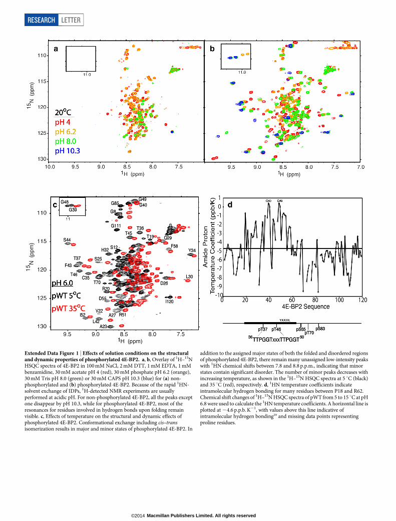
Extended Data Figure 1 | Effects of solution conditions on the structuraland dynamic properties of phosphorylated 4E-BP2. a, b, Overlay of 1H–15NHSQC spectra of 4E-BP2 in 100 mM NaCl, 2 mM DTT, 1 mM EDTA, 1 mMbenzamidine, 30 mM acetate pH 4 (red), 30 mM phosphate pH 6.2 (orange),30 mM Tris pH 8.0 (green) or 30 mM CAPS pH 10.3 (blue) for (a) non-phosphorylated and (b) phosphorylated 4E-BP2. Because of the rapid 1HN-solvent exchange of IDPs, 1H-detected NMR experiments are usuallyperformed at acidic pH. For non-phosphorylated 4E-BP2, all the peaks exceptone disappear by pH 10.3, while for phosphorylated 4E-BP2, most of theresonances for residues involved in hydrogen bonds upon folding remainvisible. c, Effects of temperature on the structural and dynamic effects ofphosphorylated 4E-BP2. Conformational exchange including cis–transisomerization results in major and minor states of phosphorylated 4E-BP2. In
addition to the assigned major states of both the folded and disordered regionsof phosphorylated 4E-BP2, there remain many unassigned low-intensity peakswith 1HN chemical shifts between 7.8 and 8.8 p.p.m., indicating that minorstates contain significant disorder. The number of minor peaks decreases withincreasing temperature, as shown in the 1H–15N HSQC spectra at 5 uC (black)and 35 uC (red), respectively. d, 1HN temperature coefficients indicateintramolecular hydrogen bonding for many residues between P18 and R62.Chemical shift changes of 1H–15N HSQC spectra of pWT from 5 to 15 uC at pH6.8 were used to calculate the 1HN temperature coefficients. A horizontal line isplotted at 24.6 p.p.b. K21, with values above this line indicative ofintramolecular hydrogen bonding29 and missing data points representingproline residues.
RESEARCH LETTER
Macmillan Publishers Limited. All rights reserved©2014
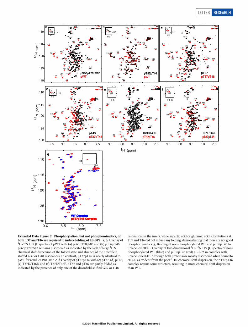
Extended Data Figure 2 | Phosphorylation, but not phosphomimetics, ofboth T37 and T46 are required to induce folding of 4E-BP2. a, b, Overlay of1H–15N HSQC spectra of pWT with (a) pS65pT70pS83 and (b) pT37pT46.pS65pT70pS83 remains disordered as indicated by the lack of large 1HNchemical shift dispersion of the folded state and absence of the downfieldshifted G39 or G48 resonances. In contrast, pT37pT46 is nearly identical topWT for residues P18–R62. c–f, Overlay of pT37pT46 with (c) pT37, (d) pT46,(e) T37D/T46D and (f) T37E/T46E. pT37 and pT46 are partly folded asindicated by the presence of only one of the downfield shifted G39 or G48
resonances in the insets, while aspartic acid or glutamic acid substitutions atT37 and T46 did not induce any folding, demonstrating that these are not goodphosphomimetics. g, Binding of non-phosphorylated WT and pT37pT46 tounlabelled eIF4E. Overlay of two-dimensional 1H–15N HSQC spectra of non-phosphorylated WT (blue) and pT37pT46 (red) 4E-BP2 in complex withunlabelled eIF4E. Although both proteins are mostly disordered when bound toeIF4E, as evident from the poor 1HN chemical shift dispersion, the pT37pT46complex retains some structure, resulting in more chemical shift dispersionthan WT.
LETTER RESEARCH
Macmillan Publishers Limited. All rights reserved©2014

Extended Data Figure 3 | Structural models of phosphorylated 4E-BP2calculated with CS-Rosetta and NMR data. a, Alignment of ribbon diagramsof the lowest energy structures from , 20,000 structural models using differentinputs: 1HN, 15N, Ca and Cb chemical shifts (grey), 1HN, 15N, 13Ca, 13Cb and13CO chemical shifts (blue), and 1HN, 15N, 13Ca, 13Cb and 13CO chemical shiftsas well as NOEs (red). b, Superposition of the final 20 lowest energy structurescalculated using all chemical shifts and NOEs. Residues P18–K57 are shown
using a rainbow colour spectrum from amino (N) to C termini. c, Examples of1HN–1HN NOEs within the folded 4E-BP2. Shown are strips from 15N-edited15N-NOESY demonstrating both short- and long-range interactions. Note thatresidues within the long loop connecting strandsb1 andb2 contain no short- orlong-range NOEs, indicating that it is very dynamic, consistent with low[1H]–15N NOEs. Not surprisingly, this loop shows the largest variation in themodels (see a, b).
RESEARCH LETTER
Macmillan Publishers Limited. All rights reserved©2014

Extended Data Figure 4 | CS-Rosetta scores for calculations of the foldedstate of phosphorylated 4E-BP2 using different input data. CS-Rosetta2 wasused to create models three separate times as new input data were acquired,each time with the observation that low Rosetta energy models converge to thesame topology. Over these three sequential runs each addition of new dataserved primarily to drive sampling towards a previously observed energyminimum. a–c, CS-Rosetta energy for ,20,000 structural models as a functionof Ca r.m.s.d. to the structure with the lowest energy point in the final ensembleusing (a) 1HN, 15N, 13Ca and 13Cb chemical shifts, (b) 1HN, 15N, 13Ca, 13Cband 13CO chemical shifts, and (c) all chemical shifts and NOEs (finalensemble). In green are the best 5% of structures on the basis of agreement with
NOEs, whereas in red are the 20 structures with the lowest NOE violations usedto generate Extended Data Fig. 3b. Note that the CS-Rosetta energy plotted hereis the empirical Rosetta energy value without chemical shift or NOE terms,reflecting the intrinsic energy rather than the fit to experimental data.d, Histograms showing the percentage distribution of structures with Car.m.s.d. as a function of Ca r.m.s.d. (going out to 6 A) for the different CS-Rosetta input data: 1HN, 15N, 13Ca and 13Cb chemical shifts (grey), 1HN, 15N,13Ca, 13Cb and 13CO chemical shifts (blue), 1HN, 15N, 13Ca, 13Cb, and 13COchemical shifts and NOEs without filtering (red) and with the final set of filters(cyan) for (1) the hydrogen bonding observed from the temperature coefficientmeasurements and (2) the best 5% by number of NOE satisfied.
LETTER RESEARCH
Macmillan Publishers Limited. All rights reserved©2014

Extended Data Figure 5 | Chemical shifts and calculated secondarystructure of phosphorylated 4E-BP2. Secondary chemical shifts define thetopology of phosphorylated 4E-BP2 and validate the CS-Rosetta approach forstructure determination. a–c, Fractional secondary structure as calculated by
Talos1 as a function of residues number for (a) strand, (b) helix and (c) loopfor residues 1–75 of phosphorylated 4E-BP2. d, Secondary chemical shifts forthe folded region of phosphorylated 4E-BP2 from Talos1 as a function ofresidue number for 13CO, 1Ha, 13Ca, 15N, 13Cb and 1HN shifts.
RESEARCH LETTER
Macmillan Publishers Limited. All rights reserved©2014

Extended Data Figure 6 | NOE violations for calculated structure ofphosphorylated 4E-BP2. a, Contact map showing observed NOEs for eachpair of residues for satisfied NOE restraints in blue and unsatisfied NOErestraints in red, with the areas of the circles proportional to the total number ofNOEs in each case. Violations were calculated using distance boundaries of5/6/7A for strong, medium and weak NOEs, and the number of violations foreach residue pair was either averaged across the ensemble (above the x 5 y line)or by only counting restraints that were never satisfied in any of the modelsin the ensemble (below the x 5 y line). The secondary structure of the protein is
represented on the diagonal in green (b-strand), yellow (310 helix) and black(turn) bars. b–d, Examples of NOE violations consistent with dynamicconformational exchange. A detailed look at individual NOE pairs (satisfiedshown in yellow dashed lines, unsatisfied in red dashed lines) supports theconclusion that minor conformations contribute to the high number ofviolations, as consistently violated restraints conflict with the majority of thedata that define the major conformation. For more information about the NOEviolations and conformational exchange within phosphorylated 4E-BP2, seeSupplementary Information.
LETTER RESEARCH
Macmillan Publishers Limited. All rights reserved©2014

Extended Data Figure 7 | ITC binding profiles of several 4E-BP2 constructs to eIF4E at 20 6C. a, WT; b, pT37; c, pT46; d, pT37pT46; e, T37D/T46D; f, T37E/T46E; g, pG39VG48V); h, pWT using competition with pS65pT70pS83.
RESEARCH LETTER
Macmillan Publishers Limited. All rights reserved©2014

Extended Data Figure 8 | Resonance assignments of phosphorylatedresidues. a, Overlay of 1H–15N HSQC spectra of pWT in red withphosphorylated S83A (pT37pT46pS65pT70) in black, showing the absence ofthe pS83 peak in pWT and other local changes. The blue arrow indicates theposition of A83. b, Serine 13Ca (red) and 13Cb (blue) chemical shifts inphosphorylated 4E-BP2. Although they did not show significant downfield
shifts in the 1HN–15N HSQC spectrum (Fig. 1), S65 and S83 showed significantdeviations from the random coil values compared with the other serines,consistent with phosphorylation. S25 and S44 also showed deviations as a resultof the interactions within the folded domain (close in space to otherphosphates), but not to the degree expected for a phosphorylated serine.
LETTER RESEARCH
Macmillan Publishers Limited. All rights reserved©2014

Extended Data Table 1 | Structural and energetic properties of phosphorylated 4E-BP2
RESEARCH LETTER
Macmillan Publishers Limited. All rights reserved©2014

Extended Data Table 2 | ITC binding parameters for 4E-BP2 constructs to eIF4E
Enthalpy changes (DH), entropy changes (DS) and dissociation constants (Kd) from ITC measurements as well as combined binding and unfolding free energies derived from Kd values using DG 5 2RTln(1/Kd),with R the ideal gas constant and T the temperature, and estimated unfolding free energies using WT as a reference for pT37, pT46, pT37pT46 and pWT.aDG values, which may include contributions from binding, unfolding of the b-sheet structure encompassing residues P18–R62 and folding of the helix encompassing the YXXXXLW sequence, were calculated fromDG 5DH 2 TDS 5 2RTln(1/Kd). The estimated unfolding free energy was obtained using DG 2DGWT, as for WT there was no contribution from unfolding.bThe enhanced stability of the fivefold phosphorylated (pWT) over pT37pT46, 4.8 versus 2.6 kcal mol21 estimated unfolding free energy, is possibly due to long-range transient electrostatic interactions betweenthe phosphorylated acidic C terminus with the basic folded domain. The predicted isoelectric point (pI) of non-phosphorylated full-length 4E-BP2 is 6.16; however, the region involved in the phosphorylation-induced folding is very basic (pI 5 9.77), while the C terminus (S65–I120) is acidic (pI 5 4.68). Phosphorylation is expected to drop the pI even further, potentially causing stabilizing transient electrostaticinteractions between the folded region and the phosphorylated C terminus.
LETTER RESEARCH
Macmillan Publishers Limited. All rights reserved©2014