for dental research 2015 a systematic review and … · for dental research 2015 ... of the caries...
TRANSCRIPT
Journal of Dental Research2015, Vol. 94(4) 522 –533© International & American Associations for Dental Research 2015Reprints and permissions: sagepub.com/journalsPermissions.navDOI: 10.1177/0022034515571184jdr.sagepub.com
Clinical Review
IntroductionConventionally, treatment of caries lesions has been performed by removing carious hard tissues and restoring the resulting cavity. This invasive approach is increasingly challenged, as removal of the lesion is seen as symptomatic, not causal, treat-ment, while biologically grounded treatments, such as non- or microinvasive strategies, aim at controlling the disease pro-cess: Noninvasive strategies avoid any removal of hard tissues and focus, for example, on influencing the equilibrium of de- and remineralization or removing/controlling the biofilm activity. Microinvasive strategies involve conditioning of den-tal hard tissues and are thus not completely noninvasive; how-ever, only few micrometers of enamel or dentin are removed. These strategies aim at establishing a diffusion barrier for acids, minerals, or carbohydrates via sealing the lesion (Kidd 2004; Kidd and Fejerskov 2004; van Amerongen et al. 2008).
On the one hand, non- or microinvasive therapies might be able to avoid or postpone invasive treatments and the associ-ated cycle of retreatments, which could help to retain teeth lon-ger at lower costs (Brantley et al. 1995; Griffin et al. 2002; Frencken et al. 2012; Schwendicke et al. 2014). On the other,
noninvasive treatments greatly depend on patients’ compli-ance, while microinvasively treated (sealed) lesions might require frequent resealing. Thus, it could be argued that mini-mally invasive treatment might remain a viable option, espe-cially for treating lesions in pits and fissures—that is, the majority of dental caries lesions (Hugoson et al. 2000; Marthaler 2004; Hopcraft et al. 2009): Such minimally inva-sive treatments include preventive resin restorations, sealant restorations, or enameloplasty and do not follow the principle
571184 JDRXXX10.1177/0022034515571184Journal of Dental ResearchTreating Caries: A Systematic Reviewreview-article2015
1Department of Operative and Preventive Dentistry, Charité–Universitätsmedizin Berlin, Germany2Institute of Epidemiology and Preventive Medicine, College of Public Health, National Taiwan University, Taipei, Taiwan
A supplemental appendix to this article is published electronically only at http://jdr.sagepub.com/supplemental.
Corresponding Author:F. Schwendicke, Department for Operative and Preventive Dentistry, Charité Centre for Dental Medicine, Aßmannshauser Str. 4-6, 14197 Berlin, Germany. Email: [email protected]
Treating Pit-and-Fissure Caries: A Systematic Review and Network Meta-analysis
F. Schwendicke1, A.M. Jäger1, S. Paris1, L.Y. Hsu2, and Y.K. Tu2
AbstractFor shallow or moderately deep pit-and-fissure lesions, various treatment options are available: (1) noninvasive treatments (e.g., fluoride application, antibacterial treatments, oral hygiene advice) avoid any dental hard tissue removal; (2) microinvasive treatments (e.g., sealing) remove only a few micrometers of hard tissues by etching; and minimally invasive methods (e.g., “preventive” resin/sealant restoration) remove carious dentin but avoid sacrificing sound tissues. We aimed at systematically reviewing and comparing these strategies for treating pit-and-fissure lesions in permanent teeth using network meta-analysis. Randomized or nonrandomized clinical trials investigating shallow or moderately deep primary caries lesions in fissured or pitted surfaces were included. We compared the risk of requiring invasive treatments or any retreatments in noninvasive, microinvasive, and minimally invasive treated lesions; untreated lesions were used as controls. Five electronic databases were systematically screened up to September 2013 and cross-referencing performed. Pairwise and network meta-analyses were performed and odds ratios and 95% confidence intervals (CI) calculated. Certainty of estimates was evaluated via GRADE criteria. From a total of 2,214 identified records, 14 studies representing 1,440 patients with 3,551 treated lesions were included. Pairwise meta-analysis found microinvasive and minimally invasive treated lesions to require less invasive retreatments than control lesions (odds ratios [95% confidence intervals]: 0.13 [0.07 to 0.26], 0.13 [0.03 to 0.50], respectively), whereas the estimate for noninvasively treated lesions remained nonsignificant (0.64 [0.39 to 1.06]). These findings were reflected in the strategy ranking stemming from network meta-analysis (first, minimally invasive; second, microinvasive; third, noninvasive). However, microinvasive treatment required significantly more total retreatments (including resealing) than minimally or noninvasive treatments. Due to limited study quality, the evidence was graded as low or very low. Clinical treatment decisions should consider the long-term sequelae and costs stemming from different therapies as well as their subjective impact on the patient. Available treatment options seem suitable for treating shallow or moderately deep pit-and-fissure lesions in permanent teeth; further conclusions are not possible.
Keywords: dental, enamel, dentin, fluoride, Monte Carlo method, dental restoration failure
at UNIV LIBRARY AT IUPUI on March 28, 2015 For personal use only. No other uses without permission.jdr.sagepub.comDownloaded from
© International & American Associations for Dental Research 2015
Treating Caries: A Systematic Review 523
of “extension for prevention” but are guided by the extension of the caries lesion and aim at preserving hard tissues. Occlusally, they can be performed without greatly sacrificing sound tooth tissue (which is a difference to treatment of proximal lesions), while the effectiveness of the resulting restoration is largely independent from patients’ compliance, with potentially long retention times of such minimally invasive restorations.
The treatment of deep caries lesions has been recently sub-ject of 2 systematic reviews (Ricketts et al. 2013; Schwendicke et al. 2013), while the last systematic review focusing on shal-low or moderately deep lesions was published in 2008 (Griffin et al. 2008). Moreover, the latter review focused on sealing and its efficacy for arresting lesions; it did not compare all 3 dis-cussed treatments with one another. Thus, there is the need for a comprehensive and updated review. We conducted a system-atic review and network meta-analysis to compare noninva-sive, microinvasive, and minimally invasive treatments for pit-and-fissure lesions in permanent teeth. Network meta-anal-ysis provides a statistical framework to compare more than 2 interventions simultaneously according to all available direct and indirect evidence.
Materials and MethodsThis review follows international guidelines for performing and reporting systematic reviews and pairwise or network meta-analysis (Moher et al. 2009; Hoaglin et al. 2011; Jansen et al. 2011). We aimed at answering the following question: In patients with shallow or moderately deep pit-and-fissure caries lesions, are non- or microinvasive, instead of minimally inva-sive, treatments more efficacious for avoiding retreatments?
Selection Criteria
Studies. Randomized, quasi-randomized, or nonrandomized controlled trials published in 1967 or later with a minimum follow-up of 6 mo were considered. Nonrandomized trials were included only if lesions treated in different groups were comparable. Including nonrandomized studies was performed to increase comprehensiveness of the review and has been shown to not generally introduce systematic bias (Deeks et al. 2003).
Participants. Participants included humans with primary caries lesions in fissured or pitted surfaces of permanent posterior teeth. Caries was to be limited to enamel or the outer two-thirds of the dentin—that is, deep caries lesions were not to be included. If both sound and caries surfaces had been treated, separate calculation for only carious surfaces needed to be feasible.
Intervention and control. Only studies that compared a mini-mum of 2 of the following strategies were included:
•• Noninvasive treatment—such as remineralization (e.g., nonstandard highly concentrated fluoride toothpastes,
fluoride rinses, gels, varnishes, or other remineraliza-tion agents), antibacterial treatments, oral hygiene edu-cation, or professional oral hygiene. We did not include ozone treatment, since we assumed it to be neither stan-dard nor generally available in primary care.
•• Microinvasive treatment—that is, caries sealing using resin-based or glass ionomer cement sealants. Laser therapy was not included.
•• Minimally invasive treatment—that is, restorative treat-ment including caries removal but aiming at preserva-tion of sound dental hard tissues (e.g., “preventive” resin restorations, sealant restorations, restorations after enameloplasty or fissurotomy, minimally invasive res-torations using various materials).
•• Control—that is, no active or placebo treatment and standard oral home care. Note that this option is not a “treatment” according to the outlined groups (noninva-sive, microinvasive, minimally invasive) but was included in the review as comparator for most strategies and to allow construction of a network).
Outcomes. We assessed clinically or radiographically deter-mined need for retreatment. Need for retreatment was defined as any complication that could have required mending (e.g., progression of the lesion, sealant loss, secondary caries or frac-ture of restorations, pulpal complications).
We chose need to retreat as outcome, as it integrates various outcome parameters used in studies evaluating noninvasive, microinvasive, and minimally invasive treatments. We should highlight that the need to retreat was based on the complica-tion, not on the reporting of performed retreatments, as most studies did not explicitly describe handling of complications. Need to retreat was further analyzed as need for invasive retreatments (e.g., due to lesion progression, restoration loss, secondary caries, pulpal complications) and need for any retreatments (including invasive but also all other retreatments).
Search StrategyIdentification of studies was based on a search strategy for each electronic database (Cochrane Central Register of Controlled Trials, Medline via PubMed, Embase); the search was carried out September 23, 2013. For screening procedures, we used a 3-pronged approach and employed a restriction to title/abstract searches without controlled vocabularies (e.g., MeSH; Fig. 1, Appendix Table 1). Cross-referencing from retrieved full-text studies identified further articles. Two addi-tional databases were searched: www.clinicaltrials.gov for unpublished trials and opengrey.eu for gray literature. The search was not restricted regarding the language, and studies published in languages other than English or German were planned to be translated. Neither authors nor journals were blinded to reviewers. Title and abstract of identified studies were screened by 2 calibrated reviewers (F.S., A.M.J.) for eli-gibility. Consensus was obtained by discussion or consulting a third reviewer (S.P.).
at UNIV LIBRARY AT IUPUI on March 28, 2015 For personal use only. No other uses without permission.jdr.sagepub.comDownloaded from
© International & American Associations for Dental Research 2015
524 Journal of Dental Research 94(4)
Data Extraction and Evaluation
Data from eligible studies were independently extracted by 2 reviewers (F.S., A.M.J.) using piloted electronic spread-sheets. Data were recorded according to guidelines outlined by the Cochrane Collaboration (Higgins and Green 2011). For data extraction, we assumed that cavitated lesions always involved dentin, while noncavitated lesions were assumed to be limited to enamel as long as not stated otherwise. We extracted the proportion of lesions that had received or required invasive treatment or retreatment (i.e., lesions that were restored/rerestored or required restoration/rerestora-tion) as well as the proportion of lesions that had received or required any retreatment (i.e., lesions that were retreated or required retreatment of any kind; e.g., resealing). Note that for noninvasive treatment, reapplication of, for example, flu-oride varnish was not counted as retreatment but part of the ongoing therapy.
Case definitions (diagnostic criteria used to assess car-ies), lesion depths, caries risk, and methods used to assess caries progression or failure were also extracted. If data were missing or methodological issues were to be addressed, study authors were contacted via e-mail and—if appropri-ate—information added to the data extraction. Missing data (e.g., sample size at baseline, numbers of patients who received different treatments) were estimated as outlined below.
Risk of Bias Assessment
Selection bias (sequence genera-tion, allocation concealment), per-formance and detection bias (blinding of participants, operators, examiners), attrition bias (loss to follow-up and missing values or participants), and reporting bias (selective reporting, unclear with-drawals, missing outcomes) were recorded, assessed, and classified according to Cochrane guidelines (Higgins and Green 2011).
Pairwise and Network Meta-analysis
Traditional pairwise meta-analysis makes direct comparisons for each pair of treatments, while network meta-analysis includes both direct and indirect evidence for multiple treatment comparisons. The differ-ences in the results could then be used to detect potential inconsis-tency between direct and indirect evidence. Whenever appropriate, we used Cochrane’s Q and I2 to
evaluate the heterogeneity in the pairwise comparisons within the network.
The unit of analysis for meta-analysis was the patient. When patients contributed to >1 tooth/lesion, adjustments were made by converting the number of teeth to the number of patients by assuming that teeth were evenly distributed among patients. Pairwise meta-analysis was performed using a ran-dom effect model via Comprehensive Meta-Analysis 2.2.64 (Biostat, Englewood, NJ, USA), with odds ratios (ORs) and 95% confidence intervals (CIs). Heterogeneity was assessed quantitatively using I2 statistics (Higgins and Thompson 2002). Funnel plot analysis and Egger test were performed to assess small study effects or publication bias (Egger et al. 1997; Higgins and Green 2011). Evidence for each pairwise esti-mates was graded using Grade Profiler 3.6 according to GRADE guidelines (Atkins et al. 2004).
Network meta-analysis was performed (Hoaglin et al. 2011) using Bayesian random effects hierarchical models and a Markov chain Monte Carlo simulation via WinBugs, imple-mented in R 3.0.2 (R Foundation, Vienna, Austria). To fit the model, we used a noninformative uniform prior distribution and assessed convergence based on the Brooks-Gelman-Rubin criteria (Brooks and Gelman 1998) and inspection of history plots. Simulations were performed with 50,000 tuning itera-tions and further 50,000 simulations per chain at a thinning interval of 50 with 3 chains of initial values. We calculated posterior medians and 95% credibility intervals, equaling the
2206 records identified through searching of electronic databases
128 articles screened full-text for eligibility
8 records identified by hand search
2078 records excluded
114 full-text articles excluded
• 56 no defined comparisons
• 27 no caries or unable to
separate sound from caries
• 12 allocated treatment
according to lesion depth
• 18 no clinical or primary data
• 1 not retrievable
136 articles screened full-text for eligibility
22 articles included
14 studies included
Scre
enin
g of
tit
le a
ndab
stra
ctSc
reen
ing
of
full-
text
art
icle
sSearch(((((((caries[Title/Abstract]) ORcarious[Title/Abstract]) ORdecay[Title/Abstract]) ORcavity[Title/Abstract]) ORcavitated[Title/Abstract]) ORcavitation[Title/Abstract]) ORdecayed[Title/Abstract]) ORdemineralized[Title/Abstract]
yielded 97369 entries
Search((((((((((((((enameloplasty[Title/Abstract]) ORodontomy [Title/Abstract]) OR fissurotomy[Title/Abstract]) OR sealant [Title/Abstract]) OR resin[Title/Abstract]) OR composite [Title/Abstract]) ORrestoration [Title/Abstract]) OR glass ionomer[Title/Abstract]) OR glassionomer[Title/Abstract]) ORsealing [Title/Abstract]) OR fluoride[Title/Abstract]) ORremineralization [Title/Abstract]) OR chlorhexidine[Title/Abstract]) OR varnish[Title/Abstract]) ORhygiene [Title/Abstract] yielded 252687 entries
Search((((((fissure[Title/Abstract]) ORpit[Title/Abstract]) ORfissures[Title/Abstract]) ORpits[Title/Abstract]) ORocclusal[Title/Abstract]) ORpremolars[Title/Abstract]) ORmolars[Title/Abstract])
yielded 97451 entriesSear
ch o
fele
ctro
nic
data
base
s
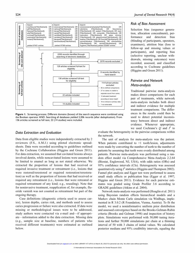
Figure 1. Screening process. Different domains (boxes) of the search sequence were combined using the Boolean operator AND. Searching all databases yielded 2,206 records (after deduplication). From 136 articles screened as full text, 22 (14 studies) were included.
at UNIV LIBRARY AT IUPUI on March 28, 2015 For personal use only. No other uses without permission.jdr.sagepub.comDownloaded from
© International & American Associations for Dental Research 2015

Treating Caries: A Systematic Review 525
range of estimated parameters after exclusion of extreme val-ues (Tu et al. 2012). The consistency between direct and indi-rect results was evaluated using the statistical software Stata command ifplot (Chaimani et al. 2013) and treatments ranked according to their probability of being the best choice (Salanti et al. 2011).
Subgroup Analyses
Subgroup analyses were performed to evaluate effects of participants’ age (<18 vs. >18 y), lesion depths (enamel vs. dentinal lesions), surface integrity (not cavitated vs. cavi-tated) and sealant materials (resin based vs. glass ionomer cement sealant) on effect estimates. Similarly, we explored the effects of study design (randomized vs. nonrandomized controlled trial) and case definition (questionable vs. certain presence of caries lesions). Last, effect estimates were based on calculations using the intention-to-treat-principle, count-ing all lost cases as failure, thereby accounting for imbal-anced attrition.
Results
Results of the Search
Through electronic databases, 2206 studies were found to be possibly eligible. No ongoing trials were identified; 128 stud-ies were analyzed full-text; and 8 further studies were retrieved by cross-referencing and screened for eligibility. In total, 136 studies were investigated full-text. Authors of 13 studies were contacted, and 8 replied. Eventually, 14 studies reported in 22 articles were found eligible for this review (Fig. 1). Reasons for study exclusion at the full-text stage can be found in Appendix Table 2.
Included Studies
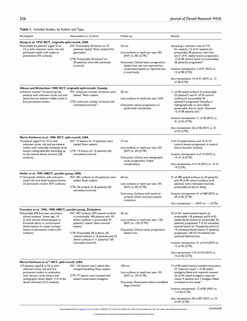
The 14 included studies represented 1,440 patients with 3,551 treated lesions. The majority of studies compared microinva-sive treatments with control, while no study compared nonin-vasive versus minimally invasive treatment (Fig. 2).
From the included studies, 5 were performed in the United States, 4 in Brazil, and 1 each in China, Canada, Zimbabwe, Albania, and Denmark. Ten and 4 studies used random and nonrandom allocation of lesions to groups, respectively (Table 1), and all studies were published in English. Trials were reported between 1976 and 2012, with a follow-up range up to 10 y and with dropout rates between 0% and 18% per year. Most patients were children or adolescents, with 3 studies investigating adult patients as well (Mertz-Fairhurst et al. 1998; Bakhshandeh et al. 2012; Liu et al. 2012). The majority of studies analyzed noncavitated caries, with 5 studies evaluat-ing treatment of cavitated lesions (Going et al. 1976; Mertz-Fairhurst et al. 1986; Frencken et al. 1998; Mertz-Fairhurst et al. 1998; Maltz et al. 2003) and 3 more trials analyzing
treatment of lesions radiographically extending into dentin (Borges et al. 2010; Bakhshandeh et al. 2012; Borges et al. 2012; da Silveira et al. 2012). The case definition for a caries lesion differed greatly among studies (Table 1).
Pairwise Comparison of Interventions
Effect estimates were calculated for pairwise comparisons of interventions, evaluating the risk of requiring invasive or any retreatment. Performing microinvasive or minimally invasive compared with controls significantly reduced the risk of inva-sive retreatment being required (OR [95% CI]: 0.13 [0.07 to 0.26] or 0.13 [0.03 to 0.50], respectively), while no significant effect could be shown for noninvasive treatment versus control (Fig. 3a). Micro- and noninvasive or minimally and microinva-sive treatments did not significantly differ from one another with regard to the risk of requiring invasive retreatments. No pairwise estimate could be calculated to compare noninvasive versus minimally invasive treatments.
Microinvasively treated lesions required any retreatments sig-nificantly more often (OR [95% CI]: 17.8 [5.94 to 53.5]) than noninvasively treated lesions (Fig. 3b). Similarly, lesions in con-trol groups required retreatment more frequently than minimal invasively treated lesions. The effect estimate comparing nonin-vasive treatment versus control did not reach statistical signifi-cance (0.64 [0.39 to 1.06]). No significant differences were found between micro- and minimally invasive treatments or microinva-sive treatment and control. No effect estimate was calculated for the comparison between minimally and noninvasive treatments.
Network Comparisons of Interventions
Comparing noninvasive treatment versus control did not reveal significantly reduced risks of requiring invasive retreatment (OR [95% credibility interval]: 0.58 [0.16 to 2.02]).
Figure 2. Network of the comparisons for the Bayesian network meta-analysis. The size of the nodes is proportional to the number of lesions (in parentheses) receiving each treatment. The width of the lines is proportional to the number of trials (beside the line) comparing the connected treatments. Note that the network meta-analysis used patients as the unit of analysis.
at UNIV LIBRARY AT IUPUI on March 28, 2015 For personal use only. No other uses without permission.jdr.sagepub.comDownloaded from
© International & American Associations for Dental Research 2015
526 Journal of Dental Research 94(4)
Table 1. Included Studies, by Author and Type.
Participants Intervention vs. Control Follow-up Results
Going et al. 1976: RCT, originally split-mouth, USAPresumably 63 patientsa (aged 10 to
14 y) with unknown caries risk and permanent teeth with explorer penetration (93 surfaces).
INT: Presumably 49 lesions (in 33 patients) sealed.a Resin sealant first generation.
CTR: Presumably 45 lesionsa (in 30 patients) were left untreated (control).
24 mo.
Lost surfaces or teeth per year: 8% (INT) vs. 8% (CTR).a
Outcomes: Clinical lesion progression. Sealant loss was not reported but recalculated based on reported loss in total study.
Assuming a retention rate of 71% for sealants, 12 of 41 sealants (in presumably 28 patients) were lost and 7 of 41 sealed lesions progressed. 13 of 38 control lesion (in presumably 26 patients) progressed.a
Invasive retreatment: 7 of 41 (INT) vs. 13 of 98 (CTR)
Any retreatment: 19 of 41 (INT) vs. 13 of 38 (CTR)
Gibson and Richardson 1980: RCT, originally split-mouth, CanadaUnknown numberb of second-grade
patients with unknown caries risk and sticky fissures without visible caries in first permanent molars.
INT: Unknown number of lesions were sealed.b Resin sealant.
CTR: Unknown number of lesions left untreated (control).b
30 mo.
Lost surfaces or teeth per year: N/A.
Outcomes: Lesion progression or performed retreatment.
11 of 58 sealed surfaces (in presumably 25 patientsb) and 41 of 53 control surfaces (in presumably 24 patientsb) progressed clinically or radiographically or were filled, presumably due to caries. Assumed 15 of 58 sealants lost.b
Invasive retreatment: 11 of 58 (INT) vs. 41 of 53 (CTR).
Any retreatment: 26 of 58 (INT) vs. 41 of 53 (CTR).
Mertz-Fairhurst et al. 1986: RCT, split-mouth, USA14 patients (aged 9 to 19 y) with
unknown caries risk and permanent molars with minimally cavitated caries lesions radiographically extending up to the enamel dentin junction (28 surfaces).
INT: 14 lesions (in 14 patients) were sealed. Resin sealant.
CTR: 14 lesions (in 14 patients) left untreated (control).
12 mo.
Lost surfaces or teeth per year: 0% (INT) vs. 0% (CTR).
Outcomes: Clinical and radiographic caries progression. Pulpal complications.
4 of 14 sealed lesions and 14 of 14 control lesions progressed. 6 control lesions became sensitive.
Invasive retreatment: 4 of 14 (INT) vs. 14 of 14 (CTR).
Any retreatment: 4 of 14 (INT) vs. 14 of 14 (CTR).
Heller et al. 1995: NRCT, parallel group, USA71 first-grade children with unknown
caries risk and initial incipient lesions on permanent molars (437 surfaces).
INT: 381 surfaces (in 64 patients) were sealed. Resin sealant.
CTR: 56 surfaces (in 8 patients) left untreated (control).
60 mo.
Lost surfaces or teeth per year: 0% (INT) vs. 0% (CTR).
Outcomes: Surfaces with caries or surfaces which received invasive treatment.
41 of 380 sealed surfaces (in 63 patients) and 29 of 56 control surfaces (in 8 patients) were retreated invasively, presumably all due to decay.
Invasive retreatment: 41 of 380 (INT) vs. 29 of 56 (CTR).
Any retreatment: — (INT) vs. — (CTR).
Frencken et al. 1996, 1998: NRCT, parallel group, ZimbabwePresumably 258 first-year secondary
school studentsc (mean age, 14 y) with enamel (white/opaque or brownish-dark) or noncavitated dentin lesions in mostly occlusal lesions in permanent molars (551 surfaces).
INT: 493 surfaces (392 enamel surfaces in presumably 184 patients and 101 dentin surfaces in presumably 47 patients)c sealed. Glass ionomer sealant.
CTR: Presumably 58 surfaces (35 enamel surfaces in 16 patients and 23 dentin surfaces in 11 patients)c left untreated (control).
36 mo.
Lost surfaces or teeth per year: 12% (INT) vs. 12% (CTR).c
Outcomes: Clinical caries progression. Sealant loss.
23 of 251 sealed enamel lesions (in presumably 118 patients) and 8 of 63 sealed dentin lesions (in presumably 30 patients) progressed. 7 of 22 untreated enamel lesions (in 10 patients) and 6 of 14 untreated dentin lesions (7 patients) progressed. 144 of 314 sealants lost, assumed balanced loss.
Invasive retreatment: 31 of 314 (INT) vs. 13 of 36 (CTR).
Any retreatment: 175 of 314 (INT) vs. 13 of 36 (CTR).
Mertz-Fairhurst et al.d: RCT, split-mouth, USA123 patients (aged 8 to 52 y) with
unknown caries risk and 312 permanent molars or premolars with obvious caries lesions and radiographic lesion depth <1/2 of the dentin thickness (312 surfaces).
INT: 156 lesions were sealed after marginal bevelling. Resin sealant.
CTR: 77 lesions were treated with sealed conservative amalgams.
120 mo.
Lost surfaces or teeth per year: 4% (INT) vs. 3% (CTR).
Outcomes: Restoration failure (modified Ryge criteria).e
12 of 85 sealed lesions needed restoration, 37 required reseal. 1 of 44 sealed amalgams failed and required renewal, 22 of 44 sealed amalgams required reseal. 4 sealants and 1 amalgam failed unrelated to the study.e
Invasive retreatment: 12 of 85 (INT) vs. 1 of 44 (CTR).
Any retreatment: 49 of 85e (INT) vs. 23 of 44e (CTR).
(continued) at UNIV LIBRARY AT IUPUI on March 28, 2015 For personal use only. No other uses without permission.jdr.sagepub.comDownloaded from
© International & American Associations for Dental Research 2015
Treating Caries: A Systematic Review 527
Participants Intervention vs. Control Follow-up Results
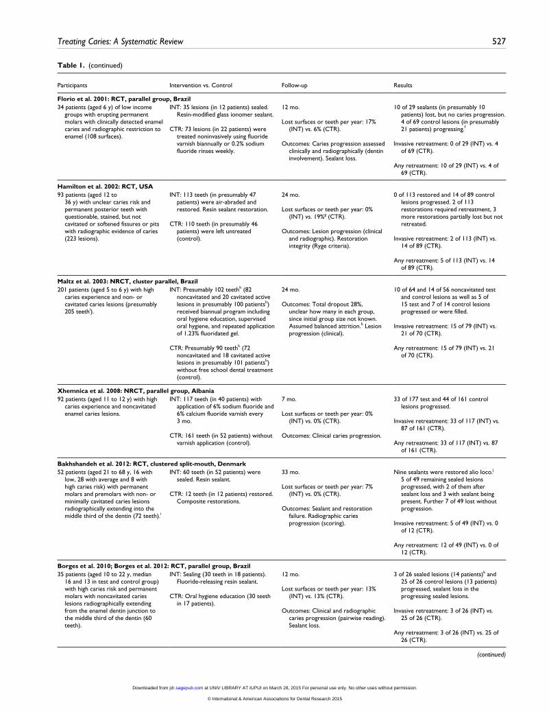
Florio et al. 2001: RCT, parallel group, Brazil34 patients (aged 6 y) of low income
groups with erupting permanent molars with clinically detected enamel caries and radiographic restriction to enamel (108 surfaces).
INT: 35 lesions (in 12 patients) sealed. Resin-modified glass ionomer sealant.
CTR: 73 lesions (in 22 patients) were treated noninvasively using fluoride varnish biannually or 0.2% sodium fluoride rinses weekly.
12 mo.
Lost surfaces or teeth per year: 17% (INT) vs. 6% (CTR).
Outcomes: Caries progression assessed clinically and radiographically (dentin involvement). Sealant loss.
10 of 29 sealants (in presumably 10 patients) lost, but no caries progression. 4 of 69 control lesions (in presumably 21 patients) progressing.f
Invasive retreatment: 0 of 29 (INT) vs. 4 of 69 (CTR).
Any retreatment: 10 of 29 (INT) vs. 4 of 69 (CTR).
Hamilton et al. 2002: RCT, USA93 patients (aged 12 to
36 y) with unclear caries risk and permanent posterior teeth with questionable, stained, but not cavitated or softened fissures or pits with radiographic evidence of caries (223 lesions).
INT: 113 teeth (in presumably 47 patients) were air-abraded and restored. Resin sealant restoration.
CTR: 110 teeth (in presumably 46 patients) were left untreated (control).
24 mo.
Lost surfaces or teeth per year: 0% (INT) vs. 19%g (CTR).
Outcomes: Lesion progression (clinical and radiographic). Restoration integrity (Ryge criteria).
0 of 113 restored and 14 of 89 control lesions progressed. 2 of 113 restorations required retreatment, 3 more restorations partially lost but not retreated.
Invasive retreatment: 2 of 113 (INT) vs. 14 of 89 (CTR).
Any retreatment: 5 of 113 (INT) vs. 14 of 89 (CTR).
Maltz et al. 2003: NRCT, cluster parallel, Brazil201 patients (aged 5 to 6 y) with high
caries experience and non- or cavitated caries lesions (presumably 205 teethi).
INT: Presumably 102 teethh (82 noncavitated and 20 cavitated active lesions in presumably 100 patientsh) received biannual program including oral hygiene education, supervised oral hygiene, and repeated application of 1.23% fluoridated gel.
CTR: Presumably 90 teethh (72 noncavitated and 18 cavitated active lesions in presumably 101 patientsh) without free school dental treatment (control).
24 mo.
Outcomes: Total dropout 28%, unclear how many in each group, since initial group size not known. Assumed balanced attrition.h Lesion progression (clinical).
10 of 64 and 14 of 56 noncavitated test and control lesions as well as 5 of 15 test and 7 of 14 control lesions progressed or were filled.
Invasive retreatment: 15 of 79 (INT) vs. 21 of 70 (CTR).
Any retreatment: 15 of 79 (INT) vs. 21 of 70 (CTR).
Xhemnica et al. 2008: NRCT, parallel group, Albania92 patients (aged 11 to 12 y) with high
caries experience and noncavitated enamel caries lesions.
INT: 117 teeth (in 40 patients) with application of 6% sodium fluoride and 6% calcium fluoride varnish every 3 mo.
CTR: 161 teeth (in 52 patients) without varnish application (control).
7 mo.
Lost surfaces or teeth per year: 0% (INT) vs. 0% (CTR).
Outcomes: Clinical caries progression.
33 of 177 test and 44 of 161 control lesions progressed.
Invasive retreatment: 33 of 117 (INT) vs. 87 of 161 (CTR).
Any retreatment: 33 of 117 (INT) vs. 87 of 161 (CTR).
Bakhshandeh et al. 2012: RCT, clustered split-mouth, Denmark52 patients (aged 21 to 68 y, 16 with
low, 28 with average and 8 with high caries risk) with permanent molars and premolars with non- or minimally cavitated caries lesions radiographically extending into the middle third of the dentin (72 teeth).i
INT: 60 teeth (in 52 patients) were sealed. Resin sealant.
CTR: 12 teeth (in 12 patients) restored. Composite restorations.
33 mo.
Lost surfaces or teeth per year: 7% (INT) vs. 0% (CTR).
Outcomes: Sealant and restoration failure. Radiographic caries progression (scoring).
Nine sealants were restored alio loco.j 5 of 49 remaining sealed lesions progressed, with 2 of them after sealant loss and 3 with sealant being present. Further 7 of 49 lost without progression.
Invasive retreatment: 5 of 49 (INT) vs. 0 of 12 (CTR).
Any retreatment: 12 of 49 (INT) vs. 0 of 12 (CTR).
Borges et al. 2010; Borges et al. 2012: RCT, parallel group, Brazil35 patients (aged 10 to 22 y, median
16 and 13 in test and control group) with high caries risk and permanent molars with noncavitated caries lesions radiographically extending from the enamel dentin junction to the middle third of the dentin (60 teeth).
INT: Sealing (30 teeth in 18 patients). Fluoride-releasing resin sealant.
CTR: Oral hygiene education (30 teeth in 17 patients).
12 mo.
Lost surfaces or teeth per year: 13% (INT) vs. 13% (CTR).
Outcomes: Clinical and radiographic caries progression (pairwise reading). Sealant loss.
3 of 26 sealed lesions (14 patients)k and 25 of 26 control lesions (13 patients) progressed, sealant loss in the progressing sealed lesions.
Invasive retreatment: 3 of 26 (INT) vs. 25 of 26 (CTR).
Any retreatment: 3 of 26 (INT) vs. 25 of 26 (CTR).
Table 1. (continued)
(continued)
at UNIV LIBRARY AT IUPUI on March 28, 2015 For personal use only. No other uses without permission.jdr.sagepub.comDownloaded from
© International & American Associations for Dental Research 2015
528 Journal of Dental Research 94(4)
Participants Intervention vs. Control Follow-up Results
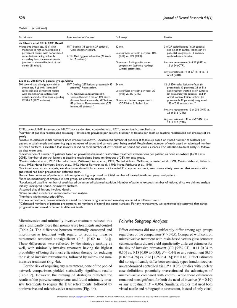
da Silveira et al. 2012: RCT, Brazil44 patients (mean age, 13 y) with
moderate to high caries risk and 61 permanent molars with noncavitated caries lesions radiographically extending from the enamel dentin junction to the middle third of the dentin (61 teeth).
INT: Sealing (33 teeth in 27 patients). Glass ionomer sealant.
CTR: Oral hygiene education (28 teeth in 17 patients).
12 mo.
Lost surfaces or teeth per year: 18% (INT) vs. 14% (CTR).
Outcomes: Radiographic caries progression (pairwise reading). Clinical sealant loss.
3 of 27 sealed lesions (in 24 patients) and 12 of 24 control lesions (in 14 patients) progressed. 11 sealants replaced once, 5 twice.
Invasive retreatment: 3 of 27 (INT) vs. 12 of 24 (CTR).
Any retreatment: 19 of 27l (INT) vs. 12 of 24 (CTR).
Liu et al. 2012: RCT, parallel group, China501 second- and third-grade children
(mean age, 9 y) with “spreaded” caries risk and permanent molars with enamel caries surfaces with opacities and discolorations, equalling ICDAS 2 (1076 surfaces).
INT: Sealing (257 lesions, presumably 43 patients)n Resin sealant.
CTR: Noninvasive treatment (5% sodium fluoride 6 mo or 38% silver diamine fluoride annually, 547 lesions, 88 patients). Placebo treatment (272 lesions, 45 patients).l
24 mo.
Lost surfaces or teeth per year: 0% (INT) vs. 3% (CTR).
Outcomes: Lesion progression to ICDAS 4 to 6. Sealant loss.
12 of 256 sealed lesion surfaces (in presumably 43 patients), 23 of 512 noninvasively treated lesion surfaces (in presumably 86 patients), and 24 of 251 control lesion surfaces (in presumably 42 patients) progressed. 132 of 256 sealants lost.m
Invasive retreatment: 12 of 256 (INT) vs. 23 of 512 (CTR).
Any retreatment: 144 of 256m (INT) vs. 23 of 512 (CTR).
CTR, control; INT, intervention; NRCT, nonrandomized controlled trial; RCT, randomized controlled trial.aNumber of patients recalculated assuming 1.49 sealants provided per patient. Number of lesions per teeth at baseline recalculated per dropout of 8% yearly.bUnable to calculate initial numbers since dropout unknown. Recalculated number of patients at follow-up based on stated number of sealants per patient in total sample and assuming equal numbers of sound and carious teeth being sealed. Recalculated number of teeth based on tabulated number of sealed surfaces. Calculated lost sealants based on total number of lost sealants on sound and caries surfaces. For intention-to-treat analysis, follow-up data were used.cRecalculation of number of patients based on provided atraumatic restorative treatment restorations per patient, as done elsewhere (Griffin et al. 2008). Number of control lesions at baseline recalculated based on dropout of 38% for test group.dMertz-Fairhurst et al., 1987; Mertz-Fairhurst, Williams, Pierce, et al., 1991; Mertz-Fairhurst, Williams, Schuster, et al., 1991; Mertz-Fairhurst, Richards, et al., 1992; Mertz-Fairhurst, Smith, et al., 1992; Mertz-Fairhurst et al., 1995; Mertz-Fairhurst et al., 1998.eFor intention-to-treat analysis, loss due to unrelated failures were not included. For any retreatment, we conservatively assumed that rerestoration and reseal had been provided for different teeth.fRecalculated number of patients at follow-up in each group based on initial number of treated teeth per group and patient.gSince no mentioning of dropout in test group, no attrition assumed.hRecalculated baseline number of teeth based on assumed balanced attrition. Number of patients exceeds number of lesions, since we did not analyze initially unerupted, sound, or inactive surfaces.iAssumed that all lesions involved dentin.jWere counted as failure in intention-to-treat analysis.kNumbers within manuscript differ.lFor any retreatment, conservatively assumed that caries progression and resealing occurred in different teeth.mCalculated numbers of patients proportional to numbers of sound and caries surfaces. For any retreatment, we conservatively assumed that caries progression and reseal occurred in different teeth.
Table 1. (continued)
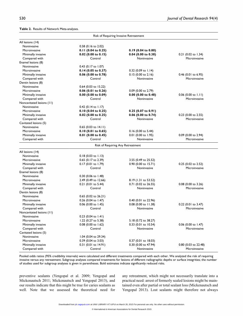
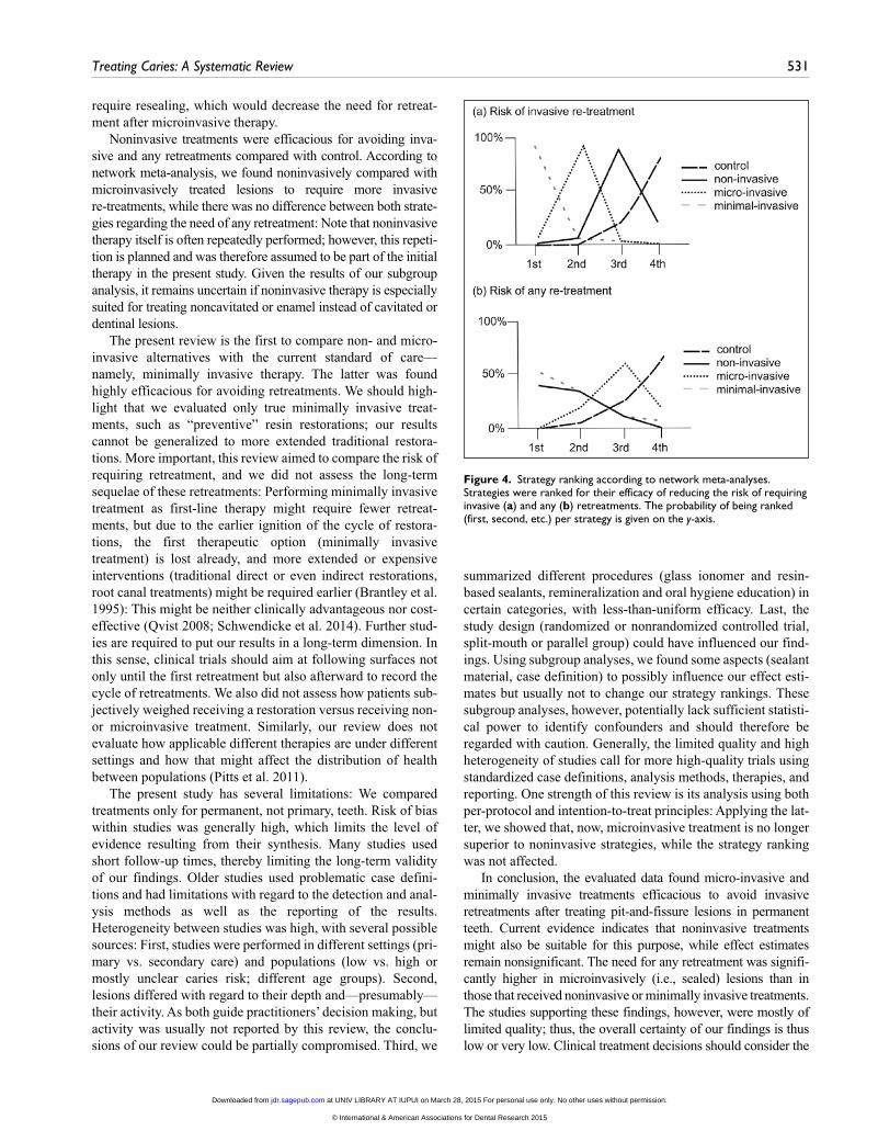
Microinvasive and minimally invasive treatment reduced this risk significantly more than noninvasive treatments and control (Table 2). The difference between minimally compared and microinvasive treatment with regard to requiring invasive retreatment remained nonsignificant (0.21 [0.02 to 1.34]). These differences were reflected by the strategy ranking as well, with minimally invasive treatment having the highest probability of being the most efficacious therapy for reducing the risk of invasive retreatments, followed by micro- and non-invasive treatment (Fig. 4a).
For the risk of requiring any retreatment, none of the pooled network comparisons yielded statistically significant results (Table 2). However, the ranking of strategies reflected the results of the pairwise comparisons and found minimally inva-sive treatments to require the least retreatments, followed by noninvasive and microinvasive treatments (Fig. 4b).
Pairwise Subgroup Analyses
Effect estimates did not significantly differ among age groups regardless of the comparison (P > 0.05). Compared with control, microinvasive treatment with resin-based versus glass ionomer cement sealants did not yield significantly different estimates for the risk of invasive retreatment (OR [95% CI]: 0.11 [0.04 to 0.30] vs. 0.18 [0.09 to 0.35]; P = 0.44) or any retreatment (0.30 [0.02 to 4.78] vs. 2.26 [1.25 to 4.16]; P = 0.16). Effect estimates did not significantly differ between study types (randomized vs. nonrandomized controlled trial, P > 0.05). Studies with unclear case definitions potentially overestimated the advantages of microinvasive compared with control, while these differences remained nonsignificant for both the need of invasive (P = 0.19) or any retreatment (P = 0.06). Similarly, studies that used both visual-tactile and radiographic assessment, instead of only visual
at UNIV LIBRARY AT IUPUI on March 28, 2015 For personal use only. No other uses without permission.jdr.sagepub.comDownloaded from
© International & American Associations for Dental Research 2015

Treating Caries: A Systematic Review 529
assessment, to detect caries progression potentially judged microinvasive ther-apy more advantageously, but estimates remained nonsignificant for the need of invasive (P = 0.10) or any retreatment (P = 0.06) when compared with nonin-vasive therapy.
Network Subgroup Analyses
Subgroup analyses for lesions of differ-ent depths or surface integrities did not indicate significant differences among groups. Compared with control and noninvasive therapy, microinvasive and minimally invasive treatments tended to be more advantageous in dentinal than enamel lesions (Table 2). In contrast, noninvasive treatment was more efficacious in enamel than den-tinal lesions and not efficacious for treating cavitated lesions.
Applying the intention-to-treat-prin-ciple to our analyses—that is, when counting losses to follow-up as failure (requiring retreatment)—we found noninvasive treatment significantly superior to control for reducing the risk of invasive re-retreatments but not sig-nificantly different from microinvasive treatment anymore (Appendix Table 3). Microinvasive treatment was now found significantly inferior to mini-mally invasive treatment regarding the need for invasive and any retreatment (Appendix Table 3).
Risk of Bias and Evidence Grading
We did not detect publication bias via statistical (P > 0.05, Egger test) or graphical means. Risk of bias was gen-erally found high for all included stud-ies (Appendix Table 4). The resulting overall evidence was graded as very low for all but 2 effect estimates (Appendix Tables 5 and 6).
DiscussionThere is increasing evidence that both non- and microinvasive therapy are suitable to treat shallow and moder-ately deep pit-and-fissure lesions. We found microinvasive treatment (i.e., caries sealing) efficacious in preventing invasive treatments or retreatments, corroborating previous results (Griffin et al. 2008). However, such microinvasive therapies seem to require
other retreatments (e.g., resealing) more often than noninva-sive or minimally invasive alternatives. Such lost sealants do not necessarily lead to caries development, as demonstrated for
Figure 3. Results of pairwise meta-analyses regarding the risk of requiring invasive (a) and any (b) retreatments. Data were synthesized with random effects meta-analysis, with odds ratios (OR) and 95% confidence intervals (CIs) being calculated. Pooled effect estimates are indicated by diamonds, with estimates for heterogeneity (I2) and significance of the overall effect being given.
at UNIV LIBRARY AT IUPUI on March 28, 2015 For personal use only. No other uses without permission.jdr.sagepub.comDownloaded from
© International & American Associations for Dental Research 2015
530 Journal of Dental Research 94(4)
Table 2. Results of Network Meta-analyses.
Risk of Requiring Invasive Retreatment
All lesions (14) Noninvasive 0.58 (0.16 to 2.02) Microinvasive 0.11 (0.04 to 0.25) 0.19 (0.04 to 0.80) Minimally invasive 0.02 (0.00 to 0.15) 0.04 (0.00 to 0.38) 0.21 (0.02 to 1.34) Compared with Control Noninvasive MicroinvasiveEnamel lesions (8) Noninvasive 0.43 (0.17 to 1.07) Microinvasive 0.14 (0.05 to 0.37) 0.32 (0.09 to 1.14) Minimally invasive 0.06 (0.00 to 0.78) 0.15 (0.00 to 2.16) 0.46 (0.01 to 6.95) Compared with Control Noninvasive MicroinvasiveDentin lesions (8) Noninvasive 0.64 (0.03 to 15.22) Microinvasive 0.06 (0.01 to 0.26) 0.09 (0.00 to 2.79) Minimally invasive 0.00 (0.00 to 0.09) 0.00 (0.00 to 0.48) 0.06 (0.00 to 1.11) Compared with Control Noninvasive MicroinvasiveNoncavitated lesions (11) Noninvasive 0.42 (0.14 to 1.17) Microinvasive 0.10 (0.04 to 0.25) 0.25 (0.07 to 0.91) Minimally invasive 0.02 (0.00 to 0.25) 0.06 (0.00 to 0.74) 0.23 (0.00 to 2.55) Compared with Control Noninvasive MicroinvasiveCavitated lesions (5) Noninvasive 0.65 (0.03 to 14.11) Microinvasive 0.10 (0.01 to 0.65) 0.16 (0.00 to 5.44) Minimally invasive 0.01 (0.00 to 0.45) 0.01 (0.00 to 1.95) 0.09 (0.00 to 2.94) Compared with Control Noninvasive Microinvasive
Risk of Requiring Any Retreatment
All lesions (14) Noninvasive 0.18 (0.03 to 1.13) Microinvasive 0.65 (0.17 to 2.39) 3.55 (0.49 to 25.52) Minimally invasive 0.17 (0.01 to 1.79) 0.90 (0.00 to 15.71) 0.25 (0.02 to 2.52) Compared with Control Noninvasive MicroinvasiveEnamel lesions (8) Noninvasive 0.30 (0.06 to 1.48) Microinvasive 2.49 (0.49 to 12.66) 8.19 (1.31 to 53.52) Minimally invasive 0.21 (0.01 to 5.44) 0.71 (0.02 to 26.55) 0.08 (0.00 to 3.26) Compared with Control Noninvasive MicroinvasiveDentin lesions (8) Noninvasive 0.65 (0.02 to 26.21) Microinvasive 0.26 (0.04 to 1.47) 0.40 (0.01 to 22.96) Minimally invasive 0.06 (0.00 to 1.45) 0.08 (0.00 to 11.38) 0.22 (0.01 to 3.47) Compared with Control Noninvasive MicroinvasiveNoncavitated lesions (11) Noninvasive 0.23 (0.04 to 1.41) Microinvasive 1.22 (0.27 to 5.38) 5.18 (0.72 to 38.27) Minimally invasive 0.08 (0.00 to 1.62) 0.33 (0.01 to 10.54) 0.06 (0.00 to 1.47) Compared with Control Noninvasive MicroinvasiveCavitated lesions (5) Noninvasive 1.04 (0.04 to 29.34) Microinvasive 0.39 (0.04 to 3.03) 0.37 (0.01 to 18.03) Minimally invasive 0.31 (0.01 to 14.91) 0.30 (0.00 to 47.94) 0.80 (0.03 to 22.48) Compared with Control Noninvasive Microinvasive
Pooled odds ratios (95% credibility intervals) were calculated and different treatments compared with each other. We analyzed the risk of requiring invasive versus any retreatment. Subgroup analyses compared treatments for lesions of different radiographic depths or surface integrities; the number of studies used for subgroup analyses is given in parentheses. Bold estimates indicate significantly reduced risks.
preventive sealants (Yengopal et al. 2009; Yengopal and Mickenautsch 2011; Mickenautsch and Yengopal 2013), and our results indicate that this might be true for caries sealants as well. Note that we assessed the theoretical need for
any retreatment, which might not necessarily translate into a practical need: arrest of formerly sealed lesions might be main-tained even after partial or total sealant loss (Mickenautsch and Yengopal 2013). Lost sealants might therefore not always
at UNIV LIBRARY AT IUPUI on March 28, 2015 For personal use only. No other uses without permission.jdr.sagepub.comDownloaded from
© International & American Associations for Dental Research 2015
Treating Caries: A Systematic Review 531
require resealing, which would decrease the need for retreat-ment after microinvasive therapy.
Noninvasive treatments were efficacious for avoiding inva-sive and any retreatments compared with control. According to network meta-analysis, we found noninvasively compared with microinvasively treated lesions to require more invasive re-treatments, while there was no difference between both strate-gies regarding the need of any retreatment: Note that noninvasive therapy itself is often repeatedly performed; however, this repeti-tion is planned and was therefore assumed to be part of the initial therapy in the present study. Given the results of our subgroup analysis, it remains uncertain if noninvasive therapy is especially suited for treating noncavitated or enamel instead of cavitated or dentinal lesions.
The present review is the first to compare non- and micro-invasive alternatives with the current standard of care—namely, minimally invasive therapy. The latter was found highly efficacious for avoiding retreatments. We should high-light that we evaluated only true minimally invasive treat-ments, such as “preventive” resin restorations; our results cannot be generalized to more extended traditional restora-tions. More important, this review aimed to compare the risk of requiring retreatment, and we did not assess the long-term sequelae of these retreatments: Performing minimally invasive treatment as first-line therapy might require fewer retreat-ments, but due to the earlier ignition of the cycle of restora-tions, the first therapeutic option (minimally invasive treatment) is lost already, and more extended or expensive interventions (traditional direct or even indirect restorations, root canal treatments) might be required earlier (Brantley et al. 1995): This might be neither clinically advantageous nor cost-effective (Qvist 2008; Schwendicke et al. 2014). Further stud-ies are required to put our results in a long-term dimension. In this sense, clinical trials should aim at following surfaces not only until the first retreatment but also afterward to record the cycle of retreatments. We also did not assess how patients sub-jectively weighed receiving a restoration versus receiving non- or microinvasive treatment. Similarly, our review does not evaluate how applicable different therapies are under different settings and how that might affect the distribution of health between populations (Pitts et al. 2011).
The present study has several limitations: We compared treatments only for permanent, not primary, teeth. Risk of bias within studies was generally high, which limits the level of evidence resulting from their synthesis. Many studies used short follow-up times, thereby limiting the long-term validity of our findings. Older studies used problematic case defini-tions and had limitations with regard to the detection and anal-ysis methods as well as the reporting of the results. Heterogeneity between studies was high, with several possible sources: First, studies were performed in different settings (pri-mary vs. secondary care) and populations (low vs. high or mostly unclear caries risk; different age groups). Second, lesions differed with regard to their depth and—presumably—their activity. As both guide practitioners’ decision making, but activity was usually not reported by this review, the conclu-sions of our review could be partially compromised. Third, we
summarized different procedures (glass ionomer and resin-based sealants, remineralization and oral hygiene education) in certain categories, with less-than-uniform efficacy. Last, the study design (randomized or nonrandomized controlled trial, split-mouth or parallel group) could have influenced our find-ings. Using subgroup analyses, we found some aspects (sealant material, case definition) to possibly influence our effect esti-mates but usually not to change our strategy rankings. These subgroup analyses, however, potentially lack sufficient statisti-cal power to identify confounders and should therefore be regarded with caution. Generally, the limited quality and high heterogeneity of studies call for more high-quality trials using standardized case definitions, analysis methods, therapies, and reporting. One strength of this review is its analysis using both per-protocol and intention-to-treat principles: Applying the lat-ter, we showed that, now, microinvasive treatment is no longer superior to noninvasive strategies, while the strategy ranking was not affected.
In conclusion, the evaluated data found micro-invasive and minimally invasive treatments efficacious to avoid invasive retreatments after treating pit-and-fissure lesions in permanent teeth. Current evidence indicates that noninvasive treatments might also be suitable for this purpose, while effect estimates remain nonsignificant. The need for any retreatment was signifi-cantly higher in microinvasively (i.e., sealed) lesions than in those that received noninvasive or minimally invasive treatments. The studies supporting these findings, however, were mostly of limited quality; thus, the overall certainty of our findings is thus low or very low. Clinical treatment decisions should consider the
Figure 4. Strategy ranking according to network meta-analyses. Strategies were ranked for their efficacy of reducing the risk of requiring invasive (a) and any (b) retreatments. The probability of being ranked (first, second, etc.) per strategy is given on the y-axis.
at UNIV LIBRARY AT IUPUI on March 28, 2015 For personal use only. No other uses without permission.jdr.sagepub.comDownloaded from
© International & American Associations for Dental Research 2015
532 Journal of Dental Research 94(4)
long-term sequelae stemming from any of these retreatments, as well as the costs and subjective impacts of different therapies. Available treatment options seem suitable for treating shallow or moderately deep pit-and-fissure lesions in permanent teeth, while this review cannot make further conclusions.
Author Contributions
F. Schwendicke, contributed to conception, design, data acquisi-tion, analysis, and interpretation, drafted and critically revised the manuscript; A.M. Jäger, contributed to conception, design, data acquisition, analysis, and interpretation, critically revised the man-uscript; S. Paris, contributed to design and data interpretation, drafted and critically revised the manuscript; L.Y. Hsu, contrib-uted to data analysis and interpretation, critically revised the man-uscript; Y.K. Tu, contributed to design, data acquisition, analysis, and interpretation, drafted and critically revised the manuscript. All authors gave final approval and agree to be accountable for all aspects of the work.
Acknowledgments
This study was funded by the authors and their institutions. Dr Falk Schwendicke receives a grant from the German Research Foundation (DFG Schw 1766/2-1). Dr Yu-Kang Tu is also funded by a grant from the Ministry of Science & Technology in Taiwan (NSC 101-2314-B-002-197-MY2). The authors declare no poten-tial conflicts of interest with respect to the authorship and/or pub-lication of this article.
ReferencesAtkins D, Best D, Briss PA, Eccles M, Falck-Ytter Y, Flottorp S, Guyatt GH,
Harbour RT, Haugh MC, Henry D, et al. 2004. Grading quality of evidence and strength of recommendations. BMJ. 328(7454):1490.
Bakhshandeh A, Qvist V, Ekstrand K. 2012. Sealing occlusal caries lesions in adults referred for restorative treatment: 2–3 years of follow-up. Clin Oral Investig. 16:521–529.
Borges BC, Campos GB, da Silveira AD, de Lima KC, Pinheiro IV. 2010. Efficacy of a pit and fissure sealant in arresting dentin non-cavitated caries: a 1-year follow-up, randomized, single-blind, controlled clinical trial. Am J Dent. 23:311–316.
Borges BC, de Souza Borges J, Braz R, Montes MA, de Assunção Pinheiro IV. 2012. Arrest of non-cavitated dentinal occlusal caries by sealing pits and fis-sures: a 36-month, randomised controlled clinical trial. Int Dent J. 62:251–255.
Brantley C, Bader J, Shugars D, Nesbit S. 1995. Does the cycle of rerestoration lead to larger restorations? J Am Dent Assoc. 126:1407–1413.
Brooks SP, Gelman A. 1998. General methods for monitoring convergence of itera-tive simulations. Journal of Computational and Graphical Statistics. 7:434–455.
Chaimani A, Higgins JP, Mavridis D, Spyridonos P. 2013. Graphical tools for network meta-analysis in STATA. PloS One. 8(10):e76654.
da Silveira AD, Borges BC, de Almeida Varela H, de Lima KC, Pinheiro IV. 2012. Progression of non-cavitated lesions in dentin through a nonsurgical approach: a preliminary 12-month clinical observation. Eur J Dent. 6:34–42.
Deeks JJ, Dinnes J, D’Amico R, Sowden AJ, Sakarovitch C, Song F, Petticrew M, Altman DG; International Stroke Trial Collaborative Group; European Carotid Surgery Trial Collaborative Group. 2003. Evaluating non-ran-domised intervention studies. Health Technol Assess. 7(27):iii–x, 1–173.
Egger M, Smith GD, Schneider M, Minder C. 1997. Bias in meta-analysis detected by a simple, graphical test. BMJ. 315(7109):629–634.
Florio FM, Pereira AC, Meneghim Mde C, Ramacciato JC. 2001. Evaluation of non-invasive treatment applied to occlusal surfaces. ASDC J Dent Child. 68:326–331.
Frencken J, Leal S, de Lima Navarro M. 2012. 25 years atraumatic restorative treatment (ART) approach: a contemporary overview. Clin Oral Investig. 16:1337–1346.
Frencken JE, Makoni F, Sithole WD. 1996. Atraumatic restorative treatment and glass-ionomer sealants in a school oral health programme in Zimbabwe: evaluation after 1 year. Caries Res. 30:428–433.
Frencken JE, Makoni F, Sithole WD. 1998. ART restorations and glass iono-mer sealants in Zimbabwe: survival after 3 years. Community Dent Oral Epidemiol. 26:372–381.
Gibson GB, Richardson AS. 1980. Sticky fissure management. 30-month report. J Can Dent Assoc. 46:255–258.
Going RE, Conti AJ, Haugh LD, Grainger DA. 1976. Two-year clinical evalu-ation of a pit and fissure sealant: part II. Caries initiation and progression. J Am Dent Assoc. 92:578–585.
Griffin SO, Griffin PM, Gooch BF, Barker LK. 2002. Comparing the costs of three sealant delivery strategies. J Dent Res. 81:641–645.
Griffin SO, Oong E, Kohn W, Vidakovic B, Gooch BF, CDC Dental Sealant Systematic Review Work Group, Bader J, Clarkson J, Fontana MR, Meyer DM, Rozier RG, Weintraub JA, Zero DT. 2008. The effectiveness of seal-ants in managing caries lesions. J Dent Res. 87:169–174.
Hamilton JC, Dennison JB, Stoffers KW, Gregory WA, Welch KB. 2002. Early treatment of incipient carious lesions: a two-year clinical evaluation. J Am Dent Asso. 133:1643–1651.
Heller KE, Reed SG, Bruner FW, Eklund SA, Burt BA. 1995. Longitudinal evaluation of sealing molars with and without incipient dental caries in a public health program. J Public Health Dent. 55:148–153.
Higgins JP, Green S, editors. 2011. Cochrane handbook for systematic reviews of interventions. Version 5.10. Updated March 2011. Oxford (UK): Cochrane Collaboration.
Higgins JP, Thompson SG. 2002. Quantifying heterogeneity in a meta-analysis. Stat Med. 21:1539–1558.
Hoaglin DC, Hawkins N, Jansen JP. 2011. Conducting indirect-treatment- comparison and network-meta-analysis studies: report of the ISPOR Task Force on Indirect Treatment Comparisons Good Research Practices. Part 2. Value Health. 14:429–437.
Hopcraft MS, Yapp KE, Mahoney G, Morgan MV. 2009. Dental caries experi-ence in young Australian Army recruits 2008. Aust Dent J. 54:316–322.
Hugoson A, Koch G, Hallonsten A-L, Norderyd J, Åberg A. 2000. Caries prev-alence and distribution in 3–20-year-olds in Jönköping, Sweden, in 1973, 1978, 1983, and 1993. Community Dent Oral Epidemiol. 28:83–89.
Jansen JP, Fleurence R, Devine B, Itzler R, Barrett A, Hawkins N, Lee K, Boersma C, Annemans L, Cappelleri JC. 2011. Interpreting indirect treat-ment comparisons and network meta-analysis for health-care decision mak-ing: report of the ISPOR Task Force on Indirect Treatment Comparisons Good Research Practices. Part 1. Value Health. 14:417–428.
Kidd EA. 2004. How “clean” must a cavity be before restoration? Caries Res. 38:305–313.
Kidd EA, Fejerskov O. 2004. What constitutes dental caries? Histopathology of carious enamel and dentin related to the action of cariogenic biofilms. J Dent Res. 83 Spec No. C:C35–C38.
Liu BY, Lo EC, Chu CH, Lin HC. 2012. Randomized trial on fluorides and sealants for fissure caries prevention. J Dent Res. 91:753–758.
Maltz M, Barbachan e Silva B, Carvalho DQ, Volkweis A. 2003. Results after two years of non-operative treatment of occlusal surface in children with high caries prevalence. Braz Dent J. 14:48–54.
Marthaler TM. 2004. Changes in dental caries 1953–2003. Caries Res. 38:173–181.Mertz-Fairhurst EJ, Adair SM, Sams DR, Curtis JW Jr, Ergle JW, Hawkins
KI, Mackert JR Jr, O’Dell NL, Richards EE, Rueggeberg F, et al. 1995. Cariostatic and ultraconservative sealed restorations: nine-year results among children and adults. ASDC J Dent Child. 62:97–107.
Mertz-Fairhurst EJ, Call-Smith KM, Shuster GS, Williams JE, Davis QB, Smith CD, Bell RA, Sherrer JD, Myers DR, Morse PK, et al. 1987. Clinical per-formance of sealed composite restorations placed over caries compared with sealed and unsealed amalgam restorations. J Am Dent Assoc. 115:689–694.
Mertz-Fairhurst EJ, Curtis JW, Ergle JW, Rueggeberg FA, Adair SM. 1998. Ultraconservative and cariostatic sealed restorations: results at year 10. J Am Dent Assoc. 129:55–66.
Mertz-Fairhurst EJ, Richards EE, Williams JE, Smith CD, Mackert JR Jr, Schuster GS, Sherrer JD, O’Dell NL, Pierce KL, Wenner KK, et al. 1992. Sealed restorations: 5-year results. Am J Dent. 5:5–10.
Mertz-Fairhurst EJ, Schuster GS, Fairhurst CW. 1986. Arresting caries by seal-ants: results of a clinical study. J Am Dent Assoc. 112:194–197.
Mertz-Fairhurst EJ, Smith CD, Williams JE, Sherrer JD, Mackert JR Jr, Richards EE, Schuster GS, O’Dell NL, Pierce KL, Kovarik RE, et al. 1992. Cariostatic and ultraconservative sealed restorations: six-year results. Quintessence Int. 23:827–838.
Mertz-Fairhurst EJ, Williams JE, Pierce KL, Smith CD, Schuster GS, Mackert JR Jr, Sherrer JD, Wenner KK, Richards EE, Davis QB, et al. 1991. Sealed restora-tions: 4-year results. Am J Dent. 4:43–49. Erratum in: Am J Dent. 1991;4:122.
Mertz-Fairhurst EJ, Williams JE, Schuster GS, Smith CD, Pierce KL, Mackert JR Jr, Sherrer JD, Wenner KK, Davis QB, Garman TA, et al. 1991. Ultraconservative sealed restorations: three-year results. J Public Health Dent. 51:239–250.
at UNIV LIBRARY AT IUPUI on March 28, 2015 For personal use only. No other uses without permission.jdr.sagepub.comDownloaded from
© International & American Associations for Dental Research 2015
Treating Caries: A Systematic Review 533
Mickenautsch S, Yengopal V. 2013. Validity of sealant retention as surrogate for caries prevention: a systematic review. PloS One. 8(10):e77103.
Moher D, Liberati A, Tetzlaff J, Altman DG; PRISMA Group. 2009. Preferred reporting items for systematic reviews and meta-analyses: the PRISMA statement. PLoS Med. 6(6):e1000097.
Pitts N, Amaechi B, Niederman R, Acevedo AM, Vianna R, Ganss C, Ismail A, Honkala E. 2011. Global oral health inequalities: Dental Caries Task Group—research agenda. Adv Dent Res. 23:211–220.
Qvist V. 2008. Longevity of restorations: the “death spiral.” In: Fejerskov O, Kidd EAM, editors. Dental caries: the disease and its clinical management. Oxford (UK): Blackwell Munksgaard. p. 444–455.
Ricketts D, Lamont T, Innes NP, Kidd E, Clarkson JE. 2013. Operative car-ies management in adults and children. Cochrane Database Syst Rev. 3:CD003808.
Salanti G, Ades AE, Ioannidis JP. 2011. Graphical methods and numerical summaries for presenting results from multiple-treatment meta-analysis: an overview and tutorial. J Clin Epidemiol. 64:163–171.
Schwendicke F, Dörfer CE, Paris S. 2013. Incomplete caries removal: a sys-tematic review and meta-analysis. J Dent Res. 92:306–314. Erratum in: J Dent Res. 2013;92:759.
Schwendicke F, Meyer-Lueckel H, Stolpe M, Dörfer CE, Paris S. 2014. Costs and effectiveness of treatment alternatives for proximal caries lesions. PloS One. 9(1):e86992.
Tu YK, Needleman I, Chambrone L, Lu HK, Faggion CM Jr. 2012. A Bayesian network meta-analysis on comparisons of enamel matrix derivatives, guided tissue regeneration and their combination therapies. J Clin Periodontol. 39:303–314.
van Amerongen P, van Amerongen E, Watson T, Opdam N, Roeters J, Bittermann D, Kidd E. 2008. Restoring the tooth: “the seal is the deal.” In: Fejerskov O, Kidd EA, editors. Dental caries: the disease and its clinical management. Oxford (UK): Blackwell Munksgaard. p. 386–426.
Xhemnica L, Sulo D, Rroco R, Hysi D. 2008. Fluoride varnish application: a new prophylactic method in Albania. Effect on enamel carious lesions in permanent dentition. Eur J Paediatr Dent. 9:93–96.
Yengopal V, Mickenautsch S, Bezerra AC, Leal SC. 2009. Caries-preventive effect of glass ionomer and resin-based fissure sealants on permanent teeth: a meta analysis. J Oral Sci. 51:373–382.
Yengopal V, Mickenautsch S. 2011. Caries-preventive effect of resin-modified glass-ionomer cement (RM-GIC) versus composite resin: a quantitative systematic review. Eur Arch Paediatr Dent. 12:5–14.
at UNIV LIBRARY AT IUPUI on March 28, 2015 For personal use only. No other uses without permission.jdr.sagepub.comDownloaded from
© International & American Associations for Dental Research 2015