forcing stem cells to behave: a biophysical perspective of...
TRANSCRIPT

BB41CH23-Chen ARI 3 April 2012 16:11
Forcing Stem Cells to Behave:A Biophysical Perspective of theCellular MicroenvironmentYubing Sun,1,2 Christopher S. Chen,3
and Jianping Fu1,2,4
1Integrated Biosystems and Biomechanics Laboratory, 2Department of MechanicalEngineering, and 4Department of Biomedical Engineering, University of Michigan, Ann Arbor,Michigan 48109; email: [email protected] of Bioengineering, University of Pennsylvania, Philadelphia, Pennsylvania 19104;email: [email protected]
Annu. Rev. Biophys. 2012. 41:519–42
First published online as a Review in Advance onFebruary 23, 2012
The Annual Review of Biophysics is online atbiophys.annualreviews.org
This article’s doi:10.1146/annurev-biophys-042910-155306
Copyright c© 2012 by Annual Reviews.All rights reserved
1936-122X/12/0609-0519$20.00
Keywords
stem cell fate, mechanotransduction, cellular microenvironment,mechanical forces, microtechnology/nanotechnology
Abstract
Physical factors in the local cellular microenvironment, including cell shapeand geometry, matrix mechanics, external mechanical forces, and nanotopo-graphical features of the extracellular matrix, can all have strong influences onregulating stem cell fate. Stem cells sense and respond to these insoluble bio-physical signals through integrin-mediated adhesions and the force balancebetween intracellular cytoskeletal contractility and the resistant forces orig-inated from the extracellular matrix. Importantly, these mechanotransduc-tion processes can couple with many other potent growth-factor-mediatedsignaling pathways to regulate stem cell fate. Different bioengineering toolsand microscale/nanoscale devices have been successfully developed to en-gineer the physical aspects of the cellular microenvironment for stem cells,and these tools and devices have proven extremely powerful for identifyingthe extrinsic physical factors and their downstream intracellular signalingpathways that control stem cell functions.
519
Ann
u. R
ev. B
ioph
ys. 2
012.
41:5
19-5
42. D
ownl
oade
d fr
om w
ww
.ann
ualr
evie
ws.
org
by U
nive
rsity
of
Mic
higa
n -
Ann
Arb
or o
n 06
/26/
12. F
or p
erso
nal u
se o
nly.
BB41CH23-Chen ARI 3 April 2012 16:11
Microenvironment:the soluble andinsoluble surroundingsof a cell
Stem cell niche:specific stem cellmicroenvironmentthat regulates howstem cells participatein tissue generation,maintenance, andrepair
Extracellular matrix(ECM): a meshworkof proteins,polysaccharides, andglycoproteins thatprovides structural andadhesive support tocells and tissues
Mechanotransduc-tion: the processeswhereby cells convertphysiologicalmechanical stimuli tointracellularbiochemical responses
hESCs: humanembryonic stem cells
Contents
INTRODUCTION . . . . . . . . . . . . . . . . . . . . . . . . . . . . . . . . . . . . . . . . . . . . . . . . . . . . . . . . . . . . . . . 520MECHANICAL CONTROL OF STEM CELL FATE . . . . . . . . . . . . . . . . . . . . . . . . . . . . 522
Cell Shape and Control of Cytoskeletal Tension . . . . . . . . . . . . . . . . . . . . . . . . . . . . . . . . . 522Nanotopography: Integrins Making Sense . . . . . . . . . . . . . . . . . . . . . . . . . . . . . . . . . . . . . . . 525Mechanical Forces and Matrix Mechanics: A Balanced Tensional Homeostasis . . . . . 527
MECHANOTRANSDUCTION PATHWAYS TO REGULATESTEM CELL FATE . . . . . . . . . . . . . . . . . . . . . . . . . . . . . . . . . . . . . . . . . . . . . . . . . . . . . . . . . . . 530Integrin Signaling . . . . . . . . . . . . . . . . . . . . . . . . . . . . . . . . . . . . . . . . . . . . . . . . . . . . . . . . . . . . . . 531Mechanosensitive Ion Channels . . . . . . . . . . . . . . . . . . . . . . . . . . . . . . . . . . . . . . . . . . . . . . . . . 534
SUMMARY AND OUTLOOK . . . . . . . . . . . . . . . . . . . . . . . . . . . . . . . . . . . . . . . . . . . . . . . . . . . 534
INTRODUCTION
Stem cells are critical players during development, tissue regeneration, and healthy homeostaticcell turnover, and they are an important driving force for the developing fields of functional tis-sue engineering and regenerative medicine owing to their self-renewal capacity and pluripotency(28). Collectively, all stem cells share the ability to self-renew and differentiate into specific lin-eages. Embryonic stem (ES) cells—which are derived from the inner cell mass of the developingblastocyst—are pluripotent, whereas stem cells derived from adult tissues generally maintain amore limited, tissue-specific, regenerative potential (28, 93). Owing to their ability to generatetissue de novo following disease or injury, there is a widespread hope of developing stem cell–basedtherapies for various degenerative diseases (71, 76). A key aspect in the enabling of these stemcell–based therapies will be the ability to manipulate interactions between stem cells and theirlocal microenvironment (a setting in vivo known as the stem cell niche) in order to regulate anddirect stem cell fate (61, 89, 113).
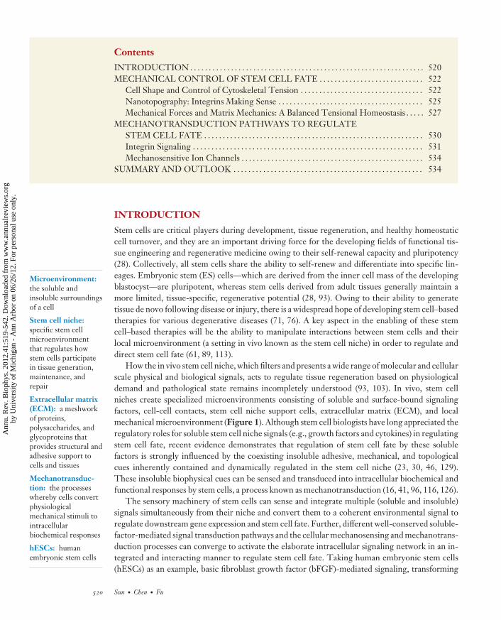
How the in vivo stem cell niche, which filters and presents a wide range of molecular and cellularscale physical and biological signals, acts to regulate tissue regeneration based on physiologicaldemand and pathological state remains incompletely understood (93, 103). In vivo, stem cellniches create specialized microenvironments consisting of soluble and surface-bound signalingfactors, cell-cell contacts, stem cell niche support cells, extracellular matrix (ECM), and localmechanical microenvironment (Figure 1). Although stem cell biologists have long appreciated theregulatory roles for soluble stem cell niche signals (e.g., growth factors and cytokines) in regulatingstem cell fate, recent evidence demonstrates that regulation of stem cell fate by these solublefactors is strongly influenced by the coexisting insoluble adhesive, mechanical, and topologicalcues inherently contained and dynamically regulated in the stem cell niche (23, 30, 46, 129).These insoluble biophysical cues can be sensed and transduced into intracellular biochemical andfunctional responses by stem cells, a process known as mechanotransduction (16, 41, 96, 116, 126).
The sensory machinery of stem cells can sense and integrate multiple (soluble and insoluble)signals simultaneously from their niche and convert them to a coherent environmental signal toregulate downstream gene expression and stem cell fate. Further, different well-conserved soluble-factor-mediated signal transduction pathways and the cellular mechanosensing and mechanotrans-duction processes can converge to activate the elaborate intracellular signaling network in an in-tegrated and interacting manner to regulate stem cell fate. Taking human embryonic stem cells(hESCs) as an example, basic fibroblast growth factor (bFGF)-mediated signaling, transforming
520 Sun · Chen · Fu
Ann
u. R
ev. B
ioph
ys. 2
012.
41:5
19-5
42. D
ownl
oade
d fr
om w
ww
.ann
ualr
evie
ws.
org
by U
nive
rsity
of
Mic
higa
n -
Ann
Arb
or o
n 06
/26/
12. F
or p
erso
nal u
se o
nly.
BB41CH23-Chen ARI 3 April 2012 16:11
Strain forces
Focal adhesionFocal adhesion
CytoskeletonCell-celladhesion
Receptors
ECM rigidityECM rigidity
β α
Talin
FexternalFexternal
Fcell
Mechanosensitiveion channels
Ions
Signaling molecules
Soluble factors
Physical stimuli
Mechanosensors
Vin
PaxFAK
Zyxin
VASP
ECM
Shear stress
Figure 1Schematic showing biophysical signals in the stem cell niche and the intricate reciprocal molecular interactions between stem cells andtheir microenvironment to regulate stem cell fate. The extracellular microenvironment of stem cells is a hydrated protein- andproteoglycan-based gel network comprising soluble and physically bound signals as well as signals arising from cell-cell interactions.Biophysical signals in the stem cell niche include matrix rigidity and topography, flow shear stress, strain forces, and other mechanicalforces exerted by adjacent support cells (blue text). Stem cells can sense these biophysical stimuli through mechanosensors such as ionchannels, focal adhesions, cell surface receptors, actin cytoskeleton, and cell-cell adhesions (red text). A magnified view of the focaladhesion structure is also shown, which includes transmembrane heterodimeric integrin, paxillin (Pax), talin, focal adhesion kinase(FAK), vinculin (Vin), zyxin, and vasodilator-stimulated phosphoprotein (VASP). Abbreviation: ECM, extracellular matrix.
TGF-β: transforminggrowth factor β
MAPK: mitogen-activated proteinkinase
ROCK: Rho-associated coiled-coil-containingprotein kinase
growth factor-β (TGF-β)/activin/Nodal-mediated signaling, and canonical Wnt (wingless)/β-catenin-mediated signaling are all central for the self-renewal of hESCs, while bone morpho-genetic proteins (BMPs) induce differentiation of hESCs (33, 138, 139). bFGF activates themitogen-activated protein kinase (MAPK)/extracellular-signal regulated kinase (ERK) signalingcascade in hESCs, also known to be a central mechanotransduction pathway for adaptive cellularresponses to mechanical stimuli from the cellular microenvironment (63, 75). Extracellular me-chanical forces stimulate expressions of TGF-β, activin, and Nodal, providing an autocrine orparacrine signaling mechanism to promote maintenance of the pluripotency of hESCs (94, 110).β-catenin, which is a critical component of the canonical Wnt signaling pathway, plays an impor-tant role in cell-cell adhesions by mediating cytoskeletal attachment of E-cadherin to the actincytoskeleton. β-catenin-mediated E-cadherin-based cell-cell adhesions are mechanosensitive anddepend on nonmuscle myosin II (NMMII) activity in hESCs (74). Further, β-catenin is criticalfor the mechanical induction of Twist expression in Drosophila; Twist is a transcription factorassociated with regulation of skeletal development (36).
Moreover, recent studies show that the RhoA-GTPase/Rho-associated coiled coil-containingkinase (ROCK)/myosin-II signaling axis, which is the major biochemical pathway mediating theactin cytoskeleton tension in nonmuscle cells (35, 106), plays a critical role in regulating survival andcloning efficiency of single hESCs (17, 128, 131). Blocking RhoA/ROCK-mediated cytoskeletontension using drug inhibitors reduces dissociation-induced apoptosis of hESCs, suggesting that
www.annualreviews.org • A Biophysical Perspective of the Cellular Microenvironment 521
Ann
u. R
ev. B
ioph
ys. 2
012.
41:5
19-5
42. D
ownl
oade
d fr
om w
ww
.ann
ualr
evie
ws.
org
by U
nive
rsity
of
Mic
higa
n -
Ann
Arb
or o
n 06
/26/
12. F
or p
erso
nal u
se o
nly.

BB41CH23-Chen ARI 3 April 2012 16:11
Nanotopography:surfaces and structureswith nanoscaletopological features
hyperactivation of cytoskeleton tension, triggered by hESC cell dissociation, is the upstreamregulator and direct cause of hESC apoptosis. Importantly, RhoA/ROCK-mediated cytoskeletontension plays a critical role in the mechanotransduction process (16, 58). All together, biophysicalsignals in the cellular microenvironment of stem cells can have extensive potential to regulate andsynergize with classical signal transduction pathways induced by soluble factors to control stemcell fate.
With recent major advances in understanding how the insoluble biophysical signals in thecellular microenvironment regulate stem cell fate, tissue engineering and regenerative medicine arebecoming increasingly oriented toward biologically inspired in vitro cellular microenvironmentsdesigned to guild stem cell growth, differentiation, and functional assembly (78, 127). The premiseis that, to unlock the full potential of stem cells, at least some aspects of the dynamic cellularmicroenvironment that are associated with their renewal, differentiation, and assembly in nativetissues need to be reconstructed.
A major goal of this review is therefore to offer a perspective on this new trend of designingsynthetic artificial in vitro stem cell niches and their promise for stem cell research and to enablenew, clinically relevant strategies for tissue regeneration. In particular, we focus on discussing thebiophysical signals in the synthetic stem cell niche and their functional effects on stem cell fate.To do so, we take the approach of highlighting some illustrative examples of using bioengineeringstrategies for controllable synthetic cellular microenvironments developed through the interac-tions of stem cell biology, tissue engineering, and microtechnology/nanotechnology at multiplelength scales. We first stress different biophysical factors in the cellular microenvironment thatare critical for the fate decisions of stem cells, such as ECM geometry, nanotopography, and me-chanics, and describe how these biophysical factors affect cell signaling and function. We discussthe mechanosensory machinery and mechanotransduction mechanisms stem cells can use to senseand respond to these biophysical factors and how these mechanotransduction pathways convergewith classical signal transduction pathways to control stem cell fate. We discuss different versatileand powerful bioengineering and microtechnology/nanotechnology strategies and methods thatcan be used for constructing the synthetic stem cell niche. We offer some perspectives on potentialresearch directions and opportunities for engineering stem cell functions using well-controlledcellular microenvironments.
MECHANICAL CONTROL OF STEM CELL FATE
Functional regulation of stem cells in vivo normally plays out in the context of embryonic devel-opment, tissue regeneration, and the wound healing response, in which extracellular mechanicalforces abound and the mechanical environment surrounding the stem cells changes dynamically.Plenty of evidence exists to suggest that these biophysical signals from the local stem cell nicheinstruct the subsequent behaviors of stem cells. There are other extensive reviews on the topicof stem cell niche signals regulating stem cell fate, especially for in vivo organismal developmentsettings (65, 93, 103); here we provide illustrative examples using novel bioengineering and micro-fabrication/nanofabrication approaches to control the local stem cell niche and, where evidencesuggests, mechanical control of stem cell fate through synergistic regulations of stem cell shape,cytoskeleton tension, and integrin-mediated adhesion signaling.
Cell Shape and Control of Cytoskeletal Tension
Cell shape is a potent regulator of cell growth and physiology (39). Cells adapt and optimize theirshape for their specific functions. For example, adipocytes are spherical in shape to maximize
522 Sun · Chen · Fu
Ann
u. R
ev. B
ioph
ys. 2
012.
41:5
19-5
42. D
ownl
oade
d fr
om w
ww
.ann
ualr
evie
ws.
org
by U
nive
rsity
of
Mic
higa
n -
Ann
Arb
or o
n 06
/26/
12. F
or p
erso
nal u
se o
nly.
BB41CH23-Chen ARI 3 April 2012 16:11
hMSCs: humanmesenchymal stemcells
Actomyosincontractility:intracellular forcesgenerated by thedynamic interaction ofmyosin motors andactin filaments
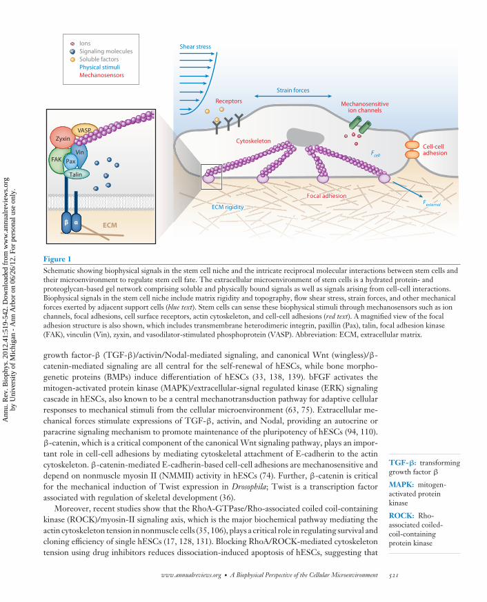
lipid storage, whereas neurons have long axons to deliver signals rapidly over a long distance. Infact, many events associated with stem cell differentiation during embryonic development andtissue regeneration are designed to change cell shape, and those changes in shape can influencetissue structure and function (56, 81, 141). Thus, a natural converse question for stem cells arises:whether their fate can be regulated by their intrinsic and dynamically regulated cell shape. Com-pelling studies to support cell shape as a key regulator of stem cell fate came from experiments usingbioengineering strategies to pattern the spreading and morphology of human mesenchymal stemcells (hMSCs). hMSCs are isolated from bone marrow and can differentiate into multiple lineagesof mesenchymal tissues (15, 104). By using microcontact printing to coat flat polydimethylsilox-ane surfaces with distinct patterns of adhesive ECM islands, McBeath et al. (86) reported that inresponse to a bipotential differentiation medium that contained inducers for both the adipogenicand osteogenic differentiations, single hMSCs confined to small ECM islands selectively under-went adipogenesis, whereas single hMSCs on large ECM islands were biased toward osteogenesis(Figure 2a).
This osteogenic-adipogenic switch in well-spread versus poorly spread hMSCs required gen-eration of cytoskeleton tension through RhoA-dependent actomyosin contractility. RhoA is amember of Rho family small GTPases involved in cellular signaling and cytoskeletal organization,and it stimulates cytoskeleton tension through its effector, ROCK, which directly phosphorylatesboth NMMII regulatory myosin light chain (MLC) and MLC phosphatase to synergistically in-crease MLC phosphorylation and thus myosin II contractility (6, 48). Inhibition of cytoskeletontension using either cytochalasin D (an actin depolymerization agent) or Y-27632 (a ROCK in-hibitor) promoted adipogenesis, mimicking the phenotype of poorly spread hMSCs. Moreover,manipulation of the RhoA pathway could override the effects of soluble differentiation factors,such that dominant-negative RhoA induced adipogenesis even in the context of pure osteogenicmedium, whereas constitutively active RhoA triggered osteogenesis in pure adipogenic medium.These findings highlight RhoA activity as a potential convergence point for mechanical and sol-uble factor signaling in the control of stem cell differentiation. Importantly, McBeath et al. alsodemonstrated that expression of constitutively active ROCK rescued osteogenic differentiationof poorly spread MSCs, and this effect required myosin II activity, indicating that cell shape andRhoA regulate osteogenic-adipogenic switching through the development of cytoskeleton tension.
Ruiz & Chen (107) confirmed the importance of cytoskeleton tension in regulating stem cell fatein the setting of multicellular structures. Ruiz & Chen applied microscale patterning approachesto control the geometries of both two-dimensional (2D) and three-dimensional (3D) multicellularstructures of hMSCs (Figure 2b). The authors reported that in the presence of soluble factorspermitting both osteogenic and adipogenic differentiations, hMSCs at the edge of the multicel-lular structures selectively differentiated into the osteogenic lineage, whereas those in the centerbecame adipocytes. Using some microfabricated cellular traction force sensors (31, 124), Ruiz &Chen further demonstrated that a gradient of traction stress across the 2D multicellular hMSCstructures could precede and mirror the patterns of multicellular differentiation, where regionsof high stress were concentrated with osteogenesis of hMSCs, whereas hMSCs in regions of lowstress differentiated to adipocytes. Inhibition of cytoskeleton tension using blebbistatin (a myosinII inhibitor), Y-27632, or ML-7 (an inhibitor of MLC kinase) suppressed the spatial patterns ofmulticellular differentiation of osteogenesis versus adipogenesis, for both 2D and 3D multicellularstructures of hMSCs. Interestingly, in addition to the overall cell shape, cell geometry also playsan important role in regulating stem cell fate (66, 77, 119). Kilian et al. (66) demonstrated that inresponse to a bipotential differentiation medium that contained inducers for both the adipogenicand osteogenic differentiations, single hMSCs cultured in rectangles with increasing aspect ra-tio and in shapes with pentagonal symmetry but with different subcellular curvature—and with
www.annualreviews.org • A Biophysical Perspective of the Cellular Microenvironment 523
Ann
u. R
ev. B
ioph
ys. 2
012.
41:5
19-5
42. D
ownl
oade
d fr
om w
ww
.ann
ualr
evie
ws.
org
by U
nive
rsity
of
Mic
higa
n -
Ann
Arb
or o
n 06
/26/
12. F
or p
erso
nal u
se o
nly.

BB41CH23-Chen ARI 3 April 2012 16:11
a
Hoechst
Oct4pSmad1
Gro
wth
me
dia
Ost
eo
/ad
ipo
me
dia
c
1,024 μm2 10,000 μm2
D = 800 μmD = 400 μmD = 200 μm
50 μm 250 μm
b
Figure 2Microcontact printing to manipulate the cell shape and colony size of stem cells to control their fate.(a) Brightfield micrographs of single hMSCs plated on different sized adhesive ECM islands. hMSCs werestained for alkaline phosphatase activity (ALP, blue) and lipid droplet accumulation (Lip, red ) after 7 days ofculture in either the growth (top row) or the bipotential differentiation (bottom row) medium. Adapted fromReference 86, Copyright c© 2004, with permission from Elsevier. (b) Brightfield micrographs of differentshaped multicellular hMSC colonies stained for ALP (blue) and Lip (red ) after 14 days of culture in thebipotential differentiation medium. Adapted from Reference 107, Copyright c© 2008, with permission fromJohn Wiley and Sons. (c) Immunofluorescent images showing different sized hESC colonies (H9) cultured inXV media after withdrawal of all exogenous growth factors for 48 h. hESCs were stained for Hoechst, Oct4,and pSmad1 to indicate the effect of colony size on the pluripotency maintenance of hESCs. Adapted fromReference 102, Copyright c© 2007, with permission from Nature Publishing Group. Abbreviations: hMSC,human mesenchymal stem cells; ECM, extracellular matrix; hESC, human embryonic stem cell.
each occupying the same area—displayed different adipogenesis and osteogenesis profiles. Usingcytoskeleton-disrupting pharmacological agents, Kilian et al. further confirmed a causal role forcytoskeleton tension in modulating the shape-based trends in lineage commitment of hMSCs.Taken together, the aforementioned studies demonstrate a causal role of cell shape and geometryin regulating stem cell fate. Importantly, there is a common theme emerging from all these studies:
524 Sun · Chen · Fu
Ann
u. R
ev. B
ioph
ys. 2
012.
41:5
19-5
42. D
ownl
oade
d fr
om w
ww
.ann
ualr
evie
ws.
org
by U
nive
rsity
of
Mic
higa
n -
Ann
Arb
or o
n 06
/26/
12. F
or p
erso
nal u
se o
nly.
BB41CH23-Chen ARI 3 April 2012 16:11
Focal adhesion (FA):cell adhesion sites fortheir attachment toECM, whereintracellular actinfilaments can link toECM proteins throughtransmembraneproteins such asintegrins
Cell shape signal seems to converge with soluble inductive factors on the actin cytoskeleton andthe RhoA/ROCK-mediated cytoskeleton tension to regulate stem cell fate.
Peerani et al. (102) demonstrated the effect of the cellular microenvironment on hESC fateby patterning hESC colonies onto defined adhesive islands with a controlled colony diameter(Figure 2c). Peerani et al. showed that larger colonies with a high local cell density microenviron-ment would promote the maintenance of pluripotency in hESCs through a niche size-dependentspatial gradient of BMP-mediated Smad1 signaling generated as a result of antagonistic inter-actions between hESCs and hESC-derived extraembryonic endoderm. Thus, the effect of thiscolony size on the pluripotency maintenance of hESCs appears to be mediated by interactions be-tween exogenously controlled parameters and autocrine and paracrine secretion of endogenouslyproduced factors from hESCs. Even though there was no mechanotransduction mechanism re-vealed specifically in the study by Peerani et al. (102), combining their results together with theobservations from hMSCs discussed above, it is reasonable to speculate that stem cell fate is me-diated by a combination of soluble factors and insoluble biophysical signals in the local stem cellmicroenvironment. Thus, structural and mechanical cues associated with cytoskeletal reorganiza-tion appear to be integrated with several developmental signaling pathways critical for stem cellfate determination, and the integrated mechanochemical networks provide a mechanism for stemcells to orchestrate the many structural and mechanical changes associated with morphogenesis todirect the downstream genetic programs required to give rise to the appropriate spatiotemporalpatterns of stem cell differentiation.
Nanotopography: Integrins Making Sense
During embryonic development and tissue regeneration, stem cells interact not only with eachother but also with the 3D porous network of the ECM, which comprises fibrillar networks ofproteins such as collagen and laminin interlaced with proteoglycan. Although the characteristicpore size of the ECM might provide a direct physical constraint on the stem cell size and shape,the microscale and nanoscale topography, structure, and architecture of the fibrous ECM are alsoimportant biophysical signals that can regulate stem cell adhesion and cytoskeletal organizationand thus stem cell behaviors such as proliferation, migration, and differentiation (25, 44, 120,135). Equipped with advanced sub-100-nm microfabrication/nanofabrication techniques, mate-rials scientists and applied physicists have successfully teamed up with stem cell biologists andtissue engineers to generate well-controlled molecular and cellular scale topography on 2D planarsubstrates to investigate their independent effect on stem cell fate. Existing studies on nanoto-pography have suggested that instead of directly affecting cytoskeleton tension as in the case forcell shape, nanotopographical cues appear to elicit their effect on stem cells by directly modu-lating the molecular arrangement, dynamic organization, and signaling of the cellular adhesionmachinery.
Adhesion of stem cells to the nanotopographical ECM is mediated via heterodimeric trans-membrane receptors, namely, α- and β-integrins (Figure 1). Combinations of among 18 α-chainsand 8 β-chains form different heterodimers to yield a rich diversity of ECM receptors, enablingdifferential cell-type-specific responses to variations in the ECM. Upon binding ECM, integrinscan cluster to form dynamic adhesion structures called focal adhesions (FAs). On the cytoplasmicside of FAs, integrins can interact, via their cytoplasmic tails, with different adaptor and signalingproteins. Among these molecules, talin, vinculin, paxillin, and α-actinin are adaptor proteins thatprovide a direct physical linkage to the actin cytoskeleton. Importantly, binding of integrins tothe ECM proteins can activate tyrosine kinase and phosphatase signaling to elicit downstreambiochemical signals important for regulation of gene expression and stem cell fate. Thus, it is
www.annualreviews.org • A Biophysical Perspective of the Cellular Microenvironment 525
Ann
u. R
ev. B
ioph
ys. 2
012.
41:5
19-5
42. D
ownl
oade
d fr
om w
ww
.ann
ualr
evie
ws.
org
by U
nive
rsity
of
Mic
higa
n -
Ann
Arb
or o
n 06
/26/
12. F
or p
erso
nal u
se o
nly.
BB41CH23-Chen ARI 3 April 2012 16:11
FAK: focal adhesionkinase
speculated that nanotopographical signals intrinsically contained in the ECM surrounding stemcells can regulate stem cell fate through their direct effect on integrin-mediated FA signaling.
As reported by Arnold et al. (4), by using block-copolymer micelle nanolithography to patterngold nanodots coated with adhesive peptides, when the spacing between these nanodots exceededapproximately 70 nm, cell adhesion and spreading, FA, and actin stress fiber formations weresignificantly impaired, likely owing to the restricted clustering of integrin molecules by the distancebetween the adjacent gold nanodots. In another relevant study using nanoimprint lithographyto pattern gold nanodots functionalized with binding ligand RGD (Arg-Gly-Asp), Schvartzmanet al. (115) reported a drastic increase in the spreading efficiency of cells on arrays of differentgeometric arrangements of the nanodots when at least four liganded sites were spaced no morethan 60 nm apart, with no dependence on global density. This interesting observation pointedto the existence of a minimum of four integrin adhesion units required for initial growth andmaturation of nascent FAs on fibronectin as defined in space and stoichiometry. Together, thesetwo studies based on well-controlled cell-ECM interactions demonstrate the molecular sensitivityand dynamic organization of FAs regulated by the local nanotopographical cue.
Given the potent influence of local nanotopography on regulating molecular organization ofFAs, it is not surprising that nanotopography can significantly affect stem cell fate. Yim et al. (143)showed that hMSCs cultured on the nanoscale gratings on the polydimethylsiloxane surface tendedto align and elongate their actin cytoskeleton and nuclei along the nanogratings. Gene profilingand immunostaining by Yim et al. further showed significant upregulation of neuronal markerssuch as microtubule-associated protein 2 for hMSCs cultured on the nanogratings, compared withunpatterned flat controls, and the combination of nanotopography and biochemical inductive fac-tors such as retinoic acid further enhanced the expressions of neuronal markers. Importantly, Yimet al. (143) further demonstrated that nanotopography showed a stronger independent effect com-pared with biochemical cues (in this case, retinoic acid for neurogenic differentiation of hMSCs)alone on unpatterned control surfaces. In a follow-up study, Yim et al. (142) found that on thenanogratings, expressions of most integrins except α3 and β5 were considerably downregulatedand that the aligned actin cytoskeleton on the nanogratings was not as prominent or dense ason flat surface controls. Further, distributions of vinculin and focal adhesion kinase (FAK), twoprominent FA proteins, on the nanogratings were different from those on flat surfaces. Combinedtogether, studies from Yim et al. suggest that the local nanotopographical cues could affect themolecular organization and composition and signaling of FAs and that such modified FA signalingmight further influence cytoskeleton structure to mediate stem cell fate.
More recently, Dalby et al. (27) applied electron beam lithography and hot embossing to pat-tern nanoscale pits of different symmetry and with varying degrees of disorder in the polymethyl-methacrylate or polycaprolactone substrates. Dalby et al. (27) first reported that the nanoscaledisorder in the nanopit array stimulated hMSCs to produce bone mineral in vitro, even in theabsence of osteogenic supplements. Interestingly, Dalby et al. further showed that hMSCs platedon perfectly ordered or totally random arrays of nanopits produced much less osteoblastic differ-entiation. A more recent study from the same authors demonstrated another intriguing effect ofnanotopography on hMSC fate regulation. McMurray et al. (87) showed that the perfectly or-dered arrays of nanopits, even though not efficient to promote osteogenic differentiation of hMSCsas shown in their previous work, were conducive to hMSC growth while permitting prolongedretention of their multipotency and differentiation potential.
The aforementioned studies strongly suggest the potential of nanoscale structured surfacesas noninvasive tools to control the local stem cell microenvironment to regulate stem cell fate,even though the mechanisms by which stem cells can sense and respond to the nanotopographicalsignal is not yet clear. But as discussed above, molecular arrangement and dynamic organization
526 Sun · Chen · Fu
Ann
u. R
ev. B
ioph
ys. 2
012.
41:5
19-5
42. D
ownl
oade
d fr
om w
ww
.ann
ualr
evie
ws.
org
by U
nive
rsity
of
Mic
higa
n -
Ann
Arb
or o
n 06
/26/
12. F
or p
erso
nal u
se o
nly.

BB41CH23-Chen ARI 3 April 2012 16:11
of integrin-mediated FAs appear to be sensitive to the local arrangement and presentation of thenanotopographical signal. Thus, it is likely that integrin-mediated FA signaling, critical for manycellular functions (42, 117) and strongly dependent on their dynamic characteristics and molecularprocesses (52, 101, 145), plays an important functional role in regulating the stem cell sensitivityto nanotopography.
Mechanical Forces and Matrix Mechanics: A Balanced Tensional Homeostasis
During embryonic development and tissue regeneration, cells are exposed not only to structuralchanges in the surrounding ECM, but also to many mechanical stresses. There are local changesin mechanical forces during development, caused by the addition or removal of cells, cell move-ments associated with morphogenesis, muscle contraction, and relaxation, as well as during bonecompression and decompression. Therefore, stem cells are constantly subjected to and adjust toexternal force fluctuations from their local microenvironment. Another physical signal importantfor regulating stem cell fate is the intrinsic elastic modulus of the ECM surrounding stem cells.Stem cells sense and respond to changes of the elastic modulus of the ECM by modulating theirendogenous cytoskeleton contractility, balanced by resistant forces generated by the deforma-tion of the ECM, the magnitude of which is determined by the ECM elastic modulus. Thus, itappears that stem cells are mechanosensitive and mechanoresponsive to mechanical forces andmatrix mechanics through a modulated delicate force balance between endogenous cytoskeletoncontractility and external mechanical forces transmitted across the cell-ECM adhesions. Indeed,such tensional homeostasis in the intracellular cytoskeleton has a key role in the regulation ofbasic cellular functions, such as cell proliferation, apoptosis, adhesion, and migration (80, 136).Deregulation of the tensional homeostasis in cells contributes to the pathogenesis of several humandiseases, such as atherosclerosis, osteoarthritis and osteoporosis, and cancer (12, 47, 57).
External forces and matrix mechanics have a key role in the regulation of stem cell fate. Thedetailed molecular picture of the mechanotransduction process for stem cells to sense and re-spond to external forces and changes in matrix mechanics has yet to be identified. The force bal-ance transmitted across the mechanical continuum of ECM-integrin-cytoskeleton can regulateintegrin-mediated adhesion signaling (such as FAK and Src signaling) to coordinate downstreamintegrated stem cell function. These biophysical signals are sensed at the FA sites in which in-tegrins provide the mechanical linkage between the ECM and the actin cytoskeleton. Exposingstem cells to mechanical strain or fluid shear stress or plating stem cells on substrates with varyingelastic moduli activates integrins, which promote recruitment of scaffold and signaling proteinsto strengthen FAs and to transmit biochemical signals into the cell. These mechanotransductionpathways establish positive-feedback loops in which integrin engagement activates actomyosincytoskeleton contractility, which in turn reinforces FAs (16, 41). Thus, the level of cytoskeletoncontractility generated inside the cell is directly proportional to the adhesion strength and thematrix elastic modulus and dictates the cellular responses of stem cells.
Effects of external forces including mechanical strain, compression, and fluid shear stress oncellular functions have long been studied for cardiovascular tissues, skeletal muscles, and adultstem cells such as hMSCs (19, 29, 97, 129). Evidence related to regulation of pluripotent stemcell fate by mechanical forces in vitro has only recently begun to emerge. Saha et al. showed thatin the presence of mouse embryonic fibroblast (MEF) conditioned medium, under cyclic equib-iaxial strain, hESC differentiation was reduced and self-renewal was promoted without selectingagainst survival of differentiated or undifferentiated cells (109). A more recent study by the sameauthors further showed that the TGF-β/activin/Nodal signaling pathway played a crucial role inrepressing hESC differentiation under mechanical strain (110). Saha et al. showed that mechanical
www.annualreviews.org • A Biophysical Perspective of the Cellular Microenvironment 527
Ann
u. R
ev. B
ioph
ys. 2
012.
41:5
19-5
42. D
ownl
oade
d fr
om w
ww
.ann
ualr
evie
ws.
org
by U
nive
rsity
of
Mic
higa
n -
Ann
Arb
or o
n 06
/26/
12. F
or p
erso
nal u
se o
nly.
BB41CH23-Chen ARI 3 April 2012 16:11
b
0 1 24 48
Static Shear stress
a
Magnetic field
Stem cell
Shear stress
Stem cell
Bri
gh
tfie
ldO
ct3
/4
10 μm
Time (h)
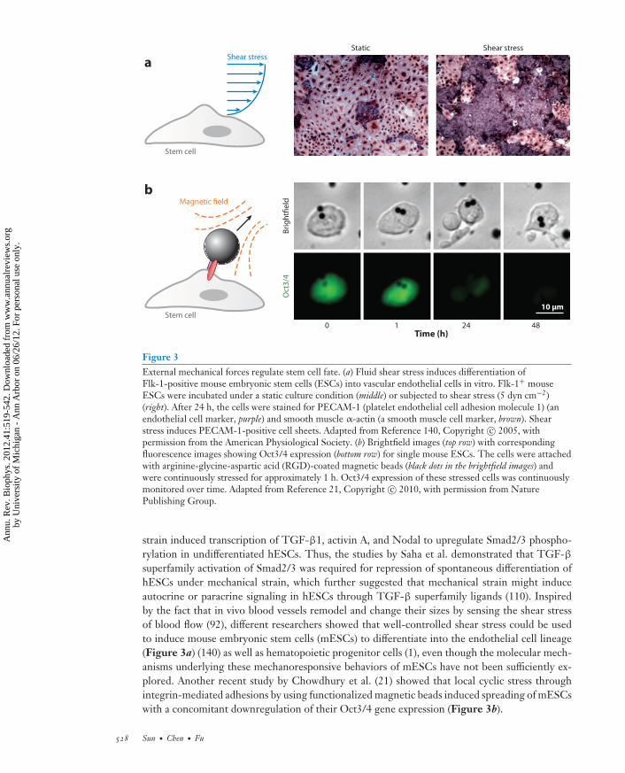
Figure 3External mechanical forces regulate stem cell fate. (a) Fluid shear stress induces differentiation ofFlk-1-positive mouse embryonic stem cells (ESCs) into vascular endothelial cells in vitro. Flk-1+ mouseESCs were incubated under a static culture condition (middle) or subjected to shear stress (5 dyn cm−2)(right). After 24 h, the cells were stained for PECAM-1 (platelet endothelial cell adhesion molecule 1) (anendothelial cell marker, purple) and smooth muscle α-actin (a smooth muscle cell marker, brown). Shearstress induces PECAM-1-positive cell sheets. Adapted from Reference 140, Copyright c© 2005, withpermission from the American Physiological Society. (b) Brightfield images (top row) with correspondingfluorescence images showing Oct3/4 expression (bottom row) for single mouse ESCs. The cells were attachedwith arginine-glycine-aspartic acid (RGD)-coated magnetic beads (black dots in the brightfield images) andwere continuously stressed for approximately 1 h. Oct3/4 expression of these stressed cells was continuouslymonitored over time. Adapted from Reference 21, Copyright c© 2010, with permission from NaturePublishing Group.
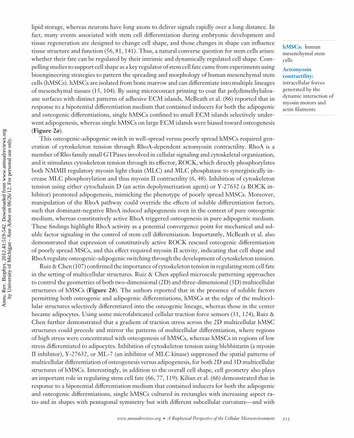
strain induced transcription of TGF-β1, activin A, and Nodal to upregulate Smad2/3 phospho-rylation in undifferentiated hESCs. Thus, the studies by Saha et al. demonstrated that TGF-βsuperfamily activation of Smad2/3 was required for repression of spontaneous differentiation ofhESCs under mechanical strain, which further suggested that mechanical strain might induceautocrine or paracrine signaling in hESCs through TGF-β superfamily ligands (110). Inspiredby the fact that in vivo blood vessels remodel and change their sizes by sensing the shear stressof blood flow (92), different researchers showed that well-controlled shear stress could be usedto induce mouse embryonic stem cells (mESCs) to differentiate into the endothelial cell lineage(Figure 3a) (140) as well as hematopoietic progenitor cells (1), even though the molecular mech-anisms underlying these mechanoresponsive behaviors of mESCs have not been sufficiently ex-plored. Another recent study by Chowdhury et al. (21) showed that local cyclic stress throughintegrin-mediated adhesions by using functionalized magnetic beads induced spreading of mESCswith a concomitant downregulation of their Oct3/4 gene expression (Figure 3b).
528 Sun · Chen · Fu
Ann
u. R
ev. B
ioph
ys. 2
012.
41:5
19-5
42. D
ownl
oade
d fr
om w
ww
.ann
ualr
evie
ws.
org
by U
nive
rsity
of
Mic
higa
n -
Ann
Arb
or o
n 06
/26/
12. F
or p
erso
nal u
se o
nly.
BB41CH23-Chen ARI 3 April 2012 16:11
The ability of stem cells to sense matrix mechanics has been demonstrated only recently, yet itsimplications for functional tissue engineering and regenerative medicine have already generatedtremendous excitement. In a landmark study, Engler et al. demonstrated that in the absence ofexogenous soluble cues, plating hMSCs on polyacrylamide gels of varying elasticity was sufficientto induce hMSCs to differentiate into different tissue types corresponding to the tissues’ relativemechanical elasticity in vivo (Figure 4a,b) (34). As discussed above, adherent cells sense matrixmechanics through a force balance between intracellular actomyosin contractility and the resistantforce of the ECM determined by its elastic deformation. Thus, the level of cytoskeleton tensiongenerated inside stem cells is directly proportional to the elastic modulus of the substrate stem cellsadhere to. Indeed, this positive correlation between the elastic modulus of the substrate and intra-cellular cytoskeleton contractility was reported by Engler et al. and others for hMSCs and manyother mechanosensitive adherent cells (34, 40). Importantly, to demonstrate that this cytoskeletoncontractility indeed played a causal role for regulating matrix mechanics-dependent changes inhMSC differentiation, Engler et al. showed that addition of blebbistatin to block intracellularcytoskeleton tension generation in hMSCs obliterated matrix mechanics-driven differentiation.Recently, Huebsch et al. (55) extended the in vitro study of mechanobiology in MSCs to a 3Dmicroenvironment setting by using a 3D hydrogel synthetic ECM formed by alginate polymersthat presented integrin-binding RGD peptides (Figure 4c) (55). Using this 3D cell culture sys-tem with well-controlled elastic modulus encapsulating mouse MSCs (mMSCs), Huebsch et al.showed that osteogenesis of mMSCs occurred predominantly at 11–30 kPa, comparable to thenative tissue stiffness of precalcified bone (30). Because mMSCs were encapsulated in the 3Dhydrogel, their morphology appeared to be independent of the elastic modulus of the hydrogel.Still, Huebsch et al. demonstrated that matrix stiffness regulated integrin binding as well as re-organization of adhesion ligands on the nanoscale, both of which were cytoskeleton contractilitydependent and correlated with osteogenic commitment of mMSCs, again highlighting the impor-tance of intracellular cytoskeleton contractility and its force balance with deforming surroundingECM in regulating stem cell fate.
Other types of adult stem cells, including skeletal muscle stem cells (Figure 4d ) (43),hematopoietic stem cells (HSCs) (53), and adult neuron stem cells (8, 73, 108), have also beenstudied for their mechanoresponsive behaviors to matrix mechanics in both 2D and 3D cellularmicroenvironments. The most definitive experimental evidence to demonstrate mechanosensitiv-ity of pluripotent ESCs to matrix mechanics was shown in a recent study by Chowdhury et al.(20), who reported that mESCs could maintain their pluripotency on soft polyacrylamide gels(∼500 Pa) even under long-term culture conditions (at least 15 passages) without exogenousleukemia inhibitory factor (a soluble factor critical for maintenance of pluripotency of mESCs),in sharp contrast to mESCs seeded on the conventional rigid tissue culture plates. Importantly,traction force measurements of these mESCs demonstrated that their cytoskeleton contractilitywas mechanosensitive and correlated positively with the elastic modulus of the polyacrylamide gels(20), implicating involvement of cytoskeleton contractility in regulating their mechanosensitivityto changes in matrix mechanics.
Collectively, a few common observations can be drawn from the aforementioned studies ofthe mechanosensitivity of stem cells. All the studies have explicitly or implicitly suggested theinvolvement of cytoskeleton contractility in regulating the mechanosensitivity of stem cells,suggesting the importance of the force balance along the mechanical axis of the ECM-integrin-cytoskeleton linkage and their regulation by the mechanical signals in the stem cell niche.Moreover, strong evidence suggests that the differentiation potentials of stem cells towarddistinct lineages can be maximized if the cells are cultured in the mechanical microenvironmentmimicking their tissue elasticity in vivo (Figure 4a). This observation is important for both
www.annualreviews.org • A Biophysical Perspective of the Cellular Microenvironment 529
Ann
u. R
ev. B
ioph
ys. 2
012.
41:5
19-5
42. D
ownl
oade
d fr
om w
ww
.ann
ualr
evie
ws.
org
by U
nive
rsity
of
Mic
higa
n -
Ann
Arb
or o
n 06
/26/
12. F
or p
erso
nal u
se o
nly.

BB41CH23-Chen ARI 3 April 2012 16:11
functional tissue engineering and developmental biology because it anticipates a major role ofdynamic control for matrix mechanics in controlling stem cell function and tissue development.Indeed, dynamic regulation of matrix mechanics has emerged as a critical regulator of differen-tiation and morphogenesis (22, 88, 144). An emerging hypothesis has further suggested a rolefor the long-lived cytoskeleton structures as epigenetic memories to determine responses of stemcell shape, function, and fate to changes of matrix mechanics (38).
MECHANOTRANSDUCTION PATHWAYS TO REGULATESTEM CELL FATE
Stem cells can sense and respond to local biophysical signals through integrin-mediated FA sig-naling, and such signaling can be regulated by the force balance between endogenous cytoskeletoncontractility and external mechanical forces transmitted across the cell-ECM adhesions. In thissection, we discuss how this force balance across the mechanical continuum of ECM-integrin-cytoskeleton can be further transduced into the intracellular space of stem cells to mediate signalingmolecules important for stem cell fate (Figure 5).
My
oD
Neurogenic Myogenic Osteogenic
0.1−1 8−17 25−40
b
a Soft tissue elasticity, E (kPa)
Matrix elasticity, E (kPa) 1 10 100
1.0
0.5
0.0
Neurogenic
Myogenic
Osteogenic
Brain Fat Muscle
Cartilage
Precalcified bone
1 10 100
Re
lati
ve
diff
ere
nti
ati
on
ma
rke
r e
xp
ress
ion
E (kPa)
CB
Fα
1β
3-t
ub
uli
n
5 μm
5 μm
5 μm
2.5 20 110
LipALP
DAPI
OCN
c d
0
25
50
2 12 42 106
*
E (kPa) E (kPa)
En
gra
ftm
en
t (%
)
20 μm 20 μm 20 μm
100 μm 100 μm100 μm
2 12 42 106
E (kPa)
2 4 6 8 10
(photons s–1 cm–2 sr–1 × 10⁵)
530 Sun · Chen · Fu
Ann
u. R
ev. B
ioph
ys. 2
012.
41:5
19-5
42. D
ownl
oade
d fr
om w
ww
.ann
ualr
evie
ws.
org
by U
nive
rsity
of
Mic
higa
n -
Ann
Arb
or o
n 06
/26/
12. F
or p
erso
nal u
se o
nly.
BB41CH23-Chen ARI 3 April 2012 16:11
Integrin Signaling
Ras/MAPK signaling. Stem cells can sense and respond to biophysical signals through integrin-mediated FA signaling. Indeed, forces transmitted through FAs, generated either internally bycytoskeleton contractility or externally by mechanical forces, can trigger both mechanical andbiochemical responses in cells. Forces at FAs activate several kinases involved in regulation ofcellular functions (111, 116, 133, 137). Perhaps the most important players in this mechanotrans-duction system are FAK and Src family kinases such as Fyn (45, 72, 100, 132). One major down-stream signaling pathway following FAK/Src activation is the Ras-Raf-MEK-ERK pathway (onebranch of the MAPK pathway), and the exact molecular mechanism of how integrins regulateMAPK is not yet well defined. Several possible pathways have been proposed, including integrin-FAK-Grb2-SOS-Ras (114) and integrin-Fyn-Shc-Grb2-SOS-Ras (130), as well as through theepidermal growth factor receptor (13). ERK is then translocated to the nucleus to regulate geneexpression by activating different transcription factors.
The Ras/MAPK pathway plays a critical role in stem cell fate. For example, MAPK signalingis required for stemness maintenance of both neural stem cells (NSCs) (14) and human epidermalstem cells (147). Interestingly, the FGF/MAPK cascade plays a functional role in promotingdifferentiation of mESCs, thus inhibition of MAPK signaling can support self-renewal of mESCs(105). In contrast, FGF/MAPK signaling promotes self-renewal of hESCs, indicating that hESCsmay have cellular responses to the biophysical signals opposite those of mESCs.
MAPK-mediated stem cell fate is dynamically required during different stages of stem celldifferentiation. For instance, activated MAPK signaling is required for the early linage specifi-cation of mESCs to adipocytes, whereas the MAPK pathway has to be shut down during theirterminal differentiation (11). This observation further suggests that spatial and temporal dynamicmodulation of the biophysical signals in the stem cell niche can be necessary for optimizing stemcell behaviors.
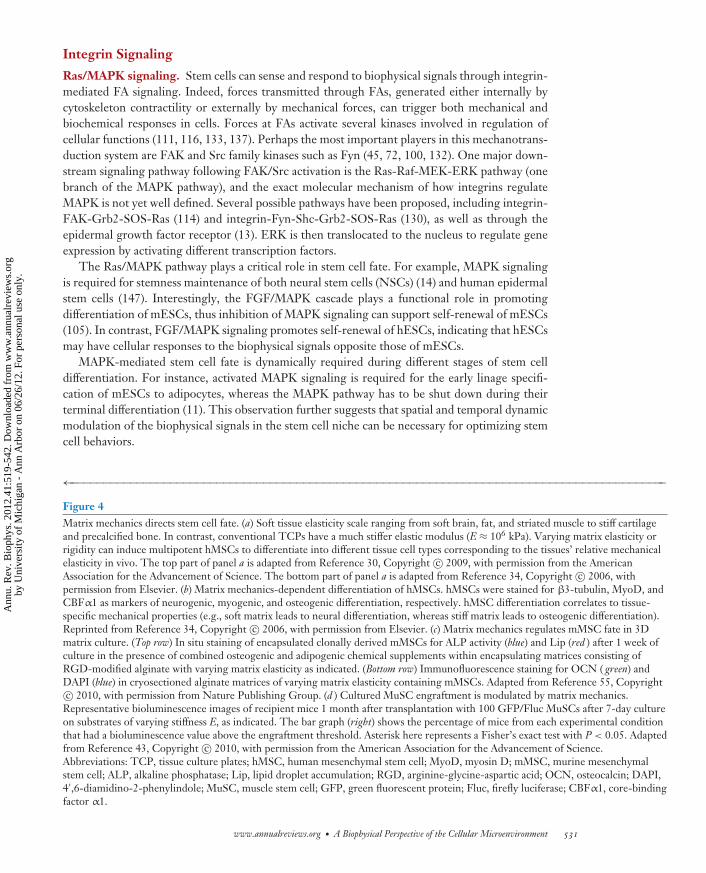
←−−−−−−−−−−−−−−−−−−−−−−−−−−−−−−−−−−−−−−−−−−−−−−−−−−−−−−−−−−−−−−−−−−−−−−−−−−−−−−−−−−−−−−−−−−Figure 4Matrix mechanics directs stem cell fate. (a) Soft tissue elasticity scale ranging from soft brain, fat, and striated muscle to stiff cartilageand precalcified bone. In contrast, conventional TCPs have a much stiffer elastic modulus (E ≈ 106 kPa). Varying matrix elasticity orrigidity can induce multipotent hMSCs to differentiate into different tissue cell types corresponding to the tissues’ relative mechanicalelasticity in vivo. The top part of panel a is adapted from Reference 30, Copyright c© 2009, with permission from the AmericanAssociation for the Advancement of Science. The bottom part of panel a is adapted from Reference 34, Copyright c© 2006, withpermission from Elsevier. (b) Matrix mechanics-dependent differentiation of hMSCs. hMSCs were stained for β3-tubulin, MyoD, andCBFα1 as markers of neurogenic, myogenic, and osteogenic differentiation, respectively. hMSC differentiation correlates to tissue-specific mechanical properties (e.g., soft matrix leads to neural differentiation, whereas stiff matrix leads to osteogenic differentiation).Reprinted from Reference 34, Copyright c© 2006, with permission from Elsevier. (c) Matrix mechanics regulates mMSC fate in 3Dmatrix culture. (Top row) In situ staining of encapsulated clonally derived mMSCs for ALP activity (blue) and Lip (red ) after 1 week ofculture in the presence of combined osteogenic and adipogenic chemical supplements within encapsulating matrices consisting ofRGD-modified alginate with varying matrix elasticity as indicated. (Bottom row) Immunofluorescence staining for OCN ( green) andDAPI (blue) in cryosectioned alginate matrices of varying matrix elasticity containing mMSCs. Adapted from Reference 55, Copyrightc© 2010, with permission from Nature Publishing Group. (d ) Cultured MuSC engraftment is modulated by matrix mechanics.
Representative bioluminescence images of recipient mice 1 month after transplantation with 100 GFP/Fluc MuSCs after 7-day cultureon substrates of varying stiffness E, as indicated. The bar graph (right) shows the percentage of mice from each experimental conditionthat had a bioluminescence value above the engraftment threshold. Asterisk here represents a Fisher’s exact test with P < 0.05. Adaptedfrom Reference 43, Copyright c© 2010, with permission from the American Association for the Advancement of Science.Abbreviations: TCP, tissue culture plates; hMSC, human mesenchymal stem cell; MyoD, myosin D; mMSC, murine mesenchymalstem cell; ALP, alkaline phosphatase; Lip, lipid droplet accumulation; RGD, arginine-glycine-aspartic acid; OCN, osteocalcin; DAPI,4′,6-diamidino-2-phenylindole; MuSC, muscle stem cell; GFP, green fluorescent protein; Fluc, firefly luciferase; CBFα1, core-bindingfactor α1.
www.annualreviews.org • A Biophysical Perspective of the Cellular Microenvironment 531
Ann
u. R
ev. B
ioph
ys. 2
012.
41:5
19-5
42. D
ownl
oade
d fr
om w
ww
.ann
ualr
evie
ws.
org
by U
nive
rsity
of
Mic
higa
n -
Ann
Arb
or o
n 06
/26/
12. F
or p
erso
nal u
se o
nly.
BB41CH23-Chen ARI 3 April 2012 16:11
PI3K:phosphatidylinositol3-kinase
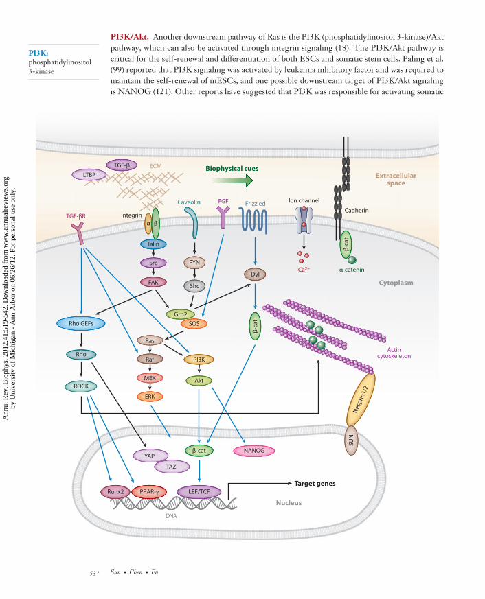
PI3K/Akt. Another downstream pathway of Ras is the PI3K (phosphatidylinositol 3-kinase)/Aktpathway, which can also be activated through integrin signaling (18). The PI3K/Akt pathway iscritical for the self-renewal and differentiation of both ESCs and somatic stem cells. Paling et al.(99) reported that PI3K signaling was activated by leukemia inhibitory factor and was required tomaintain the self-renewal of mESCs, and one possible downstream target of PI3K/Akt signalingis NANOG (121). Other reports have suggested that PI3K was responsible for activating somatic
Frizzled
TGF-βR Integrin
Ion channel
Cadherin
FGFCaveolin
FYN
Shc
Src
FAK
Grb2
SOS
Ras
ERK
Raf
MEK
Nucleus
PI3K
Akt
ECM
Dvl
NANOG
Rho GEFs
Rho
ROCK
Actincytoskeleton
LTBP
TGF-β
Ca2+
Nes
pri
n1/
2
YAP
TAZ
DNA
Target genesLEF/TCF
Cytoplasm
α β
β-c
at
β-c
at
SU
N
Extracellularspace
Talin
Biophysical cues
Runx2 PPAR-γ
β-cat
α-catenin
532 Sun · Chen · Fu
Ann
u. R
ev. B
ioph
ys. 2
012.
41:5
19-5
42. D
ownl
oade
d fr
om w
ww
.ann
ualr
evie
ws.
org
by U
nive
rsity
of
Mic
higa
n -
Ann
Arb
or o
n 06
/26/
12. F
or p
erso
nal u
se o
nly.

BB41CH23-Chen ARI 3 April 2012 16:11
stem cells, such as HSCs (146) and intestinal stem cells (51), to exit from their quiescent states.There is also signaling cross-talk between the PI3K/Akt pathway and the Wnt signaling pathway,a central pathway that controls the fate decisions of many different stem cells (69).
RhoA/ROCK. By acting through its effector ROCK, RhoA is a key molecular regulator of actincytoskeleton tension and FA formation (i.e., upstream regulator of integrin). RhoA/ROCK sig-naling also acts as a downstream target of integrin-mediated signaling through activated FAK(32). RhoA can be activated by different growth factors and cytokines as well as the biophysi-cal signals from the cellular microenvironment. The functional role of RhoA/ROCK-mediatedcytoskeleton contractility is well appreciated in the lineage commitments of hMSCs. ActivatingRhoA promotes osteogenesis of hMSCs by upregulating Runx2 expression, whereas inhibitionof RhoA leads to adipogenesis of hMSCs (5, 86). In response to activated RhoA/ROCK signal-ing, intact actin cytoskeleton structure is required for mechanoresponsive hMSC differentiations.RhoA/ROCK-mediated cytoskeleton contractility can directly regulate certain gene expressionsof transcription factors (e.g., PPAR-γ and Sox-9) to influence stem cell differentiation.
Wnt/β-catenin. Wnt/β-catenin signaling can regulate fate decisions of different stem cell types,including ESCs, HSCs, MSCs, and NSCs (10, 26, 84, 91). In the canonical Wnt pathway, theexpression and nuclear translocation and accumulation of β-catenin are regulated through Dvl.The role of Wnt/β-catenin signaling in regulating stem cell fates can be complicated. For example,for mESCs, Wnt signaling is necessary for maintaining their pluripotency (7, 112); however,overexpression of β-catenin can also promote neural lineage commitment of mESCs (98). Thesignaling cross-talk between Wnt and integrin has been identified, and two different modelsinvolving integrin-linked kinase and FAK have been proposed. In the first model (95), integrin-linked kinase is suggested to stabilize and/or promote the nuclear accumulation of β-catenin; inthe second model (24), Grb2 integrated integrin signaling through FAK with Wnt signaling viaDvl and JNK, a downstream kinase of Grb2, and promoted translocation of β-catenin into thenucleus.
Direct regulation of Wnt signaling by biophysical signals has been demonstrated in osteoblasts.Data show that mechanical loading could regulate Wnt signaling in a time-dependent manner(60). In this study, after a 15-min cyclic stretch, Wnt signaling in human osteoblasts was ultimatelydownregulated despite an initial increase of β-catenin expression.
TGF-β. TGF-β is a secreted protein that belongs to the TGF-β superfamily. It binds to a latentTGF-β-binding protein that is linked to ECM; therefore, TGF-β is stored in extracellular space(3). The most remarkable role of TGF-β is to inhibit cell proliferation. Given the fact that many
←−−−−−−−−−−−−−−−−−−−−−−−−−−−−−−−−−−−−−−−−−−−−−−−−−−−−−−−−−−−−−−−−−−−−−−−−−−−−−−−−−−−−−−−−−−Figure 5Schematic of signaling cross-talk between the mechanotransductive processes (black arrows) and other known soluble factor-mediatedsignaling pathways regulating the fate decisions of stem cells (blue arrows). Abbreviations: TGF-β, transforming growth factor β;LTPB, latent TGF-β-binding protein; TGF-βR, transforming growth factor β receptor; Rho GEFs, Rho guanine nucleotideexchange factors; ROCK, Rho-associated kinase; FAK, focal adhesion kinase; Grb2, growth factor receptor-bound protein 2; SOS, Sonof sevenless; PI3K, phosphoinositide 3-kinase; FGF, fibroblast growth factor; Dvl, Dishevelled; β-cat, β-catenin; YAP, Yes-associatedprotein; TAZ, transcriptional coactivator with PDZ-binding motif; Runx2, Runt-related transcription factor 2; PPAR-γ, peroxisomeproliferator-activated receptor γ; LEF, lymphoid enhancer factor; Ca2+, calcium ion; ECM, extracellular matrix; Src, Rous sarcomaoncogene cellular homolog; Shc, SH2-containing collagen-related proteins; FYN, a Src family tyrosine-protein kinase; SUN, Sad1pand UNC-84 homology; MEK, MAPK/Erk kinase.
www.annualreviews.org • A Biophysical Perspective of the Cellular Microenvironment 533
Ann
u. R
ev. B
ioph
ys. 2
012.
41:5
19-5
42. D
ownl
oade
d fr
om w
ww
.ann
ualr
evie
ws.
org
by U
nive
rsity
of
Mic
higa
n -
Ann
Arb
or o
n 06
/26/
12. F
or p
erso
nal u
se o
nly.

BB41CH23-Chen ARI 3 April 2012 16:11
adult stem cells need to be kept in a quiescent state, TGF-β plays important roles in this process.For example, TGF-β can inhibit expansion of NSCs and keep HSCs in their quiescent state(3, 10), and some studies have shown that TGF-β is critical for maintaining the pluripotency ofhESCs via Smad2/3 signaling (59, 125). In addition to the canonical pathway via Smad2/3, TGF-βactivates multiple major signaling pathways including MAPK, PI3K, and Rho/ROCK (85, 90).
The signaling cross-talk between integrin and TGF-β has been extensively studied. The reg-ulation of TGF-β activation by integrin has been reviewed in detail by Margadant & Sonnenberg(82). Certain types of integrins can directly regulate activation of TGF-β either through cellulartraction forces exerted by actin cytoskeleton or through some G-protein-coupled receptors. Inaddition, integrin can indirectly control the expression of the components in the TGF-β pathway,and it has also been shown that external mechanical forces can activate release of TGF-β fromECM (79, 134). TGF-β can also regulate actin cytoskeleton through the RhoA/ROCK pathway,which has been well recognized in the epithelial to mesenchymal transition process of tumor cells(9). Taken together, forces transmitted through integrins generated either internally by cytoskele-ton contractility or externally by mechanical forces can activate TGF-β signaling, which in turnregulates stem cell fate. Several studies have confirmed this important signaling cross-talk betweenintegrin and TGF-β. For example, the pluripotency of hESCs can be improved by directly ap-plying a cyclic mechanical strain or indirectly using stiff substrates to activate latent TGF-β fromECM or fibroblasts as feeder cells (2, 110, 134).
Mechanosensitive Ion Channels
In addition to integrin signaling, mechanosensitive ion channels can also regulate mechanore-sponsiveness of stem cells (83). Interestingly, based on the tethered model, mechanosensitiveion channels can be linked with ECM and/or cytoskeleton, and the relative displacement ofchannels with respect to ECM or cytoskeleton is responsible for the gating of channels (49).Thus, mechanosensitive ion channels can be directly activated by external forces or intracellularcytoskeleton contractility (50, 70, 122).
The major downstream effect of the activation of mechanosensitive ion channels is the changesof the cytoplasmic Ca2+ concentration as well as their oscillations (70). Ca2+ oscillations have beenobserved in MSCs and are considered as both an indicator and a regulator for MSC differentia-tion (64, 123). This Ca2+ concentration oscillation is influenced by substrate stiffness (68). Ca2+
oscillations have also been found in mESCs, human preadipocytes, and human cardiac progenitorcells (37, 54, 62), indicating that mechanical forces might have the potential to directly regulatethe fates of these cell types through modulating calcium signals.
SUMMARY AND OUTLOOK
The molecular mechanisms by which stem cells maintain their self-renewal ability and control theirdifferentiation need to be determined in order for these cells to be used effectively for functionaltissue engineering and regenerative medicine. Much of our effort until now has focused on thebiochemical components and soluble factors in the stem cell microenvironment that are criticalfor their self-renewal and differentiation. Yet, recent evidence demonstrates that stem cells are alsoheavily influenced by coexisting insoluble adhesive, mechanical, and topological cues containedwithin the dynamic stem cell niche. Experimental evidence has clearly suggested that insolublebiophysical signals, such as cell shape and geometry, external forces and matrix mechanics, andnanotopography can elicit intracellular programs to regulate stem cell fates, likely through theintegrin-mediated FA signaling and the force balance across the mechanical continuum of ECM-integrin-cytoskeleton.
534 Sun · Chen · Fu
Ann
u. R
ev. B
ioph
ys. 2
012.
41:5
19-5
42. D
ownl
oade
d fr
om w
ww
.ann
ualr
evie
ws.
org
by U
nive
rsity
of
Mic
higa
n -
Ann
Arb
or o
n 06
/26/
12. F
or p
erso
nal u
se o
nly.
BB41CH23-Chen ARI 3 April 2012 16:11
The molecular mechanisms for stem cells to sense and respond to different biophysical signalsare not yet clear and likely would be specific to cell type and involve different mechanisms workingin concert. It also appears that the dominant effect of different biophysical signals on stem cellfunctions depends on different experimental settings, and that stem cell fate is mediated by theintricate interactions and interdependencies between soluble factors and insoluble biophysicalsignals in their local cellular microenvironment.
aaa
8 μm8 μm
bb
50 μm
1 × 10⁻³ μL min−1
1.1 μL min−1
Input
Loadingoutput
Logarithmicoutput
Cell culturechamber
c
dLoad green
cells upwardTransfer cells
downwardLoad red cells
downward
3 mm
50 μm
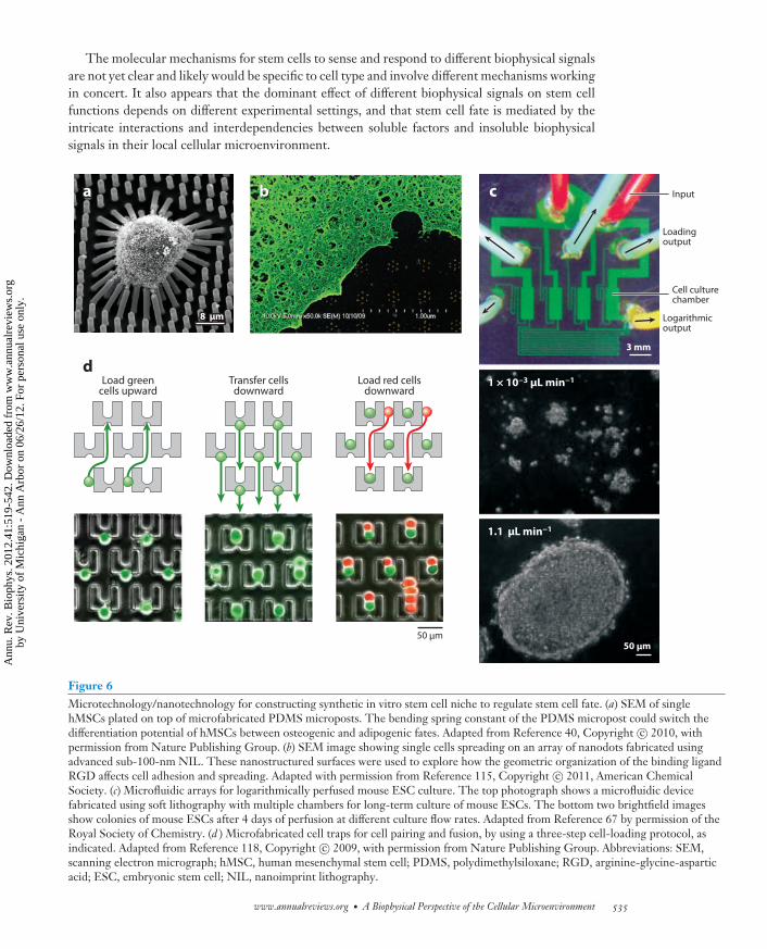
Figure 6Microtechnology/nanotechnology for constructing synthetic in vitro stem cell niche to regulate stem cell fate. (a) SEM of singlehMSCs plated on top of microfabricated PDMS microposts. The bending spring constant of the PDMS micropost could switch thedifferentiation potential of hMSCs between osteogenic and adipogenic fates. Adapted from Reference 40, Copyright c© 2010, withpermission from Nature Publishing Group. (b) SEM image showing single cells spreading on an array of nanodots fabricated usingadvanced sub-100-nm NIL. These nanostructured surfaces were used to explore how the geometric organization of the binding ligandRGD affects cell adhesion and spreading. Adapted with permission from Reference 115, Copyright c© 2011, American ChemicalSociety. (c) Microfluidic arrays for logarithmically perfused mouse ESC culture. The top photograph shows a microfluidic devicefabricated using soft lithography with multiple chambers for long-term culture of mouse ESCs. The bottom two brightfield imagesshow colonies of mouse ESCs after 4 days of perfusion at different culture flow rates. Adapted from Reference 67 by permission of theRoyal Society of Chemistry. (d ) Microfabricated cell traps for cell pairing and fusion, by using a three-step cell-loading protocol, asindicated. Adapted from Reference 118, Copyright c© 2009, with permission from Nature Publishing Group. Abbreviations: SEM,scanning electron micrograph; hMSC, human mesenchymal stem cell; PDMS, polydimethylsiloxane; RGD, arginine-glycine-asparticacid; ESC, embryonic stem cell; NIL, nanoimprint lithography.
www.annualreviews.org • A Biophysical Perspective of the Cellular Microenvironment 535
Ann
u. R
ev. B
ioph
ys. 2
012.
41:5
19-5
42. D
ownl
oade
d fr
om w
ww
.ann
ualr
evie
ws.
org
by U
nive
rsity
of
Mic
higa
n -
Ann
Arb
or o
n 06
/26/
12. F
or p
erso
nal u
se o
nly.

BB41CH23-Chen ARI 3 April 2012 16:11
Moving forward, it is important to recognize that tissue development from stem cells in vivois a long-term process in which dynamic changes in the chemical and physical environments sur-rounding the cells abound. How we can generate in vitro stem cell microenvironments to mimicthe dynamic nature and complexity of the in vivo stem cell niche is currently a significant chal-lenge. Researchers from different disciplines have devised different bioengineering strategies andmicroscale/nanoscale tools that can provide good controls of different aspects of the stem cell mi-croenvironment. Some of these techniques have already been mentioned in the examples discussedabove, which include microcontact printing, synthetic hydrogels, microfluidics, and microfabri-cation/nanofabrication (Figure 6). These tools, which span different scales, from molecular tocellular to organ levels, have proven to be extremely powerful in allowing stem cell biologists andtissue engineers to identify the extrinsic physical factors and their independent effects on stemcell fates. We envisage that in the future, these tools will be further polished and used in differentcombinations to allow researchers to generate dynamic and complex synthetic cellular microenvi-ronments, with the molecular, structural, hydrodynamic, and mechanical cues well controlled inconjunction with their spatial and temporal levels and combinations. Given the complexity of thestem cell niche signals, it is also important to utilize high-throughput tools that can help screendifferent combinations of the environmental signals to elicit the desired stem cell behaviors. Suchhigh-throughput screening assays no doubt can benefit from more in-depth understanding of themolecular mechanisms that regulate stem cell fate.
SUMMARY POINTS
1. Physical signals in the local cellular microenvironment can strongly influence stem cellfate.
2. By controlling cytoskeleton tension, cell shape is a key regulator of stem cell fate.
3. Nanotopographical cues can control stem cell behaviors by modulating the moleculararrangement, dynamic organization, and signaling of the cellular adhesion machinery.
4. External mechanical forces and matrix mechanics can regulate stem cell behaviorsthrough the force balance along the mechanical continuum of the ECM-integrin-cytoskeleton linkage, and their regulation by the mechanical signals in the stem cellniche.
5. The force balance across the ECM-integrin-cytoskeleton linkage can be further trans-duced into the intracellular space of stem cells to mediate signaling molecules importantfor stem cell fate, such as those mediated by integrin signaling and mechanosensitive ionchannels.
DISCLOSURE STATEMENT
The authors are not aware of any affiliations, memberships, funding, or financial holdings thatmight be perceived as affecting the objectivity of this review.
ACKNOWLEDGMENTS
We acknowledge valuable comments and suggestions on the manuscript by group members of theIntegrated Biosystems and Biomechanics Laboratory (R. Lam, W. Chen, S. Weng, and J. Mann).Work in J. Fu’s lab is supported by the National Science Foundation (CMMI 1129611), the
536 Sun · Chen · Fu
Ann
u. R
ev. B
ioph
ys. 2
012.
41:5
19-5
42. D
ownl
oade
d fr
om w
ww
.ann
ualr
evie
ws.
org
by U
nive
rsity
of
Mic
higa
n -
Ann
Arb
or o
n 06
/26/
12. F
or p
erso
nal u
se o
nly.
BB41CH23-Chen ARI 3 April 2012 16:11
National Institutes of Health (UL1RR024986), and the Department of Mechanical Engineeringat the University of Michigan, Ann Arbor. Work in C.S. Chen’s lab is supported by grants from theNational Institutes of Health (EB00262, EB001046, HL73305, GM74048) and the Penn Centerfor Musculoskeletal Disorders. Finally, we extend our apologies to all our colleagues in the fieldwhose work we were unable to discuss or cite formally because of space constraints and imposedreference limitations.
LITERATURE CITED
1. Adamo L, Naveiras O, Wenzel PL, McKinney-Freeman S, Mack PJ, et al. 2009. Biomechanical forcespromote embryonic haematopoiesis. Nature 459:1131–35
2. Ahamed J, Burg N, Yoshinaga K, Janczak CA, Rifkin DB, Coller BS. 2008. In vitro and in vivo evidencefor shear-induced activation of latent transforming growth factor-β1. Blood 112:3650–60
3. Aigner L, Bogdahn U. 2008. TGF-β in neural stem cells and in tumors of the central nervous system.Cell Tissue Res. 331:225–41
4. Arnold M, Cavalcanti-Adam EA, Glass R, Blummel J, Eck W, et al. 2004. Activation of integrin functionby nanopatterned adhesive interfaces. Chem. Phys. Chem. 5:383–88
5. Arnsdorf EJ, Tummala P, Kwon RY, Jacobs CR. 2009. Mechanically induced osteogenicdifferentiation—the role of RhoA, ROCKII and cytoskeletal dynamics. J. Cell Sci. 122:546–53
6. Aspenstrom P. 1999. Effectors for the Rho GTPases. Curr. Opin. Cell Biol. 11:95–1027. Aubert J, Dunstan H, Chambers I, Smith A. 2002. Functional gene screening in embryonic stem cells
implicates Wnt antagonism in neural differentiation. Nat. Biotechnol. 20:1240–458. Banerjee A, Arha M, Choudhary S, Ashton RS, Bhatia SR, et al. 2009. The influence of hydrogel modulus
on the proliferation and differentiation of encapsulated neural stem cells. Biomaterials 30:4695–999. Bhowmick NA, Ghiassi M, Bakin A, Aakre M, Lundquist CA, et al. 2001. Transforming growth factor-
β1 mediates epithelial to mesenchymal transdifferentiation through a RhoA-dependent mechanism. Mol.Biol. Cell 12:27–36
10. Blank U, Karlsson G, Karlsson S. 2008. Signaling pathways governing stem-cell fate. Blood 111:492–50311. Bost F, Aouadi M, Caron L, Binetruy B. 2005. The role of MAPKs in adipocyte differentiation and
obesity. Biochimie 87:51–5612. Butcher DT, Alliston T, Weaver VM. 2009. A tense situation: forcing tumour progression. Nat. Rev.
Cancer 9:108–2213. Cabodi S, Moro L, Bergatto E, Boeri Erba E, Di Stefano P, et al. 2004. Integrin regulation of epidermal
growth factor (EGF) receptor and of EGF-dependent responses. Biochem. Soc. Trans. 32:438–4214. Campos LS, Leone DP, Relvas JB, Brakebusch C, Fassler R, et al. 2004. β1 integrins activate a MAPK
signalling pathway in neural stem cells that contributes to their maintenance. Development 131:3433–4415. Caplan AI. 1991. Mesenchymal stem cells. J. Orthop. Res. 9:641–5016. Chen CS. 2008. Mechanotransduction—a field pulling together? J. Cell Sci. 121:3285–9217. Chen G, Hou Z, Gulbranson DR, Thomson JA. 2010. Actin-myosin contractility is responsible for the
reduced viability of dissociated human embryonic stem cells. Cell Stem Cell 7:240–4818. Chen HC, Guan JL. 1994. Association of focal adhesion kinase with its potential substrate phosphatidyli-
nositol 3-kinase. Proc. Natl. Acad. Sci. USA 91:10148–5219. Chien S. 2007. Mechanotransduction and endothelial cell homeostasis: the wisdom of the cell. Am. J.
Physiol. Heart Circ. Physiol. 292:H1209–2420. Demonstrates thatpluripotent stem cellsare sensitive to subtlechanges in externalmechanical forces andmatrix mechanics (alsosee Reference 21).
20. Chowdhury F, Li Y, Poh Y-C, Yokohama-Tamaki T, Wang N, Tanaka TS. 2010. Soft substratespromote homogeneous self-renewal of embryonic stem cells via downregulating cell-matrixtractions. PLoS One 5:e15655
21. Chowdhury F, Na S, Li D, Poh Y-C, Tanaka TS, et al. 2010. Material properties of the cell dictatestress-induced spreading and differentiation in embryonic stem cells. Nat. Mater. 9:82–88
22. Chun T-H, Hotary KB, Sabeh F, Saltiel AR, Allen ED, Weiss SJ. 2006. A pericellular collagenase directsthe 3-dimensional development of white adipose tissue. Cell 125:577–91
www.annualreviews.org • A Biophysical Perspective of the Cellular Microenvironment 537
Ann
u. R
ev. B
ioph
ys. 2
012.
41:5
19-5
42. D
ownl
oade
d fr
om w
ww
.ann
ualr
evie
ws.
org
by U
nive
rsity
of
Mic
higa
n -
Ann
Arb
or o
n 06
/26/
12. F
or p
erso
nal u
se o
nly.

BB41CH23-Chen ARI 3 April 2012 16:11
23. Cohen DM, Chen CS. 2008. Mechanical control of stem cell differentiation. In StemBook, ed. S Bhatia,J Polak. Cambridge, MA: Harvard Stem Cell Institute
24. Crampton SP, Wu B, Park EJ, Kim JH, Solomon C, et al. 2009. Integration of the β-catenin-dependentWnt pathway with integrin signaling through the adaptor molecule Grb2. PLoS One 4:e7841
25. Curtis ASG, Wilkinson CDW. 1998. Reactions of cells to topography. J. Biomater. Sci. Polym. Ed.9:1313–29
26. Czyz J, Wobus AM. 2001. Embryonic stem cell differentiation: the role of extracellular factors.Differentiation 68:167–74
27. Dalby MJ, Gadegaard N, Tare R, Andar A, Riehle MO, et al. 2007. The control of human mesenchymalcell differentiation using nanoscale symmetry and disorder. Nat. Mater. 6:997–1003
28. Daley GQ, Scadden DT. 2008. Prospects for stem cell-based therapy. Cell 132:544–4829. Davies PF. 1995. Flow-mediated endothelial mechanotransduction. Physiol. Rev. 75:519–6030. Discher DE, Mooney DJ, Zandstra PW. 2009. Growth factors, matrices, and forces combine and control
stem cells. Science 324:1673–7731. du Roure O. 2005. Force mapping in epithelial cell migration. Proc. Natl. Acad. Sci. USA 102:2390–9532. DuFort CC, Paszek MJ, Weaver VM. 2011. Balancing forces: architectural control of mechanotrans-
duction. Nat. Rev. Mol. Cell Biol. 12:308–1933. Dvorak P, Dvorakova D, Koskova S, Vodinska M, Najvirtova M, et al. 2005. Expression and potential
role of fibroblast growth factor 2 and its receptors in human embryonic stem cells. Stem Cells 23:1200–11
34. Demonstrates thatmatrix elasticity issufficient to directcommitments of adultstem cells towarddifferent lineages.
34. Engler AJ, Sen S, Sweeney HL, Discher DE. 2006. Matrix elasticity directs stem cell lineagespecification. Cell 126:677–89
35. Etienne-Manneville S, Hall A. 2002. Rho GTPases in cell biology. Nature 420:629–3536. Farge E. 2003. Mechanical induction of Twist in the Drosophila foregut/stomodeal primordium.
Curr. Biol. 13:1365–7737. Ferreira-Martins J, Rondon-Clavo C, Tugal D, Korn JA, Rizzi R, et al. 2009. Spontaneous calcium
oscillations regulate human cardiac progenitor cell growth. Circ. Res. 105:764–7438. Fletcher DA, Mullins RD. 2010. Cell mechanics and the cytoskeleton. Nature 463:485–9239. Folkman J, Moscona A. 1978. Role of cell shape in growth control. Nature 273:345–4940. Fu J, Wang Y-K, Yang MT, Desai RA, Yu X, et al. 2010. Mechanical regulation of cell function using
geometrically modulated elastomeric substrates. Nat. Methods 7:733–6641. Geiger B, Spatz JP, Bershadsky AD. 2009. Environmental sensing through focal adhesions. Nat. Rev.
Mol. Cell Biol. 10:21–3342. Giancotti FG, Ruoslahti E. 1999. Integrin signaling. Science 285:1028–33
43. Demonstrates thatthe regenerativecapability of stem cellsin vivo can be stronglyinfluenced by the matrixmechanics of thesubstrates where thecells are cultured invitro.
43. Gilbert PM, Havenstrite KL, Magnusson KEG, Sacco A, Leonardi NA, et al. 2010. Substrateelasticity regulates skeletal muscle stem cell self-renewal in culture. Science 329:1078–81
44. Griffith LG, Swartz MA. 2006. Capturing complex 3D tissue physiology in vitro. Nat. Rev. Mol. Cell Biol.7:211–24
45. Guan J-L. 1997. Focal adhesion kinase in integrin signaling. Matrix Biol. 16:195–20046. Guilak F, Cohen DM, Estes BT, Gimble JM, Liedtke W, Chen CS. 2009. Control of stem cell fate by
physical interactions with the extracellular matrix. Cell Stem Cell 5:17–2647. Hahn C, Schwartz MA. 2009. Mechanotransduction in vascular physiology and atherogenesis. Nat. Rev.
Mol. Cell Biol. 10:53–6248. Hall A. 1998. Rho GTPases and the actin cytoskeleton. Science 279:509–1449. Hamill OP, McBride DW Jr. 1997. Induced membrane hypo/hyper-mechanosensitivity: a limitation of
patch-clamp recording. Annu. Rev. Physiol. 59:621–3150. Hayakawa K, Tatsumi H, Sokabe M. 2008. Actin stress fibers transmit and focus force to activate
mechanosensitive channels. J. Cell Sci. 121:496–50351. He XC, Yin T, Grindley JC, Tian Q, Sato T, et al. 2007. PTEN-deficient intestinal stem cells initiate
intestinal polyposis. Nat. Genet. 39:189–9852. Hoffman BD, Grashoff C, Schwartz MA. 2011. Dynamic molecular processes mediate cellular mechan-
otransduction. Nature 475:316–23
538 Sun · Chen · Fu
Ann
u. R
ev. B
ioph
ys. 2
012.
41:5
19-5
42. D
ownl
oade
d fr
om w
ww
.ann
ualr
evie
ws.
org
by U
nive
rsity
of
Mic
higa
n -
Ann
Arb
or o
n 06
/26/
12. F
or p
erso
nal u
se o
nly.

BB41CH23-Chen ARI 3 April 2012 16:11
53. Holst J, Watson S, Lord MS, Eamegdool SS, Bax DV, et al. 2010. Substrate elasticity provides mechanicalsignals for the expansion of hemopoietic stem and progenitor cells. Nat. Biotechnol. 28:1123–28
54. Hu R, He ML, Hu H, Yuan BX, Zang WJ, et al. 2009. Characterization of calcium signaling pathwaysin human preadipocytes. J. Cell. Physiol. 220:765–70
55. Demonstrates thatmatrix mechanics canregulate lineagecommitments of stemcells in a 3D setting.
55. Huebsch N, Arany PR, Mao AS, Shvartsman D, Ali OA, et al. 2010. Harnessing traction-mediatedmanipulation of the cell/matrix interface to control stem-cell fate. Nat. Mater. 9:518–26
56. Ingber D. 1991. Extracellular matrix and cell shape: potential control points for inhibition of angiogenesis.J. Cell. Biochem. 47:236–41
57. Ingber DE. 2003. Mechanobiology and diseases of mechanotransduction. Ann. Med. 35:564–7758. Ingber DE. 2006. Cellular mechanotransduction: putting all the pieces together again. FASEB J. 20:811–
2759. James D, Levine AJ, Besser D, Hemmati-Brivanlou A. 2005. TGFβ/activin/nodal signaling is necessary
for the maintenance of pluripotency in human embryonic stem cells. Development 132:1273–8260. Jansen JH, Eijken M, Jahr H, Chiba H, Verhaar JA, et al. 2010. Stretch-induced inhibition of Wnt/β-
catenin signaling in mineralizing osteoblasts. J. Orthop. Res. 28:390–9661. Jones DL, Wagers AJ. 2008. No place like home: anatomy and function of the stem cell niche. Nat. Rev.
Mol. Cell Biol. 9:11–2162. Kapur N, Mignery GA, Banach K. 2007. Cell cycle-dependent calcium oscillations in mouse embryonic
stem cells. Am. J. Physiol. Cell Physiol. 292:C1510–1863. Katsumi A, Orr AW, Tzima E, Schwartz MA. 2004. Integrins in mechanotransduction. J. Biol. Chem.
279:12001–464. Kawano S, Otsu K, Shoji S, Yamagata K, Hiraoka M. 2003. Ca2+ oscillations regulated by Na+-Ca2+
exchanger and plasma membrane Ca2+ pump induce fluctuations of membrane currents and potentialsin human mesenchymal stem cells. Cell Calcium 34:145–56
65. Keller G. 2005. Embryonic stem cell differentiation: emergence of a new era in biology and medicine.Genes Dev. 19:1129–55
66. Kilian KA, Bugarija B, Lahn BT, Mrksich M. 2010. Geometric cues for directing the differentiation ofmesenchymal stem cells. Proc. Natl. Acad. Sci. USA 107:4872–77
67. Kim L, Vahey MD, Lee H-Y, Voldman J. 2006. Microfluidic arrays for logarithmically perfused embry-onic stem cell culture. Lab Chip 6:394–406
68. Kim TJ, Seong JH, Ouyang MX, Sun J, Lu SY, et al. 2009. Substrate rigidity regulates Ca2+ oscillationvia RhoA pathway in stem cells. J. Cell. Physiol. 218:285–93
69. Kimura T, Nakano T. 2009. Regulation of stem cell system by PI3K/Akt signaling. In Regulatory Networksin Stem Cells, ed. VK Rajasekhar, MC Vemuri, pp. 309–18. Clifton, NJ: Humana Press
70. Kobayashi T, Sokabe M. 2010. Sensing substrate rigidity by mechanosensitive ion channels with stressfibers and focal adhesions. Curr. Opin. Cell Biol. 22:669–76
71. Laflamme MA, Murry CE. 2011. Heart regeneration. Nature 473:326–3572. Lehoux S, Esposito B, Merval R, Tedgui A. 2005. Differential regulation of vascular focal adhesion kinase
by steady stretch and pulsatility. Circulation 111:643–4973. Leipzig ND, Shoichet MS. 2009. The effect of substrate stiffness on adult neural stem cell behavior.
Biomaterials 30:6867–7874. Li D, Zhou J, Wang L, Shin ME, Su P, et al. 2010. Integrated biochemical and mechanical signals
regulate multifaceted human embryonic stem cell functions. J. Cell Biol. 191:631–4475. Li J, Wang G, Wang C, Zhao Y, Zhang H, et al. 2007. MEK/ERK signaling contributes to the mainte-
nance of human embryonic stem cell self-renewal. Differentiation 75:299–30776. Lindvall O, Kokaia Z. 2006. Stem cells for the treatment of neurological disorders. Nature 441:1094–9677. Luo W, Jones SR, Yousaf MN. 2008. Geometric control of stem cell differentiation rate on surfaces.
Langmuir 24:12129–3378. Lutolf MP, Gilbert PM, Blau HM. 2009. Designing materials to direct stem-cell fate. Nature 462:433–4179. Maeda T, Sakabe T, Sunaga A, Sakai K, Rivera Alexander L, et al. 2011. Conversion of mechanical force
into TGF-β-mediated biochemical signals. Curr. Biol. 21:933–4180. Mammoto T, Ingber DE. 2010. Mechanical control of tissue and organ development. Development
137:1407–20
www.annualreviews.org • A Biophysical Perspective of the Cellular Microenvironment 539
Ann
u. R
ev. B
ioph
ys. 2
012.
41:5
19-5
42. D
ownl
oade
d fr
om w
ww
.ann
ualr
evie
ws.
org
by U
nive
rsity
of
Mic
higa
n -
Ann
Arb
or o
n 06
/26/
12. F
or p
erso
nal u
se o
nly.

BB41CH23-Chen ARI 3 April 2012 16:11
81. Manasek FJ, Burnside MB, Waterman RE. 1972. Myocardial cell shape change as a mechanism ofembryonic heart looping. Dev. Biol. 29:349–71
82. Margadant C, Sonnenberg A. 2010. Integrin-TGF-β crosstalk in fibrosis, cancer and wound healing.EMBO Rep. 11:97–105
83. Martinac B. 2004. Mechanosensitive ion channels: molecules of mechanotransduction. J. Cell Sci.117:2449–60
84. Maruyama T, Mirando AJ, Deng CX, Hsu W. 2010. The balance of WNT and FGF signaling influencesmesenchymal stem cell fate during skeletal development. Sci. Signal. 3:ra40
85. Massague J, Blain SW, Lo RS. 2000. TGFβ signaling in growth control, cancer, and heritable disorders.Cell 103:295–309
86. Demonstrates thatcell shape plays a criticalrole in lineagecommitments of stemcells by regulatingRhoA-mediatedcytoskeletoncontractility.
86. McBeath R, Pirone DM, Nelson CM, Bhadriraju K, Chen CS. 2004. Cell shape, cytoskeletaltension, and RhoA regulate stem cell lineage commitment. Dev. Cell 6:483–95
87. McMurray RJ, Gadegaard N, Tsimbouri PM, Burgess KV, McNamara LE, et al. 2011. Nanoscalesurfaces for the long-term maintenance of mesenchymal stem cell phenotype and multipotency. Nat.Mater. 10:637–44
88. Moore KA, Polte T, Huang S, Shi B, Alsberg E, et al. 2005. Control of basement membrane remodelingand epithelial branching morphogenesis in embryonic lung by Rho and cytoskeletal tension. Dev. Dyn.232:268–81
89. Morrison SJ, Spradling AC. 2008. Stem cells and niches: mechanisms that promote stem cell maintenancethroughout life. Cell 132:598–611
90. Moustakas A, Heldin C-H. 2005. Non-Smad TGF-β signals. J. Cell Sci. 118:3573–8491. Muroyama Y, Kondoh H, Takada S. 2004. Wnt proteins promote neuronal differentiation in neural
stem cell culture. Biochem. Biophys. Res. Commun. 313:915–2192. Murray CD. 1926. The physiological principle of minimum work: I. The vascular system and the cost
of blood volume. Proc. Natl. Acad. Sci. USA 12:207–1493. Murry CE, Keller G. 2008. Differentiation of embryonic stem cells to clinically relevant populations:
lessons from embryonic development. Cell 132:661–8094. O’Callaghan CJ, Williams B. 2000. Mechanical strain-induced extracellular matrix production by human
vascular smooth muscle cells: role of TGF-β1. Hypertension 36:319–2495. Oloumi A, Syam S, Dedhar S. 2006. Modulation of Wnt3a-mediated nuclear β-catenin accumulation
and activation by integrin-linked kinase in mammalian cells. Oncogene 25:7747–5796. Orr AW, Helmke BP, Blackman BR, Schwartz MA. 2006. Mechanisms of mechanotransduction.
Dev. Cell 10:11–2097. Osol G. 1995. Mechanotransduction by vascular smooth muscle. J. Vasc. Res. 32:275–9298. Otero JJ, Fu W, Kan L, Cuadra AE, Kessler JA. 2004. β-catenin signaling is required for neural differ-
entiation of embryonic stem cells. Development 131:3545–5799. Paling NR, Wheadon H, Bone HK, Welham MJ. 2004. Regulation of embryonic stem cell self-renewal
by phosphoinositide 3-kinase-dependent signaling. J. Biol. Chem. 279:48063–70100. Parsons JT. 2003. Focal adhesion kinase: the first ten years. J. Cell Sci. 116:1409–16101. Parsons JT, Horwitz AR, Schwartz MA. 2010. Cell adhesion: integrating cytoskeletal dynamics and
cellular tension. Nat. Rev. Mol. Cell Biol. 11:633–43102. Peerani R, Rao BM, Bauwens C, Yin T, Wood GA, et al. 2007. Niche-mediated control of human
embryonic stem cell self-renewal and differentiation. EMBO J. 26:4744–55103. Pera MF, Tam PPL. 2010. Extrinsic regulation of pluripotent stem cells. Nature 465:713–20104. Pittenger MF, Mackay AM, Beck SC, Jaiswal RK, Douglas R, et al. 1999. Multilineage potential of adult
human mesenchymal stem cells. Science 284:143–47105. Qi X, Li TG, Hao J, Hu J, Wang J, et al. 2004. BMP4 supports self-renewal of embryonic stem cells by
inhibiting mitogen-activated protein kinase pathways. Proc. Natl. Acad. Sci. USA 101:6027–32106. Riento K, Ridley AJ. 2003. ROCKs: multifunctional kinases in cell behaviour. Nat. Rev. Mol. Cell Biol.
4:446–56107. Ruiz SA, Chen CS. 2008. Emergence of patterned stem cell differentiation within multicellular structures.
Stem Cells 26:2921–27
540 Sun · Chen · Fu
Ann
u. R
ev. B
ioph
ys. 2
012.
41:5
19-5
42. D
ownl
oade
d fr
om w
ww
.ann
ualr
evie
ws.
org
by U
nive
rsity
of
Mic
higa
n -
Ann
Arb
or o
n 06
/26/
12. F
or p
erso
nal u
se o
nly.
BB41CH23-Chen ARI 3 April 2012 16:11
108. Saha K, Keung AJ, Irwin EF, Li Y, Little L, et al. 2008. Substrate modulus directs neural stem cellbehavior. Biophys. J. 95:4426–38
109. Saha S, Ji L, de Pablo JJ, Palecek SP. 2006. Inhibition of human embryonic stem cell differentiation bymechanical strain. J. Cell. Physiol. 206:126–37
110. Saha S, Ji L, de Pablo JJ, Palecek SP. 2008. TGF β/activin/nodal pathway in inhibition of humanembryonic stem cell differentiation by mechanical strain. Biophys. J. 94:4123–33
111. Sastry SK, Burridge K. 2000. Focal adhesions: a nexus for intracellular signaling and cytoskeletal dy-namics. Exp. Cell Res. 261:25–36
112. Sato N, Meijer L, Skaltsounis L, Greengard P, Brivanlou AH. 2004. Maintenance of pluripotency inhuman and mouse embryonic stem cells through activation of Wnt signaling by a pharmacologicalGSK-3-specific inhibitor. Nat. Med. 10:55–63
113. Scadden DT. 2006. The stem-cell niche as an entity of action. Nature 441:1075–79114. Schlaepfer DD, Hanks SK, Hunter T, van der Geer P. 1994. Integrin-mediated signal transduction
linked to Ras pathway by Grb2 binding to focal adhesion kinase. Nature 372:786–91115. Schvartzman M, Palma M, Sable J, Abramson J, Hu XA, et al. 2011. Nanolithographic control of the
spatial organization of cellular adhesion receptors at the single-molecule level. Nano Lett. 11:1306–12116. Schwartz MA, DeSimone DW. 2008. Cell adhesion receptors in mechanotransduction. Curr. Opin. Cell
Biol. 20:551–56117. Schwartz MA, Ginsberg MH. 2002. Networks and crosstalk: integrin signalling spreads. Nat. Cell Biol.
4:E65–E8118. Skelley AM, Kirak O, Suh H, Jaenisch R, Voldman J. 2009. Microfluidic control of cell pairing and
fusion. Nat. Methods 6:147–52119. Song W, Lu HX, Kawazoe N, Chen GP. 2011. Adipogenic differentiation of individual mesenchymal
stem cell on different geometric micropatterns. Langmuir 27:6155–62120. Stevens MM, George JH. 2005. Exploring and engineering the cell surface interface. Science 310:1135–38121. Storm MP, Bone HK, Beck CG, Bourillot PY, Schreiber V, et al. 2007. Regulation of NANOG expres-
sion by phosphoinositide 3-kinase-dependent signaling in murine embryonic stem cells. J. Biol. Chem.282:6265–73
122. Sukharev S, Corey DP. 2004. Mechanosensitive channels: multiplicity of families and gating paradigms.Sci. STKE 2004:re4
123. Sun S, Liu YM, Lipsky S, Cho M. 2007. Physical manipulation of calcium oscillations facilitates osteod-ifferentiation of human mesenchymal stem cells. FASEB J. 21:1472–80
124. Tan JL, Tien J, Pirone DM, Gray DS, Bhadriraju K, Chen CS. 2003. Cells lying on a bed of micronee-dles: an approach to isolate mechanical force. Proc. Natl. Acad. Sci. USA 100:1484–89
125. Valdimarsdottir G, Mummery C. 2005. Functions of the TGFβ superfamily in human embryonic stemcells. APMIS 113:773–89
126. Vogel V, Sheetz M. 2006. Local force and geometry sensing regulate cell functions. Nat. Rev. Mol. CellBiol. 7:265–75
127. Vunjak-Novakovic G, Scadden DT. 2011. Biomimetic platforms for human stem cell research. Cell StemCell 8:252–61
128. Walker A, Su H, Conti MA, Harb N, Adelstein RS, Sato N. 2010. Non-muscle myosin II regulatessurvival threshold of pluripotent stem cells. Nat. Commun. 1:71
129. Wang JHC, Thampatty BP. 2008. Mechanobiology of adult and stem cells. Int. Rev. Cell Mol. Biol.271:301–46
130. Wary KK, Mariotti A, Zurzolo C, Giancotti FG. 1998. A requirement for caveolin-1 and associatedkinase Fyn in integrin signaling and anchorage-dependent cell growth. Cell 94:625–34
131. Watanabe K, Ueno M, Kamiya D, Nishiyama A, Matsumura M, et al. 2007. A ROCK inhibitor permitssurvival of dissociated human embryonic stem cells. Nat. Biotechnol. 25:681–86
132. Webb DJ, Donais K, Whitmore LA, Thomas SM, Turner CE, et al. 2004. FAK-Src signalling throughpaxillin, ERK and MLCK regulates adhesion disassembly. Nat. Cell Biol. 6:154–61
133. Wehrle-Haller B, Imhof BA. 2002. The inner lives of focal adhesions. Trends Cell Biol. 12:382–89134. Wipff PJ, Rifkin DB, Meister JJ, Hinz B. 2007. Myofibroblast contraction activates latent TGF-β1 from
the extracellular matrix. J. Cell Biol. 179:1311–23
www.annualreviews.org • A Biophysical Perspective of the Cellular Microenvironment 541
Ann
u. R
ev. B
ioph
ys. 2
012.
41:5
19-5
42. D
ownl
oade
d fr
om w
ww
.ann
ualr
evie
ws.
org
by U
nive
rsity
of
Mic
higa
n -
Ann
Arb
or o
n 06
/26/
12. F
or p
erso
nal u
se o
nly.
BB41CH23-Chen ARI 3 April 2012 16:11
135. Wong JY, Leach JB, Brown XQ. 2004. Balance of chemistry, topography, and mechanics at the cell-biomaterial interface: issues and challenges for assessing the role of substrate mechanics on cell response.Surf. Sci. 570:119–33
136. Wozniak MA, Chen CS. 2009. Mechanotransduction in development: a growing role for contractility.Nat. Rev. Mol. Cell Biol. 10:34–43
137. Wozniak MA, Modzelewska K, Kwong L, Keely PJ. 2004. Focal adhesion regulation of cell behavior.Biochim. Biophys. Acta 1692:103–19
138. Xiao L, Yuan X, Sharkis SJ. 2006. Activin A maintains self-renewal and regulates fibroblast growth factor,Wnt, and bone morphogenic protein pathways in human embryonic stem cells. Stem Cells 24:1476–86
139. Xu RH, Peck RM, Li DS, Feng X, Ludwig T, Thomson JA. 2005. Basic FGF and suppression of BMPsignaling sustain undifferentiated proliferation of human ES cells. Nat. Methods 2:185–90
140. Yamamoto K, Sokabe T, Watabe T, Miyazono K, Yamashita JK, et al. 2005. Fluid shear stress inducesdifferentiation of Flk-1-positive embryonic stem cells into vascular endothelial cells in vitro. Am. J.Physiol. Heart Circ. Physiol. 288:H1915–24
141. Yang Y, Relan NK, Przywara DA, Schuger L. 1999. Embryonic mesenchymal cells share the potentialfor smooth muscle differentiation: myogenesis is controlled by the cell’s shape. Development 126:3027–33
142. Yim EK, Darling EM, Kulangara K, Guilak F, Leong KW. 2010. Nanotopography-induced changes infocal adhesions, cytoskeletal organization, and mechanical properties of human mesenchymal stem cells.Biomaterials 31:1299–306
143. Yim EKF, Pang SW, Leong KW. 2007. Synthetic nanostructures inducing differentiation of humanmesenchymal stem cells into neuronal lineage. Exp. Cell Res. 313:1820–29
144. Young JL, Engler AJ. 2011. Hydrogels with time-dependent material properties enhance cardiomyocytedifferentiation in vitro. Biomaterials 32:1002–9
145. Zaidel-Bar R, Geiger B. 2010. The switchable integrin adhesome. J. Cell Sci. 123:1385–88146. Zhang J, Grindley JC, Yin T, Jayasinghe S, He XC, et al. 2006. PTEN maintains haematopoietic stem
cells and acts in lineage choice and leukaemia prevention. Nature 441:518–22147. Zhu AJ, Haase I, Watt FM. 1999. Signaling via β1 integrins and mitogen-activated protein kinase
determines human epidermal stem cell fate in vitro. Proc. Natl. Acad. Sci. USA 96:6728–33
542 Sun · Chen · Fu
Ann
u. R
ev. B
ioph
ys. 2
012.
41:5
19-5
42. D
ownl
oade
d fr
om w
ww
.ann
ualr
evie
ws.
org
by U
nive
rsity
of
Mic
higa
n -
Ann
Arb
or o
n 06
/26/
12. F
or p
erso
nal u
se o
nly.