forensic toxicology laboratory office of chief … · 2015-09-17 · page 1 of 17 forensic...
TRANSCRIPT

Page1of17
FORENSIC TOXICOLOGY LABORATORY OFFICE OF CHIEF MEDICAL EXAMINER
CITY OF NEW YORK
TRAMADOL, O-DESMETHYLTRAMADOL, N-DESMETHYLTRAMADOL BY SPE WITH GCMS/NPD ANALYSIS
PRINCIPLE
Tramadol is an analgesic used to treat moderate pain. It undergoes N-and O-demethylation to N-desmethyltramadol (NDMT) and O-desmethyltramadol (ODMT). The ODMT is an active metabolite which breaks down rapidly on column. Derivatization with proprionic anhydride allows for the metabolites to hold stable and are well resolved from each other. Solid phase extraction (SPE) is used to extract tramadol, ODMT and NDMT. In SPE, the drug is temporarily bound to a sorbent in the solid phase column as the prepared sample is poured through these columns. The column is washed to remove interfering compounds, followed by elution of the drug. The eluent is evaporated, derivatized with propionic anhydride and then reconstituted with THI. The resulting THI solution is analyzed by GCMS-NPD.
Qualitative analysis is performed by SIM, ( selective ion monitoring), by GCMS using a single point calibrator. Methapyrilene, MPY, is used as the internal standard.
SAFETY
The handling of all biological specimens and reagents is performed within the guidelines which are detailed in the Safety and Health manual.
SPECIMEN PREPARATION
The procedure is routinely applied to the following biological specimens and their aliquots unless otherwise specified:
Blood 1.0 mL of undiluted specimen
Urine 1.0 mL of undiluted specimen
Brain 1.0 mL of a 1:3 homogenate
Gastric Contents 1.0 mL of a 1:10 dilution
Liver 1.0 mL of a 1:5 homogenate
Vitreous Humor 1.0 mL of the undiluted specimen
DILUTION OF SPECIMENS
Specimens are diluted as follows:
Brain 1:3 5.0 g of brain homogenized with 10 mL of deionized water.
Liver 1:5 5.0 g of liver homogenized with 20 mL of deionized water.
Gastric 1:10 2.0 mL of liquid q.s. to 20 mL of deionized water, or 2.0 g of a solid
Page2of17
specimen homogenized with 18 mL of deionized water.
Note: Do not use homogenates older than two weeks unless low sample size requires it.
Discuss with supervisor and note in case record.
Note: In preparing gastric homogenates, the entire submitted amount of gastric contents must
be homogenized prior to sampling; total weight must be documented on reports.
REAGENTS AND MATERIALS
All chemicals should be analytical reagent grade or better.
1. Deionized water (DI H2O)
2. Negative Matrices
Certified negative matrices (blood, brain, and liver from calf or sheep) are obtained from outside source and validated in-house. These are stored frozen (-10 o C or lower).
3. Sodium acetate trihydrate. Certified ACS. Fisher Scientific or equivalent.
100mM sodium acetate buffer (pH 4.5)
1. Dissolve 2.93g sodium acetate trihydrate in 400mL DI H2O; add 1.62mL glacial acetic acid. Dilute to 500mL using DI H2O. Mix. Adjust pH to 4.5 + 0.1 with sodium acetate or acetic acid.
Storage: in glass; room temperature.
Stability: 6 months; inspect daily before use for contamination.
4. pH 9.0 buffer
Dissolve 20g KHCO3 in 800mL DI H2O; add 10g K2CO3. Dilute to 1L using DI H2O. Mix. Adjust pH to 9.0 ± 0.1 with KHCO3 to lower the pH, or K2CO3 to raise the pH.
Storage: room temperature in glass.
Stability: 6 months. Inspect for contamination each day of use.
5. HCl, concentrated, 12.1N. Fisher Scientific or equivalent.
Hydrochloric Acid, 100mM
Add 8.4mL concentrated HCl to 800mL Deionized water. Dilute to 1000mL with deionized H2O.
Store at room temperature in glass or plastic.
Stability: 6 months. Inspect each day before use for contamination.
6. Methanol (Fisher Scientific - ACS Certified)
Page3of17
7. Propionic anhydride (Sigma-Aldrich)
8. Ethyl Acetate, Fisher Scientific Optima or equivalent.
9. Ammonium Hydroxide, NH4OH (Fisher Scientific)
Eluting solvent: Ethyl acetate/NH4OH (98/2). Prepare fresh daily for use.
Note: Ammonium hydroxide will break down to ammonia and water. The ammonia will
evaporate if the container is not kept closed. This will cause a pH decrease, making the
reagent unsuitable for solid phase extraction. Use small lots of working solution (3 mL
bottles). If the solution appears old, discard and use a fresh bottle. Always keep ammonium
hydroxide refrigerated.
10. Preparation of Standards (Calibrators and Controls) The following standards are prepared separately to yield a final concentration of 100mg/L:
Drug (manufacturer, or equivalent) Amount Total Volume Solvent Used
Tramadol HCl(Pharm Res) 0.0056g 50mL Methanol
O-desmethyl tramadol (Cerillient) 1mL from 1mg/mL stock
10mL Methanol
N-desmethyl tramadol (Cerillient) 1mL from 1mg/mL stock
10mL Methanol
11. Polycrom Clin II Solid Phase Extraction Columns, CEREX
12. System 48 (or CEREX 48) Processor connected to nitrogen source.
13. Waste Rack, SPE Rack, Collection Tube Rack.
14. Concentrator (Turbovap or CEREX) connected to a nitrogen source.
15. Toluene/Heptane/Isoamyl Alcohol, 39:10:1(THI)
Add 780mL of toluene, 200mL of heptane and 20mL of isoamyl alcohol to a 1000mL graduated cylinder. Briefly swirl by hand, then transfer to a 1L repipeter.
EXTRACTION PROCEDURE
Calibrator and control are prepared by adding validated stock solutions to 1.0mL certified negative matrix in appropriately labeled 16 x 125 mm tubes. A single point calibrator is prepared at 1.0 mg/L. A control is also prepared at 0.2 mg/L.
1. Calibrator: Using the 100 mg/L validated calibrator stock solution add the following amount to the appropriately labeled test tube
Add 10µL to the 1.0mg/L cal test tube
2. Control: Using the 100 mg/L validated control stock solution add the following amount to the appropriately labeled test tube
Add 2 µL to the 0.2 mg/L qc test tube

Page4of17
3. After the calibrator and control have been prepared, aliquot the case specimens after they have equilibrated to room temperature.. Homogenize specimen if needed. Make certain that the toxicology number on the specimen bottle exactly matches the number on the tube. Mix each specimen container at room temperature by gentle inversion a couple of times and pipet 1.0mL of each specimen into the appropriately labeled 16 x 125 mm culture test tube. After sampling all the cases, immediately return specimens back to storage.
4. Add 10µL of Methapyrilene Internal Standard (50 mg/L) to all tubes (blank, calibrator, control, and cases) then vortex.
5. Add 6mL 100 mM sodium acetate buffer pH 4.5 to the tubes. Mix by Vortex for 30 seconds then sonicate for 15 minutes. Centrifuge sample for 15 minutes at ≈ 3000rpm.
6. Decant the supernatant into the appropriately labeled Polychrom Clin II column and apply nitrogen at a pressure of 2-4 psi; apply manual positive pressure as necessary.
7. Prepare the eluting solvent by pouring 98 mL of Ethyl Acetate into a glass flask with stopper; add 2 mL of refrigerated NH4OH. Place flask on stir plate and allow eluting solvent to mix while completing the remaining steps of the extraction.
Note: Prepare fresh eluting solvent each day of use.
8. Wash Column (all wash steps are pressurized at 2-4psi); Ensure that all reagents have washed through the column; apply manual positive pressure as necessary.
Pour 2 mL pH 9.0 buffer onto column. Pour 2 mL 100 mM HCl onto column. Pour 2 mL CH3OH onto column. Pour 2 mL ethyl acetate onto column. Dry column for 1 minute at ≈ 25 psi (Max Flow)
9. Elute Drugs
Place labeled 10mL conical centrifuge under each column to collect eluate. Elute with 2 mL eluting solvent (ethyl acetate/NH4OH, 98/2); collect eluent at 1 to 2 mL/minute. Apply pressure as necessary to achieve the desired flow rate.
10. Dry under nitrogen at room temperature to absolute dryness using a concentrator.
11. Derivatize samples
Add 3 µL of propionic anhydride to the tubes with a 10 µL syringe. Cap the tubes. Mix by vortex and heat the samples at 70 °C for 22 minutes in an oven.
12. Remove the tubes from heat and allow tubes to cool to room temperature. Add 1.0 mL of the sodium carbonate solution and 1.0 mL of pH 9.8 buffer.
13. Add 200 µL of THI solution and recap tubes. Briefly vortex. Centrifuge for 10 minutes at ≈ 3000 RPM. Using a Pasteur pipet, transfer the upper THI layer to a glass insert in an appropriately labeled vial. After each transfer, immediately seal each vial with an aluminum seal, by using a hand crimper, to avoid possible contamination from other samples. Samples may also be transferred into screw cap vials and capped immediately. Do not wait until all transfers have been made to seal the vials.
14. Run on GC-MS/NPD by using the tramando,nmetab.M method.
15. Enter the date in the Dataease database on the same day that analysis completed so that the samples are not duplicated by another analyst.

Page5of17
INSTRUMENTATION
Agilent 6890 gas chromatograph with Agilent 5973 mass spectrometer and an Agilent 6890 Series nitrogen-phosphorous detector equipped with a 7683A auto-sampler with controller and a RTX-50 (Crossbonded 50% phenyl-50% methyl polysiloxane) column (15m x 0.25mm x 0.25 µm film thickness). A Deans switch is used to split the output of the analytical column to the two detectors. A computer with Agilent Chemstation software is used to acquire the raw data and Chemstation Enhance Data Analysis software is used to process the data.
INSTRUMENT SETUP
6890 Series II Gas Chromatogram
1. Ensure that the selected GCMS is operational and is not in need of repair. If maintenance is required, consult the appropriate manual and notify the supervisor.
2. The Deans switch uses restrictors to control the split ratio. For all instruments, the restrictor is 0.25mm ID deactivated fused silica. The approximate length of the restrictor from the Dean splitter to the MSD is 84 cm. The approximate length of the restrictor from the Dean splitter to the NPD detector is 27 cm.
3. Make sure GCMS power on.
4. Check that the NPD bead voltage is set to current value as listed on the instrument.
5. Perform a Mass Spectrometric (MS) Tune to check if the instrument is operable. See the “Mass Spectrometric Tuning (Autotune)” SOP for specifications.
6. The Chemstation method file for tramadol metabolite analysis is named MSxTRAMANDO,NMETAB.M, where the x refers to the instrument number. There should be a method file with the appropriate name in each method subdirectory. Load the appropriate method for the analysis. Consult supervisor if correct method is unclear.
7. On Chemstation verify that all other GCMS parameters are set correctly; go to Method, Edit Entire Method. Wait for “Ready”.
Method Section to Edit:
(X) Method Information
(X) Instrument/Acquisition
Method Information
(X) Save copy of method with data
(X) Data Acquisition
( ) Data analysis
Inlet and Injection Parameters
Sample Inlet: GC
Injection Source: GC ALS
Page6of17
Mass Spectrometer: Enabled
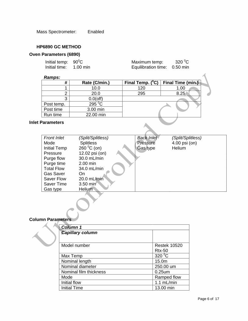
HP6890 GC METHOD
Oven Parameters (6890)
Initial temp: 900C Maximum temp: 320 0C Initial time: 1.00 min Equilibration time: 0.50 min
Ramps:
# Rate (C/min.) Final Temp. (0C) Final Time (min.)
1 10.0 120 1.00
2 20.0 295 8.25
3 0.0(off)
Post temp. 295 0C
Post time 3.00 min
Run time 22.00 min
Inlet Parameters
Front Inlet (Split/Splitless) Back Inlet (Split/Splitless) Mode Splitless Pressure 4.00 psi (on) Initial Temp 260 0C (on) Gas type Helium Pressure 12.02 psi (on) Purge flow 30.0 mL/min Purge time 2.00 min Total Flow 34.0 mL/min Gas Saver On Saver Flow 20.0 mL/min Saver Time 3.50 min Gas type Helium
Column Parameters
Column 1
Capillary column
Model number Restek 10520 Rtx-50
Max Temp 320 0C
Nominal length 15.0m
Nominal diameter 250.00 um
Nominal film thickness 0.25um
Mode Ramped flow
Initial flow 1.1 mL/min
Initial Time 13.00 min
Page7of17
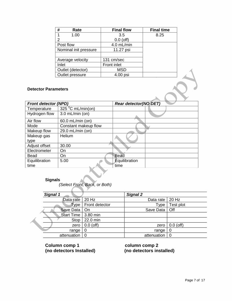
# Rate Final flow Final time
1 1.00 3.5 8.25 2 0.0 (off)
Post flow 4.0 mL/min
Nominal init pressure 11.27 psi
Average velocity 131 cm/sec
Inlet Front inlet
Outlet (detector) MSD
Outlet pressure 4.00 psi
Detector Parameters
Signals (Select Front, Back, or Both)
Signal 1 Signal 2
Data rate 20 Hz Data rate 20 Hz
Type Front detector Type Test plot
Save Data On Save Data Off
Start Time 3.80 min
Stop 22.0 min
zero 0.0 (off) zero 0.0 (off)
range 0 range 0
attenuation 0 attenuation 0
Column comp 1 column comp 2 (no detectors Installed) (no detectors installed)
Front detector (NPD) Rear detector(NO DET)
Temperature 325 0C mL/min(on)
Hydrogen flow 3.0 mL/min (on)
Air flow 60.0 mL/min (on)
Mode Constant makeup flow
Makeup flow 29.0 mL/min (on)
Makeup gas type
Helium
Adjust offset 30.00
Electrometer On
Bead On Bead
Equilibration time
5.00 Equilibration time
Page8of17
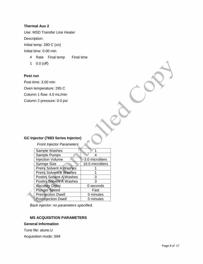
Thermal Aux 2
Use: MSD Transfer Line Heater
Description:
Initial temp: 280 C (on)
Initial time: 0.00 min
# Rate Final temp Final time
1 0.0 (off)
Post run
Post time: 3.00 min
Oven temperature: 295 C
Column 1 flow: 4.0 mL/min
Column 2 pressure: 0.0 psi
GC Injector (7683 Series Injector)
Front Injector Parameters
Sample Washes 1
Sample Pumps 4
Injection Volume 3.0 microliters
Syringe Size 10.0 microliters
PreInj Solvent A Washes 1
PreInj Solvent B Washes 1
PostInj Solvent A Washes 3
PostInj Solvent A Washes 3
Viscosity Delay 0 seconds
Plunger Speed Fast
PreInjection Dwell 0 minutes
PostInjection Dwell 0 minutes
Back injector: no parameters specified.
MS ACQUISITION PARAMETERS
General Information
Tune file: atune.U
Acquisition mode: SIM
Page9of17
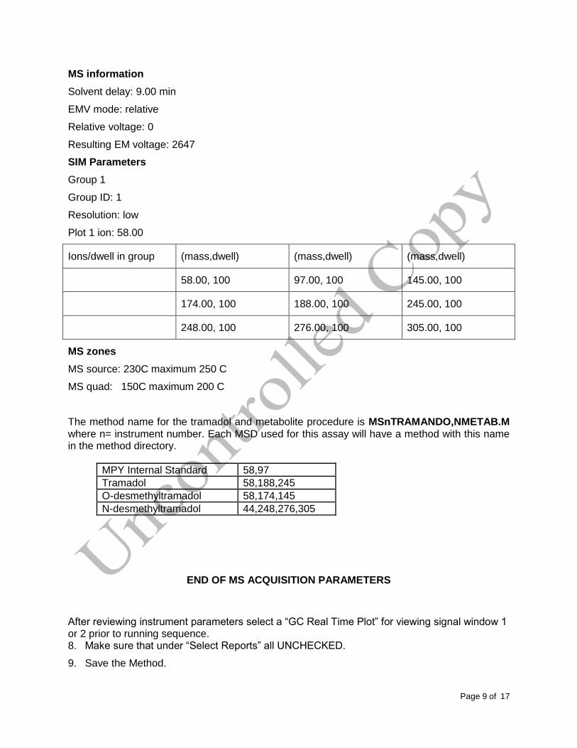
MS information
Solvent delay: 9.00 min
EMV mode: relative
Relative voltage: 0
Resulting EM voltage: 2647
SIM Parameters
Group 1
Group ID: 1
Resolution: low
Plot 1 ion: 58.00
Ions/dwell in group (mass,dwell) (mass,dwell) (mass,dwell)
58.00, 100 97.00, 100 145.00, 100
174.00, 100 188.00, 100 245.00, 100
248.00, 100 276.00, 100 305.00, 100
MS zones
MS source: 230C maximum 250 C
MS quad: 150C maximum 200 C
The method name for the tramadol and metabolite procedure is MSnTRAMANDO,NMETAB.M where n= instrument number. Each MSD used for this assay will have a method with this name in the method directory.
MPY Internal Standard 58,97
Tramadol 58,188,245
O-desmethyltramadol 58,174,145
N-desmethyltramadol 44,248,276,305
END OF MS ACQUISITION PARAMETERS
After reviewing instrument parameters select a “GC Real Time Plot” for viewing signal window 1 or 2 prior to running sequence. 8. Make sure that under “Select Reports” all UNCHECKED.
9. Save the Method.

Page10of17
INSTRUMENT SETUP
1. An acceptable autotune must be obtained prior to batch analysis.
2. All autosampler syringe wash vials are filled with methanol.
3. Prepare a sequence using the following steps.
When Chemstation is opened, the Openlab ECM Login screen appears, Enter the instrument name (ms6, ms7, etc.) as appropriate for username and the current password. Verify that Account field says production and Domain field says Built-In. If Chemstation is already running, it may be necessary to log out and re-log in. Using the Chemstation software, at the top Method and Run toolbar under ECM, select Logon to ECM. Follow the instructions above to log on.
1. Under Sequence toolbar, select Load Sequence. Select default.s. Click on Select.
2. Under Sequence toolbar, select Edit Sequence. At the top of the screen under Data Path, click on Browse. Under Select Data Path, click on the msdchem folder. The click 1 and then click on the Data folder to highlight it. In lower left of dialog box, select Make New Folder. A folder with the name New Folder is created under DATA. Right click on New Folder and Rename or double click to highlight the folder name and change the entry. Rename the file using the format MSNMMDDYYx, where “N” is instrument number, MM = month, DD = day, YY= year and x = a letter indicating the batch being run, e.g. MS1041013a. Click OK which will take you back to the sample log table.
3. Type-in all information under each column (i.e. Type, Vial, Sample, Method/Keyword, Datafile, Comments/Keywordstring). For case samples, include aliquot number, laboratory number, specimen source, dilution if any (i.e., 2-13-2432 fem). If the sample has been diluted, enter the appropriate dilution factor in the Multiplier column.
4. In the Vial column, click in the cell with the number 1, hold down the left mouse button and drag to the last vial number in the sequence (cells will be highlighted). Right click and select Fill Column and Increment. Verify that the vial numbers are correct.
5. In the Method column, verify that the correct method (i.e. TRAMANDO,NMETAB.M) is loaded in the first cell. Then click on the first cell containing the method name, hold down the left mouse and drag to the last vial number in the sequence (cells will be highlighted). Right click and select Fill Column, No Increment. Verify that the method for each vial is correct.
6. In the Data File column, in the cell corresponding to the first vial, enter the data file name in the format MSNMMDDx001, where N = instrument number, MM = month, DD = day, and x = a letter indicating the batch being run, i.e. MS10410a001a. Click on this cell, hold down the left mouse and drag to the last vial number of the sequence (the cells will be highlighted). Right click and select Fill Column and Increment. Verify that the data file information for each vial is correct.
7. In the Comment column, enter any additional information for the vials.
8. In the Level column, verify that the correct level numbers are entered for calibrators in this batch.
9. Verify No Update is selected for all vials under Update Rf and Update Rt.
Page11of17
10. Review the information typed for the sequence. Correct any information as needed. Verify that the Data Path is C:MSDCHEM\1\Data\current sequence name. Verify that the Method Path is C:MSDCHEM\1\METHODS. Then Click ok.
11. Empty solvent wash bottles on the instrument and replace it with fresh methanol before starting the sequence.
12. Use the printed sequence list to load the vials in the correct position on the autosampler tray. Edit the sequence as necessary to correct any errors; save the sequence. Reprint sequence if necessary.
13. Document the “Chain of Custody” and the “Vials Loaded” labels.
14. Document the “Instrument Status Logsheet” that hangs on the instrument’s oven door.
15. Start sequence by selecting Sequence Run Sequence.
16. When the batch is complete, remove all vials using the sequence list. Once again document the “Vials Loaded” label.
Note: Occasionally, it will be desirable to run several subsequences in one batch. Use the
instructions below to accomplish this.
Setting-Up a Subsequence
On the Sample Log Table:
1. Select “Keyword” for “Type”
2. Select “DataPath” for “Method/Keyword”
3. Under “Comment/KeywordString” type in the new data path for your subsequence i.e.: C:\MSDCHEM\1\DATA\MSNMMDDYYx where “N” is the instrument’s number and “x” is the letter designated to the subsequence (it must be different than that of the original sequence).
4. The suffix of the data files must be different, or “y”, from that of the original; i.e.:
MSNMMDDy001; the subsequence data files must start with 1 again.
5. After typing in the entire sequence, save sequence accordingly.
6. Go to Sequence Simulate Sequence Run Sequence.
7. A dialog box will pop-up: DataPath C:\MSDCHEM\1\DATA\MSNMMDDYYy does not exist. Edit Sample Log Table? Click No if the sequence was set up correctly.
8. A 2nd dialog box will pop-up: Create C:\MSDCHEM\1\DATA\MSNMMDDYYy? Click
Yes.
9. A 3rd dialog box will pop-up: Sequence Verification Done! View it? Click Yes or No.
Page12of17
Setting-Up a Subsequence with a Different Method
On the Sample Log Table:
1. After inserting the DataPath keywords and Commands, Insert a Row.
2. Select “Keyword” for “Type”
3. Select “MethodPath” for “Method/Keyword”, under “Comment/KeywordString” type in the new method name for your subsequence.
4. Check that the method for each sample is changed to the new method.
5. Follow Steps 4-9 in the “Setting-Up a Subsequence” Section (see previous section).
6. On the Method and Run toolbar, under Sequence, select Run Sequence. In the dialog box under Sequence Comment enter the initials of the individual who has entered the sequence in the Operator Field (e.g., MPM). Under Data File Directory verify that the data file path is C:MSDCHEM/1\DATA\batch name.
7. Under Sequence, select Save Sequence As. Under File Name, type in the name of the folder that the batch will be saved to under DATA, MSNMMDDYYx (e.g. MS1041013a) Select Save. The extension “.s” will automatically be added.
THE FOLLOWING APPLIES TO ALL SEQUENCE SET-UPS (i.e. sequence, sequence with same method, or sequence with a different method):
1. Under Sequence select Print Sequence. Verify that Brief Format is selected and click on OK. The sequence will be printed. Apply the preprinted labels for documenting verification of process steps to the printed sequence list.
2. Use the printed sequence list to load vials into the appropriate autosampler positions as indicated by the order on the printed sequence list. Check vial information against the sequence list and ensure that the vial is inserted in the correct numeric position in the autosampler as indicated on the sequence list. Document correct positioning by dating and initialing the appropriate line on the sequence list.
3. Under Sequence select Run Sequence. Verify that the Sequence comments and Data Field information are correct (i.e. verify that the proper sequence is loaded. If not, load the proper sequence). Click on Run Sequence.
4. After the batch is finished, unload the vials. Compare the vial information to the sequence list as they are removed, to verify that the correct vial was in the correct position. Date and initial the sequence list when this is completed. Annotate discrepancies if necessary
DATA TRANSFER AND PROCESSING – EXPORTING RAW DATA FOR PROCESSING
All processing and review of the processed data are performed on a processing computer.
Page13of17
SAVE METHOD TO ECM
After the run finishes, the raw data files will be in the data subdirectory on the local chemstation and also automatically transferred to ECM. From the acquiring computer, make sure the proper method, the one used to acquire the data, is loaded. On the top toolbar under ECM click-on Save Method to ECM.
Click on the GCMS, the correct instrument name folder, locate and double click with mouse on the sequence that just ran under the appropriate month, the method will be saved.
RETRIEVE SEQUENCE FROM ECM – RAW DATA FOR PROCESSING
1. At the processing computer, click on Processing Data Analysis. Log on using your OCME network username and password.
2. On Enhanced Data Analysis screen, click on ECM at the top toolbar and select Retrieve entire sequence from ECM.
This will open up the Openlab ECM screen. Select GCMS, then the appropriate instrument, month, and sequence to be retrieved. On the status line at the bottom of the screen, notice that all datafiles along with the method of the entire sequence is being retrieved. The sequence will be downloaded to the following location: C:\msdchem\1\ECM\Retrieve\”sequence name” DATA REVIEW
There are three levels of review; the first level of review is the transference and processing of the
raw data, this may be performed by any trained analyst; the second level of review is performed
by an experienced analyst who is trained and signed off in data review, he / she will review the
processed data; the third level of review is considered the final level of review, this can only be
performed by the Laboratory Manager. He/she will review the data for the entire case ensuring
that screening, confirmatory and qualitative and / or quantitative analysis on the case have been
completed and reported accurately. As needed, he/she will also schedule additional analysis and
contact the Medical Examiner on the case to discuss any findings and / or review case history.
LOAD METHOD AND SEQUENCE – FIRST LEVEL REVIEW
1. On the left side of the screen under the C drive, open C:\msdchem\1\ecm\retrieve
2. Under retrieve, click on the batch for processing. When all files have been downloaded to the processing computer, verify that the appropriate method is present in the batch.
3. To load the method, right click on the method under the batch being processed and select load. This will bring up “Be sure changes are saved. Load now?”, click yes. If the method is not present, load the method by retrieving the method from ECM.
4. Click on any file in the batch to load it.
Page14of17
PERFORM BATCH CALIBRATION – PROCESSING – FIRST LEVEL REVIEW
Under enhanced data analysis:
1. Load the calibrator datafile. Process the calibrator by typing-in “QT 1” in the space provided, click-on “Execute”.
2. Select the View, select Q-Edit Quant Result. Review the integration of each compound, if necessary re-integrate peaks (i.e. the baseline is the most scientifically accurate one that can be drawn). Double check each of the compound’s retention time and that each compound has a response. All auto- integrated compounds should have an “x” next to their name. Click Exit when finished, save any changes made.
3. Update the existing calibration table (level 1, which is for a 1.0mg/L sample). Select Calibrate, Update One Level. When asked to requant file before update, select NO. Select replace responses and replace retention time and Replace Qualifier Ion Relative Responses, and choose the existing level ID (#1). Click Do Update.
4. Review the Compound database. Double click on the internal standard listed on the left to reveal the compounds quantitated with it. Select the calibration tab to reveal compound responses. Click OK or Cancel when review is complete
5. Requant the calibrator by typing-in “QT 2” in the space provided; click-on “Execute”, to update calibrator. Review with QEDIT. Check the responses, retention times and ion ratios.
6. Save Method before proceeding. Select Method from the toolbar, save method, make sure that the path is correct. Save to OpenLab ECM at this time.
7. Process controls and cases. Select Tools from the toolbar, DoLIST, Quant, No Report (QT 1), Add, and OK. Select appropriate files. Click the → Arrow and Process. Review each file using QEDIT.
8. When review is complete, select report format by choosing Quantitate from the toolbar, Report Options. Check SIM style report and uncheck Internal Standards. Press OK.
9. To print reports, select Tools from the toolbar, DoLIST, Profile Quant w/o Calculations (QT 0,1,’P’), Add, and OK. Select files to print, click the → Arrow and Process.
10. Print the calibration table for the current batch by clicking Calibrate on the command line. Select List, Calibrate Report and click OK. The Calibration report will print to the screen. Review the r2 values, then right click on the screen report to print it.
Page15of17
11. Save files to ECM. Select ECM from the toolbar, select “Save multiple data files to ECM”. Select all data files on the left and move them to the right empty space, the click Process. Allow each data file to be successfully uploaded to ECM before exiting Data Analysis.
12. Save method to ECM. Select ECM from the toolbar, Save Method to ECM. Make sure data path is correct.
13. It is now safe to delete the batch from the processing computer.
BATCH CLEAN UP
Select “my computer”. Find the batch on the C drive at C:\msdchem\1\ecm\retrieve\batch. Right click on the batch to be deleted and select delete. Do not delete a batch that has not been successfully uploaded to ECM.
ACCEPTANCE CRITERIA – APPLIES TO ALL LEVELS OF REVIEW
1. Check chromatography of all injections. Examine the peak shape and evaluate any peaks show non-Gaussian shape. Identify unresolved peaks and peaks with shoulders on either side. Consult a supervisor about any unusual events.
2. Ion ratios must be within ± 20% of the calibrator for blood samples. For tissues, the ion ratios must be within ± 30% of the calibrator.
3. The blank must not contain detectable amounts of target analytes or significant interfering peaks.
4. Check that all significant peaks in the chromatogram are integrated. Check if the baseline used to integrate is appropriate.
5. For each analyte the base ion must be present and two additional significant ions must be present (these may be the ions designated in the method or additional known ions from reference materials or libraries.)
6. Retention times of the target analytes should match those of the calibrator or controls within ± 2% if this data is available.
SECOND LEVEL REVIEW
The Second Level Reviewer will review the processed data in its entirety according to the
acceptance criteria section of this SOP.

Page16of17
REPORTING
After the batch has undergone second level review and has been printed, either the first level reviewer or the second level reviewer may report the data in the appropriate case file
Report using the following guidelines:
1. Each case printout must have a copy attached of the accepted calibrator and controls and the sequence list.
2. Enter the sequence with tune attached in the sequence logbook for the appropriate MS.
3. Evaluate and review data in accordance with guidelines in the Acceptance Criteria section of this SOP. Annotate the results on the case file reporting sheet, and include the case chromatogram and control package in the appropriate case folder.
4. Tramadol metabolites are reported as “Detected” or “Not Detected”.
5. Compare your results to the results already reported for the case, to check if results are consistent with all other data. Notify supervisor if any inconsistencies are noticed.
6. Submit the case file with all reports for Third Level Review.
THIRD LEVEL REVIEW (FINAL REVIEW)
The third and final level review can only be performed by the Laboratory Managers. He/she will review the data for the entire case according to all established criteria. They will ensure that screening, confirmatory and quantitative analysis on the case have been completed and reported accurately. As needed, they will also schedule additional analysis and contact the Medical Examiner on the case to discuss any findings and / or review case history.
REFERENCES
SPEware Corp. Cerex Applications Manual.
System 48 Processor. Users Guide.
Turbovap. Users Guide.
REVISION HISTORY
Ver 04.20.2015 1. Revision history implemented.
Ver 08.31.2015 1. Added internal standard acceptance criteria.
Ver 09.04.2015 1. Defined the level of review(s)
Ver 09.16.2015
1. Removed internal standard acceptance criteria.
2. Minor grammatical changes.

Page17of17