formapure dna · pn b44690kf 3 protocol for dna isolation contents • introduction, page 3 • kit...
TRANSCRIPT

Instructions For Use
FormaPure DNA:
Extended Protocol for DNAIsolation from FFPE Sample
March 2019
Beckman Coulter, Inc.250 S. Kraemer Blvd.Brea, CA 92821 U.S.A.
PN B4
4690KF
FormaPure DNA:Extended Protocol for DNA Isolation from FFPE SamplePN B44690KF (March 2019)
© 2019 Beckman Coulter, Inc.All rights reserved.
Beckman Coulter, the stylized logo, and the Beckman Coulter product and service marks mentioned herein are trademarks or registered trademarks of Beckman Coulter, Inc. in the United States and other countries.
All other trademarks, service marks, products, or services are trademarks or registered trademarks of their respective holders.
Contact Us • For questions regarding this protocol, call Technical
Support at Beckman Coulter at 1-800-369-0333. • For additional information, or if damaged product is
received, call Beckman Coulter Customer Service at 800-742-2345 (USA or Canada) or contact your local Beckman Coulter Representative.
• Refer to www.beckman.com/techdocs for updated protocols.
Glossary of Symbols is available at www.beckman.com/techdocs (PN C05838).
Product Availability B89230 — FormaPure DNA, 50 Prep Kit B89231 — FormaPure DNA, 96 Prep Kit B89232 — FormaPure DNA, 384 Prep Kit
Find us on the World Wide Web at:www.beckman.com
Beckman Coulter Eurocenter S.A.22, rue Juste-OlivierCase Postale 1044CH - 1260 Nyon 1, SwitzerlandTel: +41 (0) 22 365 36 11
Printed in USA

Protocol for DNA Isolation
Contents
• Introduction, page 3 • Kit Specifications, page 4 • Warnings and Precautions, page 4 • Materials Supplied, page 5 • Materials Required but not Supplied, page 6 • Process Overview, page 7 • DNA Extraction Protocol, page 7 • Troubleshooting Guide, page 11 • Revision History, page 15
Introduction
The FormaPure DNA extraction and purification kit utilizes the patented Beckman Coulter SPRI paramagnetic bead-based technology to isolate DNA from formalin-fixed, paraffin-embedded (FFPE) tissue without the use of xylene. This kit has been optimized for use with downstream sequencing and genotyping assays. Specifically, genomic DNA isolated with FormaPure DNA is compatible with the following downstream applications: • Targeted amplicon NGS • Targeted capture NGS • Whole exome sequencing • Whole genome sequencing • Endpoint or qPCRFormaPure DNA isolates DNA from tissue sections totaling a thickness of up to 3 x 10 microns. The protocol can be performed in both 96-well plates (manually and automated) and in 1.5 mL tubes (manually only). Nucleic acid extraction begins with the solubilization of the paraffin from the tissue slices in tubes. An enzymatic lysis step digests the tissue and releases the nucleic acids, followed by decrosslinking at a high temperature. The remaining protocol can be carried out in plates or tubes. RNA is removed from the sample and a binding solution is added to immobilize the nucleic acids to the surface of the SPRI beads. Contaminants are rinsed away using a simple washing procedure and the nucleic acids are eluted with water.
PN B44690KF 3

Protocol for DNA IsolationKit Specifications
Kit Specifications
Warnings and Precautions
Read and observe the following safety information.IMPORTANT The symbol indicates a potential safety risk involving the material, action, or equipment
required for executing a procedural action; when you see the symbol, return to this section to review relevant safety information.
Kit Type Number of Preps
Small kit, PN B89230 50Medium kit, PN B89231 96Large kit, PN B89232 384
DANGER
Proteinase KH315 Causes skin irritation.H319 Causes serious eye irritation.H334 May cause allergy or asthma symptoms or breathing difficulties if inhaled.H335 May cause respiratory irritation.P261 Avoid breathing vapors.P280 Wear protective gloves, protective clothing and eye/face protection.P284 In case of inadequate ventilation, wear respiratory protection.
P304+P340 IF INHALED: Remove person to fresh air and keep at rest in a position comfortable for breathing.
P342+P311 If experiencing respiratory symptoms: Call a POISON CENTER or doctor/physician.
Safety Data Sheet is available at www.beckman.com/techdocs.
PN B44690KF 4

Protocol for DNA IsolationMaterials Supplied
CAUTIONRisk of chemical injury from Proteinase K. To avoid contact with Proteinase K, wear appropriate personal protective equipment, including protective eyewear, gloves, and suitable laboratory attire. Refer to the Safety Data Sheet for details about chemical exposure before using the chemical.
CAUTIONRisk of burning from hot liquid splattering into your eyes or onto your skin. Wear appropriate personal protective equipment while incubating the samples. Place tube cap locks on the tubes to prevent the tops of the tubes from opening during incubation.
Materials Supplied
The following reagents are supplied in the FormaPure DNA kit. The reagent icon is included in the instructions as a visual aid to ensure the correct reagent is used. NOTE Refer to the product labels for expiration dates.
Reagent Icon Storage Conditions
Mineral Oil 15 to 30 °C
Lysis 15 to 30 °C
Bind 15 to 30 °C
Wash 15 to 30 °C
RNase A - 15 to 30 °CProteinase K - 15 to 30 °C
PN B44690KF 5

Protocol for DNA IsolationMaterials Required but not Supplied
Materials Required but not Supplied
Required Reagents
Required EquipmentFormaPure DNA processing can be done in a 96-well plate or tube format. Refer to the tables below for the hardware and consumables required for this procedure.
Reagent Supplier Part Number
100% ethanol AmericanBio AB00138 (or equivalent)Nuclease-free water Thermo Fisher AM9932 (or equivalent)
Table 1 Required Hardware and Accessories
Hardware and Accessories Format
Pipettes (P20, P200, P1000 multi or single channel as needed)
plate and tube
Adjustable heat source (for example, a water bath or a heat block). Two are recommended.
plate and tube
Vortexer plate and tubeBeckman Coulter Microcentrifuge 16, or equivalent plate and tubeBeckman Coulter Agencourt SPRIPlate 96R Ring Super Magnet Plate, PN A32782
plate
Beckman Coulter Agencourt SPRIStand Magnetic 6-tube Stand (for 1.5, 1.7, or 2.0 mL tubes), PN A29182
tube
Table 2 Required Consumables
Consumables Format
Barrier tips for P20, P200, and P1000 pipettes plate and tube1.5-1.7 mL microcentrifuge tubes plate and tubeMicrocentrifuge tube cap locks plate and tubeThermo Fisher 1.2 mL 96-well Plate, PN AB1127, or equivalent
plate
200 μL 96-well storage plate platePCR Adhesive Seals plate
PN B44690KF 6
Protocol for DNA IsolationProcess Overview
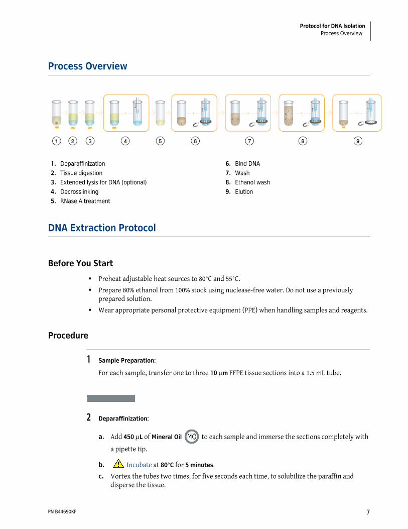
Process Overview
DNA Extraction Protocol
Before You Start • Preheat adjustable heat sources to 80°C and 55°C. • Prepare 80% ethanol from 100% stock using nuclease-free water. Do not use a previously
prepared solution. • Wear appropriate personal protective equipment (PPE) when handling samples and reagents.
Procedure
1 Sample Preparation:For each sample, transfer one to three 10 m FFPE tissue sections into a 1.5 mL tube.
2 Deparaffinization:
a. Add 450 L of Mineral Oil to each sample and immerse the sections completely with a pipette tip.
b. Incubate at 80°C for 5 minutes. c. Vortex the tubes two times, for five seconds each time, to solubilize the paraffin and
disperse the tissue.
1. Deparaffinization2. Tissue digestion3. Extended lysis for DNA (optional)4. Decrosslinking5. RNase A treatment
6. Bind DNA7. Wash8. Ethanol wash9. Elution
PN B44690KF 7

Protocol for DNA IsolationDNA Extraction Protocol
3 Tissue Digestion:
a. Add 200 L of Lysis to each sample.
NOTE Do not vortex the tubes as this may cause the mineral oil and lysis to emulsify.
b. Centrifuge the tubes at 10,000 × g for 15 seconds. The mineral oil forms a separate upper phase.NOTE Incubate the tubes for 3 more minutes at 80°C if the mineral oil layer starts turning
cloudy or the tissue is stuck at the interface of mineral oil and lysis buffer. After the incubation, make sure to cool the tubes for 2 minutes before adding Proteinase K.
c. Add 20 μL of Proteinase K to the aqueous, lower phase and pipette mix 10 times without disrupting the upper phase.
d. Incubate the tubes for a minimum of 60 minutes at 55°C (up to 16 hours) to achieve complete lysis.
4 Decrosslinking:
a. Incubate the tubes at 80°C for 60 minutes.b. Remove the tubes from the heat source.c. Transfer as much of the lysate (lower phase) as possible to a 96-well plate, or to 1.5 mL
tubes, without disrupting the upper phase.NOTE Minimize the amount of Mineral Oil that is transferred along with the lysate. However, a
small amount of Mineral Oil carryover does not affect downstream applications.
5 RNase A Treatment:a. Add 5 L of RNase A to each sample.b. Pipette mix five times with a P200 pipette set at 150 L to thoroughly distribute the
enzyme. Mix gently to minimize the generation of bubbles.c. Incubate at room temperature for 5 minutes.
PN B44690KF 8
Protocol for DNA IsolationDNA Extraction Protocol
6 Bind DNA:
a. Fully resuspend the Bind solution by shaking or vortexing.
b. Add 300 L of Bind to each sample and mix 10 times with a P1000 pipette set at 350 L. Mix gently to minimize the generation of bubbles.NOTE DNA binds to the magnetic particles during this step. When mixing, use a mix volume that is
slightly less than the total volume in the well and pipette slowly to minimize the formation of air bubbles. Air bubbles can trap magnetic beads and prevent them from being pulled to the bottom of the plate, thus decreasing yield.
c. Incubate at room temperature for 5 minutes.d. Place the samples on the magnet for 10 minutes, or until the solution is clear, to allow the
beads to separate. Use a SPRIPlate 96R Ring Super Magnet Plate if working in a 96-well plate, or place the tubes in an Agencourt SPRIStand Magnetic 6-tube Stand if using tubes.
e. With the samples on the magnet, aspirate the supernatant without disrupting the beads. Discard the supernatant.NOTE When aspirating, place the pipette at the center of the ring, or away from the beads in the
tube, to avoid disturbing the magnetic beads. Bead loss will result in lower yields.
7 Wash:a. Remove the samples from the magnet.
b. Add 400 L of Wash to each sample.c. Using a P1000 pipette set at 250 L, mix the samples 15 times or until the beads are fully
resuspended in the solution. Mix gently to minimize the generation of bubbles.d. Place the samples back on the magnet for 10 minutes, or until the solution is clear, to allow
the beads to separate.e. With the samples on the magnet, aspirate the supernatant without disrupting the beads.
Discard the supernatant.NOTE When aspirating, place the pipette at the center of the ring, or away from the beads in the
tube, to avoid disturbing the magnetic beads. Bead loss will result in lower yields.
8 Ethanol Wash:a. Remove the samples from the magnet.
PN B44690KF 9

Protocol for DNA IsolationDNA Extraction Protocol
b. Add 750 L of freshly prepared 80% ethanol to each sample.c. Using a P1000 pipette set at 600 L, mix the sample 20 times, or until the beads are fully
resuspended.d. Place the samples back on the magnet for three minutes, or until the solution is clear, to
allow the beads to separate.e. With the samples on the magnet, aspirate the supernatant without disrupting the beads.
Discard the supernatant.NOTE Remove as much ethanol as possible, without disturbing the magnetic beads, before drying.
Dispose of Ethanol waste in accordance with the local regulations and acceptable laboratory practices.
f. Air dry the samples on the magnet for 10 minutes.
9 Elution:a. Remove the samples from the magnet.b. Add a minimum of 40 L of nuclease free water to each sample and mix 10 times with a P200
pipette set at 30 L or until beads are fully resuspended.c. Cap tubes or cover the plate with a PCR adhesive seal and incubate at 55°C for one minute.d. Place the samples back on the magnet for one minute, or until the solution is clear, to allow
the beads to separate.e. With the samples on the magnet, transfer as much of the supernatant as possible to a
96-well storage plate, or to a new tube, without disturbing the magnetic beads. f. Store at -20°C.
PN B44690KF 10

Protocol for DNA IsolationTroubleshooting Guide
Troubleshooting Guide
This troubleshooting guide may be helpful to maximize nucleic acid yield, integrity, and purity from FFPE tissues, or to solve any issues that may arise. The scientists at Beckman Coulter are available to answer any questions you may have about the information in this troubleshooting guide and the protocols in this manual (refer to Contact Us on page 2 for contact information).NOTE Visit www.Formapure.com for instructional videos and updated information.
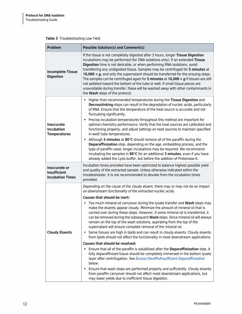
This section includes the following tables: • Table 3, Troubleshooting Low Yield • Table 4, Troubleshooting Poor Quality of Extracted Nucleic AcidsTable 3 Troubleshooting Low Yield
Problem Possible Solution(s) and Comment(s)
Poor Starting Sample Quality
The processes of formalin fixation, paraffin embedding and/or storage of FFPE tissues cause damage to the nucleic acids. While the FormaPure chemistry is designed to maximize yield and integrity for challenging FFPE samples, this chemistry cannot repair damaged nucleic acids.
Low Tissue Input or Tissue Type
• Some FFPE samples may contain very low amounts of tissue or cells, depending on the tissue and disease types; therefore, the amount of nucleic acids may be inherently low prior to extraction. If possible, increase the amount of FFPE samples to obtain the desired yield.
• Certain tissue types are more difficult to digest than others. An extended Tissue Digestion incubation can be performed (with DNA isolations only) to free up more of the nucleic acids.
Bead/Sample Loss
• Disruption of the bead pellet during supernatant removal may cause decreased yields. The pipette tip should not contact the bead pellet during aspirations. If a brown color is seen in the pipette tip during aspiration, beads are present and the solution should be dispensed back into the tube or well. Place samples back on magnet until solution is fully cleared and let the beads settle towards the magnet before aspirating again.
• Insufficient bead clearing during magnetic separation may lead to decreased yields. Ensure that the beads are completely settled to the magnet and the supernatant is clear before removing the supernatant.
• Undigested tissue can trap the beads and prevent efficient nucleic acid binding or lead to bead and sample loss. Tissue should be thoroughly digested in the Tissue Digestion step before bead addition. If undigested tissue remains after the Tissue Digestion step, avoid transferring the undigested tissue to another tube or well before proceeding to the Bind step. For additional information, see Incomplete Tissue Digestion below.
PN B44690KF 11
Protocol for DNA IsolationTroubleshooting Guide
Incomplete Tissue Digestion
If the tissue is not completely digested after 3 hours, longer Tissue Digestion incubations may be performed (for DNA isolations only). If an extended Tissue Digestion time is not desirable, or when performing RNA isolations, avoid transferring any undigested tissue. Samples may be centrifuged for 5 minutes at 10,000 × g, and only the supernatant should be transferred for the ensuing steps. The samples can be centrifuged again for 5 minutes at 10,000 × g if tissues are still not pelleted toward the bottom of the tube or well. If small tissue pieces are unavoidable during transfer, these will be washed away with other contaminants in the Wash steps of the protocol.
Inaccurate Incubation Temperatures
• Higher than recommended temperatures during the Tissue Digestion and Decrosslinking steps can result in the degradation of nucleic acids, particularly of RNA. Ensure that the temperature of the heat source is accurate and not fluctuating significantly.
• Precise incubation temperatures throughout this method are important for optimal chemistry performance. Verify that the heat sources are calibrated and functioning properly, and adjust settings on heat sources to maintain specified in-well/ tube temperatures.
• Although 5 minutes at 80°C should remove all of the paraffin during the Deparaffinization step, depending on the age, embedding process, and the type of paraffin used, longer incubations may be required. We recommend incubating the samples in 80°C for an additional 3 minutes, even if you have already added the Lysis buffer, but before the addition of Proteinase K.
Inaccurate or Insufficient Incubation Times
Incubation times provided have been optimized to balance highest possible yield and quality of the extracted sample. Unless otherwise indicated within the troubleshooter, it is not recommended to deviate from the incubation times provided.
Cloudy Eluents
Depending on the cause of the cloudy eluent, there may or may not be an impact on downstream functionality of the extracted nucleic acids.Causes that should be inert: • Too much mineral oil carryover during the lysate transfer and Wash steps may
make the eluents appear cloudy. Minimize the amount of mineral oil that is carried over during these steps. However, if some mineral oil is transferred, it can be removed during the subsequent Wash steps. Since mineral oil will always remain on the top of the wash solutions, aspirating from the top of the supernatant will ensure complete removal of the mineral oil.
• Some tissues are high in lipids and can result in cloudy eluents. Cloudy eluents from lipids should not affect the functionality in most downstream applications.
Causes that should be resolved: • Ensure that all of the paraffin is solubilized after the Deparaffinization step. A
fully deparaffinized tissue should be completely immersed in the bottom lysate layer after centrifugation. See Excess Paraffin/Insufficient Deparaffinization below.
• Ensure that wash steps are performed properly and sufficiently. Cloudy eluents from paraffin carryover should not affect most downstream applications, but may lower yields due to inefficient tissue digestion.
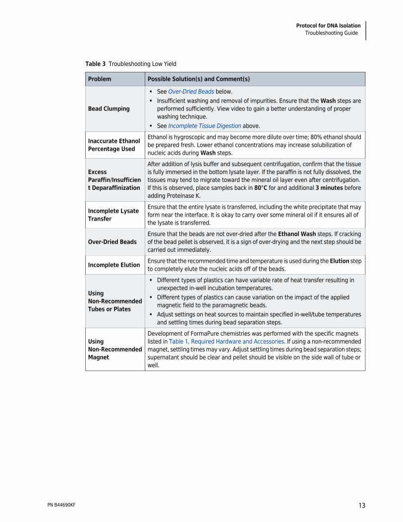
Table 3 Troubleshooting Low Yield
Problem Possible Solution(s) and Comment(s)
PN B44690KF 12
Protocol for DNA IsolationTroubleshooting Guide
Bead Clumping
• See Over-Dried Beads below. • Insufficient washing and removal of impurities. Ensure that the Wash steps are
performed sufficiently. View video to gain a better understanding of proper washing technique.
• See Incomplete Tissue Digestion above.
Inaccurate Ethanol Percentage Used
Ethanol is hygroscopic and may become more dilute over time; 80% ethanol should be prepared fresh. Lower ethanol concentrations may increase solubilization of nucleic acids during Wash steps.
Excess Paraffin/Insufficient Deparaffinization
After addition of lysis buffer and subsequent centrifugation, confirm that the tissue is fully immersed in the bottom lysate layer. If the paraffin is not fully dissolved, the tissues may tend to migrate toward the mineral oil layer even after centrifugation. If this is observed, place samples back in 80°C for and additional 3 minutes before adding Proteinase K.
Incomplete Lysate Transfer
Ensure that the entire lysate is transferred, including the white precipitate that may form near the interface. It is okay to carry over some mineral oil if it ensures all of the lysate is transferred.
Over-Dried BeadsEnsure that the beads are not over-dried after the Ethanol Wash steps. If cracking of the bead pellet is observed, it is a sign of over-drying and the next step should be carried out immediately.
Incomplete Elution Ensure that the recommended time and temperature is used during the Elution step to completely elute the nucleic acids off of the beads.
Using Non-Recommended Tubes or Plates
• Different types of plastics can have variable rate of heat transfer resulting in unexpected in-well incubation temperatures.
• Different types of plastics can cause variation on the impact of the applied magnetic field to the paramagnetic beads.
• Adjust settings on heat sources to maintain specified in-well/tube temperatures and settling times during bead separation steps.
Using Non-Recommended Magnet
Development of FormaPure chemistries was performed with the specific magnets listed in Table 1, Required Hardware and Accessories. If using a non-recommended magnet, settling times may vary. Adjust settling times during bead separation steps; supernatant should be clear and pellet should be visible on the side wall of tube or well.
Table 3 Troubleshooting Low Yield
Problem Possible Solution(s) and Comment(s)
PN B44690KF 13

Protocol for DNA IsolationTroubleshooting Guide
Table 4 Troubleshooting Poor Quality of Extracted Nucleic Acids
Problem Possible Solution(s) and Comment(s)
Nucleic Acid Appears Degraded
• The processes of formalin fixation, paraffin embedding and/or storage of FFPE tissues cause degradation of nucleic acids.
• If nucleic acids are more degraded than expected, use sterile techniques to ensure that DNase and RNase are not a source of contamination during the isolation processes.
• Store the nucleic acids at -20°C, or -80°C for long-term storage.
RNAse and/or DNAse Contamination
• Use sterile techniques to ensure that DNase and RNase are not a source of contamination during the isolation processes.
• Filter tips should be used for RNA workflows so buffer sources are not contaminated.
• If all other sources of contamination are ruled out, replace reagents.
DNA Contamination with RNA
While RNase A should be active in sample lysates that contain cellular debris and Lysis buffer components, these components will inhibit DNase activity. Make sure that the ethanol washes are performed properly before DNase treatments, and remove the ethanol as much as possible as excess ethanol may also prevent DNase activity.
RNA Contaminated with DNA
Ensure temperatures are appropriate for full nuclease activity: RNase A treatments should be carried out at room temperature and DNase I treatments should be carried out at 37°C.
Poor Performance in Downstream Assays
• Ensure that the Wash steps are performed properly and sufficiently. View video to gain a better understanding of proper washing technique.
• Residual ethanol should be removed and/or air-dried before proceeding to subsequent steps.
• During supernatant removal steps after magnetic separation, make sure to remove as much of the supernatant as possible without disturbing the beads.
• Some more fibrous tissues, such as muscle, will form more extensive or tighter crosslinks upon fixation, so longer Decrosslinking incubations may increase nucleic acid functionality. For DNA isolations, Decrosslinking incubations can be performed for up to 3 hours at 80°C. We do not recommend extending the Decrosslinking incubations for RNA isolations as this can further degrade the RNA.
PN B44690KF 14

Protocol for DNA IsolationRevision History
Revision History
Go to www.beckman.com/techdocs to download the most recent manual for this product.Initial Issue AA, 12/2017
Revision AB, 1/2018
Revision AC, 5/2018Updates were made to the following sections: • Process Overview • Troubleshooting GuideRevision AD, 3/2019Updates were made to the following section: • Materials Supplied
PN B44690KF 15

© 2019 Beckman Coulter, Inc.All Rights Reserved
www.beckman.com